Review

Complications Associated with Oblique Lumbar Interbody Fusion: A Systematic Review

by
Quan Rui Tan
1,
Russell Andrew Wong
1,
Arun-Kumar Kaliya-Perumal
1 and
Jacob Yoong-Leong Oh
2,*
1
Lee Kong Chian School of Medicine, Nanyang Technological University, Singapore 308232, Singapore
2
Division of Spine, Department of Orthopaedic Surgery, Tan Tock Seng Hospital, Singapore 308433, Singapore
*
Author to whom correspondence should be addressed.
Surg. Tech. Dev. 2023, 12(4), 211-223; https://doi.org/10.3390/std12040020
Submission received: 5 July 2023 / Revised: 27 August 2023 / Accepted: 3 November 2023 / Published: 20 November 2023

Abstract

:
The main advantage of Oblique Lumbar Interbody Fusion (OLIF) is its ability to provide safe access to the lumbar spine while being a robust interbody fusion technique through a minimally invasive approach. This study reviews the postoperative complications of OLIF, offering a comprehensive understanding of its advantages and disadvantages. A total of 27 studies with 1275 patients were shortlisted based on our selection criteria. Complications were categorized into intra-operative, immediate post-operative, and delayed post-operative and were interpreted based on surgical procedure into stand-alone OLIF, OLIF with posterior stabilisation, and unspecified. Major complications exhibited a pooled prevalence of just 1.7%, whereas the overall pooled prevalence of complications was 24.7%. Among the subgroups, the stand-alone subgroup had the lowest prevalence of complications (14.6%) compared to the unspecified subgroup (29.6%) and the OLIF L2-5 with posterior stabilisation subgroup (25.8%). Similarly, for major complications, the stand-alone subgroup had the lowest prevalence (1.4%), while the OLIF L2-5 with posterior stabilisation subgroup (1.8%) and the unspecified OLIF L2-5 subgroup (1.6%) had higher rates. However, the differences were not statistically significant. In conclusion, the rate of major complications after OLIF is minimal, making it a safe procedure with significant benefits outweighing the risks. The advantages of OLIF L2-5 with posterior stabilisation over stand-alone OLIF L2-5 is a subject of discussion.

1. Introduction

The evolution of Oblique Lumbar Interbody Fusion (OLIF) dates back to 1997, when Mayer reported two new approaches to the lumbar spine: a retroperitoneal approach for lumbar levels L2-5, and a transperitoneal approach for L5-S1 [1]. Even though these approaches were initially termed as anterior approaches, they are now referred to as lateral approaches to the lumbar spine. Over time, the lateral retroperitoneal approach has been widely adopted for interbody fusion surgeries addressing various lumbar pathologies, especially degenerative conditions. This method has demonstrated its effectiveness as a robust technique for achieving interbody fusion through a minimally invasive approach. While fusion procedures performed using this approach can collectively be termed Lateral Lumbar Interbody Fusion (LLIF), they can be classified as either Oblique Lumbar Interbody Fusion (OLIF) or Direct Lateral Interbody Fusion (DLIF/XLIF) depending on whether the psoas muscle is retracted or split to reach the disc space (Figure 1) [2]. Recently, the lateral retroperitoneal approach has also been adapted for the L5-S1 level, which was previously considered to be only approachable through an anterior transperitoneal approach [3]. Hence, in addition to Posterior Lumbar Interbody Fusion (PLIF), Transforaminal Lumbar Interbody Fusion (TLIF), and Anterior Lumbar Interbody Fusion (ALIF), OLIF has become a popular technique for treatment of multiple lumbar spinal conditions including spondylosis, spondylolisthesis, and kyphoscoliosis.
The advantages of the lateral approach have been well established [4]. It allows for the placement of a wide-bodied interbody cage, which allows for more stability and provides a larger surface area [5]. In addition, there are other benefits such as good sagittal and coronal correction, the ability of indirect decompression, multi-level minimally invasive surgery (MIS) fusions, and an excellent fusion technique [6,7]. This is especially so in the context of revision surgeries, where scar tissue may make it difficult to conduct the operation. Among the lateral approaches, OLIF for lumbar levels L2-5 (OLIF L2-5) offers an additional advantage over the DLIF/XLIF approach, as it does not require the dissection of the psoas major muscle that risks disruption of the lumbar nerve plexus which can lead to post-operative hip flexion weakness (27.5%) and other neurologic injuries (36.07%) [8,9]. Hence, OLIF has become a safer alternative to posterior approaches, especially for lumbar levels L2-5 as the retroperitoneal corridor that is being accessed is anterior to the psoas major muscle [10], avoiding complications of damaging it, and also avoiding anatomical complications of the anterior approach [11]. This paper studies the complications of OLIF L2-5 separately from OLIF L5-S1. As OLIF L2-5 is increasingly becoming popular, it needs to be studied in detail as a separate entity and potential complications listed.
OLIF L2-5 can be performed alone or with additional posterior stabilisation with pedicle screws [12]. For patients with no comorbidities such as obesity, osteoporosis, or lumbar disc herniation, stand-alone OLIF may be sufficient and is shown to be equally stable as OLIF with posterior stabilisation due to the presence of intact posterior elements and bilateral spinal muscles [12]. However, OLIF with posterior stabilisation allows for a better dispersal of impact and therefore offers additional protection to the endplates than a stand-alone OLIF, thereby increasing fusion rates and decreasing rates of post-operative cage subsidence [13,14]. However, additional posterior stabilisation may pose a higher risk for complications as it increases operative time, tissue damage, and blood loss, and also incurs higher implant costs to patients [12,15]. With multiple studies now available in the literature, we intend to perform a review and consolidate the complication rates pertaining to OLIF L2-5 in a varied multinational population. Reported complications will be divided into three categories: intraoperative, immediate post-operative, and late post-operative complications. Additionally, OLIF with posterior stabilisation and stand-alone OLIF will be compared to determine the difference in rate of complications. This would allow patients and clinicians to make better and safer decisions.

2. Materials and Methods

2.1. Literature Search

We conducted an online search in September 2021 for relevant articles written in English from inception to September 2021. For this study, the following search strategy was employed: “OLIF” OR “oblique lumbar interbody fusion” OR “anterior-to-psoas” AND “complication”. The search returned 135 results from MEDLINE and 129 results from EMBASE, 5 results from Cochrane Library and 2 from study citations previously not found in database searches—giving 174 papers, excluding duplicates. The abstracts of all gathered studies were read, and prospective and retrospective studies concerning OLIF with outcome assessment were shortlisted. A total of 87 studies met this inclusion criteria and were read in full text. Excluded were studies which did not mention complications, those with unstated levels or including levels outside L2-5, those with surgical indications of malignancy or infection, those concerning revision OLIF surgeries, and those with patients in whom anterolateral screws were used. We also assessed for overlapping patient cohorts from studies from the same author, and one study was excluded. Ultimately, a total of 27 studies met our inclusion and exclusion criteria. The workflow for the literature search can be found in Figure 2. The Preferred Reporting Items for Systemic Reviews and Meta-Analyses (PRISMA) guidelines were followed (Table S1).

2.2. Data Extraction and Potential Biases

The following data were extracted from the final papers: author name, publisher, publication year, type of study, number of patients, type of fixation, neuromonitoring, navigation, radiologic guidance, study follow-up time, and operation details such as mean operation time, mean blood loss, and OLIF levels were tabulated. Complications were collated and categorized into intra-operative, immediate post-operative (within one-month or less, post-OLIF), and delayed post-operative (more than one-month post-OLIF). Results were categorized based on the surgical procedure into stand-alone OLIF, OLIF with posterior stabilisation, and unspecified. The whole data extraction process was completed independently by 2 reviewers, and discrepancies were verified by a third reviewer. Paper biases were reviewed using the Risk of Bias in Non-Randomised Studies of Interventions (ROBINS-I) template. Biases were also reviewed by 2 reviewers and differences mediated by a third reviewer.

2.3. Statistical Analysis
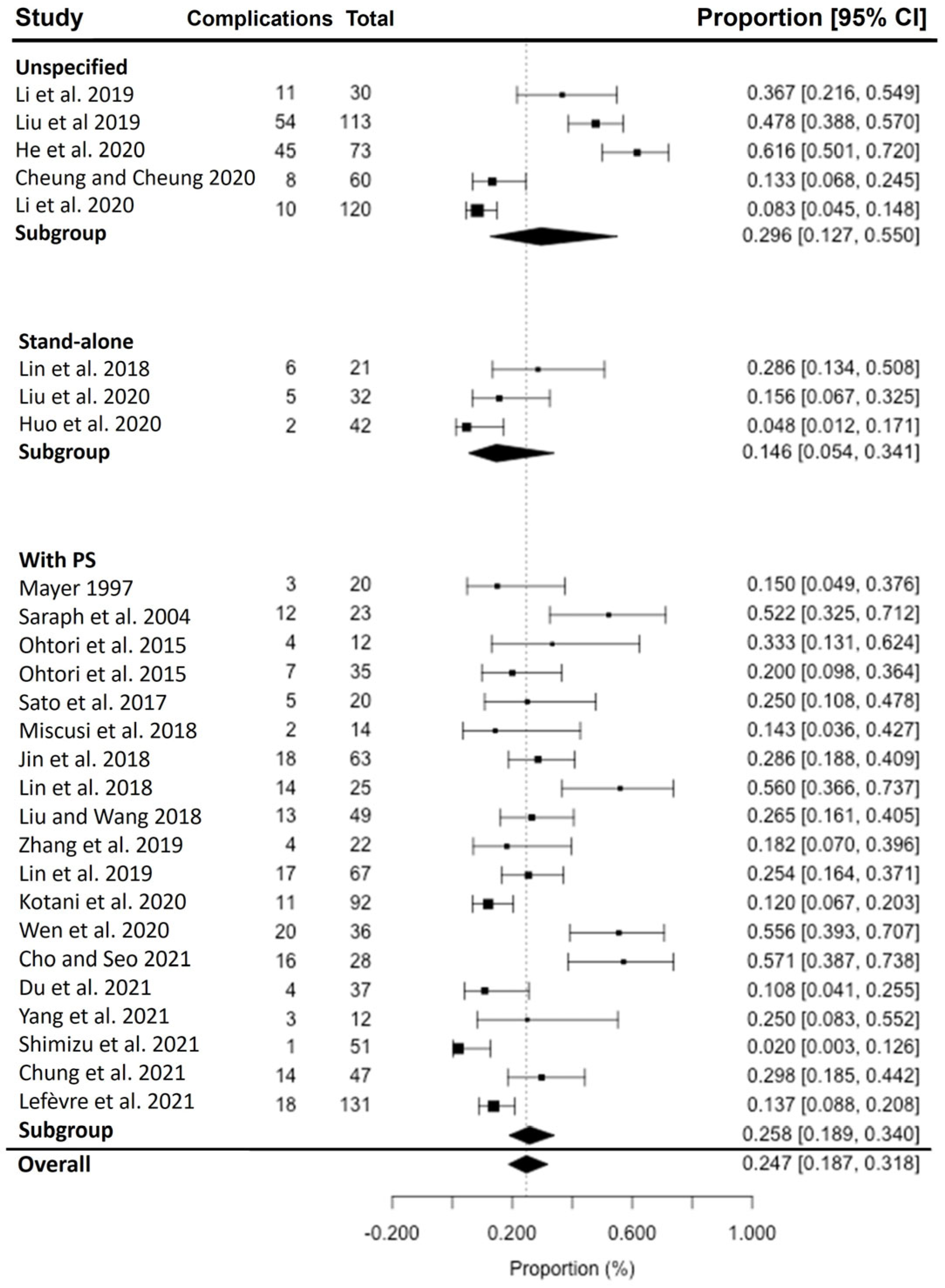
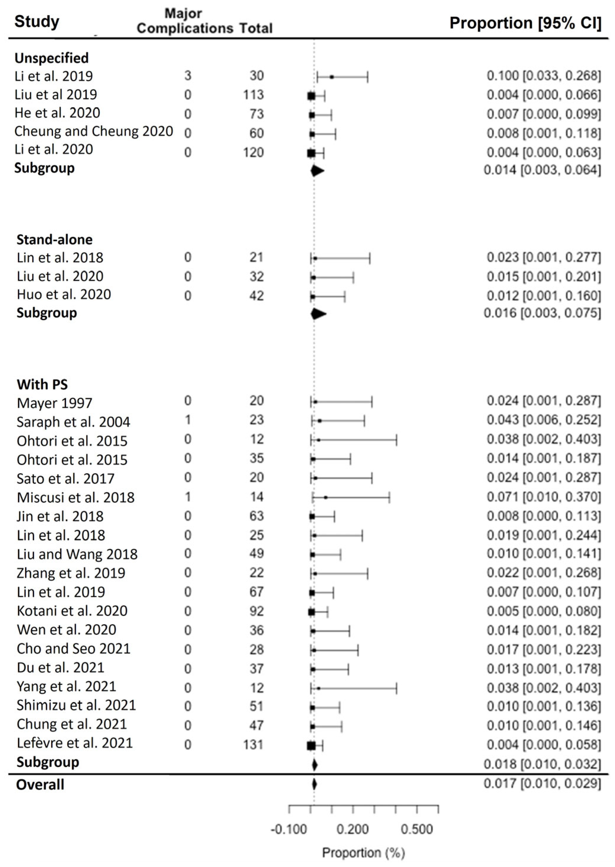
Results were analysed using Microsoft Excel and RStudio. The total numbers of each complication were extracted from 27 papers and subsequently pooled together. The data produced a pooled prevalence of complications and a forest plot based on the Random-Effects Model using the Restricted Maximum Likelihood Estimator (REML). Data were also transformed using Logit transformation to perform a single-arm proportion meta-analysis. Two subgroup analyses were also performed. The first compared stand-alone OLIF L2-5 against OLIF L2-5 with posterior stabilisation, to see whether the extra step in using posterior stabilisation significantly affected the complication rate of OLIF L2-5. Secondly, we noticed that results from earlier studies revealed a relatively high complication rate; however, it was observed that many complications were transient or minor. We thus conducted a further sub-study specifically on the major complications observed in OLIF L2-5, defined as complications that were life-threatening or debilitating long-term, which had a greater impact on patient’s quality-of-life post-surgery.

3. Results

In total, 27 studies of 1275 patients were collated from the databases, of which 21 studies were retrospective, and 6 were prospective (Table 1). Of these 27 studies, 19 studies specified OLIF L2-5 performed with posterior stabilisation [1,7,12,16,17,18,19,20,21,22,23,24,25,26,27,28,29,30,31,32,33,34,35,36,37,38,39], 3 studies specified stand-alone OLIF L2-5 [21,30,33], and 5 studies did not distinguish between these variants in their data reporting [12,26,28,29,32]. Moreover, 26 studies were performed with C-arm fluoroscopy, whereas only 1 study was performed with O-arm Computed Tomography scan (CT-Scan). One study was conducted using CT-assisted navigation [31]. The mean intra-operative blood loss was 99.4 ± 49.4 mL for single-level OLIF surgeries (n = 678, 11 studies) and 113.4 ± 69.8 ml overall (n = 1039, 22 studies). The mean operative time was noted to be 88.7 ± 30.5 min for single-level surgeries (n = 678, 11 studies) and 100.2 ± 40.3 min for all OLIF surgeries (n = 1039, 22 studies). There was a total of 327 complications, with 50 intra-operative complications, 184 immediate post-operative complications, and 93 late post-operative complications. Only three re-operations were reported among the 27 studies.
After logit transformation, the random-effects model used the Restricted Maximum Likelihood Estimator to estimate the pooled prevalence of complications based on the 27 studies. It estimated a 24.7% pooled prevalence of complications in all OLIF procedures, with or without posterior stabilisation (95% CI, 18.7 to 31.7%; 27 studies) (Figure 3). Pooled prevalence of complications was lowest in the stand-alone OLIF L2-5 subgroup at 14.6% (95% CI, 5.4 to 34.1%; 3 studies), whereas the unspecified OLIF L2-5 subgroup and the OLIF L2-5 with posterior stabilisation subgroup reported similar complication rates at 29.6% (95% CI, 12.7 to 55.0%; 5 studies) and 25.8% (95% CI, 18.9 to 34.0%; 19 studies). There was no difference between the pooled prevalence of complications among the OLIF L2-5 with posterior stabilisation subgroup compared to the stand-alone OLIF L2-5 subgroup (p = 0.747).
The heterogeneity of all three subgroups was estimated using the I2 test and REML. The unspecified OLIF L2-5 subgroup had the highest heterogeneity (I2 = 94.5%), followed by the OLIF L2-5 with posterior stabilisation subgroup (I2 = 78.1%), and lastly, the stand-alone OLIF L2-5 subgroup reported the lowest heterogeneity at I2 = 67.6%.
In 1275 patients across all 27 studies, minor vascular injuries, predominantly segmental arteries (n = 16; 1.3%) and endplate damage (n = 22; 1.7%), were the most reported intra-operative complications (Table 2). Other reported intra-operative complications with less than 1% prevalence include major vascular injury, vertebral body fracture, membrane laceration, and ureteral injury.
Transient lower limb (LL) numbness/pain (n = 80; 6.3%), transient LL weakness (n = 48; 3.8%) and nerve deficit (n = 37; 2.9%) were the most common immediate post-operative complications. Only less than 1% of patients had ileus and infection. Late post-operative complications were predominantly cage subsidence (n = 63; 5.6%; 24 studies). Other reported late post-operative complications include cage shifting/malposition/displacement (n = 2; <1%), screw malposition/breakage (n = 2; <1%), adjacent segment degeneration (n = 18, 1.4%), and pseudarthrosis (n = 8; <1%). Overall, there were three reported re-operations, all from a single study by Kotani et al. [31].
Comparing stand-alone OLIF L2-5 and OLIF L2-5 with posterior stabilisation revealed that LL numbness/pain (n = 46; 5.9%), transient LL weakness (n = 29; 3.7%), and sympathetic chain injury (n = 23; 2.9%) were the most common intra-operative complications in the OLIF L2-5 with posterior stabilisation group, compared to 3.2%, 2.1%, and 0.0% in the stand-alone OLIF L2-5 group. However, there was a high number of post-operative ileus in the stand-alone OLIF group compared to the OLIF L2-5 with posterior stabilisation group (4.2% vs. 0.8%). There were no immediate post-operative complications reported in the stand-alone OLIF L2-5 group. However, the OLIF L2-5 with posterior stabilisation group reported minor vascular injury (n = 8; 1.2%), and two cases (<1%) each of major vascular injury and ventral dural tear. Late post-operative cage subsidence rates were similar in both stand-alone OLIF L2-5 and OLIF L2-5 with posterior stabilisation (3.2% vs. 4.9%). However, only the OLIF L2-5 with posterior stabilisation group presented with pseudoarthrosis (n = 8; 1.0%) and adjacent segment disease (n = 18; 2.3%). A second sub-study of major complications in the same populations revealed that the rate of pooled major complications was 1.7% in all OLIF procedures, with or without posterior stabilisation (95% CI, 1.0 to 2.9%; 27 studies) (Figure 4).
The pooled major complication rate in stand-alone OLIF L2-5 group remained the lowest at 1.4% (95% CI, 0.3 to 6.4%; 5 studies), followed by the unspecified OLIF L2-5 group at 1.6% (95% CI, 0.3 to 7.5%; 3 studies), and then the OLIF L2-5 with posterior stabilisation group at 1.8% (95% CI, 1.0 to 3.2%%; 19 studies). There was no significant difference (p = 0.788) in major complication rates between the stand-alone OLIF L2-5 group and the OLIF L2-5 with posterior stabilisation group. The heterogeneity of all three groups was estimated using the I2 test and REML. The OLIF L2-5 with posterior stabilisation and stand-alone OLIF L2-5 subgroups had no heterogeneity (I2 = 78.05%), while the unspecified OLIF L2-5 subgroup had a relatively high heterogeneity (I2 = 56.1%).
Risk of bias assessment was performed using ROBINS-I on the 27 included studies [40]. Ten studies (37.04%) were deemed to be of moderate risk of bias, and the other seventeen studies (62.96%) were of low risk of bias (Figure 5). All studies were hence deemed to be of adequate quality to be included in the study.

4. Discussion

The pre-psoas approach for OLIF involves placing the patient in a lateral decubitus position, dissecting the abdominal wall muscles, mobilizing the peritoneum anteriorly, and then retracting the psoas muscle to reach the disc space [41,42]. This is followed by discectomy and implantation of a wide-bodied interbody cage. As discussed earlier, this stand-alone OLIF can be considered sufficient for patients without any co-morbidities and requiring short fusion [12]. However, given the stability and other advantages that a posterior fixation can offer, OLIF combined with posterior stabilisation is often performed nowadays [13,14], for which the patient is turned prone after placement of the OLIF cage and posterior instrumentation is performed with pedicle screws and rods in the usual way. This can either be completed in a single sitting or in two sittings, as per the surgeon’s discretion. Despite the various advantages that this type of fusion offers, as with any procedure, it comes with a set of reported complications that need to be considered.
In the currently available literature pertaining to OLIF, not much emphasis is given to complications and complication rates. Due to this knowledge gap, there is difficulty in advising patients pre-operatively on potential operative complications. To overcome this, our study sheds light on the complication rates following OLIF by combining existing studies to create higher levels of evidence. Moreover, our study specifically deals with OLIF L2-5, the most common levels where OLIF is performed, and analyses stand-alone OLIF and OLIF with posterior stabilisation as two separate entities to compare the complication rates.
The pooled overall prevalence of complications found in this study (24.7%) is comparable with an earlier meta-analysis on OLIF by Li et al. (26.7%) [43]. The pooled prevalence of major (1.7%) complications was also comparable to the meta-analysis by Li et al. (2.0%) [43]. Neurovascular complications are minimal due to the complete avoidance of vital neurovascular structures throughout the procedure. The reported transient neurological complications such as lower limb or groin weakness and pain can be attributed to the stretching of the nerves during manipulation of the psoas muscle; in addition, in some cases, the ilioinguinal nerve may be compromised at the iliac crest while performing OLIF L4-5 [44,45]. While injuries to major blood vessels such as the aorta, iliac vein, and ascending lumbar vein are avoided, the minor segmental arteries could still be at risk. Hence, Mayer describes the need for segmental arteries of the inferior lumbar spine to be ligated to prevent accidental rupture if encountered [1]. Mayer’s study noted an average blood loss for single-level OLIFs to be 70.3 ± 27.4 mL, 10.1 mL less than the average in this study [1]. However, it should be noted that multi-level OLIF, especially if posterior stabilisation is performed, would result in higher blood loss. Results are similar when comparing to the meta-analyses by Silvestre (2012), which included OLIF surgeries in L1-2 [44].
On the other hand, the OLIF technique has evolved, and since many surgeons have now adopted this technique, the mean operative time for single-level OLIF was found to be shorter when compared to the study by Mayer, which is 107.3 ± 14.5 min [1]. The technique can also be considered safe, as reflected by the pooled major complications rate of just 1.7% inferred in this study. Of the five major complications which were deemed to be life-threatening or had long-term debilitative effects, three were major vascular injuries. All three had left common iliac vein injury, which was ligated and repaired intraoperatively. The other two were spinal nerve root injuries. In addition, there were 16 minor complications involving the segmental arteries or the lumbar venous plexus, which are associated with lower morbidity and mortality.
Even though none of the patients required a re-operation, especially due to implant related issues, adjacent segment disease (ASD) led to re-operation in three cases, reported by Kotani et al., where initially OLIF with posterior fixation was performed [31]. Overall, we noted ASD to be reported in five studies with an incidence of 2.8%. Being a common complication following spinal fusion occurring due to increased mechanical stress on adjacent discs [46,47], it needs to be monitored in follow-up radiographs. If need be, re-operation is required to extend the fusion and instrumentation to include the newly degenerated level [48].
Our further analysis by comparing stand-alone OLIF L2-5 and OLIF L2-5 with posterior stabilisation showed that there was no significant difference in pooled prevalence of complications in general, as well as pertaining to major complications specifically. This corroborates with He et al.’s study, wherein they concluded that there was no significant difference in complications between the two OLIF subtypes [12]. Given that the addition of posterior pedicle screws does not lead to any additional major complications, considering the stability it offers, it can be performed safely if deemed necessary by the surgeon [12]. Biomechanical studies highlight the significant stability that the addition of posterior stabilisation could offer to OLIF [49]. Looking at cage subsidence, OLIF L2-5 with posterior stabilisation had almost a similar subsidence rate of 4.5% compared to 3.2% with stand-alone OLIF L2-5. This also indicates that, if cases are chosen appropriately, stand-alone OLIF offers adequate stability, probably due to the intact posterior elements and bilateral spinal muscles, which could help stabilize the spine. However, it should be noted that not all studies reported subsidence based on number of patients. Three studies reported based on number of levels rather than patients, and hence were excluded from our analysis [20,25,27]. Considering the clinical outcomes at two years post-op, He et al. showed that there is no significant difference between stand-alone OLIF L2-5 and OLIF L2-5 with posterior stabilisation [12].
Furthermore, scrutinizing the follow-up times between stand-alone-only and posterior stabilisation-only studies shows that the stand-alone-only studies may not have had a long-enough follow-up period with patients to detect cage subsidence. All three stand-alone-only OLIF papers reported results up to a maximum of 3–12 months post-operatively [21,30,33]. Compared to the other studies collated, where there were results reported longer post-operatively, the short time of reported results may have caused bias in the data collected, hence skewing the numbers regarding complications post-OLIF. This is even more pertinent for late post-operative complications such as cage subsidence, where other studies have indicated that average time-to-subsidence is 3.7 months post-OLIF [50]. As such, the risk–benefit analysis of using OLIF L2-5 with posterior stabilisation compared to stand-alone OLIF L2-5 is still up to debate, and surgeons should be wary of these limitations.
Our study has certain limitations due to the inconsistent reporting of complications in various studies. Not all complications, both major and minor, were considered by every study. Hence, some of the studies were excluded in the analysis of specific complications. A standardized method of reporting complications is required to overcome this difficulty and build evidence for a subsequent meta-analysis. Furthermore, choosing the procedure that works best for the patient requires consideration of multiple aspects, especially the fusion rates and clinical outcomes. These aspects were not evaluated in detail in this study, as the primary focus was to list out the incidence and prevalence of complications pertaining to OLIF. While our study sheds light on complications, ways to avoid such complications can be strategized to increase the safety and effectiveness of the OLIF procedure.

5. Conclusions

Our study sought to provide an accurate analysis of the complication rates of OLIF in general, as well as its variants, stand-alone OLIF L2-5 and OLIF L2-5 with posterior stabilisation. Even though the overall results indicated a complication rate of 24.7%, major complications were very minimal at 1.7%. Hence, OLIF can be considered safe, wherein the benefits largely outweigh the risks. Currently, the benefits of employing OLIF L2-5 with posterior stabilisation in contrast to performing stand-alone OLIF L2-5 can be a subject for discussion. However, as the body of evidence on performing OLIF L2-5 with posterior stabilisation grows, further studies and meta-analysis could unveil the specifics concerning the supplementary posterior stabilisation. Furthermore, studies explicitly stating all complications, be it major or minor, are necessary to establish precise evidence regarding the benefits of OLIF in comparison to other fusion techniques. This will allow surgeons to make informed decisions along with patients prior to surgery.

Supplementary Materials

The following supporting information can be downloaded at: https://www.mdpi.com/article/10.3390/std12040020/s1, Table S1: PRISMA 2020 Checklist.

Author Contributions

Conceptualization, Q.R.T., R.A.W., A.-K.K.-P. and J.Y.-L.O.; methodology, Q.R.T., R.A.W. and A.-K.K.-P.; formal analysis, Q.R.T. and R.A.W.; investigation, Q.R.T. and R.A.W.; resources, Q.R.T. and R.A.W.; data curation, Q.R.T. and R.A.W.; writing—original draft preparation, Q.R.T., R.A.W. and A.-K.K.-P.; writing—review and editing, A.-K.K.-P. and J.Y.-L.O.; supervision, J.Y.-L.O. All authors have read and agreed to the published version of the manuscript.

Funding

This research received no external funding.

Institutional Review Board Statement

Not applicable.

Informed Consent Statement

Not applicable.

Data Availability Statement

All data are included in this article.

Conflicts of Interest

The authors declare no conflict of interest.

References

  1. Mayer, H.M. A New Microsurgical Technique for Minimally Invasive Anterior Lumbar Interbody Fusion. Spine 1997, 22, 691–700. [Google Scholar] [CrossRef] [PubMed]
  2. Allain, J.; Dufour, T. Anterior lumbar fusion techniques: ALIF, OLIF, DLIF, LLIF, IXLIF. Orthop. Traumatol. Surg. Res. 2020, 106, S149–S157. [Google Scholar] [CrossRef]
  3. Orita, S.; Shiga, Y.; Inage, K.; Eguchi, Y.; Maki, S.; Furuya, T.; Aoki, Y.; Inoue, M.; Hynes, R.A.; Koda, M.; et al. Technical and Conceptual Review on the L5-S1 Oblique Lateral Interbody Fusion Surgery (OLIF51). Spine Surg. Relat. Res. 2021, 5, 1–9. [Google Scholar] [CrossRef] [PubMed]
  4. Salzmann, S.N.; Shue, J.; Hughes, A.P. Lateral Lumbar Interbody Fusion-Outcomes and Complications. Curr. Rev. Musculoskelet. Med. 2017, 10, 539–546. [Google Scholar] [CrossRef] [PubMed]
  5. Yuan, W.; Kaliya-Perumal, A.K.; Chou, S.M.; Oh, J.Y. Does Lumbar Interbody Cage Size Influence Subsidence? A Biomechanical Study. Spine 2020, 45, 88–95. [Google Scholar] [CrossRef] [PubMed]
  6. Patel, R.S.; Suh, S.W.; Kang, S.H.; Nam, K.Y.; Siddiqui, S.S.; Chang, D.G.; Yang, J.H. The Radiologic and Clinical Outcomes of Oblique Lateral Interbody Fusion for Correction of Adult Degenerative Lumbar Deformity. Indian J. Orthop. 2019, 53, 502–509. [Google Scholar] [CrossRef] [PubMed]
  7. Shimizu, T.; Fujibayashi, S.; Otsuki, B.; Murata, K.; Matsuda, S. Indirect decompression via oblique lateral interbody fusion for severe degenerative lumbar spinal stenosis: A comparative study with direct decompression transforaminal/posterior lumbar interbody fusion. Spine J. 2021, 21, 963–971. [Google Scholar] [CrossRef] [PubMed]
  8. Tohmeh, A.G.; Rodgers, W.B.; Peterson, M.D. Dynamically evoked, discrete-threshold electromyography in the extreme lateral interbody fusion approach. J. Neurosurg. Spine 2011, 14, 31–37. [Google Scholar] [CrossRef]
  9. Hijji, F.Y.; Narain, A.S.; Bohl, D.D.; Ahn, J.; Long, W.W.; DiBattista, J.V.; Kudaravalli, K.T.; Singh, K. Lateral lumbar interbody fusion: A systematic review of complication rates. Spine J. 2017, 17, 1412–1419. [Google Scholar] [CrossRef]
  10. Ng, J.P.; Kaliya-Perumal, A.K.; Tandon, A.A.; Oh, J.Y. The Oblique Corridor at L4-L5: A Radiographic-Anatomical Study into the Feasibility for Lateral Interbody Fusion. Spine 2020, 45, E552–E559. [Google Scholar] [CrossRef]
  11. Li, R.; Li, X.; Zhou, H.; Jiang, W. Development and Application of Oblique Lumbar Interbody Fusion. Orthop. Surg. 2020, 12, 355–365. [Google Scholar] [CrossRef] [PubMed]
  12. He, W.; He, D.; Sun, Y.; Xing, Y.; Wen, J.; Wang, W.; Xi, Y.; Liu, M.; Tian, W.; Ye, X. Standalone oblique lateral interbody fusion vs. combined with percutaneous pedicle screw in spondylolisthesis. BMC Musculoskelet. Disord. 2020, 21, 184. [Google Scholar] [CrossRef] [PubMed]
  13. Huang, S.; Min, S.; Wang, S.; Jin, A. Biomechanical effects of an oblique lumbar interbody fusion combined with posterior augmentation: A finite element analysis. BMC Musculoskelet. Disord. 2022, 23, 611. [Google Scholar] [CrossRef] [PubMed]
  14. Bereczki, F.; Turbucz, M.; Kiss, R.; Eltes, P.E.; Lazary, A. Stability Evaluation of Different Oblique Lumbar Interbody Fusion Constructs in Normal and Osteoporotic Condition—A Finite Element Based Study. Front. Bioeng. Biotechnol. 2021, 9, 749914. [Google Scholar] [CrossRef] [PubMed]
  15. Cai, K.; Luo, K.; Zhu, J.; Zhang, K.; Yu, S.; Ye, Y.; Jiang, G. Effect of pedicle-screw rod fixation on oblique lumbar interbody fusion in patients with osteoporosis: A retrospective cohort study. J. Orthop. Surg. Res. 2021, 16, 429. [Google Scholar] [CrossRef] [PubMed]
  16. Saraph, V.; Lerch, C.; Walochnik, N.; Bach, C.M.; Krismer, M.; Wimmer, C. Comparison of conventional versus minimally invasive extraperitoneal approach for anterior lumbar interbody fusion. Eur. Spine J. 2004, 13, 425–431. [Google Scholar] [CrossRef] [PubMed]
  17. Ohtori, S.; Mannoji, C.; Orita, S.; Yamauchi, K.; Eguchi, Y.; Ochiai, N.; Kishida, S.; Kuniyoshi, K.; Aoki, Y.; Nakamura, J.; et al. Mini-Open Anterior Retroperitoneal Lumbar Interbody Fusion: Oblique Lateral Interbody Fusion for Degenerated Lumbar Spinal Kyphoscoliosis. Asian Spine J. 2015, 9, 565–572. [Google Scholar] [CrossRef] [PubMed]
  18. Ohtori, S.; Orita, S.; Yamauchi, K.; Eguchi, Y.; Ochiai, N.; Kishida, S.; Kuniyoshi, K.; Aoki, Y.; Nakamura, J.; Ishikawa, T.; et al. Mini-Open Anterior Retroperitoneal Lumbar Interbody Fusion: Oblique Lateral Interbody Fusion for Lumbar Spinal Degeneration Disease. Yonsei Med. J. 2015, 56, 1051–1059. [Google Scholar] [CrossRef]
  19. Sato, J.; Ohtori, S.; Orita, S.; Yamauchi, K.; Eguchi, Y.; Ochiai, N.; Kuniyoshi, K.; Aoki, Y.; Nakamura, J.; Miyagi, M.; et al. Radiographic evaluation of indirect decompression of mini-open anterior retroperitoneal lumbar interbody fusion: Oblique lateral interbody fusion for degenerated lumbar spondylolisthesis. Eur. Spine J. 2017, 26, 671–678. [Google Scholar] [CrossRef]
  20. Jin, C.; Jaiswal, M.S.; Jeun, S.S.; Ryu, K.S.; Hur, J.W.; Kim, J.S. Outcomes of oblique lateral interbody fusion for degenerative lumbar disease in patients under or over 65 years of age. J. Orthop. Surg. Res. 2018, 13, 38. [Google Scholar] [CrossRef]
  21. Jin, J.; Ryu, K.S.; Hur, J.W.; Seong, J.H.; Kim, J.S.; Cho, H.J. Comparative Study of the Difference of Perioperative Complication and Radiologic Results: MIS-DLIF (Minimally Invasive Direct Lateral Lumbar Interbody Fusion) Versus MIS-OLIF (Minimally Invasive Oblique Lateral Lumbar Interbody Fusion). Clin. Spine Surg. 2018, 31, 31–36. [Google Scholar] [CrossRef]
  22. Lin, G.X.; Akbary, K.; Kotheeranurak, V.; Quillo-Olvera, J.; Jo, H.J.; Yang, X.W.; Mahatthanatrakul, A.; Kim, J.S. Clinical and Radiologic Outcomes of Direct Versus Indirect Decompression with Lumbar Interbody Fusion: A Matched-Pair Comparison Analysis. World Neurosurg. 2018, 119, e898–e909. [Google Scholar] [CrossRef] [PubMed]
  23. Liu, C.; Wang, J. Learning Curve of Minimally Invasive Surgery Oblique Lumbar Interbody Fusion for Degenerative Lumbar Diseases. World Neurosurg. 2018, 120, e88–e93. [Google Scholar] [CrossRef] [PubMed]
  24. Miscusi, M.; Ramieri, A.; Forcato, S.; Giuffre, M.; Trungu, S.; Cimatti, M.; Pesce, A.; Familiari, P.; Piazza, A.; Carnevali, C.; et al. Comparison of pure lateral and oblique lateral inter-body fusion for treatment of lumbar degenerative disk disease: A multicentric cohort study. Eur. Spine J. 2018, 27 (Suppl. S2), 222–228. [Google Scholar] [CrossRef] [PubMed]
  25. Zhang, C.; Wang, K.; Jian, F.; Wu, H. Efficacy of Oblique Lateral Interbody Fusion in Treatment of Degenerative Lumbar Disease. World Neurosurg. 2019, 124, e17–e24. [Google Scholar] [CrossRef] [PubMed]
  26. Li, J.; Wang, X.; Sun, Y.; Zhang, F.; Gao, Y.; Li, Z.; Ding, W.; Shen, Y.; Zhang, W. Safety Analysis of Two Anterior Lateral Lumbar Interbody Fusions at the Initial Stage of Learning Curve. World Neurosurg. 2019, 127, e901–e909. [Google Scholar] [CrossRef] [PubMed]
  27. Lin, G.X.; Kotheeranurak, V.; Zeng, T.H.; Mahatthanatrakul, A.; Kim, J.S. A longitudinal investigation of the endplate cystic lesion effect on oblique lumbar interbody fusion. Clin. Neurol. Neurosurg. 2019, 184, 105407. [Google Scholar] [CrossRef] [PubMed]
  28. Liu, C.; Wang, J.; Zhou, Y. Perioperative complications associated with minimally invasive surgery of oblique lumbar interbody fusions for degenerative lumbar diseases in 113 patients. Clin. Neurol. Neurosurg. 2019, 184, 105381. [Google Scholar] [CrossRef] [PubMed]
  29. Cheung, M.Y.; Cheung, P. Oblique lumbar interbody fusion in management of lumbar degenerative spinal stenosis in Chinese population. J. Orthop. Trauma Rehabil. 2020, 27, 119–127. [Google Scholar] [CrossRef]
  30. Huo, Y.; Yang, D.; Ma, L.; Wang, H.; Ding, W.; Yang, S. Oblique Lumbar Interbody Fusion with Stand-Alone Cages for the Treatment of Degenerative Lumbar Spondylolisthesis: A Retrospective Study with 1-Year Follow-Up. Pain Res. Manag. 2020, 2020, 9016219. [Google Scholar] [CrossRef]
  31. Kotani, Y.; Koike, Y.; Ikeura, A.; Tokunaga, H.; Saito, T. Clinical and radiologic comparison of anterior-posterior single-position lateral surgery versus MIS-TLIF for degenerative lumbar spondylolisthesis. J. Orthop. Sci. 2020, 26, 992–998. [Google Scholar] [CrossRef]
  32. Li, J.; Zhang, D.; Shen, Y.; Qi, X. Lumbar degenerative disease after oblique lateral interbody fusion: Sagittal spinopelvic alignment and its impact on low back pain. J. Orthop. Surg. Res. 2020, 15, 326. [Google Scholar] [CrossRef] [PubMed]
  33. Liu, J.; He, Y.; Huang, B.; Zhang, X.; Shan, Z.; Chen, J.; Fan, S.; Zhao, F. Reoccurring discogenic low back pain (LBP) after discoblock treated by oblique lumbar interbody fusion (OLIF). J. Orthop. Surg. Res. 2020, 15, 22. [Google Scholar] [CrossRef] [PubMed]
  34. Wen, J.; Shi, C.; Yu, L.; Wang, S.; Xi, Y.; Ye, X. Unilateral Versus Bilateral Percutaneous Pedicle Screw Fixation in Oblique Lumbar Interbody Fusion. World Neurosurg. 2020, 134, e920–e927. [Google Scholar] [CrossRef] [PubMed]
  35. Cho, M.S.; Seo, E.M. Efficacy and radiographic analysis of oblique lumbar interbody fusion in treating lumbar degenerative spondylolisthesis with sagittal imbalance. Neurosurg. Rev. 2021, 44, 2181–2189. [Google Scholar] [CrossRef] [PubMed]
  36. Chung, H.W.; Lee, H.D.; Jeon, C.H.; Chung, N.S. Comparison of surgical outcomes between oblique lateral interbody fusion (OLIF) and anterior lumbar interbody fusion (ALIF). Clin. Neurol. Neurosurg. 2021, 209, 106901. [Google Scholar] [CrossRef] [PubMed]
  37. Du, X.; She, Y.; Ou, Y.; Zhu, Y.; Luo, W.; Jiang, D. Oblique Lateral Interbody Fusion versus Transforaminal Lumbar Interbody Fusion in Degenerative Lumbar Spondylolisthesis: A Single-Center Retrospective Comparative Study. Biomed. Res. Int. 2021, 2021, 6693446. [Google Scholar] [CrossRef] [PubMed]
  38. Lefevre, E.; d’Astorg, H.; Fiere, V.; Gauthe, R.; Vieira, T.D.; Ould Slimane, M.; Szadkowski, M. Treatment of one-level degenerative lumbar spondylolisthesis with severe stenosis by oblique lateral interbody fusion: Clinical and radiological results after a minimal 1-year follow-up. Interdiscip. Neurosurg. 2021, 26, 101321. [Google Scholar] [CrossRef]
  39. Yang, S.L.; Liu, X.Y.; Ma, R.; Zhang, J.Q.; Liang, S.M.; Chen, Z.; Pan, Z.; Ma, Z.J.; Ding, X.L.; Kang, Y.; et al. Treatment of Degenerative Lumbar Scoliosis with Oblique Lumbar Interbody Fusion in Conjunction with Unilateral Pedicle Screw Fixation via the Wiltse Approach. Orthop. Surg. 2021, 13, 1181–1190. [Google Scholar] [CrossRef]
  40. Sterne, J.A.; Hernan, M.A.; Reeves, B.C.; Savovic, J.; Berkman, N.D.; Viswanathan, M.; Henry, D.; Altman, D.G.; Ansari, M.T.; Boutron, I.; et al. ROBINS-I: A tool for assessing risk of bias in non-randomised studies of interventions. BMJ 2016, 355, i4919. [Google Scholar] [CrossRef]
  41. Woods, K.R.; Billys, J.B.; Hynes, R.A. Technical description of oblique lateral interbody fusion at L1-L5 (OLIF25) and at L5-S1 (OLIF51) and evaluation of complication and fusion rates. Spine J. 2017, 17, 545–553. [Google Scholar] [CrossRef] [PubMed]
  42. Davis, T.T.; Hynes, R.A.; Fung, D.A.; Spann, S.W.; MacMillan, M.; Kwon, B.; Liu, J.; Acosta, F.; Drochner, T.E. Retroperitoneal oblique corridor to the L2-S1 intervertebral discs in the lateral position: An anatomic study. J. Neurosurg. Spine 2014, 21, 785–793. [Google Scholar] [CrossRef] [PubMed]
  43. Li, H.M.; Zhang, R.J.; Shen, C.L. Differences in radiographic and clinical outcomes of oblique lateral interbody fusion and lateral lumbar interbody fusion for degenerative lumbar disease: A meta-analysis. BMC Musculoskelet. Disord. 2019, 20, 582. [Google Scholar] [CrossRef] [PubMed]
  44. Silvestre, C.; Mac-Thiong, J.M.; Hilmi, R.; Roussouly, P. Complications and Morbidities of Mini-open Anterior Retroperitoneal Lumbar Interbody Fusion: Oblique Lumbar Interbody Fusion in 179 Patients. Asian Spine J. 2012, 6, 89–97. [Google Scholar] [CrossRef] [PubMed]
  45. Li, J.X.; Phan, K.; Mobbs, R. Oblique Lumbar Interbody Fusion: Technical Aspects, Operative Outcomes, and Complications. World Neurosurg. 2017, 98, 113–123. [Google Scholar] [CrossRef] [PubMed]
  46. Lee, J.C.; Choi, S.W. Adjacent Segment Pathology after Lumbar Spinal Fusion. Asian Spine J. 2015, 9, 807–817. [Google Scholar] [CrossRef] [PubMed]
  47. Donnally III, C.J.; Patel, P.D.; Canseco, J.A.; Divi, S.N.; Goz, V.; Sherman, M.B.; Shenoy, K.; Markowitz, M.; Rihn, J.A.; Vaccaro, A.R. Current incidence of adjacent segment pathology following lumbar fusion versus motion-preserving procedures: A systematic review and meta-analysis of recent projections. Spine J. 2020, 20, 1554–1565. [Google Scholar] [CrossRef]
  48. Burch, M.B.; Wiegers, N.W.; Patil, S.; Nourbakhsh, A. Incidence and risk factors of reoperation in patients with adjacent segment disease: A meta-analysis. J. Craniovertebr. Junction Spine 2020, 11, 9–16. [Google Scholar] [CrossRef]
  49. Fang, G.; Lin, Y.; Wu, J.; Cui, W.; Zhang, S.; Guo, L.; Sang, H.; Huang, W. Biomechanical Comparison of Stand-Alone and Bilateral Pedicle Screw Fixation for Oblique Lumbar Interbody Fusion Surgery-A Finite Element Analysis. World Neurosurg. 2020, 141, e204–e212. [Google Scholar] [CrossRef]
  50. Kotheeranurak, V.; Jitpakdee, K.; Lin, G.X.; Mahatthanatrakul, A.; Singhatanadgige, W.; Limthongkul, W.; Yingsakmongkol, W.; Kim, J.S. Subsidence of Interbody Cage Following Oblique Lateral Interbody Fusion: An Analysis and Potential Risk Factors. Glob. Spine J. 2021, 13, 1981–1991. [Google Scholar] [CrossRef]
Figure 1. Various lateral interbody fusion approaches. ALIF is conducted through the anterior abdominal wall, bypassing the aorta and inferior vena cava. DLIF/XLIF approach is through the psoas major muscle. OLIF uses the oblique corridor anterior to the psoas major muscle. ALIF: Anterior Lumbar Interbody Fusion; OLIF: Oblique Lumbar Interbody fusion; DLIF: Direct Lateral Interbody fusion; XLIF: Extreme Lateral Interbody Fusion; TLIF: Transforaminal Lumbar Interbody Fusion; PLIF: Posterior Lumbar Interbody Fusion.
Std 12 00020 g001
Figure 2. Flow chart of literature search.
Std 12 00020 g002
Figure 3. Forest plot of single proportion random-effects meta-analysis of overall complications, using the Restricted Maximum Likelihood Estimator [1,7,12,16,17,18,19,20,21,22,23,24,25,26,27,28,29,30,31,32,33,34,35,36,37,38,39]. CI: confidence interval; PS: posterior stabilisation.
Std 12 00020 g003
Figure 4. Forest plot of single proportion random-effects meta-analysis of major complications in the same study population [1,7,12,16,17,18,19,20,21,22,23,24,25,26,27,28,29,30,31,32,33,34,35,36,37,38,39]. CI: confidence interval; PS: posterior stabilisation.
Std 12 00020 g004
Figure 5. Quality of studies assessed by Risk of Bias in Non-Randomised Studies of Interventions (ROBINS-I) and visualised by Risk-Of-Bias VISualization (ROBVIS.33) [40].
Std 12 00020 g005
Table 1. Summarized details of the 27 studies. In total, there were 327 complications reported in the 27 studies with only 3 re-operations noted.
ParameterNumber
Total no. of studies included27
Total no. of patients1275
Type of OLIF L2-519 (with PS); 3 (Stand-alone); 5 (Unspecified)
Type of Studies21 (Retrospective); 6 (prospective)
No. of studies with CT Navigation used1
Number of complications327 (Total); 50 (Intra-op), 184 (Post-op), 93 (Late post-op); 3 (Re-operations)
OLIF: Oblique Lumbar Interbody Fusion; PS: Posterior Stabilisation.
Table 2. Complications of OLIF performed stand-alone, with posterior stabilization (PS), and unspecified. Stand-alone procedures showed the lowest total overall complications of 13.7%, followed by OLIF L2-5 with PS at 23.7%, then unspecified OLIF at 32.3%.
Group of OLIFTotal
Complication CategoryComplication
Sub-Group
Specific
Complication
Stand-Alone,
n (%)
With Posterior Stabilisation,
n (%)
Unspecified (+/− Posterior Stabilisation), n (%)
Cumulative no. of patients957843961275
Intra-operativeVascular injuryMajor02 (0.3)1 (0.3)3 (0.2)
Minor08 (1.0)8 (2.0)16 (1.3)
Endplate damageEndplate damage0022 (5.6)22 (1.7)
Vertebral body fractureVertebral body fracture03 (0.4)03 (0.2)
Membrane LacerationPeritoneal laceration001 (0.3)1 (0.1)
Ventral dural tear02 (0.3)1 (0.3)3 (0.2)
Ureteral injury02 (0.3)02 (0.2)
Total017 (2.2)33 (8.3)50 (3.9)
Immediate post-operativeNerve deficitsSpinal nerve/Nerve root injury002 (0.5)2 (0.2)
Sympathetic chain023 (2.9)12 (3.0)35 (2.7)
Ileus4 (4.2)6 (0.8)2 (0.5)12 (0.9)
Transient Lower limb weaknessPsoas weakness2 (2.1)27 (3.4)17 (4.3)46 (3.6)
Quadriceps weakness02 (0.3)02 (0.2)
Lower Limb Numbness/painNumbness2 (2.1)36 (4.6)31 (7.8)69 (5.4)
Pain1 (1.1)10 (1.3)011 (0.9)
Local Infection1 (1.1)5 (0.6)1 (0.3)7 (0.5)
Total10 (10.5)109 (13.9)65 (16.4)184 (14.4)
Late
post-operative
Cage shifting/malpositioning/displacement01 (0.1)1 (0.3)2 (0.2)
Screw malposition/breakage02 (0.3)02 (0.2)
Adjacent Segment Degeneration018 (2.3)018 (1.4)
Subsidence 3 (3.16)31 (4.91)29 (7.3)63 (5.6)
Pseudoarthrosis08 (1.02)08 (0.6)
Total3 (3.16)60 (7.65)30 (7.6)93 (7.3)
TotalReoperations03 (0.38)03 (0.2)
Overall total complications13 (13.7)186 (23.7)128 (32.3)327 (25.6)
Disclaimer/Publisher’s Note: The statements, opinions and data contained in all publications are solely those of the individual author(s) and contributor(s) and not of MDPI and/or the editor(s). MDPI and/or the editor(s) disclaim responsibility for any injury to people or property resulting from any ideas, methods, instructions or products referred to in the content.

