
Severe Typhoid Fever Complicated by Superior Mesenteric and Splenic Vein Thrombosis
 ,
, 
Abstract
:1. Introduction
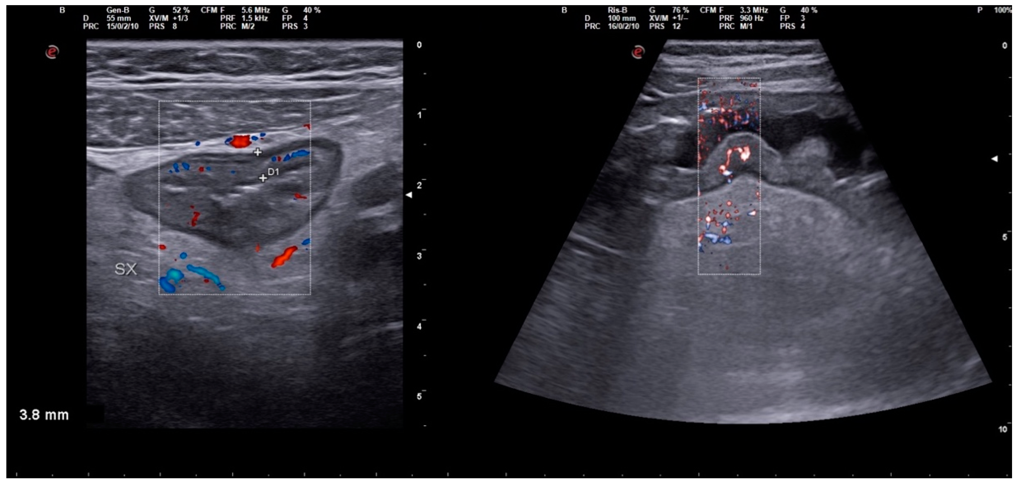
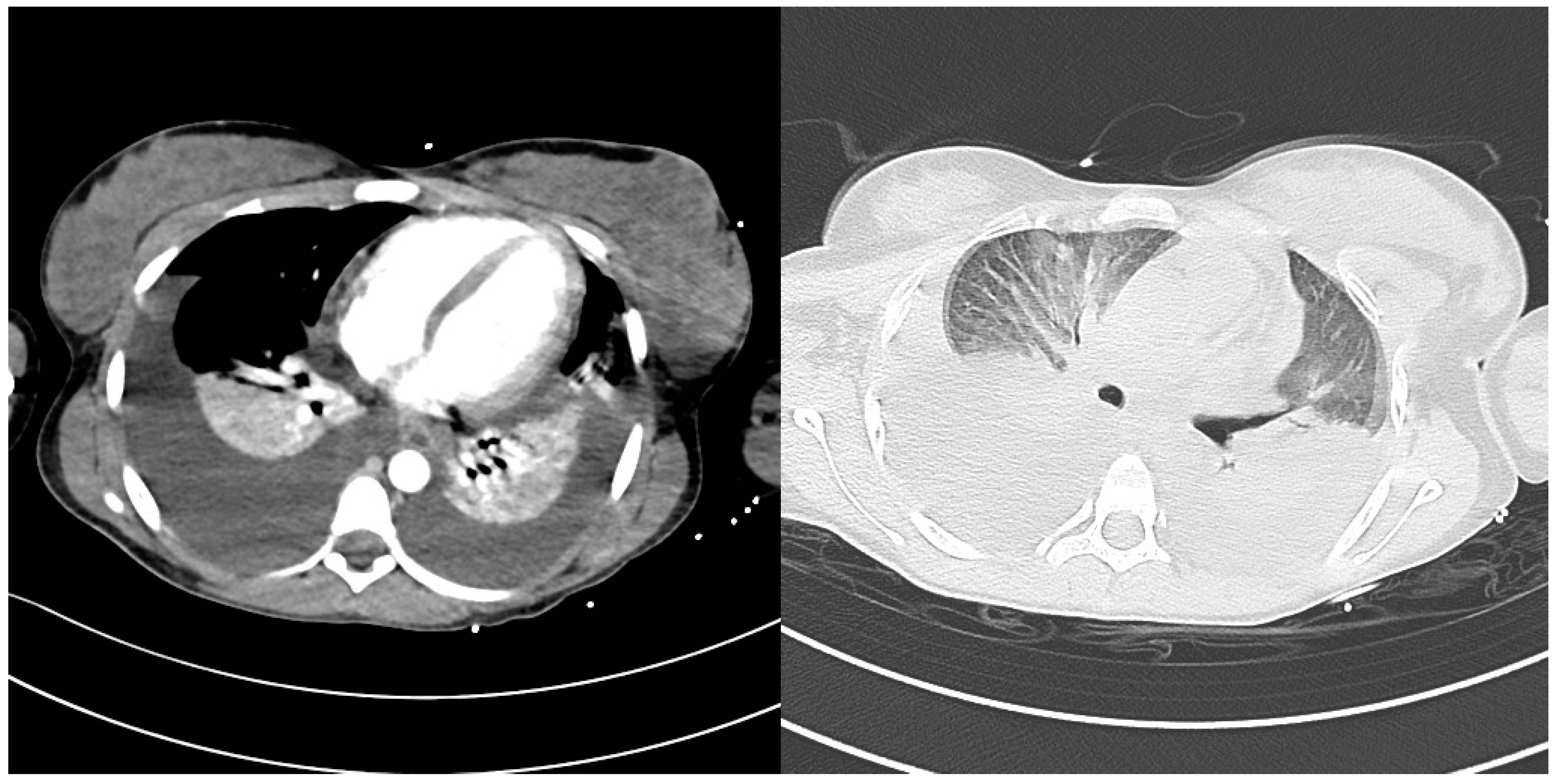
2. Case Description
3. Discussion
4. Conclusions
Author Contributions
Funding
Data Availability Statement
Conflicts of Interest
References
- Aiemjoy, K.; Seidman, J.C.; Charles, R.C.; Andrews, J.R. Seroepidemiology for Enteric Fever: Emerging Approaches and Opportunities. Open Forum Infect. Dis. 2023, 10, S21–S25. [Google Scholar] [CrossRef] [PubMed]
- Gupta, A.; Puri, S.; Aggarwal, N.P.; Randhawa, G.; Jha, P.M. Typhoid Fever Complicated by Rhabdomyolysis with Acute Hepatitis, Splenic Infarct, Pancreatitis, and Acute Kidney Injury. Indian J. Nephrol. 2023, 33, 147. [Google Scholar] [PubMed]
- Vos, T.; Abajobir, A.A.; Abate, K.H.; Abbafati, C.; Abbas, K.M.; Abd-Allah, F.; Abdulkader, R.S.; Abdulle, A.M.; Abebo, T.A.; Abera, S.F.; et al. Global, Regional, and National Incidence, Prevalence, and Years Lived with Disability for 328 Diseases and Injuries for 195 Countries, 1990–2016: A Systematic Analysis for the Global Burden of Disease Study 2016. Lancet 2017, 390, 1211–1259. [Google Scholar] [CrossRef] [PubMed] [Green Version]
- World Health Organization Typhoid. Available online: http://www.who.int/news-room/fact-sheets/detail/typhoid (accessed on 20 June 2023).
- Stanaway, J.D.; Reiner, R.C.; Blacker, B.F.; Goldberg, E.M.; Khalil, I.A.; Troeger, C.E.; Andrews, J.R.; Bhutta, Z.A.; Crump, J.A.; Im, J.; et al. The Global Burden of Typhoid and Paratyphoid Fevers: A Systematic Analysis for the Global Burden of Disease Study 2017. Lancet Infect. Dis. 2019, 19, 369–381. [Google Scholar] [CrossRef] [PubMed] [Green Version]
- Patel, T.A.; Armstrong, M.; Morris-Jones, S.D.; Wright, S.G.; Doherty, T. Imported Enteric Fever: Case Series from the Hospital for Tropical Diseases, London, United Kingdom. Am. Soc. Trop. Med. Hyg. 2010, 82, 1121–1126. [Google Scholar] [CrossRef]
- Parry, C.M.; Hien, T.T.; Dougan, G.; White, N.J.; Farrar, J.J. Typhoid Fever. N. Engl. J. Med. 2002, 347, 1770–1782. [Google Scholar] [CrossRef] [Green Version]
- Mogasale, V.; Ramani, E.; Mogasale, V.V.; Park, J. What Proportion of Salmonella Typhi Cases Are Detected by Blood Culture? A Systematic Literature Review. Ann. Clin. Microbiol. Antimicrob. 2016, 15, 32. [Google Scholar] [CrossRef] [Green Version]
- Dutta, S.; Sur, D.; Manna, B.; Sen, B.; Deb, A.K.; Deen, J.L.; Wain, J.; Von Seidlein, L.; Ochiai, L.; Clemens, J.D.; et al. Evaluation of New-Generation Serologic Tests for the Diagnosis of Typhoid Fever: Data from a Community-Based Surveillance in Calcutta, India. Diagn. Microbiol. Infect. Dis. 2006, 56, 359–365. [Google Scholar] [CrossRef]
- Wijedoru, L.; Mallett, S.; Parry, C.M. Rapid Diagnostic Tests for Typhoid and Paratyphoid (Enteric) Fever. Cochrane Database Syst. Rev. 2017, 5, 108–110. [Google Scholar] [CrossRef] [Green Version]
- Crump, J.A.; Sjölund-Karlsson, M.; Gordon, M.A.; Parry, C.M. Epidemiology, Clinical Presentation, Laboratory Diagnosis, Antimicrobial Resistance, and Antimicrobial Management of Invasive Salmonella Infections. Clin. Microbiol. Rev. 2015, 28, 901–937. [Google Scholar] [CrossRef] [Green Version]
- Naymagon, L.; Tremblay, D.; Schiano, T.; Mascarenhas, J. The Role of Anticoagulation in Pylephlebitis: A Retrospective Examination of Characteristics and Outcomes. J. Thromb. Thrombolysis 2020, 49, 325–331. [Google Scholar] [CrossRef] [PubMed]
- Jevtic, D.; Gavrancic, T.; Pantic, I.; Nordin, T.; Nordstrom, C.W.; Antic, M.; Pantic, N.; Kaljevic, M.; Joksimovic, B.; Jovanovic, M.; et al. Suppurative Thrombosis of the Portal Vein (Pylephlebits): A Systematic Review of Literature. J. Clin. Med. 2022, 11, 4992. [Google Scholar] [CrossRef]
- Balthazar, E.J.; Gollapudi, P. Septic Thrombophlebitis of the Mesenteric and Portal Veins: CT Imaging. J. Comput. Assist. Tomogr. 2000, 24, 755–760. [Google Scholar] [CrossRef]
- Soper, G.A. The Work Of A Chronic Typhoid Germ Distributor. JAMA 1907, 48, 2019–2022. [Google Scholar] [CrossRef] [Green Version]
- Pitzer, V.E.; Bowles, C.C.; Baker, S.; Kang, G.; Balaji, V.; Farrar, J.J.; Grenfell, B.T. Predicting the Impact of Vaccination on the Transmission Dynamics of Typhoid in South Asia: A Mathematical Modeling Study. PLoS Negl. Trop. Dis. 2014, 8, e2642. [Google Scholar] [CrossRef]
- European Centre for Disease Prevention and Control. Typhoid and Paratyphoid Fevers; European Centre for Disease Prevention and Control: Solna, Sweden, 2020. [Google Scholar]
- Mogasale, V.; Maskery, B.; Ochiai, R.L.; Lee, J.S.; Mogasale, V.V.; Ramani, E.; Kim, Y.E.; Park, J.K.; Wierzba, T.F. Burden of Typhoid Fever in Low-Income and Middle-Income Countries: A Systematic, Literature-Based Update with Risk-Factor Adjustment. Lancet Glob. Health 2014, 2, e570–e580. [Google Scholar] [CrossRef] [Green Version]
- LUBY, S.P.; FAIZAN, M.K.; FISHER-HOCH, S.P.; SYED, A.; MINTZ, E.D.; BHUTTA, Z.A.; MCCORMICK, J.B. Risk Factors for Typhoid Fever in an Endemic Setting, Karachi, Pakistan. Epidemiol. Infect. 1998, 120, 129–138. [Google Scholar] [CrossRef] [PubMed]
- Center for Disease Control and Prevention Information for Healthcare Professionals. Available online: https://www.cdc.gov/typhoid-fever/health-professional.html (accessed on 20 June 2023).
- Bhan, M.K.; Bahl, R.; Bhatnagar, S. Typhoid and Paratyphoid Fever. Lancet 2005, 366, 749–762. [Google Scholar] [CrossRef]
- Parry, C.M.; Thompson, C.; Vinh, H.; Chinh, N.T.; Phuong, L.T.; Ho, V.A.; Hien, T.T.; Wain, J.; Farrar, J.J.; Baker, S. Risk Factors for the Development of Severe Typhoid Fever in Vietnam. BMC Infect. Dis. 2014, 14, 73. [Google Scholar] [CrossRef] [Green Version]
- Waddington, C.S.; Darton, T.C.; Jones, C.; Haworth, K.; Peters, A.; John, T.; Thompson, B.A.V.; Kerridge, S.A.; Kingsley, R.A.; Zhou, L.; et al. An Outpatient, Ambulant-Design, Controlled Human Infection Model Using Escalating Doses of Salmonella Typhi Challenge Delivered in Sodium Bicarbonate Solution. Clin. Infect. Dis. 2014, 58, 1230–1240. [Google Scholar] [CrossRef] [Green Version]
- Butler, T.; Bell, W.R.; Levin, J.; Linh, N.N.; Arnold, K. Typhoid Fever: Studies of Blood Coagulation, Bacteremia, and Endotoxemia. Arch. Intern. Med. 1978, 138, 407–410. [Google Scholar] [CrossRef] [PubMed]
- Pineda, M.C.; LoPinto-Khoury, C. Cerebral Venous Sinus Thrombosis Secondary to Typhoid Fever: A Case Report and Brief Summary of the Literature. Neurologist 2012, 18, 202–203. [Google Scholar] [CrossRef] [PubMed]
- Belhassen-García, M.; Gomez-Munuera, M.; Pardo-Lledias, J.; Velasco-Tirado, V.; Perez-Persona, E.; Galindo-Perez, I.; Alvela-Suárez, L.; Romero-Alegría, Á.; Muñoz-Bellvis, L.; Cordero-Sánchez, M. Pylephlebitis: Incidence and Prognosis in a Tertiary Hospital. Enferm. Infecc. Microbiol. Clin. 2014, 32, 350–354. [Google Scholar] [CrossRef] [PubMed]
- Zheng, L.; Giri, B. Gastrointestinal Variant of Lemierre Syndrome: Fusobacterium Nucleatum: Bacteremia–Associated Hepatic Vein Thrombosis: A Case Report and Literature Review. Am. J. Ther. 2016, 23, e933–e936. [Google Scholar] [CrossRef]
- Rahmati, E.; She, R.C.; Kazmierski, B.; Geiseler, P.J.; Wong, D. A Case of Liver Abscess and Fusobacterium Septicemia. IDCases 2017, 9, 98–100. [Google Scholar] [CrossRef] [PubMed]
- Choudhry, A.J.; Baghdadi, Y.M.K.; Amr, M.A.; Alzghari, M.J.; Jenkins, D.H.; Zielinski, M.D. Pylephlebitis: A Review of 95 Cases. J. Gastrointest. Surg. 2016, 20, 656–661. [Google Scholar] [CrossRef] [PubMed] [Green Version]
- Stuart, B.M.; Pullen, R.L. TYPHOID: Clinical Analysis of Three Hundred and Sixty Cases. Arch. Intern. Med. 1946, 78, 629–661. [Google Scholar] [CrossRef]
- Song, J.H.; Cho, H.; Park, M.Y.; Na, D.S.; Moon, H.B.; Pai, C.H. Detection of Salmonella Typhi in the Blood of Patients with Typhoid Fever by Polymerase Chain Reaction. J. Clin. Microbiol. 1993, 31, 1439–1443. [Google Scholar] [CrossRef] [Green Version]
- Olopoenia, L.A.; King, A.L. Widal Agglutination Test − 100 Years Later: Still Plagued by Controversy. Postgrad. Med. J. 2000, 76, 80–84. [Google Scholar] [CrossRef] [Green Version]
- Levine, M.M.; Grados, O.; Gilman, R.H.; Woodward, W.E.; Solis-Plaza, R.; Waldman, W. Diagnostic Value of the Widal Test in Areas Endemic for Typhoid Fever. Am. J. Trop. Med. Hyg. 1978, 27, 795–800. [Google Scholar] [CrossRef]
- Parry, C.M.; Ribeiro, I.; Walia, K.; Rupali, P.; Baker, S.; Basnyat, B. Multidrug Resistant Enteric Fever in South Asia: Unmet Medical Needs and Opportunities. BMJ 2019, 364, k5322. [Google Scholar] [CrossRef] [PubMed] [Green Version]
- Frenck, R.W., Jr.; Nakhla, I.; Sultan, Y.; Bassily, S.B.; Girgis, Y.F.; David, J.; Butler, T.C.; Girgis, N.I.; Morsy, M. Azithromycin versus Ceftriaxone for the Treatment of Uncomplicated Typhoid Fever in Children. Clin. Infect. Dis. 2000, 31, 1134–1138. [Google Scholar] [CrossRef] [PubMed]


Disclaimer/Publisher’s Note: The statements, opinions and data contained in all publications are solely those of the individual author(s) and contributor(s) and not of MDPI and/or the editor(s). MDPI and/or the editor(s) disclaim responsibility for any injury to people or property resulting from any ideas, methods, instructions or products referred to in the content. |
© 2023 by the authors. Licensee MDPI, Basel, Switzerland. This article is an open access article distributed under the terms and conditions of the Creative Commons Attribution (CC BY) license (https://creativecommons.org/licenses/by/4.0/).
Share and Cite
Veronese, P.; Pappalardo, M.; Maffini, V.; Rubini, M.; Giacometti, A.; Ruozi, M.B.; Cella, S.; Dodi, I. Severe Typhoid Fever Complicated by Superior Mesenteric and Splenic Vein Thrombosis. Infect. Dis. Rep. 2023, 15, 377-385. https://doi.org/10.3390/idr15040038
Veronese P, Pappalardo M, Maffini V, Rubini M, Giacometti A, Ruozi MB, Cella S, Dodi I. Severe Typhoid Fever Complicated by Superior Mesenteric and Splenic Vein Thrombosis. Infectious Disease Reports. 2023; 15(4):377-385. https://doi.org/10.3390/idr15040038
Chicago/Turabian StyleVeronese, Piero, Marco Pappalardo, Valentina Maffini, Monica Rubini, Alessandra Giacometti, Maria Beatrice Ruozi, Simone Cella, and Icilio Dodi. 2023. "Severe Typhoid Fever Complicated by Superior Mesenteric and Splenic Vein Thrombosis" Infectious Disease Reports 15, no. 4: 377-385. https://doi.org/10.3390/idr15040038