Review

Differential Diagnoses and Management Approaches for Gastric Polyposis

by
Masaya Iwamuro
*,
Seiji Kawano
and
Motoyuki Otsuka
Department of Gastroenterology and Hepatology, Okayama University Graduate School of Medicine, Dentistry, and Pharmaceutical Sciences, Okayama 700-0082, Japan
*
Author to whom correspondence should be addressed.
Gastroenterol. Insights 2024, 15(1), 122-144; https://doi.org/10.3390/gastroent15010009
Submission received: 25 December 2023 / Revised: 22 January 2024 / Accepted: 26 January 2024 / Published: 27 January 2024
(This article belongs to the Special Issue Recent Advances in the Management of Gastrointestinal Disorders)

Abstract

:
Multiple gastric polyps are observed in various polyposis syndromes and conditions associated with polypoid lesion development in the stomach. Polyposis syndromes often occur concurrently with specific malignant tumors and can manifest at any point in an individual’s lifespan, thus explaining the diversity in surveillance methods. Furthermore, genetic counseling and surveillance are essential not only for the patients themselves but also for their blood relatives. Therefore, the accurate diagnosis and appropriate surveillance of multiple gastric polyps are crucial for improving patient outcomes. This review aims to provide essential information on such lesions along with representative endoscopic images of familial adenomatous polyposis, Peutz-Jeghers syndrome, Cowden syndrome, Cronkhite-Canada syndrome, juvenile polyposis syndrome, gastric adenocarcinoma and proximal polyposis of the stomach, neuroendocrine tumors in autoimmune gastritis, proton pump inhibitor-related gastric mucosal changes, and multiple submucosal heterotopic glands. We wish for this review to serve as a valuable resource for endoscopists seeking to deepen their comprehension of gastric polyposis.

1. Introduction

Gastrointestinal polyposis is characterized by the presence of numerous polyps within the gastrointestinal tract. In cases where multiple polyps are observed in the stomach, they can either be attributed to gastrointestinal polyposis syndrome or represent a distinct condition. Gastrointestinal polyposis syndromes associated with the formation of gastric polyps encompass familial adenomatous polyposis, Peutz-Jeghers syndrome, Cowden syndrome, Cronkhite-Canada syndrome, and juvenile polyposis syndrome. In contrast, other diseases characterized by the presence of multiple gastric polyps or polypoid protrusions may include neuroendocrine tumors associated with autoimmune gastritis and multiple submucosal heterotopic glands. In addition, proton pump inhibitor-related gastric mucosal changes often present as diverse lesions as well, which include multiple white and flat elevated lesions, cobblestone-like mucosa, the emergence and enlargement of the fundic gland, and hyperplastic polyps. Gastrointestinal polyposis syndromes frequently exhibit a hereditary predisposition, rendering them a significant concern for both patients and their families [1]. Furthermore, given that the organs prone to developing malignant tumors differ for each specific condition, correctly diagnosing the underlying cause of gastric polyposis and implementing appropriate treatment and surveillance measures are of paramount importance [2,3,4,5]. This review aims to describe the distinguishing attributes of gastric polyps and polypoid lesions encountered in various diseases. Additionally, we offer a comprehensive overview and concise summary of the surveillance strategies for each disease state that leads to gastric polyposis, thus facilitating clinicians’ comprehension. We have also included representative endoscopic images for illustrative purposes.

2. Gastrointestinal Polyposis Syndromes with Gastric Polyps

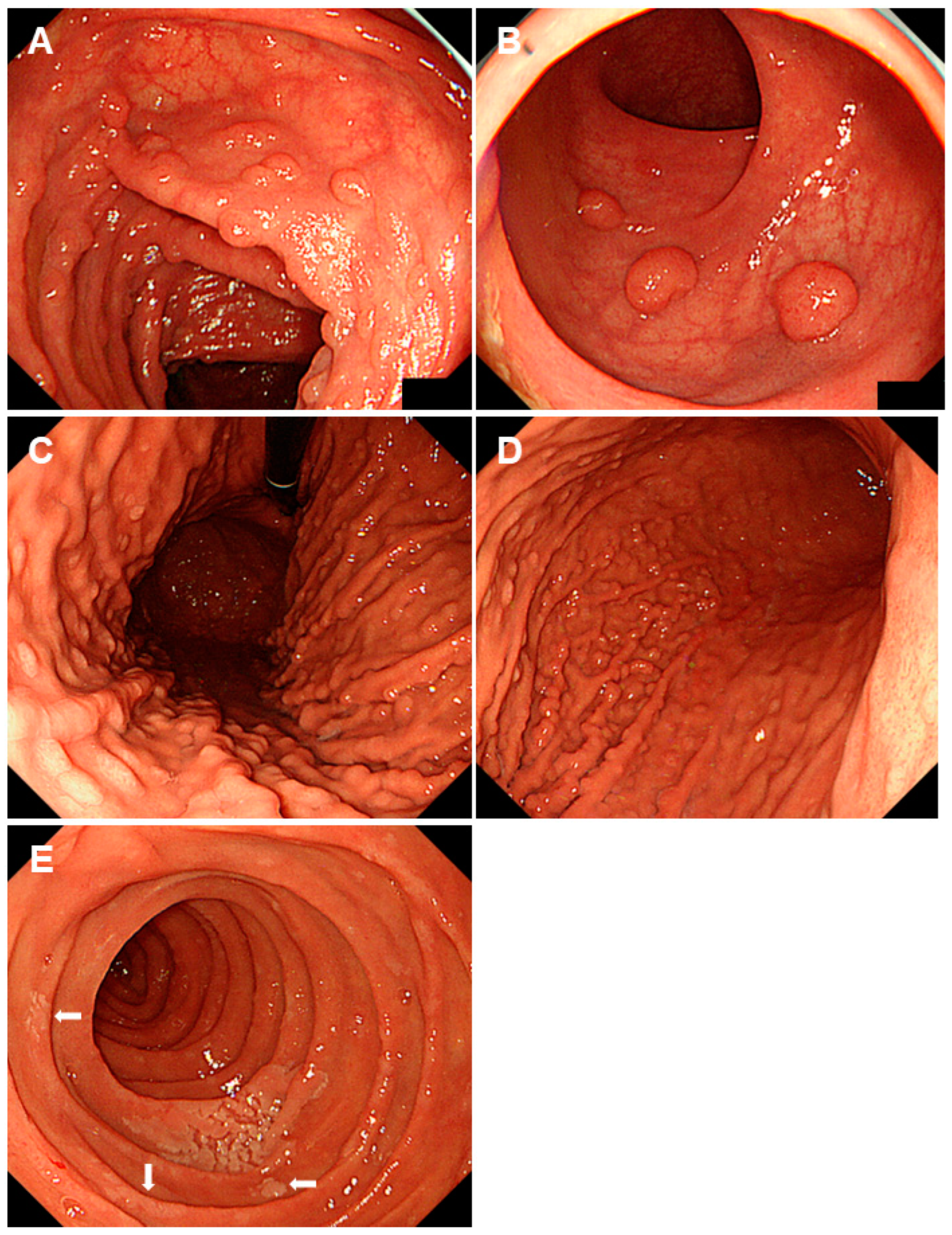
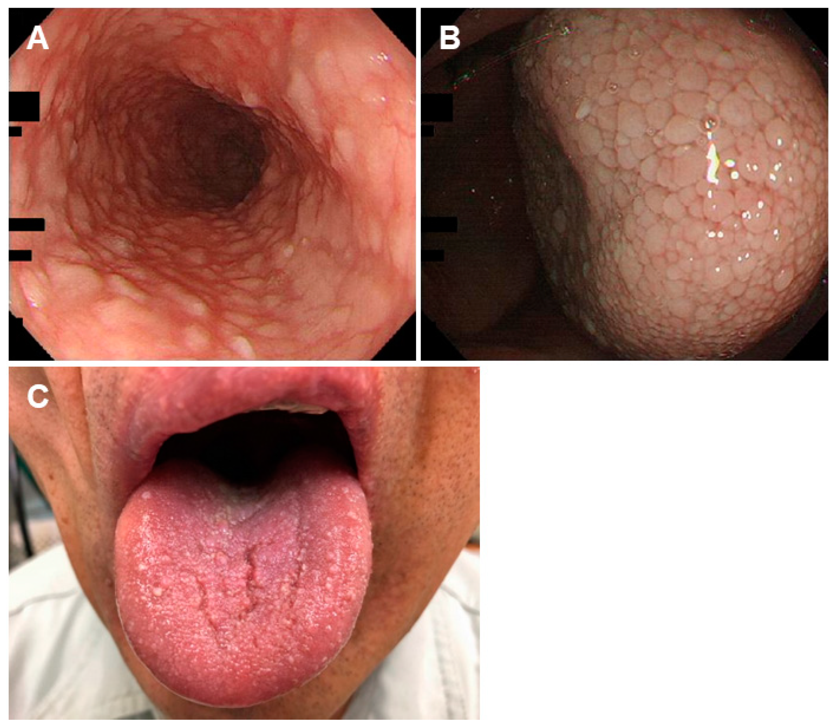
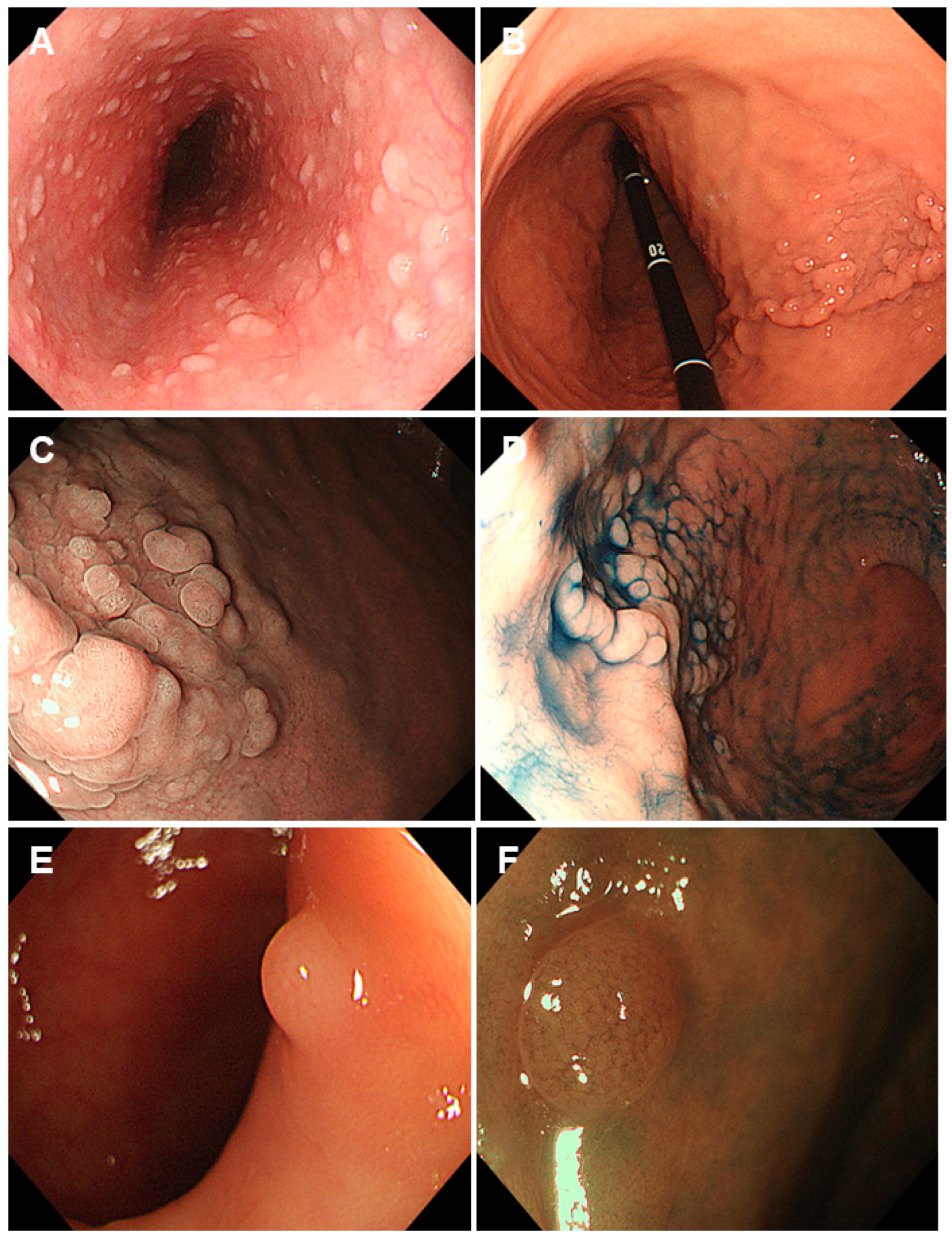
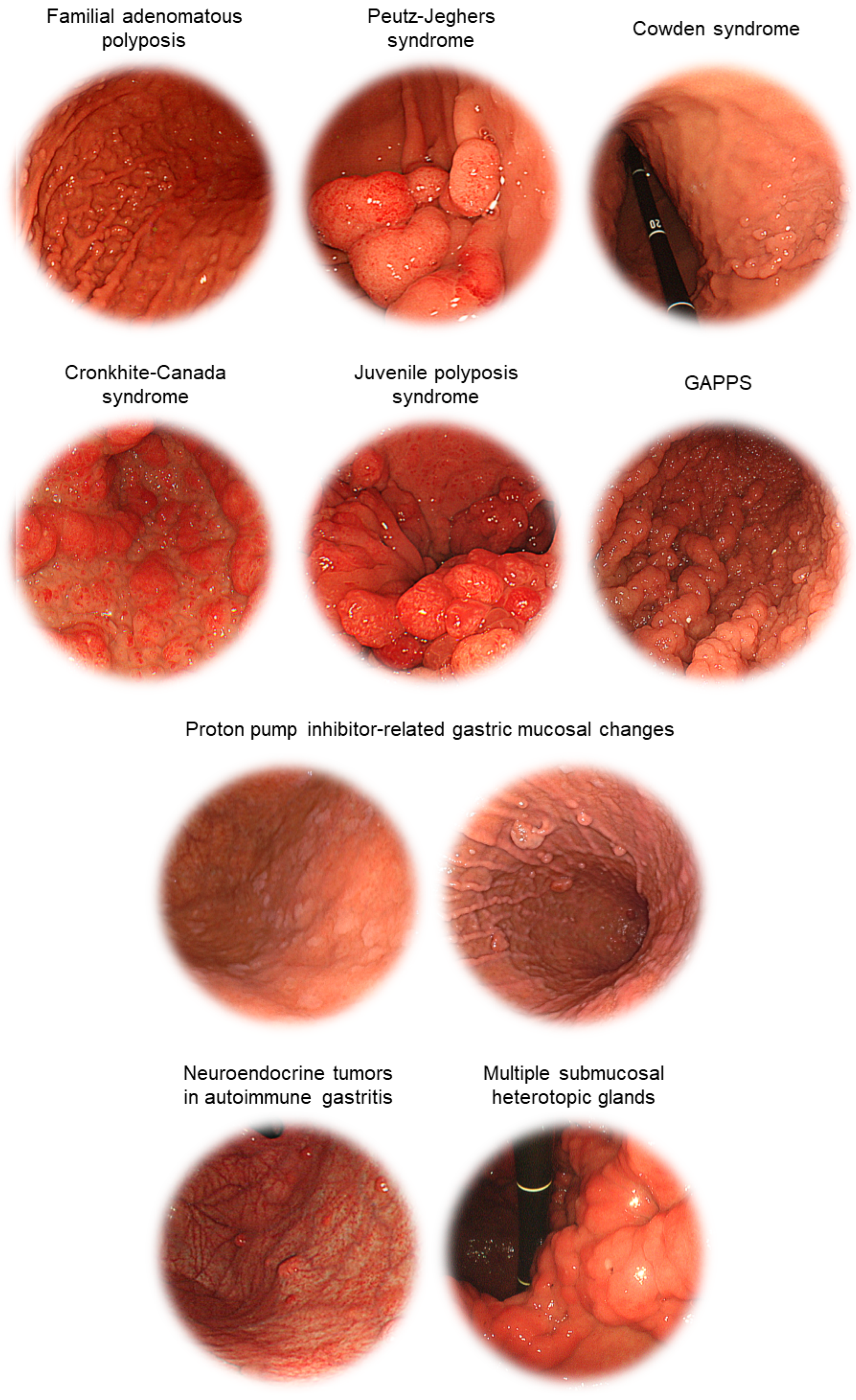
2.1. Familial Adenomatous Polyposis

Familial adenomatous polyposis is an autosomal dominant condition characterized by the presence of numerous adenomatous polyps, particularly in the colon and rectum (Figure 1A,B) [6]. If left untreated, these polyps can become malignant over time. Familial adenomatous polyposis typically arises from mutations in the APC (adenomatous polyposis coli) gene, which encodes a tumor suppressor that functions as an antagonist of the Wnt signaling pathway [6]. Individuals with familial adenomatous polyposis have a significantly increased risk of developing colorectal cancer at a young age, often in their 30s or 40s if preventive measures are not implemented [6,7,8].
Up to 88% of patients with familial adenomatous polyposis have multiple fundic gland polyps (Figure 1C,D) [7,8]. Moreover, duodenal adenomas manifest in a range spanning from 30% to 92% of afflicted individuals (Figure 1E) [9]. Consequently, when fundic gland polyposis and duodenal adenomas are detected during an esophagogastroduodenoscopy, it is imperative to include familial adenomatous polyposis as a critical element in the differential diagnosis. Unlike sporadic gastric fundic gland polyps, a subset of these polyps develop dysplasia. Although at a low frequency, previous reports have suggested a potential link to the induction of gastric carcinogenesis in fundic gland polyps [10,11,12]. Gastric adenomas are prevalent in the antrum of patients with familial adenomatous polyposis [13]. While the incidence of gastric cancer among these patients ranged from 0.6% to 1.3% in Western cohorts with no significant difference from that in the general population, the rate of gastric cancer co-occurrence in Japan was notably higher, ranging from 3.1% to 4.2% [6]. In a previous study, we examined 29 patients with familial adenomatous polyposis who underwent an esophagogastroduodenoscopy [14]. Our analysis found that eleven patients (38%) had confirmed gastric neoplasms, including twenty-three gastric adenomas and nine gastric cancers. Gastric cancer appeared as depressed, reddish lesions in the atrophic mucosa (Figure 2A,B). Gastric-type adenomas occurred in the non-atrophic mucosa, often near fundic gland polyps in the upper and middle stomach regions as flat, elevated, or protruding milky-white lesions (Figure 2C–E). In contrast, intestinal-type adenomas were mainly found in the lower stomach with elevated lesions featuring a central depression (Figure 2F,G). Notably, half of the gastric-type adenomas grew in size, whereas the intestinal-type adenomas did not. Other studies involving the analysis of a larger number of cases have reported findings indicating associations between mucin phenotypes (gastric, intestinal, or mixed) of gastric polyps in FAP and their color, morphology, and localization [13]. We believe that recognizing the distinctive endoscopic features corresponding to mucin phenotypes based on immunostaining results will accelerate the diagnosis and personalized management of gastric neoplasms in these individuals.
Genetic testing and counseling are commonly offered to families with a history of familial adenomatous polyposis to identify carriers of the gene and provide guidance on surveillance and preventive measures [6,7,8]. Individuals with familial adenomatous polyposis generally require regular colonoscopies to detect and remove polyps before they progress to cancer. Typically, colonoscopy screening for patients commences between the ages of 10 and 18 years and is performed at intervals of 1 to 2 years [6,9,15,16]. In some cases, preventive surgery, such as colectomy, may be recommended to reduce the risk of cancer. Given that adenomas frequently occur in the duodenum, including the periampullary region, an esophagogastroduodenoscopy should commence following a colectomy or around the age of 20–25 years, with the frequency of examinations determined based on the density, size, and presence of dysplasia in the duodenal adenomas [6,9,15,16]. Palpation and thyroid ultrasonography are crucial in female patients due to the relatively higher incidence of thyroid cancer. Patients with a family history of desmoid tumors should undergo abdominal and pelvic computed tomography (CT) or magnetic resonance imaging every three years after a colectomy to confirm the presence or absence of desmoids.

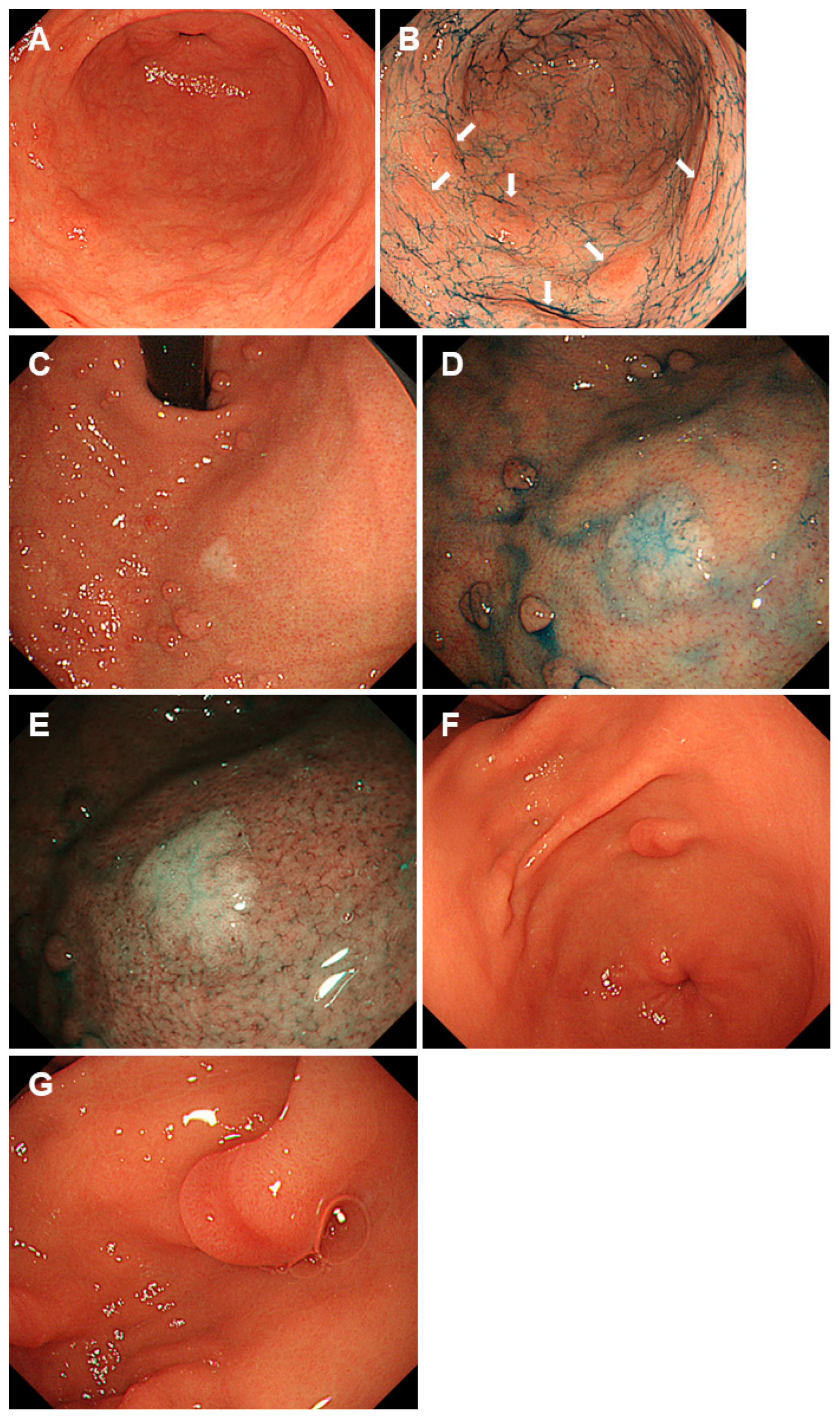
2.2. Peutz-Jeghers Syndrome

Peutz-Jeghers syndrome is an autosomal dominant genetic disorder characterized by hamartomatous polyposis throughout the gastrointestinal tract, excluding the esophagus [17]. Another distinctive feature is the presence of pigmented spots, primarily on the lips, oral mucosa, and fingertips. The hamartomatous polyps seen in this syndrome are characterized by mucosal epithelial hyperplasia and dendritic proliferation of the smooth muscle fibers from the muscularis mucosae and are referred to as Peutz-Jeghers polyps [17,18,19]. Peutz-Jeghers polyps most commonly occur in the small intestine (Figure 3A–C), but they can also appear in the stomach, colorectum (Figure 3D,E), and other parts of the digestive tract. This disease is caused by mutations in the STK11 (serine/threonine kinase 11) gene, which encodes a serine/threonine kinase that regulates cell polarity and energy metabolism and functions as a tumor suppressor [18,19,20].
Polypoid lesions are detected in the stomach of 24–91% of patients with Peutz-Jeghers syndrome [21,22]. In our retrospective study involving 11 patients, we identified two distinct types of gastric polyps [22]: (i) solitary or sporadic polyps > 5 mm, reddish, with a sessile or semi-pedunculated morphology. These included hyperplastic polyps (Figure 4A) and Peutz-Jeghers polyps (Figure 4B), which were definitively diagnosed via an endoscopic resection. (ii) Multiple sessile polyps ≤ 5 mm, mirroring the color of the peripheral mucosa, consisting of fundic gland polyps (Figure 4C,D) and hyperplastic polyps. Distinguishing fundic gland polyposis in Peutz-Jeghers syndrome from familial adenomatous polyposis is crucial to avoid a misdiagnosis. Consequently, we emphasize the necessity for the endoscopic resection of gastric polyps >5 mm in size to enable the precise histopathological identification of Peutz-Jeghers polyps.
While Peutz-Jeghers polyps are inherently benign, they can lead to complications, such as intestinal obstruction and hemorrhage. In light of the potential for childhood intussusception necessitating surgical intervention, patients are advised to undergo baseline gastrointestinal tract surveillance between the ages of 8 and 10 years or sooner if they manifest symptoms [9,23,24]. In the absence of identified polyps during the initial examination, surveillance is reinstated at 18 years of age. In conjunction with esophagogastroduodenoscopy and colonoscopy, video capsule endoscopy or magnetic resonance enterography are used for small bowel screening. Surveillance is performed every 2–3 years throughout the patient’s lifetime [9,23,24]. Polypectomy is performed on Peutz-Jeghers polyps that manifest symptomatic features or are >10 mm in size, serving as a preventive measure against the occurrence of intussusception. Owing to the STK11 mutations, patients with Peutz-Jeghers syndrome are at an increased risk of developing certain types of cancer, particularly those of the gastrointestinal tract, pancreas, breast, ovaries, testes, and lungs [17,19,25,26,27,28]. The commencement of annual pancreatic cancer surveillance is recommended at the age of 30–35 years by employing magnetic resonance cholangiopancreatography or endoscopic ultrasound [23,28]. Moreover, the initiation of surveillance for the mammary glands, ovaries, and uterus is recommended between the ages of 18 and 25 years. Furthermore, adopting a multidisciplinary approach for cancer surveillance involving diverse organs is advisable [5,23,24].

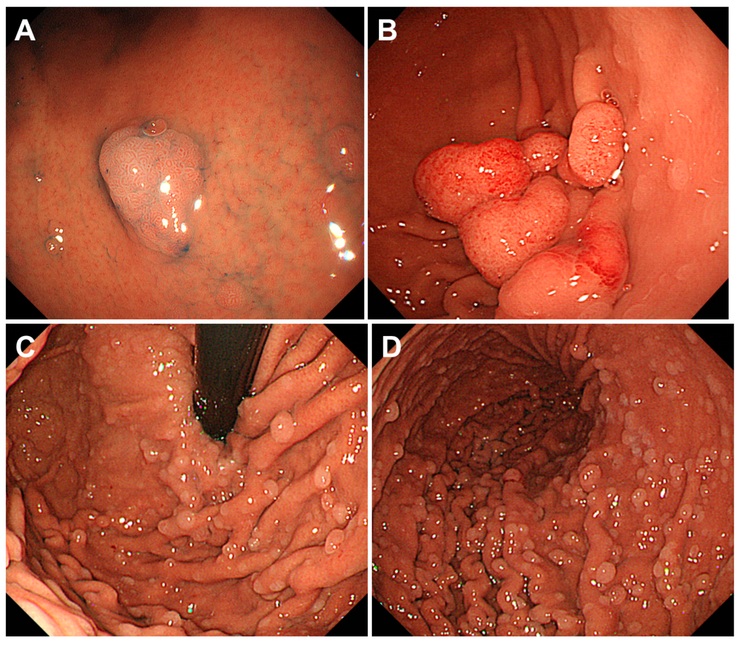
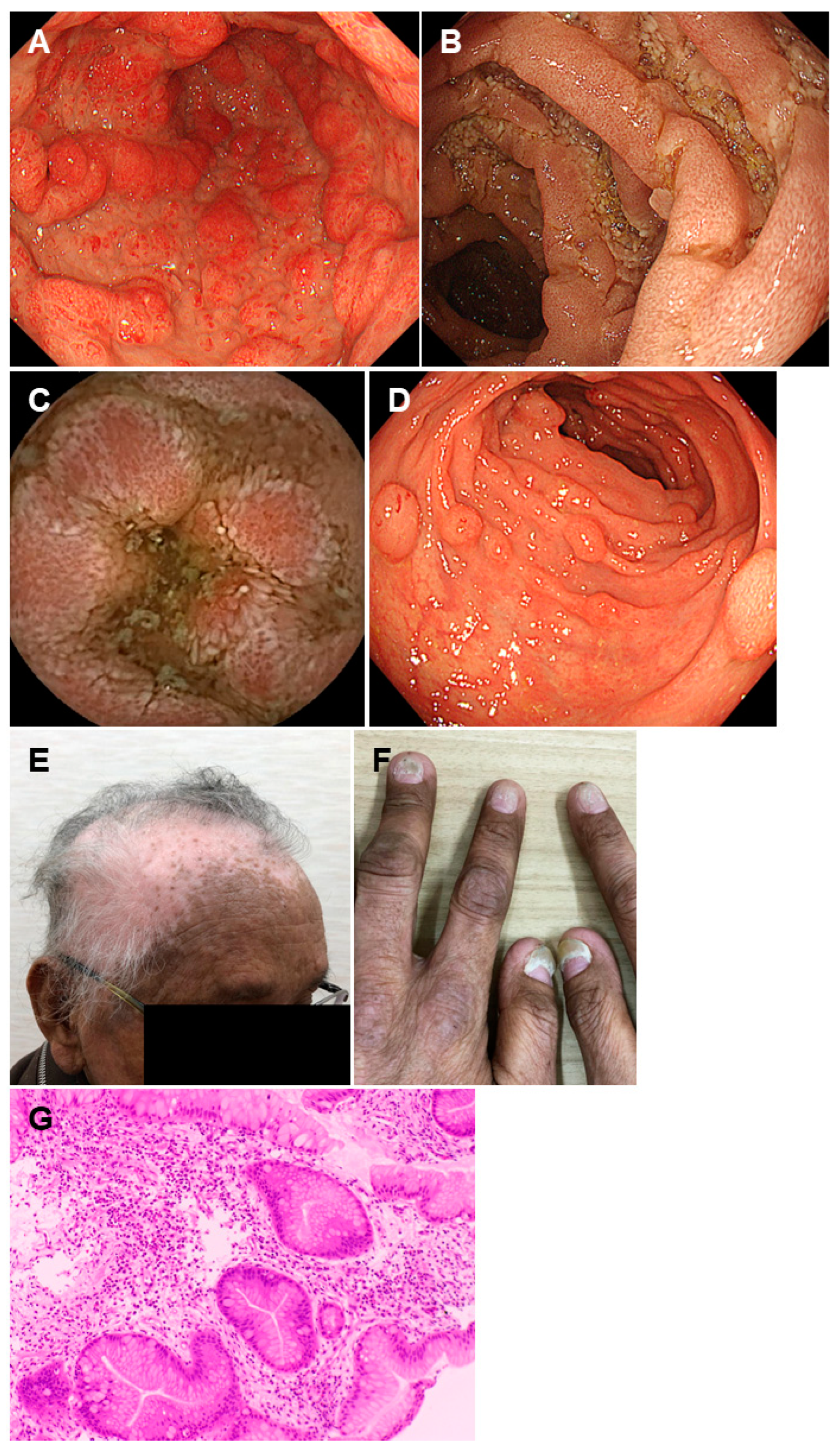
2.3. Cowden Syndrome

Cowden syndrome is a genetic condition that leads to the formation of various types of hamartomas in the skin, mucous membranes, and gastrointestinal tract. Currently, Cowden syndrome is classified as phosphatase and tensin homolog (PTEN) hamartoma tumor syndrome [5,26]. This condition results from the mutations in the PTEN gene, which is recognized as a tumor suppressor commonly mutated in various cancer types. Due to the escalation of cellular proliferation and angiogenesis, concomitant manifestations of Cowden syndrome encompass systemic vascular anomalies and malignant neoplasms [5,26,27,28]. In the revised PTEN hamartoma tumor syndrome clinical diagnostic criteria, the major criteria include breast cancer, endometrial cancer, thyroid cancer (follicular), gastrointestinal hamartomas (including ganglioneuromas), Lhermitte-Duclos disease, macrocephaly, macular pigmentation of the glans penis, and multiple mucocutaneous lesions, such as multiple trichilemmomas, acral keratoses, acral hyperkeratotic papules, mucocutaneous neuromas, and oral papillomas [29]. Minor criteria included autism spectrum disorder, colon cancer, esophageal glycogenic acanthosis, lipomas, mental retardation, renal cell carcinoma, testicular lipomatosis, thyroid cancer (other than follicular), thyroid structural lesions, such as adenoma and multinodular goiter, and vascular anomalies, including multiple intracranial developmental venous anomalies [29].
Polyposis is observed throughout the gastrointestinal tract, with esophageal diffuse glycogen acanthosis showing white flat polyposis, a characteristic manifestation of Cowden syndrome (Figure 5A and Figure 6A) [30,31]. Esophagogastroduodenoscopy may reveal white nodular lesions in the tongue, gingiva, and pharyngolaryngeal regions, leading to the identification of oral papillomas and hyperkeratotic lesions (Figure 5B,C). Gastrointestinal polyps show a color similar to that of the peripheral mucosa but may also be reddish (Figure 6B–F). These polyps are hyperplastic or hamartomatous in histology, while inflammatory, lipomatous, fibromatous, and adenomatous polyps are identified as well [32,33].
Patients with PTEN hamartoma tumor syndrome exhibit an elevated predisposition to malignancies across a spectrum of anatomical sites, manifesting cumulative lifetime risks of 85.2% for breast cancer, 35.2% for thyroid cancer, 28.2% for endometrial cancer, 9.0% for colorectal cancer, 33.6% for renal cancer, and 6.0% for melanoma [5,26]. Therefore, a multidisciplinary approach to cancer surveillance in these organs is essential [5,34].

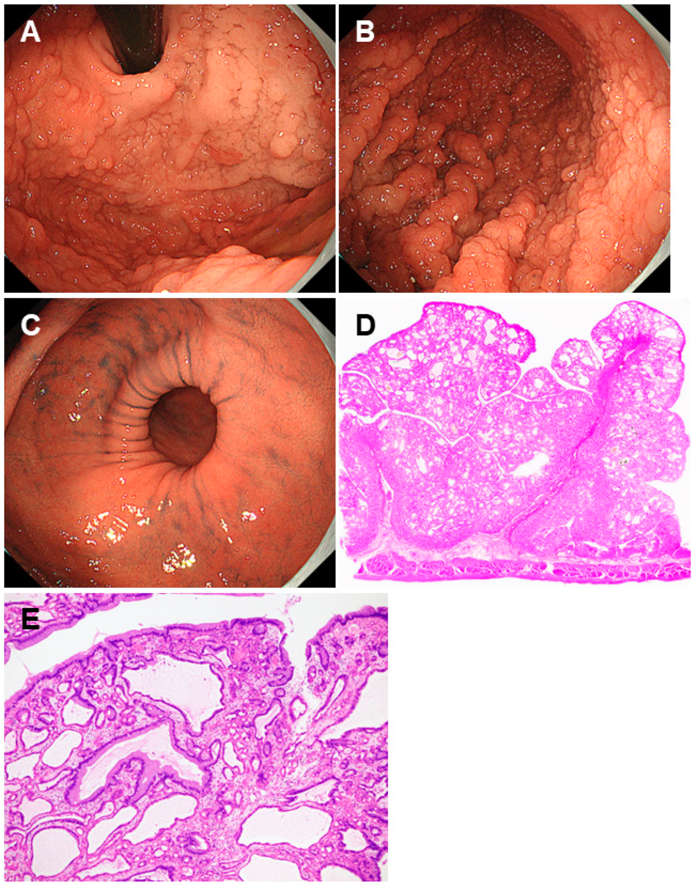
2.4. Cronkhite-Canada Syndrome

Cronkhite-Canada syndrome is a non-hereditary disorder characterized by the development of multiple non-neoplastic polyps in the digestive tract, particularly in the stomach and colon (Figure 7A–D) [35,36,37,38]. Concurrently, patients present with dermatological abnormalities, including alopecia (Figure 7E), nail atrophy (Figure 7F), and cutaneous pigmentations.
Malabsorption and protein-losing gastroenteropathy were the most frequent manifestations [35,36,37,38]. This condition is more common in middle-aged to older males. Although this is an infrequent disorder, a relatively large number of cases exist in Japan [35]. There are no universally established diagnostic criteria for Cronkhite-Canada syndrome globally; however, the Ministry of Health, Labor and Welfare in Japan has delineated the diagnostic criteria for Cronkhite-Canada syndrome as encompassing three major findings: (i) multifocal non-neoplastic polyposis within the gastrointestinal tract, prominently affecting the stomach and colon and displaying a non-hereditary etiology; (ii) gastrointestinal manifestations characterized by chronic diarrhea; and (iii) the manifestation of distinct dermatological symptoms, including alopecia, nail atrophy, and skin pigmentation [35]. The criteria included four supplementary findings: (i) low proteinemia (hypoalbuminemia) accompanied by protein leakage; (ii) dysgeusia, weight loss, and/or nutritional abnormalities; (iii) widespread non-pedunculated polyps distributed throughout the gastrointestinal tract marked by diffuse elevations with ambiguous margins, mucosal edema in the stomach, and well-defined strawberry-like polyps in the colon; and (iv) histological features corresponding to hamartomatous polyps (juvenile-like polyps) [35].
Gastric polyps are typically several millimeters to 20 mm in size and are often observed diffusely as sessile or slightly pedunculated elevations with a dense distribution [36,39]. The presence of edema with inflammation in the intervening mucosa between the polyps is crucial for the diagnosis of this disease (Figure 7G) [30].
In contrast to other polyposis syndromes, polyps in Cronkhite-Canada syndrome exhibit the potential for regression upon steroid administration, albeit with a propensity for occasional recurrence [35,36,37,38]. Although the polyps are of a non-neoplastic nature, sporadic instances of cancer development have been documented [40,41,42,43,44]. No guidelines currently exist for this disease, and surveillance methods remain undetermined.

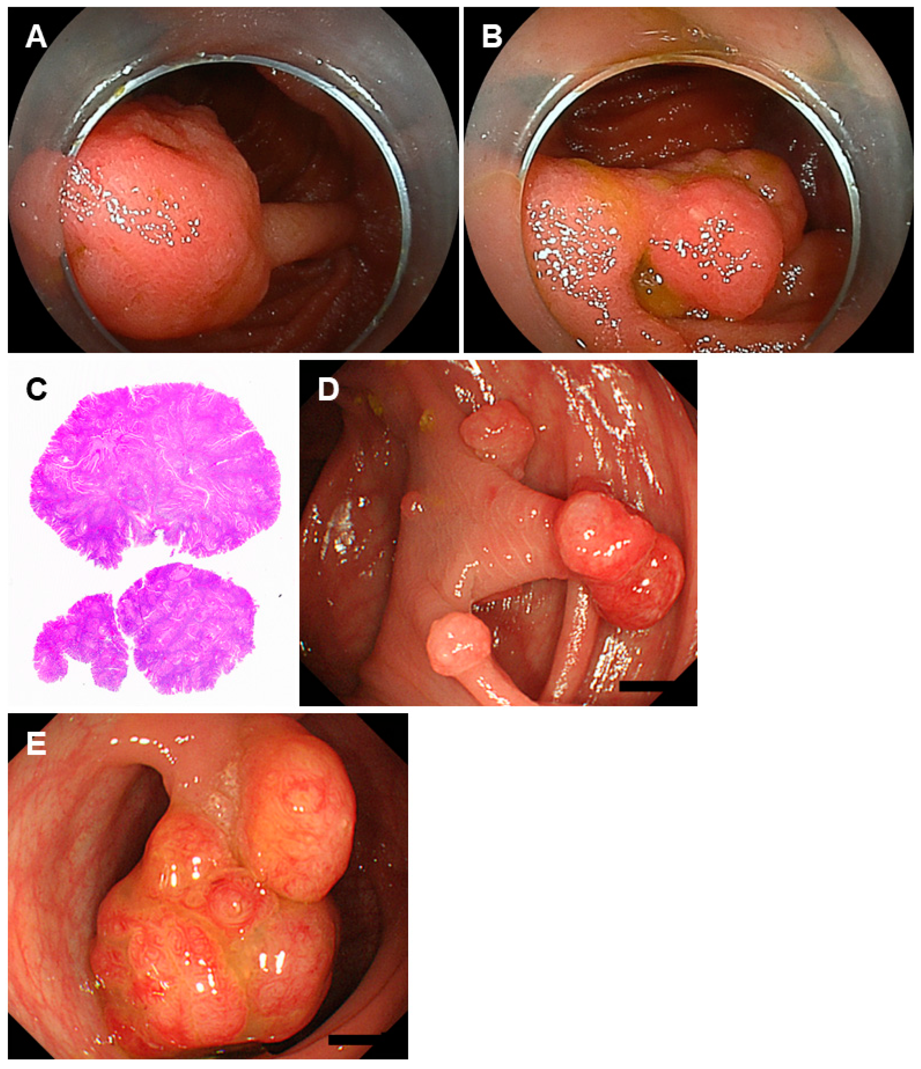
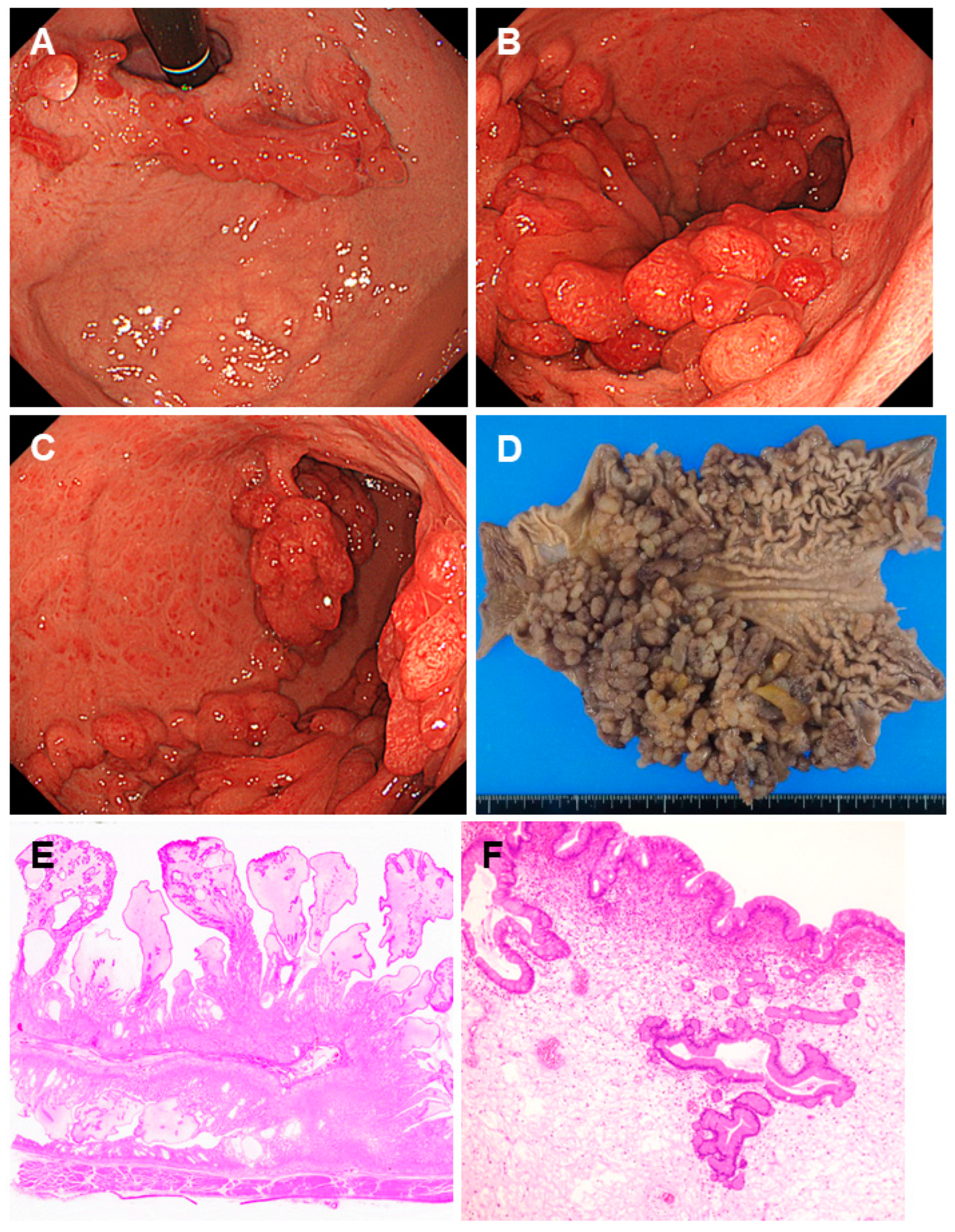
2.5. Juvenile Polyposis Syndrome

Juvenile polyposis syndrome is an autosomal dominant hereditary disorder characterized by the widespread occurrence of benign hamartomatous polyps throughout the gastrointestinal tract. Approximately 25% of the cases are sporadic and devoid of any family history [45,46]. Symptoms associated with multiple polyps in the digestive tract include intestinal obstruction due to polyp accumulation, abdominal pain, and rectal bleeding [26,45,46]. Most cases manifest before the age of 20 and are often prompted by intestinal obstruction during early adulthood. Protein-losing enteropathy, leading to hypoalbuminemia and malnutrition, also develops in patients with juvenile polyposis syndrome [45,46]. In cases presenting in infancy (neonatal/infantile onset) or early childhood (before the age of two), symptoms may include diarrhea and growth impairment, sometimes with a severe clinical course. Symptoms associated with anomalies of the central nervous system, cardiovascular system, and gastrointestinal tract, as well as complications related to hereditary hemorrhagic telangiectasia, such as hematemesis and melena, may occur. In a subset of patients, mutations have been ascertained in the SMAD4 or BMPR1A (bone morphogenetic protein receptor type 1A) genes, tumor suppressor genes implicated in the regulation of cellular proliferation and apoptosis through the transforming growth factor-β signaling pathway.
Juvenile polyps are most frequently detected in the colorectum (98%), followed by the stomach (14%), jejunum and ileum (7%), and duodenum (7%) [5]. Gastric polyps are multiple and reddish in color, with varying sizes, and exhibit swollen or edematous features. The morphology is typically papillary or tongue-like (Figure 8A–D). Pathologically, the polyps are characterized by hyperplasia of the gastric epithelium, cystic dilatation of glandular structures, and edema of the stroma (Figure 8E,F).
Although juvenile polyps are benign, patients with juvenile polyposis syndrome have an increased risk of developing gastric and colorectal cancers [45,46]. Thus, the commencement of surveillance through an esophagogastroduodenoscopy and colonoscopy is recommended at approximately the age of 12–15 years or earlier in the presence of symptoms [5,9]. The frequency of surveillance should be determined with intervals ranging from one to three years, depending on the extent of the polyp burden. Patients with a germline SMAD4 pathogenic variant often also have hereditary hemorrhagic telangiectasia. These patients should be screened for manifestations associated with hereditary hemorrhagic telangiectasia, particularly cerebral and pulmonary arteriovenous malformations.

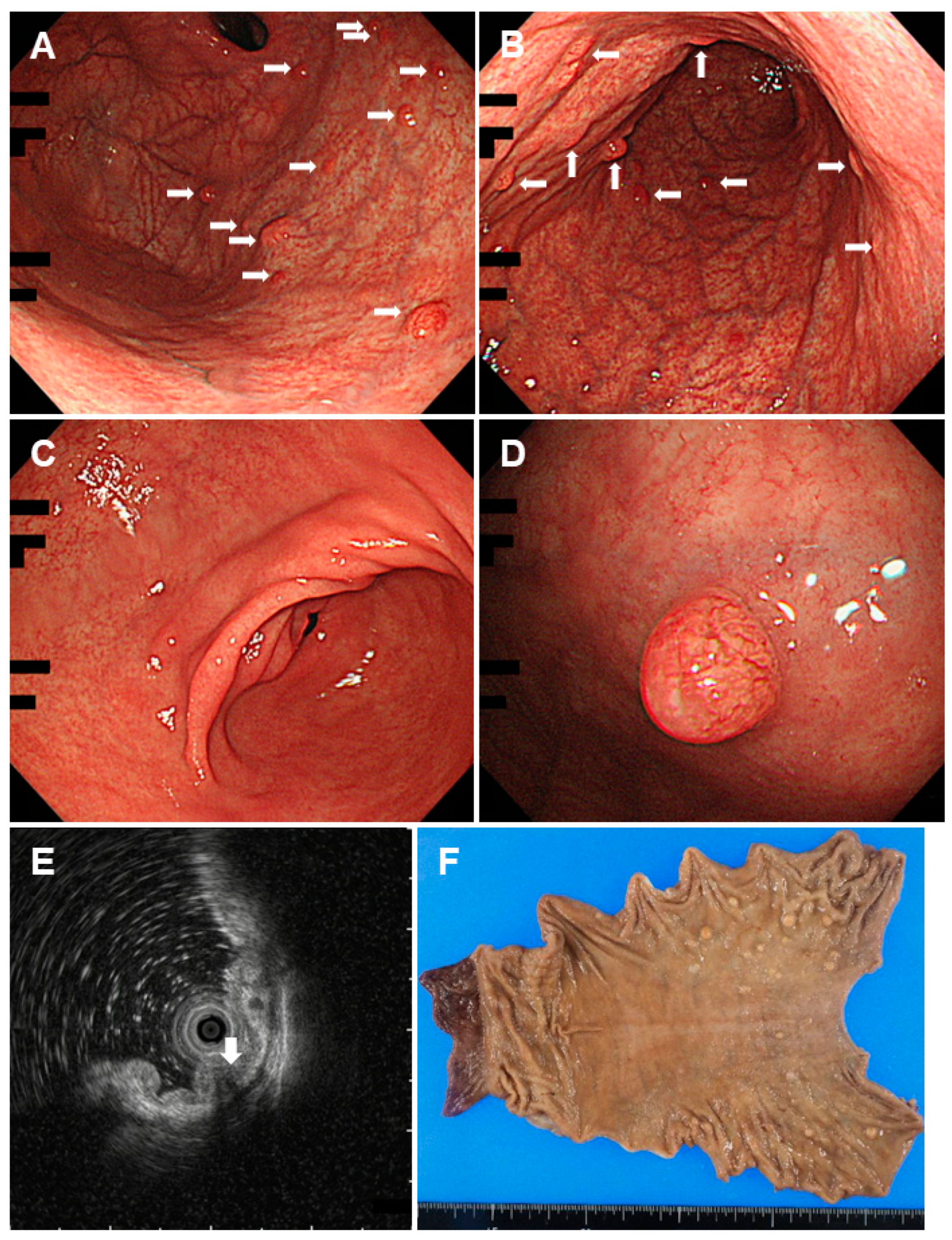
2.6. Gastric Adenocarcinoma and Proximal Polyposis of the Stomach

Gastric adenocarcinoma and proximal polyposis of the stomach (GAPPS) is a rare, autosomal dominant genetic disorder. As outlined in the disease name, GAPPS is characterized by the presence of multiple polyps in the proximal part of the stomach, i.e., the gastric fornix and body, and an increased risk of progressing to gastric adenocarcinoma [47,48]. The pathogenic variant of the APC promoter 1 B is considered a fundamental etiology of this condition [49]. The diagnosis of GAPPS pivots on the subsequent clinical and pathological criteria: (i) the exclusive presence of gastric polyps localized in the body and fornix with an absence of colorectal or duodenal polyposis; (ii) an excess of >100 polyps occurring in the proximal stomach in the index case or >30 polyps in a first-degree relative; (iii) predominantly constituted by fundic gland polyps, manifesting regions of dysplasia in some patients, or the manifestation of dysplastic fundic gland polyps or gastric adenocarcinoma in a family member; and (iv) a discernible pattern of autosomal dominant inheritance.
As described above, fundic gland polyposis is observed in the gastric fornix and body during an esophagogastroduodenoscopy, whereas the other parts remain essentially unaffected (Figure 9). Adenomas, dysplastic fundic gland polyps, and adenocarcinomas may concomitantly exist within the polyposis. Unlike familial adenomatous polyposis, the absence of polyposis in the duodenum or the colon is a crucial diagnostic indicator of GAPPS.
Given the increased risk of progression of these polyps to gastric adenocarcinoma, individuals with GAPPS require regular monitoring and surveillance, including endoscopic examinations [50]. In some cases, prophylactic gastrectomy may be considered as a preventive measure [51,52]. Owing to the infrequency of this disease, the surveillance strategy remains undetermined to date [49,53].

3. Gastric Polyposis Observed Outside of Gastrointestinal Polyposis Syndrome

3.1. Neuroendocrine Tumors in Autoimmune Gastritis

Neoplasms that manifest as multiple polypoid lesions in the stomach and require differentiation from polyposis syndrome include gastric cancers, lymphomas, and metastatic tumors. The emergence of multiple gastric neuroendocrine tumors in patients with autoimmune gastritis also falls within this category.
Autoimmune gastritis is a persistent inflammation of the stomach arising from the autoimmune system’s erroneous targeting and subsequent destruction of gastric parietal cells, resulting in reduced gastric acid secretion and vitamin B12 deficiency [54,55,56]. Reduced gastric acid production leads to elevated levels of gastrin. Over time, sustained high levels of gastrin stimulate the growth of neuroendocrine cells in the gastric mucosa, potentially leading to the development of neuroendocrine tumors. A study involving 176 patients with a median follow-up duration of 5 (3–7.5) years revealed that 33 patients manifested the emergence of a collective sum of 50 gastric neuroendocrine neoplasms with an annual cumulative incidence rate of 5.7% [57].
An analysis of 172 Japanese patients with autoimmune gastritis and histologically confirmed neuroendocrine tumors revealed that half of the patients (n = 84, 50.0%) had multiple tumors, while the other half had solitary lesions [58]. The location was most predominantly in the gastric body (n = 139, 88.5%), and the color was yellow (n = 82, 64.6%), followed by red (n = 38, 29.9%), pale (n = 5, 3.9%), and similar color to that of the background mucosa (n = 2, 1.6%). As corpus-predominant atrophy is the paramount hallmark of autoimmune gastritis on esophagogastroduodenoscopy, neuroendocrine tumors should be considered when yellow or red tumors are identified in the gastric body with marked or prevailing atrophy localized within the gastric body while sparing the gastric antrum (Figure 10).

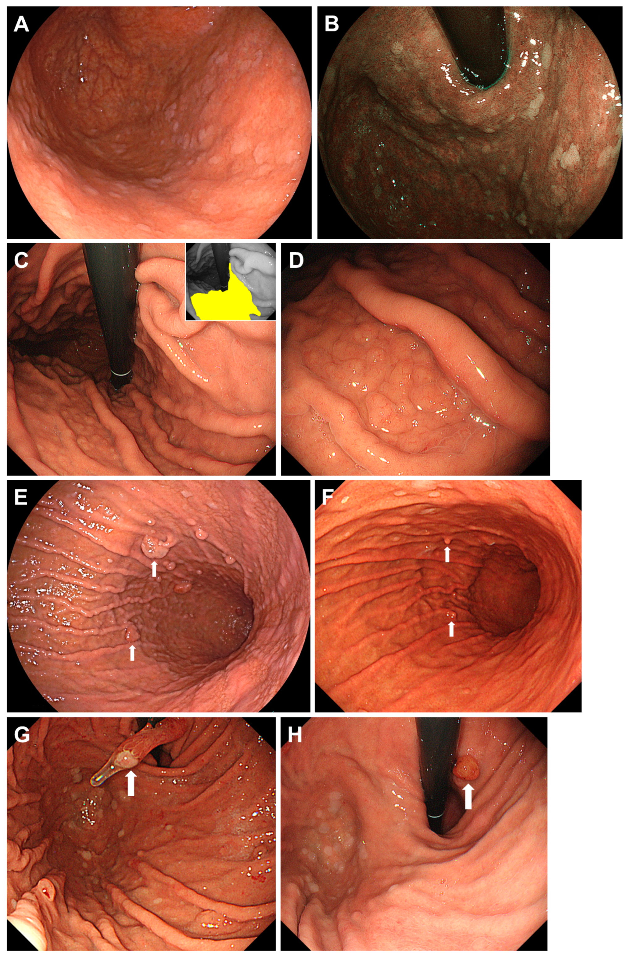
3.2. Proton Pump Inhibitor-Related Gastric Mucosal Changes

Proton pump inhibitors play a pivotal role in the management of various gastrointestinal conditions, such as gastroesophageal reflux disease and peptic ulcers. These drugs have been known to induce various morphological changes in gastric mucosa [59]. Among these, multiple polyps or polypoid-like lesions are represented by multiple white and flat elevated lesions, cobblestone-like mucosa, the emergence and enlargement of the fundic gland, and hyperplastic polyps [60].
Multiple white and flat elevated lesions result from foveolar epithelial hyperplasia. As indicated by their names, they are characterized by multiple white, flat, and elevated lesions typically located in the upper third of the stomach (Figure 11A,B). A prospective study found that the prevalence of these lesions is 10.4% [61]. Cobblestone-like mucosa was observed in the presence of numerous, approximately 3–5 mm-sized, uneven, elevated mucosal lesions within the gastric body, displaying a coloration akin to that of the surrounding mucosa, and was typically observed interspersed among the gastric folds (Figure 11C,D) [60]. An endoscopic biopsy of the cobblestone-like mucosa revealed parietal cell protrusions and the cystic dilatation of the fundic gland. Chronic use of proton pump inhibitors also induces the development and enlargement of the fundic gland and hyperplastic polyps. It is noteworthy that in some patients, the discontinuation of proton pump inhibitors leads to the reduction or disappearance of these polyps (Figure 11E–H) [62].

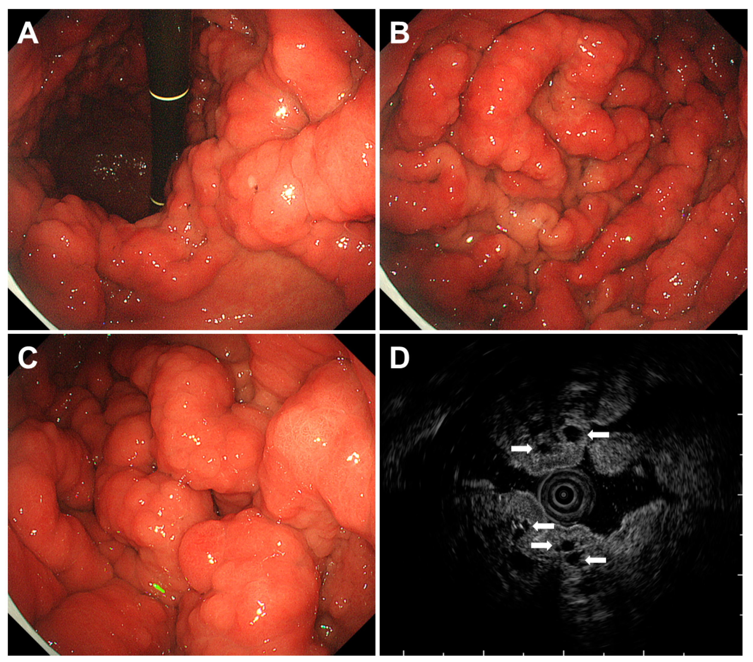
3.3. Multiple Submucosal Heterotopic Glands

Gastric multiple submucosal heterotopic glands refer to multiple aberrant or ectopic glandular structures located beneath the normal mucosal layer of the stomach. These lesions are variably termed depending on their size and include diffuse submucosal cysts, gastric cystica profunda, and hamartomatous inverted polyps [63]. The presence of these glands is thought to result from metaplastic responses to mucosal injury induced by chronic inflammation, such as H. pylori-associated gastritis [64,65]. Despite the typical non-dysplastic nature of these multiple gastric submucosal heterotopic glands, a previous investigation revealed the presence of KRAS mutations in 54% of these lesions [63], implying that their proliferative potential is incited by oncogenic mutations rather than inflammatory alterations. Although the precise risk associated with the development of dysplasia and cancer remains unclear, some authors have suggested that multiple gastric submucosal heterotopic glands represent a predisposing condition for the onset of dysplasia and carcinogenesis in the stomach [65].
Most of the multiple submucosal heterotopic glands are unexpectedly encountered within the stomach resected for malignancies, with certain lesions manifesting as polyps on endoscopy (Figure 12A-C). Given the subepithelial nature, diagnosing this condition through endoscopic observation and the histopathological analysis of biopsy samples is challenging. Endoscopic ultrasound examination proved valuable for diagnosis, showing cystic areas beneath the gastric mucosa (Figure 12D).

4. Conclusions

We present a comprehensive overview along with representative endoscopic images and a concise summary of the surveillance strategies for each disease state that leads to multiple polyps in the stomach (Figure 13). These are some potential differential diagnoses for patients with gastric polyposis and polypoid lesions. It is important to note that a patient’s medical and medication history, family history, and specific combination of symptoms are crucial for an accurate diagnosis. Genetic testing may also play a significant role in identifying the underlying conditions (Table 1). Once a diagnosis is established, tailored surveillance is imperative for each specific disorder, given the varying propensity for concomitant conditions, particularly in the organs predisposed to malignant neoplasms. Furthermore, in the context of hereditary diseases, comprehensive surveillance involving not only the affected individual but also their family along with genetic counseling, is of paramount significance. Although genetic counseling is typically advised both before and after germline testing to assist individuals in understanding the consequences of their genetic findings and in forming informed decisions regarding their own healthcare, the reduction in the costs of germline testing and enhancements in the gene set included in the panel may potentially expedite and simplify the diagnosis of gastric polyposis in the future. We hope that this review will facilitate clinicians’ understanding of gastric polyps and polypoid lesions encountered in various diseases.

Author Contributions

Conceptualization, M.I. and S.K.; formal analysis, M.I.; writing—original draft preparation, M.I.; writing—review and editing, S.K. and M.O.; supervision, M.O. All authors have read and agreed to the published version of the manuscript.

Funding

This research received no external funding.

Institutional Review Board Statement

Ethical review and approval were waived for this study due to the utilization of anonymous clinical data for image presentation.

Informed Consent Statement

Patient consent was waived due to the utilization of anonymous clinical data for image presentation.

Data Availability Statement

No new data were created.

Conflicts of Interest

The authors declare no conflicts of interest.

References

  1. Gorji, L.; Albrecht, P. Hamartomatous polyps: Diagnosis, surveillance, and management. World J. Gastroenterol. 2023, 29, 1304–1314. [Google Scholar] [CrossRef]
  2. Syngal, S.; Brand, R.E.; Church, J.M.; Giardiello, F.M.; Hampel, H.L.; Burt, R.W.; American College of Gastroenterology. ACG clinical guideline: Genetic testing and management of hereditary gastrointestinal cancer syndromes. Am. J. Gastroenterol. 2015, 110, 223–262. [Google Scholar] [CrossRef]
  3. Samadder, N.J.; Baffy, N.; Giridhar, K.V.; Couch, F.J.; Riegert-Johnson, D. Hereditary Cancer Syndromes—A Primer on Diagnosis and Management, Part 2: Gastrointestinal Cancer Syndromes. Mayo Clin. Proc. 2019, 94, 1099–1116. [Google Scholar] [CrossRef] [PubMed]
  4. Imyanitov, E.N.; Kuligina, E.S.; Sokolenko, A.P.; Suspitsin, E.N.; Yanus, G.A.; Iyevleva, A.G.; Ivantsov, A.O.; Aleksakhina, S.N. Hereditary cancer syndromes. World J. Clin. Oncol. 2023, 14, 40–68. [Google Scholar] [CrossRef] [PubMed]
  5. Boland, C.R.; Idos, G.E.; Durno, C.; Giardiello, F.M.; Anderson, J.C.; Burke, C.A.; Dominitz, J.A.; Gross, S.; Gupta, S.; Jacobson, B.C.; et al. Diagnosis and Management of Cancer Risk in the Gastrointestinal Hamartomatous Polyposis Syndromes: Recommendations from the US Multi-Society Task Force on Colorectal Cancer. Gastroenterology 2022, 162, 2063–2085. [Google Scholar] [CrossRef] [PubMed]
  6. Tomita, N.; Ishida, H.; Tanakaya, K.; Yamaguchi, T.; Kumamoto, K.; Tanaka, T.; Hinoi, T.; Miyakura, Y.; Hasegawa, H.; Takayama, T.; et al. Japanese Society for Cancer of the Colon and Rectum (JSCCR) guidelines 2020 for the Clinical Practice of Hereditary Colorectal Cancer. Int. J. Clin. Oncol. 2021, 26, 1353–1419. [Google Scholar] [CrossRef] [PubMed]
  7. Aelvoet, A.S.; Buttitta, F.; Ricciardiello, L.; Dekker, E. Management of familial adenomatous polyposis and MUTYH-associated polyposis; new insights. Best Pract. Res. Clin. Gastroenterol. 2022, 58–59, 101793. [Google Scholar] [CrossRef] [PubMed]
  8. Bianchi, L.K.; Burke, C.A.; Bennett, A.E.; Lopez, R.; Hasson, H.; Church, J.M. Fundic gland polyp dysplasia is common in familial adenomatous polyposis. Clin. Gastroenterol. Hepatol. 2008, 6, 180–185. [Google Scholar] [CrossRef] [PubMed]
  9. van Leerdam, M.E.; Roos, V.H.; van Hooft, J.E.; Dekker, E.; Jover, R.; Kaminski, M.F.; Latchford, A.; Neumann, H.; Pellisé, M.; Saurin, J.C.; et al. Endoscopic management of polyposis syndromes: European Society of Gastrointestinal Endoscopy (ESGE) Guideline. Endoscopy 2019, 51, 877–895. [Google Scholar] [CrossRef]
  10. Attard, T.M.; Giardiello, F.M.; Argani, P.; Cuffari, C. Fundic gland polyposis with high-grade dysplasia in a child with attenuated familial adenomatous polyposis and familial gastric cancer. J. Pediatr. Gastroenterol. Nutr. 2001, 32, 215–218. [Google Scholar]
  11. Garrean, S.; Hering, J.; Saied, A.; Jani, J.; Espat, N.J. Gastric adenocarcinoma arising from fundic gland polyps in a patient with familial adenomatous polyposis syndrome. Am. Surg. 2008, 74, 79–83. [Google Scholar] [CrossRef]
  12. Hofgärtner, W.T.; Thorp, M.; Ramus, M.W.; Delorefice, G.; Chey, W.Y.; Ryan, C.K.; Takahashi, G.W.; Lobitz, J.R. Gastric adenocarcinoma associated with fundic gland polyps in a patient with attenuated familial adenomatous polyposis. Am. J. Gastroenterol. 1999, 94, 2275–2281. [Google Scholar] [CrossRef]
  13. Shimamoto, Y.; Ishiguro, S.; Takeuchi, Y.; Nakatsuka, S.I.; Yunokizaki, H.; Ezoe, Y.; Nakajima, T.; Matsuno, K.; Nakahira, H.; Tanaka, K.; et al. Gastric neoplasms in patients with familial adenomatous polyposis: Endoscopic and clinicopathologic features. Gastrointest. Endosc. 2021, 94, 1030–1042.e2. [Google Scholar] [CrossRef]
  14. Kobashi, M.; Iwamuro, M.; Kuraoka, S.; Inoo, S.; Okanoue, S.; Satomi, T.; Hamada, K.; Abe, M.; Kono, Y.; Kanzaki, H.; et al. Endoscopic findings of gastric neoplasms in familial adenomatous polyposis are associated with the phenotypic variations and grades of dysplasia. Medicine 2022, 101, e30997. [Google Scholar] [CrossRef]
  15. Yang, J.; Gurudu, S.R.; Koptiuch, C.; Agrawal, D.; Buxbaum, J.L.; Abbas Fehmi, S.M.; Fishman, D.S.; Khashab, M.A.; Jamil, L.H.; Jue, T.L.; et al. American Society for Gastrointestinal Endoscopy guideline on the role of endoscopy in familial adenomatous polyposis syndromes. Gastrointest. Endosc. 2020, 91, 963–982.e2. [Google Scholar] [CrossRef] [PubMed]
  16. Monahan, K.J.; Bradshaw, N.; Dolwani, S.; Desouza, B.; Dunlop, M.G.; East, J.E.; Ilyas, M.; Kaur, A.; Lalloo, F.; Latchford, A.; et al. Guidelines for the management of hereditary colorectal cancer from the British Society of Gastroenterology (BSG)/Association of Coloproctology of Great Britain and Ireland (ACPGBI)/United Kingdom Cancer Genetics Group (UKCGG). Gut 2020, 69, 411–444. [Google Scholar] [CrossRef] [PubMed]
  17. Klimkowski, S.; Ibrahim, M.; Ibarra Rovira, J.J.; Elshikh, M.; Javadi, S.; Klekers, A.R.; Abusaif, A.A.; Moawad, A.W.; Ali, K.; Elsayes, K.M. Peutz-Jeghers Syndrome and the Role of Imaging: Pathophysiology, Diagnosis, and Associated Cancers. Cancers 2021, 13, 5121. [Google Scholar] [CrossRef] [PubMed]
  18. Gu, G.L.; Zhang, Z.; Zhang, Y.H.; Yu, P.F.; Dong, Z.W.; Yang, H.R.; Yuan, Y. Detection and analysis of common pathogenic germline mutations in Peutz-Jeghers syndrome. World J. Gastroenterol. 2021, 27, 6631–6646. [Google Scholar] [CrossRef] [PubMed]
  19. Jelsig, A.M.; van Overeem Hansen, T.; Gede, L.B.; Qvist, N.; Christensen, L.L.; Lautrup, C.K.; Frederiksen, J.H.; Sunde, L.; Ousager, L.B.; Ljungmann, K.; et al. Survival, surveillance, and genetics in patients with Peutz-Jeghers syndrome: A nationwide study. Clin. Genet. 2023, 104, 81–89. [Google Scholar] [CrossRef] [PubMed]
  20. Xu, Z.X.; Jiang, L.X.; Chen, Y.R.; Zhang, Y.H.; Zhang, Z.; Yu, P.F.; Dong, Z.W.; Yang, H.R.; Gu, G.L. Clinical features, diagnosis, and treatment of Peutz-Jeghers syndrome: Experience with 566 Chinese cases. World J. Gastroenterol. 2023, 29, 1627–1637. [Google Scholar] [CrossRef]
  21. Bartholomew, L.G.; Dahlin, D.C.; Waugh, J.M. Intestinal polyposis associated with mucocutaneous melanin pigmentation Peutz-Jeghers syndrome; review of literature and report of six cases with special reference to pathologic findings. Gastroenterology 1957, 32, 434–451. [Google Scholar] [CrossRef]
  22. Iwamuro, M.; Toyokawa, T.; Matsueda, K.; Hori, S.; Yoshioka, M.; Moritou, Y.; Tanaka, T.; Mizuno, M.; Okada, H. Two Types of Polyp Shape Observed in the Stomach of Patients with Peutz-Jeghers Syndrome. Acta Medica Okayama 2021, 75, 471–477. [Google Scholar]
  23. Yamamoto, H.; Sakamoto, H.; Kumagai, H.; Abe, T.; Ishiguro, S.; Uchida, K.; Kawasaki, Y.; Saida, Y.; Sano, Y.; Takeuchi, Y.; et al. Clinical Guidelines for Diagnosis and Management of Peutz-Jeghers Syndrome in Children and Adults. Digestion 2023, 104, 335–347. [Google Scholar] [CrossRef]
  24. Wagner, A.; Aretz, S.; Auranen, A.; Bruno, M.J.; Cavestro, G.M.; Crosbie, E.J.; Goverde, A.; Jelsig, A.M.; Latchford, A.; Leerdam, M.E.V.; et al. The Management of Peutz-Jeghers Syndrome: European Hereditary Tumour Group (EHTG) Guideline. J. Clin. Med. 2021, 10, 473. [Google Scholar] [CrossRef] [PubMed]
  25. Kim, S.H.; Kim, E.R.; Park, J.J.; Kim, E.S.; Goong, H.J.; Kim, K.O.; Nam, J.H.; Park, Y.; Lee, S.P.; Jang, H.J.; et al. Cancer risk in patients with Peutz-Jeghers syndrome in Korea: A retrospective multi-center study. Korean J. Intern. Med. 2023, 38, 176–185. [Google Scholar] [CrossRef] [PubMed]
  26. Yehia, L.; Heald, B.; Eng, C. Clinical Spectrum and Science Behind the Hamartomatous Polyposis Syndromes. Gastroenterology 2023, 164, 800–811. [Google Scholar] [CrossRef] [PubMed]
  27. Wang, Z.; Wang, Z.; Wang, Y.; Wu, J.; Yu, Z.; Chen, C.; Chen, J.; Wu, B.; Chen, Y. High risk and early onset of cancer in Chinese patients with Peutz-Jeghers syndrome. Front. Oncol. 2022, 12, 900516. [Google Scholar] [CrossRef] [PubMed]
  28. Pilarski, R.; Burt, R.; Kohlman, W.; Pho, L.; Shannon, K.M.; Swisher, E. Cowden syndrome and the PTEN hamartoma tumor syndrome: Systematic review and revised diagnostic criteria. J. Natl. Cancer Inst. 2013, 105, 1607–1616. [Google Scholar] [CrossRef]
  29. Daly, M.B.; Pilarski, R.; Yurgelun, M.B.; Berry, M.P.; Buys, S.S.; Dickson, P.; Domchek, S.M.; Elkhanany, A.; Friedman, S.; Garber, J.E.; et al. NCCN Guidelines Insights: Genetic/Familial High-Risk Assessment: Breast, Ovarian, and Pancreatic, Version 1.2020: Featured updates to the NCCN guidelines. J. Natl. Compr. Cancer Netw. 2020, 18, 380–391. [Google Scholar] [CrossRef]
  30. McGarrity, T.J.; Wagner Baker, M.J.; Ruggiero, F.M.; Thiboutot, D.M.; Hampel, H.; Zhou, X.P.; Eng, C. GI polyposis and glycogenic acanthosis of the esophagus associated with PTEN mutation positive Cowden syndrome in the absence of cutaneous manifestations. Am. J. Gastroenterol. 2003, 98, 1429–1434. [Google Scholar] [CrossRef]
  31. Ha, M.; Chung, J.W.; Hahm, K.B.; Kim, Y.J.; Lee, W.; An, J.; Kim, D.K.; Kim, M.G. A case of Cowden syndrome diagnosed from multiple gastric polyposis. World J. Gastroenterol. 2012, 18, 861–864. [Google Scholar] [CrossRef]
  32. Marra, G.; Armelao, F.; Vecchio, F.M.; Percesepe, A.; Anti, M. Cowden’s disease with extensive gastrointestinal polyposis. J. Clin. Gastroenterol 1994, 18, 42–47. [Google Scholar] [CrossRef]
  33. Stanich, P.P.; Francis, D.L.; Sweetser, S. The spectrum of findings in Cowden syndrome. Clin. Gastroenterol. Hepatol. 2011, 9, e2–e3. [Google Scholar] [CrossRef] [PubMed]
  34. Plamper, M.; Gohlke, B.; Woelfle, J. PTEN hamartoma tumor syndrome in childhood and adolescence-a comprehensive review and presentation of the German pediatric guideline. Mol. Cell. Pediatr. 2022, 9, 3. [Google Scholar] [CrossRef] [PubMed]
  35. Watanabe, C.; Komoto, S.; Tomita, K.; Hokari, R.; Tanaka, M.; Hirata, I.; Hibi, T.; Kaunitz, J.D.; Miura, S. Endoscopic and clinical evaluation of treatment and prognosis of Cronkhite-Canada syndrome: A Japanese nationwide survey. J. Gastroenterol. 2016, 51, 327–336. [Google Scholar] [CrossRef] [PubMed]
  36. Wu, Z.Y.; Sang, L.X.; Chang, B. Cronkhite-Canada syndrome: From clinical features to treatment. Gastroenterol. Rep. 2020, 8, 333–342. [Google Scholar] [CrossRef] [PubMed]
  37. Wang, W.; Shao, Y.; Zhao, D.H.; Xue, F.; Ma, X.B.; Li, Q.; Liu, C.X. Endoscopic and Pathological Characteristics of Cronkhite-Canada Syndrome: A Retrospective Analysis of 76 Cases. Turk. J. Gastroenterol. 2022, 33, 19–29. [Google Scholar] [CrossRef] [PubMed]
  38. Lu, Y.; Huang, F.; Wang, Y.; Zhou, J.; Zhao, Q.; Liu, L. Clinical and Endoscopic Characteristics of Chinese Cronkhite-Canada Syndrome Patients: A Retrospective Study of 103 Cases. Dig. Dis. 2021, 39, 488–495. [Google Scholar] [CrossRef] [PubMed]
  39. Kopáčová, M.; Urban, O.; Cyrany, J.; Laco, J.; Bureš, J.; Rejchrt, S.; Bártová, J.; Tachecí, I. Cronkhite-Canada syndrome: Review of the literature. Gastroenterol. Res. Pract. 2013, 2013, 856873. [Google Scholar] [CrossRef] [PubMed]
  40. Iqbal, U.; Chaudhary, A.; Karim, M.A.; Anwar, H.; Merrell, N. Cronkhite-Canada Syndrome: A Rare Cause of Chronic Diarrhea. Gastroenterol. Res. 2017, 10, 196–198. [Google Scholar] [CrossRef]
  41. Zhao, Y.; Lv, F.; Yang, X.; Wang, Y.; Zhang, S.; Li, P. Cronkhite-Canada Syndrome Associated with Superficial Esophageal Carcinoma: A Case Report and Literature Review. Front. Med. 2022, 9, 855336. [Google Scholar] [CrossRef]
  42. Yun, S.H.; Cho, J.W.; Kim, J.W.; Kim, J.K.; Park, M.S.; Lee, N.E.; Lee, J.U.; Lee, Y.J. Cronkhite-Canada syndrome associated with serrated adenoma and malignant polyp: A case report and a literature review of 13 cronkhite-Canada syndrome cases in Korea. Clin. Endosc. 2013, 46, 301–305. [Google Scholar] [CrossRef]
  43. Kaneko, Y.; Kato, H.; Tachimori, Y.; Watanabe, H.; Ushio, K.; Yamaguchi, H.; Itabashi, M.; Noguchi, M. Triple carcinomas in Cronkhite-Canada syndrome. Jpn. J. Clin. Oncol. 1991, 21, 194–202. [Google Scholar]
  44. Isobe, T.; Kobayashi, T.; Hashimoto, K.; Kizaki, J.; Miyagi, M.; Aoyagi, K.; Koufuji, K.; Shirouzu, K. Cronkhite-Canada syndrome complicated with multiple gastric cancers and multiple colon adenomas. Am. J. Case Rep. 2013, 14, 120–128. [Google Scholar]
  45. Brosens, L.A.; Langeveld, D.; van Hattem, W.A.; Giardiello, F.M.; Offerhaus, G.J. Juvenile polyposis syndrome. World J. Gastroenterol. 2011, 17, 4839–4844. [Google Scholar] [CrossRef] [PubMed]
  46. MacFarland, S.P.; Ebrahimzadeh, J.E.; Zelley, K.; Begum, L.; Bass, L.M.; Brand, R.E.; Dudley, B.; Fishman, D.S.; Ganzak, A.; Karloski, E.; et al. Phenotypic Differences in Juvenile Polyposis Syndrome with or without a Disease-causing SMAD4/BMPR1A Variant. Cancer Prev. Res. 2021, 14, 15–222. [Google Scholar] [CrossRef] [PubMed]
  47. Worthley, D.L.; Phillips, K.D.; Wayte, N.; Schrader, K.A.; Healey, S.; Kaurah, P.; Shulkes, A.; Grimpen, F.; Clouston, A.; Moore, D.; et al. Gastric adenocarcinoma and proximal polyposis of the stomach (GAPPS): A new autosomal dominant syndrome. Gut 2012, 61, 774–779. [Google Scholar] [CrossRef]
  48. Iwamuro, M.; Kawano, S.; Otsuka, M. An Unusual Case of Gastric Polyposis. Gastroenterology 2023, 165, 1110–1113. [Google Scholar] [CrossRef] [PubMed]
  49. Iwatsuki, M.; Matsumoto, C.; Mimori, K.; Baba, H. The comprehensive review of gastric adenocarcinoma and proximal polyposis of the stomach (GAPPS) from diagnosis and treatment. Ann. Gastroenterol. Surg. 2023, 7, 725–732. [Google Scholar] [CrossRef] [PubMed]
  50. Gullo, I.; van der Post, R.S.; Carneiro, F. Recent advances in the pathology of heritable gastric cancer syndromes. Histopathology 2021, 78, 125–147. [Google Scholar] [CrossRef] [PubMed]
  51. Foretová, L.; Navrátilová, M.; Svoboda, M.; Grell, P.; Nemec, L.; Sirotek, L.; Obermannová, R.; Novotný, I.; Sachlova, M.; Fabian, P.; et al. GAPPS–Gastric Adenocarcinoma and Proximal Polyposis of the Stomach Syndrome in 8 Families Tested at Masaryk Memorial Cancer Institute–Prevention and Prophylactic Gastrectomies. Klin Onkol 2019, 32 (Suppl. 2), 109–117. [Google Scholar] [CrossRef]
  52. Salami, A.C.; Stone, J.M.; Greenberg, R.H.; Leighton, J.C.; Miick, R.; Zavala, S.R.; Zeitzer, K.L.; Bakhis, C.T. Early Prophylactic Gastrectomy for the Management of Gastric Adenomatous Proximal Polyposis Syndrome (GAPPS). ACS Case Rev. Surg. 2022, 3, 62–68. [Google Scholar] [PubMed]
  53. Yen, T.; Stanich, P.P.; Axell, L.; Patel, S.G. APC-Associated Polyposis Conditions; Adam, M.P., Mirzaa, G.M., Pagon, R.A., Wallace, S.E., Bean, L.J.H., Gripp, K.W., Amemiya, A., Eds.; GeneReviews® [Internet]; University of Washington: Seattle, WA, USA, 2023. [Google Scholar]
  54. Dottori, L.; Pivetta, G.; Annibale, B.; Lahner, E. Update on Serum Biomarkers in Autoimmune Atrophic Gastritis. Clin. Chem. 2023, 69, 1114–1131. [Google Scholar] [CrossRef] [PubMed]
  55. Iwamuro, M.; Tanaka, T.; Otsuka, M. Update in Molecular Aspects and Diagnosis of Autoimmune Gastritis. Curr. Issues Mol. Biol. 2023, 45, 5263–5275. [Google Scholar] [CrossRef] [PubMed]
  56. Neumann, W.L.; Coss, E.; Rugge, M.; Genta, R.M. Autoimmune atrophic gastritis–pathogenesis, pathology and management. Nat. Rev. Gastroenterol. Hepatol. 2013, 10, 529–541. [Google Scholar] [CrossRef] [PubMed]
  57. Massironi, S.; Gallo, C.; Elvevi, A.; Stegagnini, M.; Coltro, L.A.; Invernizzi, P. Incidence and prevalence of gastric neuroendocrine tumors in patients with chronic atrophic autoimmune gastritis. World J. Gastrointest. Oncol. 2023, 15, 1451–1460. [Google Scholar] [CrossRef] [PubMed]
  58. Namikawa, K.; Kamada, T.; Fujisaki, J.; Sato, Y.; Murao, T.; Chiba, T.; Kaizaki, Y.; Ishido, K.; Ihara, Y.; Kurahara, K.; et al. Clinical characteristics and long-term prognosis of type 1 gastric neuroendocrine tumors in a large Japanese national cohort. Dig. Endosc. 2023, 35, 757–766. [Google Scholar] [CrossRef] [PubMed]
  59. Kiso, M.; Ito, M.; Boda, T.; Kotachi, T.; Masuda, K.; Hata, K.; Sasaki, A.; Kawamura, T.; Yoshihara, M.; Tanaka, S.; et al. Endoscopic findings of the gastric mucosa during long-term use of proton pump inhibitor—A multicenter study. Scand. J. Gastroenterol. 2017, 52, 828–832. [Google Scholar] [CrossRef]
  60. Miyamoto, S.; Kato, M.; Tsuda, M.; Matsuda, K.; Muranaka, T.; Abiko, S.; Ono, M.; Mizushima, T.; Omori, S.; Yamamoto, K.; et al. Gastric mucosal cracked and cobblestone-like changes resulting from proton pump inhibitor use. Dig. Endosc. 2017, 29, 307–313. [Google Scholar] [CrossRef]
  61. Majima, K.; Muraki, Y.; Shimamoto, T. Multiple White and Flat Elevated Lesions Observed in the Stomach: A Prospective Study of Clinical Characteristics and Risk Factors. Intern. Med. 2018, 57, 2613–2619. [Google Scholar] [CrossRef]
  62. Iwamuro, M.; Shiraha, H.; Okada, H. Gastric polyps’ regression after potassium-competitive acid blocker cessation. J. Gen. Fam. Med. 2022, 23, 358–359. [Google Scholar] [CrossRef] [PubMed]
  63. Cho, H.; Hashimoto, T.; Naka, T.; Yatabe, Y.; Oda, I.; Saito, Y.; Yoshikawa, T.; Sekine, S. Activating KRAS and GNAS mutations in heterotopic submucosal glands of the stomach. J. Gastroenterol. 2022, 57, 333–343. [Google Scholar] [CrossRef] [PubMed]
  64. Namikawa, T.; Ishida, N.; Yokota, K.; Munekage, E.; Iwabu, J.; Munekage, M.; Uemura, S.; Maeda, H.; Kitagawa, H.; Kobayashi, M.; et al. Early gastric cancer with multiple submucosal heterotopic gastric gland: A case report. Mol. Clin. Oncol. 2019, 10, 583–586. [Google Scholar] [CrossRef]
  65. Inaba, T.; Mizuno, M.; Kawai, K.; Okada, H.; Tsuji, T. Diffuse submucosal cysts of the stomach: Report of two cases in association with development of multiple gastric cancers during endoscopic follow up. J. Gastroenterol. Hepatol. 1999, 14, 1161–1165. [Google Scholar] [CrossRef] [PubMed]
Figure 1. Endoscopic images of familial adenomatous polyposis. Case 1 (A,B). Multiple adenomatous polyps are observed in the colorectum, (A) colon, (B) rectum. Case 2 (CE). (C,D) numerous fundic gland polyps in the gastric body. The color of the polyps is similar to that of the background mucosa; (E) a large, whitish, flat elevated duodenal adenoma is seen in the center. Multiple small lesions are also observed (arrows).
Gastroent 15 00009 g001
Figure 2. Endoscopic images of cancers and adenomas in familial adenomatous polyposis. Case 3 (A,B) early gastric cancers appear as reddish depressed lesions in the atrophic mucosa; (A) the lesions are difficult to recognize under white light observation; (B) indigo carmine dye spraying emphasized the redness and slight depressions (arrows). Case 4 (CE) gastric-type adenomas occurring in the non-atrophic mucosa of the gastric fornix, which is seen as a flat elevated milky-white lesion; (C) white light; (D) after indigo carmine dye spraying; (E) narrow-band imaging; (F,G) case 5. An intestinal-type adenoma located in the gastric antrum showing an elevated lesion featuring a central depression.
Gastroent 15 00009 g002
Figure 3. Endoscopic and pathological images of Peutz-Jeghers syndrome. Case 6 (AC). Pedunculated, multinodular polyps that are typical for Peutz-Jeghers syndrome observed in the small intestine; (A,B) double-balloon enteroscopy images; (C) a pathological image showing mucosal epithelial hyperplasia and the dendritic proliferation of smooth muscle fibers from the muscularis mucosae; case 7 (D,E) multiple reddish, nodular Peutz-Jeghers polyps observed in the colon.
Gastroent 15 00009 g003
Figure 4. Endoscopic images of gastric polyps in Peutz-Jeghers syndrome. Case 8 (A) a hyperplastic polyp in the stomach. Case 9 (B) a reddish-color polypoid lesion sized >5 mm identified in the gastric body. Pathologic analysis of the endoscopic mucosal resection specimen revealed characteristic features of Peutz-Jeghers polyps. Case 10 (C,D) esophagogastroduodenoscopy reveals diffuse fundic gland polyps in the stomach, which resemble familial adenomatous polyposis.
Gastroent 15 00009 g004
Figure 5. Images of Cowden syndrome. (AC) Case 11; (A) the esophagus shows diffuse glycogen acanthosis characterized by white flat polyposis, which are distinct to Cowden syndrome; (B) white nodular lesions observed in the tongue via an esophagogastroduodenoscopy; (C) oral papillomas and hyperkeratotic lesions.
Gastroent 15 00009 g005
Figure 6. Endoscopic images of Cowden syndrome. Case 12 (AF). (A) multiple glycogen acanthosis in the esophagus; (BD) gastric polyps showing a color similar to the peripheral mucosa; (B) white light; (C) narrow-band imaging; (D) after indigo carmine dye spraying; (E,F) polyps in the colon; (E) white light; (F) narrow-band imaging.
Gastroent 15 00009 g006
Figure 7. Images of Cronkhite-Canada syndrome. Case 13 (AG). (A) Reddish polyps are diffusely observed with dense distribution in the stomach; (B) duodenal polyps; (C) polyps identified in the small intestine using a video capsule enteroscopy; (D) polyps in the colon; (E) alopecia; (F) nail atrophy; (G) edema with inflammation in the intervening mucosa between the gastric polyps.
Gastroent 15 00009 g007
Figure 8. Images of juvenile polyposis syndrome. Case 14 (AF). (AC) Gastric polyps with multiple and varying sizes, appearing reddish and showing signs of swelling or edema; (D) a surgically resected specimen revealing numerous polyps; (E,F) pathology findings including hyperplasia of the gastric epithelium, cystic dilatation of glandular structures, and stromal edema.
Gastroent 15 00009 g008
Figure 9. Images of gastric adenocarcinoma and proximal polyposis of the stomach. Case 15 (AE). (AC) Dense fundic gland polyposis is observed in the gastric fornix and body with no effect on the antrum; (A) gastric fornix; (B) gastric body; (C) gastric antrum; (D,E) histologically subtle distortions and hypertrophy of the gastric foveolar epithelium are observed.
Gastroent 15 00009 g009
Figure 10. Images of neuroendocrine tumors in autoimmune gastritis. Case 16. Multiple neuroendocrine tumors are seen as red tumors in the gastric body ((A,B), arrows). Gastric atrophy is predominant in the fornix and body, while the antrum is relatively intact; (A) gastric fornix; (B) gastric body; (C) gastric antrum; (D) close-up view of a neuroendocrine tumor; (E) endoscopic ultrasonography showing a neuroendocrine tumor as a round lesion in the submucosa with low echogenicity (arrow); (F) the resected specimen shows multiple tumors in the fornix and body.
Gastroent 15 00009 g010
Figure 11. Images of proton pump inhibitor-related gastric mucosal changes. Case 17 (A,B). Multiple white and flat elevated lesions seen in the gastric fornix, which are more easily recognized via blue laser imaging; (A) white light; (B) blue laser imaging. Case 18 (C,D). Cobblestone-like mucosa observed in the gastric body; (C) the yellow area in the insert image shows cobblestone-like mucosa. Case 19 (E,F). The dimensions of fundic gland polyps (arrows) exhibit diminution subsequent to the cessation of proton pump inhibitor utilization; (E) during the administration of proton pump inhibitors; (F) following the termination of proton pump inhibitor therapy. Case 20 (G,H). A hyperplastic polyp (arrow). Concurrently, multiple white and flat elevated lesions are evident in the fornix; (G) during the course of proton pump inhibitor treatment; (H) post-cessation of proton pump inhibitor therapy.
Gastroent 15 00009 g011
Figure 12. Endoscopic images of multiple submucosal heterotopic glands. Case 21. (AC) Reddish polypoid lesions diffusely occupy the gastric body; (D) endoscopic ultrasonography showing cystic areas beneath the gastric mucosa (arrows).
Gastroent 15 00009 g012
Figure 13. Representative endoscopic images of diseases that exhibit multiple gastric polyps. GAPPS: gastric adenocarcinoma and proximal polyposis of the stomach.
Gastroent 15 00009 g013
Table 1. Causative factors of diseases exhibiting multiple gastric polyps and the associated characteristics of gastric polyps.
Disease NameCausative FactorCharacteristics of Gastric Polyps
Familial adenomatous polyposisMutations in the APC geneMultiple fundic gland polyps are typical. Intestinal-type adenomas and early gastric cancers exhibit reddish depressed lesions, while gastric-type adenomas display whitish elevated lesions.
Peutz-Jeghers syndromeMutations in the STK11 geneSolitary or sporadic polyps larger than 5 mm exhibiting a reddish color with a sessile or semi-pedunculated morphology (hyperplastic polyps and Peutz-Jeghers polyps), and multiple sessile polyps of 5 mm or smaller, mirroring the color of the peripheral mucosa (fundic gland polyps and hyperplastic polyps).
Cowden syndromeMutations in the PTEN genePolyps generally exhibit a color similar to the peripheral mucosa but may also be reddish (hyperplastic, hamartomatous, inflammatory, lipomatous, fibromatous, and adenomatous polyps).
Cronkhite-Canada syndromeNot yet elucidatedGastric polyps ranging from several millimeters to 20 mm in size are observed diffusely as sessile or slightly pedunculated elevations with a dense distribution. Edema with inflammation is present in the intervening mucosa between the polyps.
Juvenile polyposis syndromeMutations in the SMAD4 or BMPR1A genePapillary or tongue-like polyps, multiple and reddish in color of varying sizes with swollen or edematous features.
GAPPSMutations in the APC exon 1B promotor regionPolyps localized in the gastric body and fornix, predominantly constituted by fundic gland polyps manifesting as regions of dysplasia and adenocarcinoma in some patients.
Neuroendocrine tumors in autoimmune gastritisAutoimmune gastritisYellow or red tumors are identified in the gastric body with marked or prevailing atrophy localized within the gastric body while sparing the gastric antrum.
Proton pump inhibitor-related gastric mucosal changesProlonged intake of proton pump inhibitorsMultiple white and flat elevated lesions, cobblestone-like mucosa, the emergence and enlargement of the fundic gland, and hyperplastic polyps.
Multiple submucosal heterotopic glandsChronic inflammation, such as infection of Helicobacter pyloriPolypoid lesions with cystic areas beneath the gastric mucosa visualized during an endoscopic ultrasound examination.
Disclaimer/Publisher’s Note: The statements, opinions and data contained in all publications are solely those of the individual author(s) and contributor(s) and not of MDPI and/or the editor(s). MDPI and/or the editor(s) disclaim responsibility for any injury to people or property resulting from any ideas, methods, instructions or products referred to in the content.

