Next Issue
Volume 14, December
Previous Issue
Volume 14, June


Neurol. Int., Volume 14, Issue 3 (September 2022) – 19 articles

Cover Story (view full-size image):

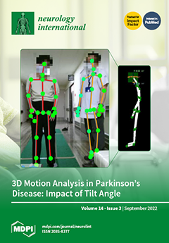
In patients with Parkinson’s disease, motor assessment is an important issue in evaluating disease progression or the efficacy of therapy. Based on the progresses of imaging capture, image processing and analyses, the quantified evaluation of postures and gaits is currently becoming increasingly popular. We used the AKIRA® system for three-dimensional evaluation of body movements of patients with Parkinson’s disease. The three-dimensional analysis of gait motion was compared with the MDS-UDPRS, and its usefulness as an evaluation index was examined. Results showed that the forward and side tilt angle of neck and trunk measured by AKIRA can be a candidate for a quantitative severity index in patients with PD. View this paper

  • Issues are regarded as officially published after their release is announced to the table of contents alert mailing list.
  • You may sign up for e-mail alerts to receive table of contents of newly released issues.
  • PDF is the official format for papers published in both, html and pdf forms. To view the papers in pdf format, click on the "PDF Full-text" link, and use the free Adobe Reader to open them.
Order results
Result details
Select all
Export citation of selected articles as:
10 pages, 696 KiB  
Article
EEG Correlation Coefficient Change with Motor Task Activation Can Be a Predictor of Functional Recovery after Hemiparetic Stroke
by Fei Zheng, Shin Sato, Kenji Mamada, Naoto Ozaki, Jin Kubo and Wataru Kakuda
Neurol. Int. 2022, 14(3), 738-747; https://doi.org/10.3390/neurolint14030062 - 19 Sep 2022
Cited by 1 | Viewed by 1632
Abstract
Background: Recently, it was reported that the extent of cortico-cortical functional connections can be estimated by the correlation coefficient based on electroencephalography (EEG) monitoring. We aimed to investigate whether the EEG correlation coefficient change with motor task activation can predict the functional outcomes [...] Read more.
Background: Recently, it was reported that the extent of cortico-cortical functional connections can be estimated by the correlation coefficient based on electroencephalography (EEG) monitoring. We aimed to investigate whether the EEG correlation coefficient change with motor task activation can predict the functional outcomes of hemiparetic stroke patients. Methods: Sixteen post-stroke hemiparetic patients admitted to our rehabilitation ward were studied. On admission, EEG recording to calculate the correlation coefficient was performed at rest and during motor task activation. For the analysis of the EEG data, the program software FOCUS (NIHON KOHDEN, Japan) was used. The motor function of paretic limbs was evaluated with the Fugl–Meyer Assessment (FMA) on admission and 4 weeks after admission. Results: Significant increases in the correlation coefficient with motor task activation were noted in C3-F3 or C4-F4, C3-F7 or C4-F8, and F3-F7 or F4-F8 of the lesional hemisphere. Among them, the rate of the correlation coefficient change in F3-F7 or F4-F8 in the lesional hemisphere was significantly correlated with the rate of the upper-limb FMA score change. Conclusion: The extent of the EEG correlation coefficient change with motor task activation in F3-F7 or F4-F8 of the lesional hemisphere may help predict the motor functional outcomes of hemiparetic upper limbs after stroke. Full article
(This article belongs to the Special Issue Stroke: From Pathophysiology to Therapy)
Show Figures

Figure 1

11 pages, 758 KiB  
Article
The Forward and Lateral Tilt Angle of the Neck and Trunk Measured by Three-Dimensional Gait and Motion Analysis as a Candidate for a Severity Index in Patients with Parkinson’s Disease
by Hirofumi Matsumoto, Makoto Shiraishi, Ariaki Higashi, Sakae Hino, Mayumi Kaburagi, Heisuke Mizukami, Futaba Maki, Junji Yamauchi, Kenichiro Tanabe, Tomoo Sato and Yoshihisa Yamano
Neurol. Int. 2022, 14(3), 727-737; https://doi.org/10.3390/neurolint14030061 - 13 Sep 2022
Cited by 1 | Viewed by 2326
Abstract
(1) Objective: To evaluate the usefulness of a three-dimensional motion-analysis system (AKIRA®) as a quantitative measure of motor symptoms in patients with Parkinson’s disease (PD). (2) Method: This study included 48 patients with PD. We measured their motion during 2 m [...] Read more.
(1) Objective: To evaluate the usefulness of a three-dimensional motion-analysis system (AKIRA®) as a quantitative measure of motor symptoms in patients with Parkinson’s disease (PD). (2) Method: This study included 48 patients with PD. We measured their motion during 2 m of walking using AKIRA®, we calculated the tilt angles of the neck and trunk, ankle height, and gait speed, then we compared these parameters with the MDS-UPDRS and the Hoehn and Yahr scale. Furthermore, we measured these AKIRA indicators before and after 1 year of observation. (3) Results: The forward tilt angle of the neck showed a strong correlation with the scores on parts II, III, and the total MDS-UPDRS, and the tilt angle of the trunk showed a moderate correlation with those measures. The lateral tilt angle of the trunk showed a moderate correlation with a freezing of the gait and a postural instability. Regarding changes over the course of 1 year (n = 34), the total scores on part III of the MDS-UPDRS and the forward tilt angle of the neck improved, while the lateral tilt angle of the trunk worsened. (4) Conclusion: Taken together, the forward and lateral tilt angles of the neck and trunk as measured by AKIRA® can be a candidate for quantitative severity index in patients with PD. Full article
(This article belongs to the Special Issue Advances in Parkinson’s Disease)
Show Figures

Figure 1

11 pages, 1189 KiB  
Systematic Review
Erdheim–Chester Disease with Isolated CNS Involvement: A Systematic Review of the Literature
by Anam Haque, Carlos A. Pérez, Thejasvi A. Reddy and Rajesh K. Gupta
Neurol. Int. 2022, 14(3), 716-726; https://doi.org/10.3390/neurolint14030060 - 05 Sep 2022
Cited by 5 | Viewed by 2220
Abstract
Erdheim–Chester disease (ECD) is a rare, sporadic, non-Langerhans cell histiocytosis that can have various presentations and higher mortality in patients presenting with neurological symptoms. We performed a systematic review to investigate and chronicle the frequency of neurological manifestations, imaging findings, treatments, and outcomes [...] Read more.
Erdheim–Chester disease (ECD) is a rare, sporadic, non-Langerhans cell histiocytosis that can have various presentations and higher mortality in patients presenting with neurological symptoms. We performed a systematic review to investigate and chronicle the frequency of neurological manifestations, imaging findings, treatments, and outcomes in published ECD patients presenting with neurological symptoms. A PubMed literature search was conducted for articles (published between January 1980 and June 2021) on ECD cases presenting with neurological manifestations. We analyzed the data of 40 patients, including our patient. Cranial neuropathies and ataxia were the most frequent clinical manifestations. A total of 50% of the symptomatic ECD CNS lesions were intraparenchymal and nearly 33% of patients died due to the disease itself or complications. CNS involvement may be the only manifestation of ECD and sometimes may require a repeat biopsy with IHC testing for excellent treatment outcomes. Full article
(This article belongs to the Collection Advances in Neurodegenerative Diseases)
Show Figures

Figure 1

9 pages, 1523 KiB  
Article
Reassessing the Diagnostic Utility of the Split Hand Index in Amyotrophic Lateral Sclerosis Patients—The Divide by Zero Problem
by Florian Antonescu, Ioana Butnariu, Marin Adam, Dana Antonescu-Ghelmez and Sorin Tuță
Neurol. Int. 2022, 14(3), 707-715; https://doi.org/10.3390/neurolint14030059 - 01 Sep 2022
Viewed by 2121
Abstract
We set out to assess the diagnostic utility of the split hand index (SHI) for amyotrophic lateral sclerosis (ALS) and also to see if and how it can be applied to severely atrophied muscles, a frequent finding in this setting. We enrolled 38 [...] Read more.
We set out to assess the diagnostic utility of the split hand index (SHI) for amyotrophic lateral sclerosis (ALS) and also to see if and how it can be applied to severely atrophied muscles, a frequent finding in this setting. We enrolled 38 patients from our clinic, 19 diagnosed with ALS and 19 controls, matched for age and sex. The SHI was calculated, on both sides, for all the patients. We calculated a SHI of 0 when the abductor pollicis brevis muscle (ABD) or first dorsal interosseous muscle (FDI) compound muscle action potentials (CMAPs) were unobtainable, and we allotted a value of 0.1 mV to abductor digiti minimi muscle (ADM) CMAP, for mathematical purposes, when the value would have been 0. The means differences were large between groups, with a significant variance heterogeneity. We performed a ROC analysis and obtained an accuracy of 0.83 for a SHI of 7.2, p-value < 0.0001. In conclusion, we reaffirm the utility of the SHI in the diagnosis of ALS, especially in limb onset cases, and we think that it can be safely extended to severely atrophied muscles with absent or very low CMAP values, without endangering the sensitivity or specificity. Full article
Show Figures

Figure 1

11 pages, 1729 KiB  
Article
Selection of Mice for Object Permanence Cognitive Task Solution
by Olga Viktorovna Perepelkina and Inga Igorevna Poletaeva
Neurol. Int. 2022, 14(3), 696-706; https://doi.org/10.3390/neurolint14030058 - 29 Aug 2022
Viewed by 1760
Abstract
The selection of mice for high (“plus”) and low (“minus”) scores in the puzzle-box test was performed over five generations. This test evaluates the success (or failure) in finding the underpass, leading to the dark part of the box, when it is blocked. [...] Read more.
The selection of mice for high (“plus”) and low (“minus”) scores in the puzzle-box test was performed over five generations. This test evaluates the success (or failure) in finding the underpass, leading to the dark part of the box, when it is blocked. This means that the mouse is either able or unable to operate the “object permanence rule” (one of the index’s cognitive abilities). For the “+” strain, animals were bred who solved the test when the underpass test blocked with a plug; the “−” strain comprised those who were unable to solve this task. In mice of the “+” strain, the proportion of animals that was able to solve “plug” stages of the test was higher than in the “−” strain and in the non-selected genetically heterogeneous population. The “+” mice ate significantly more new food in the hyponeophagia test. Animals of both strains demonstrated the ability to “manipulate” the plug blocking the underpass, touching the plug with their paws and muzzle, although the majority of “−” mice were unable to open the underpass effectively. Thus, mice of both selected strains demonstrated that they were able to understand that the underpass does exist, but only “+”-strain animals (at least the majority of them) were able to realize the solution. The selection for plug-stage solution success affected the mouse’s ability to open the hidden underpass. Full article
Show Figures

Graphical abstract

13 pages, 649 KiB  
Systematic Review
Clinical and Radiological Characteristics for Recurrence of Chronic Subdural Hematoma: A Systematic Review and Meta-Analysis
by Rakesh Mishra, Harsh Deora, William Andres Florez-Perdomo, Luis Rafael Moscote-Salazar, Ezequiel Garcia-Ballestas, Md Moshiur Rahman, Adesh Shrivastava, Sumit Raj, Vishal Chavda, Nicola Montemurro and Amit Agrawal
Neurol. Int. 2022, 14(3), 683-695; https://doi.org/10.3390/neurolint14030057 - 26 Aug 2022
Cited by 9 | Viewed by 2389
Abstract
Chronic subdural hematoma (cSDH) is one of the most studied clinical entities in the neurosurgical literature. Management of cSDH is complicated by its propensity to recurrence. Various factors for the development of recurrence of cSDH have been described in various clinical, epidemiological, and [...] Read more.
Chronic subdural hematoma (cSDH) is one of the most studied clinical entities in the neurosurgical literature. Management of cSDH is complicated by its propensity to recurrence. Various factors for the development of recurrence of cSDH have been described in various clinical, epidemiological, and observational studies, yet the evidence available is limited. A systematic review and meta-analysis as per PRISMA guidelines to identify clinical and radiological factors which can predict the development of recurrence in cSDH. A total of 14 studies were included for the systematic review and meta-analysis after a comprehensive search of the online databases. Eight studies were of high methodological quality. Age, use of anticoagulants, obesity, seizure, and liver disease were found to be statistically significant clinical risk factors for the development of recurrence in cSDH. Among the radiological parameters, the internal structure of the hematoma and the width of the hematoma was found to be significant risk factor predicting the development of recurrence. Age >75 years, use of anticoagulation therapy, liver disease, and obesity were significant risk factors for cSDH recurrence. Pneumocephalus, internal architecture of hematoma, bilateral cSDH, the width of hematoma, and the presence of bilateral cSDH are important radiological parameters of the development of recurrent cSDH Full article
(This article belongs to the Special Issue Recent Advances in Traumatic Brain Injury)
Show Figures

Figure 1

5 pages, 1191 KiB  
Case Report
A Patient with Erdheim-Chester Disease Limited to Central Nervous System
by Rajesh K. Gupta, Anam Haque, Thejasvi A. Reddy and Carlos A. Pérez
Neurol. Int. 2022, 14(3), 678-682; https://doi.org/10.3390/neurolint14030056 - 26 Aug 2022
Cited by 1 | Viewed by 1767
Abstract
Erdheim-Chester disease (ECD) is a rare, sporadic, non-Langerhans cell histiocytosis, a multisystem disorder, which has higher mortality when presented with CNS involvement. We report a 46-year-old woman who has ECD with exclusive CNS involvement. She presented with intracranial hemorrhage and had a poor [...] Read more.
Erdheim-Chester disease (ECD) is a rare, sporadic, non-Langerhans cell histiocytosis, a multisystem disorder, which has higher mortality when presented with CNS involvement. We report a 46-year-old woman who has ECD with exclusive CNS involvement. She presented with intracranial hemorrhage and had a poor response to corticosteroid and interferon. She required multiple debulking procedures and eventually responded well to cobimetinib. She has not had any other organ involvement thus far. This report highlights that CNS involvement may be the only manifestation of ECD and sometimes may require a repeat biopsy with IHC testing for excellent treatment outcomes. Full article
(This article belongs to the Collection Advances in Neurodegenerative Diseases)
Show Figures

Figure 1

5 pages, 450 KiB  
Interesting Images
Development of Parkinsonism in a Patient with Central Pontine Myelinolysis
by Annibale Antonioni, Vittorio Rispoli, Patrik Fazio, Nico Golfrè Andreasi, Vittorio Govoni and Enrico Granieri
Neurol. Int. 2022, 14(3), 673-677; https://doi.org/10.3390/neurolint14030055 - 25 Aug 2022
Viewed by 1779
Abstract
Osmotic demyelination syndrome (ODS) is caused by damage to the pons myelin sheath and nerve cells. Although the pathophysiological mechanism responsible for the damage is not yet fully understood, it is currently believed that osmotic-type changes (especially if they are massive and too [...] Read more.
Osmotic demyelination syndrome (ODS) is caused by damage to the pons myelin sheath and nerve cells. Although the pathophysiological mechanism responsible for the damage is not yet fully understood, it is currently believed that osmotic-type changes (especially if they are massive and too rapid) cause oedema that leads to compression and, subsequently, demyelination of white matter fibres. It generally manifests with acute paraparesis/tetraparesis, dysphagia, dysarthria, diplopia, and loss of consciousness, as well as hallucinations, spasms, and other neurological symptoms related to brainstem damage. In extreme cases, the locked-in syndrome may also appear. Of note, in some cases an association between osmotic demyelinating damage and the onset of movement disorders has been documented and, although the pathophysiology is still unknown, a correlation has been postulated between ODS and movement disorders. Here, we present a patient with ODS who developed parkinsonism, thus supporting the hypothesis of a correlation between these pathological events. Full article
(This article belongs to the Special Issue Advances in Parkinson’s Disease)
Show Figures

Figure 1

9 pages, 1175 KiB  
Article
Anatomical Variations of the Median Nerve: A Cadaveric Study
by Manuel Encarnacion, Renat Nurmukhametov, Rossi Evelyn Barrientos, Dmitry Melchenko, Evgeniy Goncharov, Edwin Bernard, Jose Mogorron Huerta, Jean Francois Uhl, Ibrahim E. Efe, Nicola Montemurro and Issael Ramirez
Neurol. Int. 2022, 14(3), 664-672; https://doi.org/10.3390/neurolint14030054 - 23 Aug 2022
Cited by 5 | Viewed by 2367
Abstract
Objectives: Variations in the morphological anatomy of the median nerve such as formation, distribution, and communication have been well documented. All these variations should be taken into account when practicing any surgical approach for the treatment of injuries affecting the median nerve. Furthermore, [...] Read more.
Objectives: Variations in the morphological anatomy of the median nerve such as formation, distribution, and communication have been well documented. All these variations should be taken into account when practicing any surgical approach for the treatment of injuries affecting the median nerve. Furthermore, they are of the utmost importance for interpretation of the clinical presentation. Methods: The objective of this investigation was to determine the anatomical variations in the formation of the median nerve in cadavers at the Forensic Pathology department in Central Clinical Hospital of the Academy of Sciences of the Russian Federation between January 2022 and April 2022. A descriptive, cross-sectional, and prospective information source study was conducted on 42 anatomical bodies (corpses) and 84 brachial plexuses. Results: After analyzing the results obtained in this investigation, we concluded that the median nerve presented variation in its formation in 22.6% of the investigated cases. These variations were more common in males (81.8%) than females (18.2%). The anatomical variation was unilateral in 7.1% and bilateral in 19% of all anatomical bodies examined. Conclusions: The median nerve presented a great number of variations in its formation in roughly 23% of the anatomical bodies, with male being the predominant gender. Furthermore, the most frequent region of formation was the axillary region (92.9%). For clinicians, it is important to remember these variations during surgical procedures in this area and during brachial plexus block. Full article
Show Figures

Figure 1

16 pages, 359 KiB  
Review
Novel Designer Benzodiazepines: Comprehensive Review of Evolving Clinical and Adverse Effects
by Amber N. Edinoff, Catherine A. Nix, Amira S. Odisho, Caroline P. Babin, Alyssa G. Derouen, Salim C. Lutfallah, Elyse M. Cornett, Kevin S. Murnane, Adam M. Kaye and Alan D. Kaye
Neurol. Int. 2022, 14(3), 648-663; https://doi.org/10.3390/neurolint14030053 - 22 Aug 2022
Cited by 18 | Viewed by 6010
Abstract
As tranquilizers, benzodiazepines have a wide range of clinical uses. Recently, there has been a significant rise in the number of novel psychoactive substances, including designer benzodiazepines. Flubromazolam(8-bromo-6-(2-fluorophenyl)-1-methyl-4H-[1,2,4]triazolo[4,3-a][1,4]benzodiazeZpine) is a triazolo-analogue of flubromazepam. The most common effects noted by recreational users [...] Read more.
As tranquilizers, benzodiazepines have a wide range of clinical uses. Recently, there has been a significant rise in the number of novel psychoactive substances, including designer benzodiazepines. Flubromazolam(8-bromo-6-(2-fluorophenyl)-1-methyl-4H-[1,2,4]triazolo[4,3-a][1,4]benzodiazeZpine) is a triazolo-analogue of flubromazepam. The most common effects noted by recreational users include heavy hypnosis and sedation, long-lasting amnesia, and rapid development of tolerance. Other effects included anxiolysis, muscle-relaxing effects, euphoria, loss of control, and severe withdrawals. Clonazolam, or 6-(2-chlorophenyl)-1-methyl-8-nitro-4H-[1,2,4]triazolo[4,3-α]-[1,4]-benzodiazepine, is a triazolo-analog of clonazepam. It is reported to be over twice as potent as alprazolam. Deschloroetizolam (2-Ethyl-9-methyl-4-phenyl-6H-thieno[3,2-f][1,2,4]triazolo[4,3-a][1,4]diazepine) is part of the thienodiazepine drug class, which, like benzodiazepines, stimulates GABA-A receptors. Meclonazepam ((3S)-5-(2-chlorophenyl)-3-methyl-7-nitro-1,3-dihydro-1,4-benzodiazepin-2-one) is a designer benzodiazepine with additional anti-parasitic effects. Although it has proven to be an efficacious therapy for schistosomiasis, its sedative side effects have prevented it from being marketed as a therapeutic agent. The use of DBZs has been a subject of multiple recent clinical studies, likely related to increasing presence and availability on the internet drug market and lack of regulation. Many studies have aimed to identify the prevalence of DBZs and their effects on those using them. This review discussed these designer benzodiazepines and the dangers and adverse effects that the clinician should know. Full article
10 pages, 1785 KiB  
Case Report
Relevance of Medullary Vein Sign in Neurosarcoidosis
by Richard Liberio, Emily Kramer, Anza B. Memon, Ryan Reinbeau, Parissa Feizi, Joe Joseph, Janet Wu and Shitiz Sriwastava
Neurol. Int. 2022, 14(3), 638-647; https://doi.org/10.3390/neurolint14030052 - 14 Aug 2022
Cited by 3 | Viewed by 2104
Abstract
Background: Central nervous system involvement is uncommon in patients with sarcoidosis. It remains a diagnostic challenge for clinicians, as there is a broad differential diagnosis that matches the presenting neurological signs. Often, the imaging findings also overlap with other disease entities. One understudied [...] Read more.
Background: Central nervous system involvement is uncommon in patients with sarcoidosis. It remains a diagnostic challenge for clinicians, as there is a broad differential diagnosis that matches the presenting neurological signs. Often, the imaging findings also overlap with other disease entities. One understudied finding in patients with neurosarcoidosis is the presence of medullary vein engorgement on SWI imaging, termed the “medullary vein sign”, which has been postulated to be a specific sign for neurosarcoidosis. This study aims to provide an understanding of the diagnostic potential of the medullary vein sign. Methods: Thirty-two patients who presented with neurologic signs concerning for possible neurosarcoidosis were analyzed retrospectively for the presence of the medullary vein sign. Results: Out of these cases, 7 cases of definitive neurosarcoidosis cases were found based on other imaging signs, biopsy and CSF analysis; the remaining were classified into groups as possible (16), probable (5) and (4) cases of other infectious meningoencephalitis including 2 cases of autoimmune encephalitis. Seven patients among all of these cases were found to have the medullary vein sign on imaging, with five cases with confirmed and two cases from possible neurosarcoidosis. The sensitivity of the medullary vein sign in this study was 71.4%, and the specificity was 92.3%. Discussion: The benefits of improving diagnostic criteria for neurosarcoidosis include more rapid diagnosis leading to more prompt treatment, less exposure to potentially harmful antibiotics or antifungals, and less long-term neurological effects. Our results support that the medullary vein sign will potentially fill in the diagnostic gaps that have challenged the timely diagnosis of neurosarcoidosis. Conclusions: Our findings support that the medullary vein sign has a high specificity and should be included in the diagnostic criteria for neurosarcoidosis. Full article
(This article belongs to the Collection Advances in Neurodegenerative Diseases)
Show Figures

Figure 1

10 pages, 288 KiB  
Brief Report
Self-Awareness of Cognitive Efficiency, Cognitive Status, Insight, and Financial Capacity in Patients with Mild AD, aMCI, and Healthy Controls: An Intriguing Liaison with Clinical Implications?
by Vaitsa Giannouli and Magdalini Tsolaki
Neurol. Int. 2022, 14(3), 628-637; https://doi.org/10.3390/neurolint14030051 - 30 Jul 2022
Cited by 7 | Viewed by 2010
Abstract
Objectives: This study compares objective measures of cognitive performance with subjective perception of specific performance on neuropsychological tests examining basic cognitive domains, including, for the first time, financial capacity. Additionally, differences in assessment between single- and multiple-domain aMCI, mild AD, and healthy elderly [...] Read more.
Objectives: This study compares objective measures of cognitive performance with subjective perception of specific performance on neuropsychological tests examining basic cognitive domains, including, for the first time, financial capacity. Additionally, differences in assessment between single- and multiple-domain aMCI, mild AD, and healthy elderly regarding insight are examined. Methods: Participants completed a number of neuropsychological tests and the Legal Capacity for Property Law Transactions Assessment Scale (LCPLTAS). After every test, participants were asked to complete the Clinical Insight Rating scale (CIR) and to self-evaluate their performance by comparing it to what they considered as average for people of their age and educational level. Results: These preliminary findings show significant differences in the self-assessment patterns of the four groups in measures of verbal memory, visuospatial perception and memory, executive functions, tests of attention, and financial capacity. Mild AD expressed the highest overestimations, followed by single- and multiple-domain aMCI as well as controls. Accuracy of self-report is not uniform across groups and functional areas. Conclusions: Unawareness of memory deficits in both MCI subtypes is contradictory to subjective memory complaints as being an important component for clinical diagnosis. Financial capacity is overestimated in MCI and mild AD, a finding that has a plethora of clinical and legal implications. Full article
(This article belongs to the Collection Advances in Neurodegenerative Diseases)
Show Figures

Graphical abstract

9 pages, 751 KiB  
Article
Accuracy and Sensitivity of Clinical Parameters in Predicting Successful Extubation in Patients with Acute Brain Injury
by Flávia Manhani Muzette, Rayssa Bruna Holanda Lima, Jennifer de Araújo Silva, Thamara Ferro Balsani Comin, Erlandson Ferreira Saraiva, Karla Luciana Magnani Seki and Gustavo Christofoletti
Neurol. Int. 2022, 14(3), 619-627; https://doi.org/10.3390/neurolint14030050 - 25 Jul 2022
Cited by 1 | Viewed by 2057
Abstract
Background: Brain injuries are frequent causes of intubation and mechanical ventilation. The aim of this study was to investigate the accuracy and sensitivity of clinical parameters in predicting successful extubation in patients with acute brain injury. Methods: Six hundred and forty-four patients assisted [...] Read more.
Background: Brain injuries are frequent causes of intubation and mechanical ventilation. The aim of this study was to investigate the accuracy and sensitivity of clinical parameters in predicting successful extubation in patients with acute brain injury. Methods: Six hundred and forty-four patients assisted at a high-complexity hospital were recruited. Patients were divided as for successful or failed extubation. The VISAGE score, maximum inspiratory and expiratory pressures, peak cough flow, and airway occlusion pressure at 0.1 s were used as predictors. Logistic regression analyses using ROC-curve identified values of accuracy and sensitivity. The Hosmer–Lemeshow test and the stepwise method calibrated the statistical model. Results: VISAGE score (odds ratio of 1.975), maximum inspiratory pressure (odds ratio of 1.024), and peak cough flow (odds ratio of 0.981) are factors consistent in distinguishing success from failure extubation. The ROC curve presented an accuracy of 79.7% and a sensitivity of 95.8%. Conclusions: VISAGE score, maximum inspiratory pressure and peak cough flow showed good accuracy and sensitivity in predicting successful extubation in patients with acute brain injury. The greater impact of VISAGE score indicates that patients’ neurological profile should be considered in association with ventilatory parameters in the decision of extubation. Full article
Show Figures

Figure 1

5 pages, 1445 KiB  
Case Report
Acute Carbamazepine Intoxication
by María Dolores Calabria Gallego and Mónica Alañá García
Neurol. Int. 2022, 14(3), 614-618; https://doi.org/10.3390/neurolint14030049 - 22 Jul 2022
Cited by 1 | Viewed by 2335
Abstract
Carbamazepine is an anticonvulsant drug with multiple mechanisms of action, which condition the presence of a characteristic clinical picture after the overingestion of the drug. We expose a case report about a patient who, in the context of an attempted suicide, presented acute [...] Read more.
Carbamazepine is an anticonvulsant drug with multiple mechanisms of action, which condition the presence of a characteristic clinical picture after the overingestion of the drug. We expose a case report about a patient who, in the context of an attempted suicide, presented acute intoxication by benzodiazepines and carbamazepine, presenting the characteristic clinical picture of fluctuations in the level of consciousness, even presenting gaze deconjugation, almost unreactive coma and generalized hypotonia. Full article
Show Figures

Figure 1

14 pages, 1124 KiB  
Review
Sudden Unexpected Death in Epilepsy
by Teri B. O’Neal, Sanjay Shrestha, Harsimar Singh, Ihianle Osagie, Kenechukwu Ben-Okafor, Elyse M. Cornett and Alan D. Kaye
Neurol. Int. 2022, 14(3), 600-613; https://doi.org/10.3390/neurolint14030048 - 18 Jul 2022
Cited by 12 | Viewed by 4639
Abstract
Epilepsy is a complex neurological condition with numerous etiologies and treatment options. In a subset of these patients, sudden unexpected death can occur, and to date, there are numerous explanations as to the pathophysiological mechanisms and how to mitigate these catastrophic outcomes. Approximately [...] Read more.
Epilepsy is a complex neurological condition with numerous etiologies and treatment options. In a subset of these patients, sudden unexpected death can occur, and to date, there are numerous explanations as to the pathophysiological mechanisms and how to mitigate these catastrophic outcomes. Approximately 2.3 million Americans have epilepsy, and nearly 150,000 people develop the condition each year. Sudden unexpected death in epilepsy (SUDEP) accounts for 2–18% of all epilepsy-related deaths and this is equivalent to one death in 1000 person-years of diagnosed epilepsy. It is more common in young adults aged 20–45. Seizures in the past year; the absence of terminal remission in the last five years; increased seizure frequency, particularly GTCS; and nocturnal seizures are the most potent modifiable risk factors for SUDEP. Patients not receiving any antiepileptic drug therapy are at higher risk of SUDEP. Patient education on medication compliance; care plans for seizure clusters (rescue medicines); epilepsy self-management programs; and lifestyle changes to avoid seizure-triggering factors, including avoiding excessive alcohol use and sleep deprivation, should be provided by health care providers. Continued research into SUDEP will hopefully lead to effective interventions to minimize occurrences. At present, aggressive control of epilepsy and enhanced education for individuals and the public are the most effective weapons for combating SUDEP. This narrative review focuses on updated information related to SUDEP epidemiology; pathophysiology; risk factor treatment options; and finally, a discussion of important clinical studies. We seek to encourage clinicians who care for patients with epilepsy to be aggressive in controlling seizure activity and diligent in their review of risk factors and education of patients and their families about SUDEP. Full article
Show Figures

Figure 1

19 pages, 3867 KiB  
Article
Spinal Vascular Shunts: Single-Center Series and Review of the Literature of Their Classification
by Jafeth Lizana, Nelida Aliaga, Walter Marani, Amanda Escribano and Nicola Montemurro
Neurol. Int. 2022, 14(3), 581-599; https://doi.org/10.3390/neurolint14030047 - 15 Jul 2022
Cited by 5 | Viewed by 3075
Abstract
Spinal arteriovenous shunts (sAVSs) are an uncommon disease, constituting 3 to 4% of intradural lesions; 70% of these lesions are spinal arteriovenous fistulas (sAVFs), whereas spinal arteriovenous malformations (sAVMs) are rarer. Both share the problem of their classification due to the heterogeneity of [...] Read more.
Spinal arteriovenous shunts (sAVSs) are an uncommon disease, constituting 3 to 4% of intradural lesions; 70% of these lesions are spinal arteriovenous fistulas (sAVFs), whereas spinal arteriovenous malformations (sAVMs) are rarer. Both share the problem of their classification due to the heterogeneity of their angioarchitecture. The aim of this study is to report a series of sAVSs treated in the neurosurgery department of the Hospital Nacional Guillermo Almenara during the 2018–2020 period and to present an overview of the current literature on sAVS classification. We reviewed all medical records of patients diagnosed with sAVFs and sAVMs during the 2018–2020 period, and then we analyzed images with Horos v4.0.0, illustrated some cases with Clip Studio Paint v1.10.5, and performed a descriptive statistical analysis with SPSS v25. Twelve patients were included in this study, eight of which were women (67%) and four of which were men (33%); the age range was from 3 to 74 years. Eight sAVSs were sAVFs, whereas four were sAVMs. The most frequent clinical manifestation was chronic myelopathy in seven patients (58%). Of those treated only by embolization, seven (70%) resulted in complete occlusion (five sAVFs and two sAVMs), while three (30%) remained with a residual lesion. At last follow-up, five patients (42%) improved clinically, and the seven remaining (58%) maintained the same neurological state. sAVSs require a detailed study of their angioarchitecture for proper management. The endovascular treatment is safe with acceptable cure rates. The surgical option should not be set aside. Full article
Show Figures

Graphical abstract

7 pages, 1910 KiB  
Case Report
Successful Consolidation/Maintenance Therapy with Single Agent Ibrutinib for Primary CNS Lymphoma after Initial Induction Therapy
by Steven Du, Daniela Bota and Xiao-Tang Kong
Neurol. Int. 2022, 14(3), 574-580; https://doi.org/10.3390/neurolint14030046 - 27 Jun 2022
Cited by 1 | Viewed by 2463
Abstract
Primary central nervous system lymphoma (PCNSL) is a rare and aggressive disease that originates from lymphocytes and develops in the central nervous system. There is no standard consolidation/maintenance therapy for PCNSL. While there exists a variety of options, the high chance of inferior [...] Read more.
Primary central nervous system lymphoma (PCNSL) is a rare and aggressive disease that originates from lymphocytes and develops in the central nervous system. There is no standard consolidation/maintenance therapy for PCNSL. While there exists a variety of options, the high chance of inferior outcomes for elderly patients and the risk of neurotoxicity requires exploration of alternative options for consolidation/maintenance therapy for PCNSL in the elderly population with CNS lymphoma. We treated one 77-year-old patient with single agent ibrutinib, a Bruton’s tyrosine kinase inhibitor that crosses the blood-brain-barrier, as consolidation/maintenance therapy after induction therapy with high-dose methotrexate (HD-MTX) and rituximab plus temozolomide. This treatment resulted in good tolerance, further resolution of a small residue lymphoma, and sustained remission. The patient has completed one year of consolidation/maintenance therapy and is currently under clinical and imaging surveillance. She has survived 27 months without recurrence since diagnosis. This case shows the potential effectiveness of single agent ibrutinib as consolidation/maintenance therapy for PCNSL after induction therapy. More cases are needed to confirm the findings. Full article
Show Figures

Figure 1

13 pages, 3900 KiB  
Case Report
Robot Assisted Gait Training in a Patient with Ataxia
by Gianfranco Lamberti, Gianluca Sesenna, Martina Marina, Emanuela Ricci and Gianluca Ciardi
Neurol. Int. 2022, 14(3), 561-573; https://doi.org/10.3390/neurolint14030045 - 22 Jun 2022
Cited by 2 | Viewed by 2204
Abstract
Background: Ataxia is a neurological sign characterized by motor coordination during gait/voluntary limb movements impairment. Ataxic gait leads to disability and worsening of quality of life; physiotherapy intervention is recommended to improve motor function. Recent studies showed benefits due to repetitive robotized assisted [...] Read more.
Background: Ataxia is a neurological sign characterized by motor coordination during gait/voluntary limb movements impairment. Ataxic gait leads to disability and worsening of quality of life; physiotherapy intervention is recommended to improve motor function. Recent studies showed benefits due to repetitive robotized assisted gait training using a static exoskeleton in patients affected by acquired ataxias. The aim of the study was to perform a preliminary evaluation of the short-term effects of overground UAN.GO®-assisted gait training in an adult patient with ataxia but with no clear genetic pattern. Methods: This case report study was conducted on a single male adult patient, who presented ataxic spastic gait, posterior chain tightness, pes cavus, and unstable standing position. The patient underwent two preliminary sessions to take part in the study. Treatment protocol planned 10 sessions and each one lasted 80 min, 60 of which were spent in gait training using the mobile overground exoskeleton UAN.GO®. At T1 (start of the study) and T10 (final evaluation) assessments using the Scale for the Assessment and Rating of Ataxia, Berg Balance Scale, 6-Minute Walking Test, and Likert Scale were administered. Space-time parameters of gait cycle were also evaluated: left and right step length, stance and swing percentages. Results: improvements on the Scale for the Assessment and Rating of Ataxia, Berg Balance Scale, and in the distance travelled at 6-Minute Walking Test emerged. The patient gave a positive opinion towards the treatment, showed by Likert Scale results. Kinematic gait analysis showed more physiological step length, stance and swing percentages, joint angles. The patient completed the training program with an excellent compliance. Discussion: Since these encouraging outcomes were obtained, it is possible to consider robot-assisted gait training performed with UAN.GO® as a therapeutic option to improve motor and functional performance in patients with ataxic gait. Full article
Show Figures

Figure 1

14 pages, 4080 KiB  
Article
Changes in Brain Electrical Activity after Transient Middle Cerebral Artery Occlusion in Rats
by Yuriy I. Sysoev, Veronika A. Prikhodko, Aleksandra V. Kan, Irina A. Titovich, Vadim E. Karev and Sergey V. Okovityi
Neurol. Int. 2022, 14(3), 547-560; https://doi.org/10.3390/neurolint14030044 - 21 Jun 2022
Cited by 1 | Viewed by 1978
Abstract
Objectives. Ischemic stroke is a leading cause of death and disability worldwide. To search for new therapeutic and pharmacotherapeutic strategies, numerous models of this disease have been proposed, the most popular being transient middle cerebral artery occlusion. Behavioral and sensorimotor testing, biochemical, and [...] Read more.
Objectives. Ischemic stroke is a leading cause of death and disability worldwide. To search for new therapeutic and pharmacotherapeutic strategies, numerous models of this disease have been proposed, the most popular being transient middle cerebral artery occlusion. Behavioral and sensorimotor testing, biochemical, and histological methods are traditionally used in conjunction with this model to assess the effectiveness of potential treatment options. Despite its wide overall popularity, electroencephalography/electrocorticography is quite rarely used in such studies. Materials and methods. In the present work, we explored the changes in brain electrical activity at days 3 and 7 after 30- and 45-min of transient middle cerebral artery occlusion in rats. Results. Cerebral ischemia altered the amplitude and spectral electrocorticogram characteristics, and led to a reorganization of inter- and intrahemispheric functional connections. Ischemia duration affected the severity as well as the nature of the observed changes. Conclusions. The dynamics of changes in brain electrical activity may indicate a spontaneous partial recovery of impaired cerebral functions at post-surgery day 7. Our results suggest that electrocorticography can be used successfully to assess the functional status of the brain following ischemic stroke in rats as well as to investigate the dynamics of functional recovery. Full article
Show Figures

Figure 1

Previous Issue
Next Issue
Back to TopTop