


Neurol. Int. 2023, 15(4), 1212-1226; https://doi.org/10.3390/neurolint15040076 - 28 Sep 2023
Cited by 2 | Viewed by 1025
Abstract
►
Show Figures
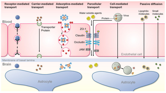
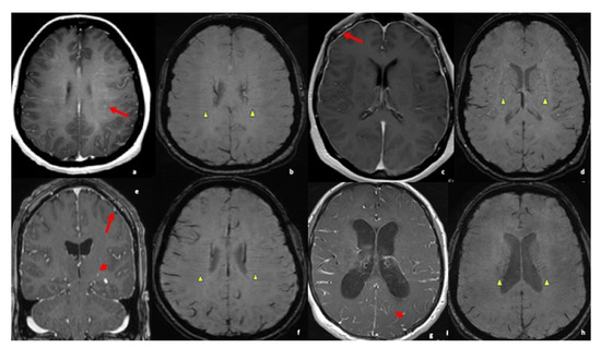
Stroke has become the first cause of functional disability and one of the leading causes of mortality worldwide. Therefore, it is of crucial importance to develop accurate biomarkers to assess stroke risk and prognosis. Emerging evidence suggests that neutrophil extracellular trap (NET) levels
[...] Read more.
Stroke has become the first cause of functional disability and one of the leading causes of mortality worldwide. Therefore, it is of crucial importance to develop accurate biomarkers to assess stroke risk and prognosis. Emerging evidence suggests that neutrophil extracellular trap (NET) levels may serve as a valuable biomarker to predict stroke occurrence and functional outcome. NETs are known to create a procoagulant state by serving as a scaffold for tissue factor (TF) and platelets inducing thrombosis by activating coagulation pathways and endothelium. A literature search was conducted in two databases (MEDLINE and Scopus) to trace all relevant studies published between 1 January 2016 and 31 December 2022, addressing the potential utility of NETs as a stroke biomarker. Only full-text articles in English were included. The current review includes thirty-three papers. Elevated NET levels in plasma and thrombi seem to be associated with increased mortality and worse functional outcomes in stroke, with all acute ischemic stroke, intracerebral hemorrhage, and subarachnoid hemorrhage included. Additionally, higher NET levels seem to correlate with worse outcomes after recanalization therapies and are more frequently found in strokes of cardioembolic or cryptogenic origin. Additionally, total neutrophil count in plasma seems also to correlate with stroke severity. Overall, NETs may be a promising predictive tool to assess stroke severity, functional outcome, and response to recanalization therapies.
Full article
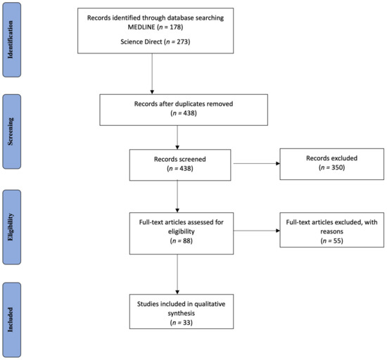
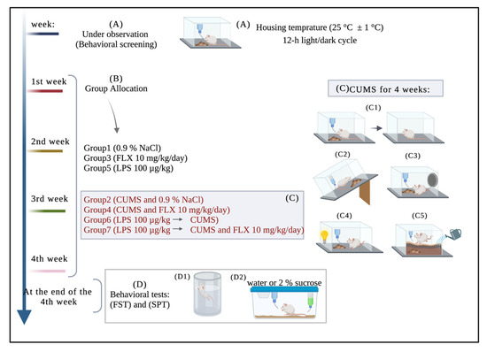
Figure 1

