

Meeting Contemporary Challenges: Development of Nanomaterials for Veterinary Medicine
 , ,
, ,
Abstract
:1. Introduction
2. Methodology
2.1. Search Strategy and Selection Criteria
2.2. Data Extraction and Analysis
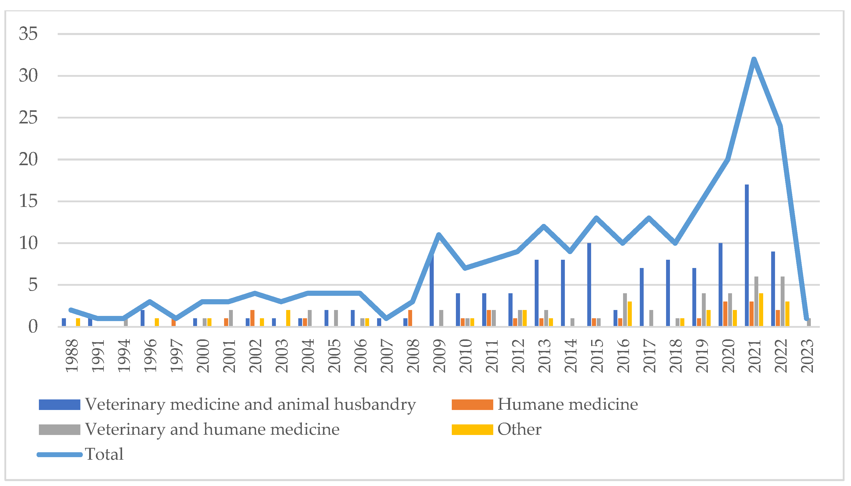
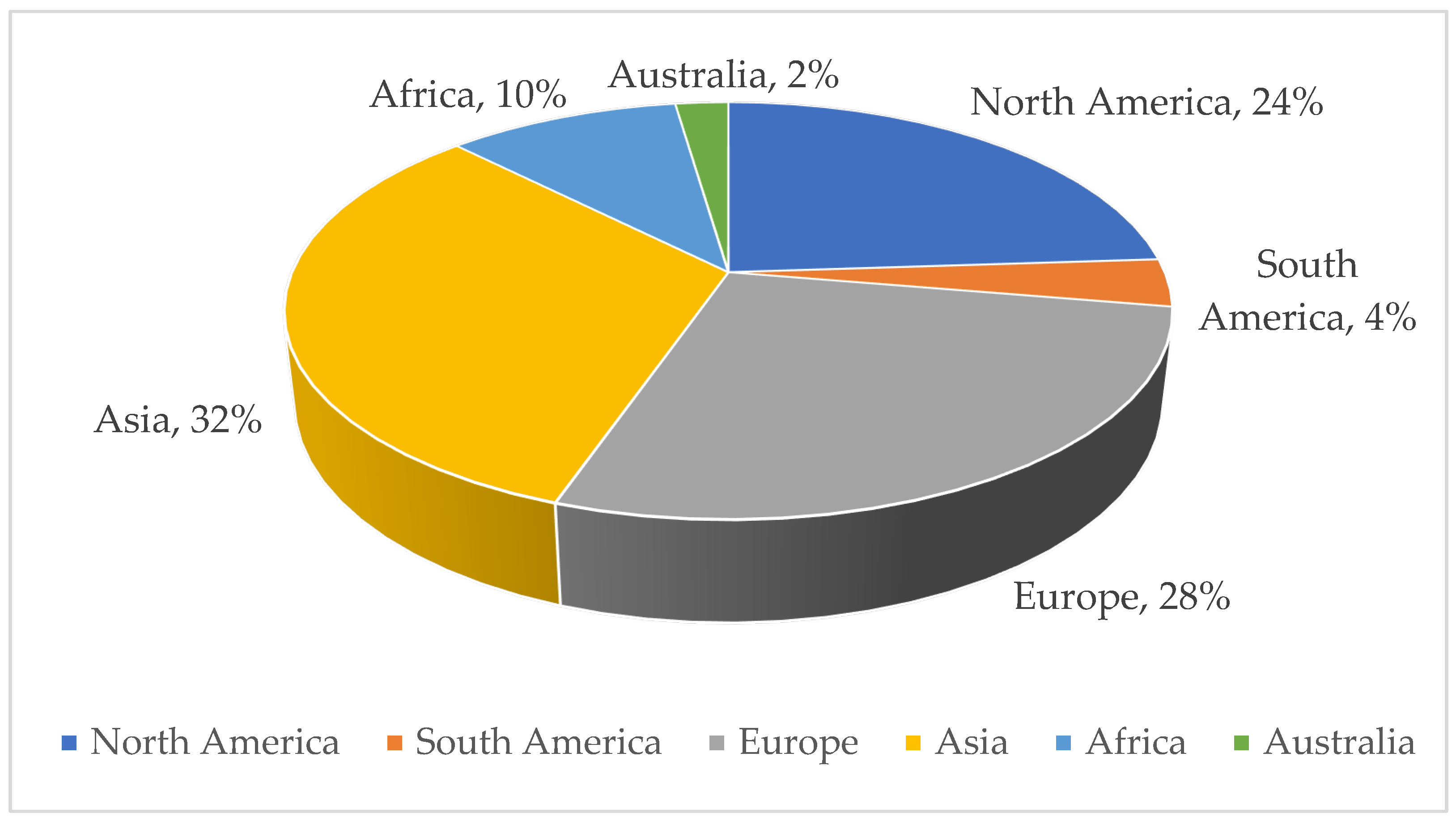
2.3. Literature Search and General Characteristics of the Studies Included
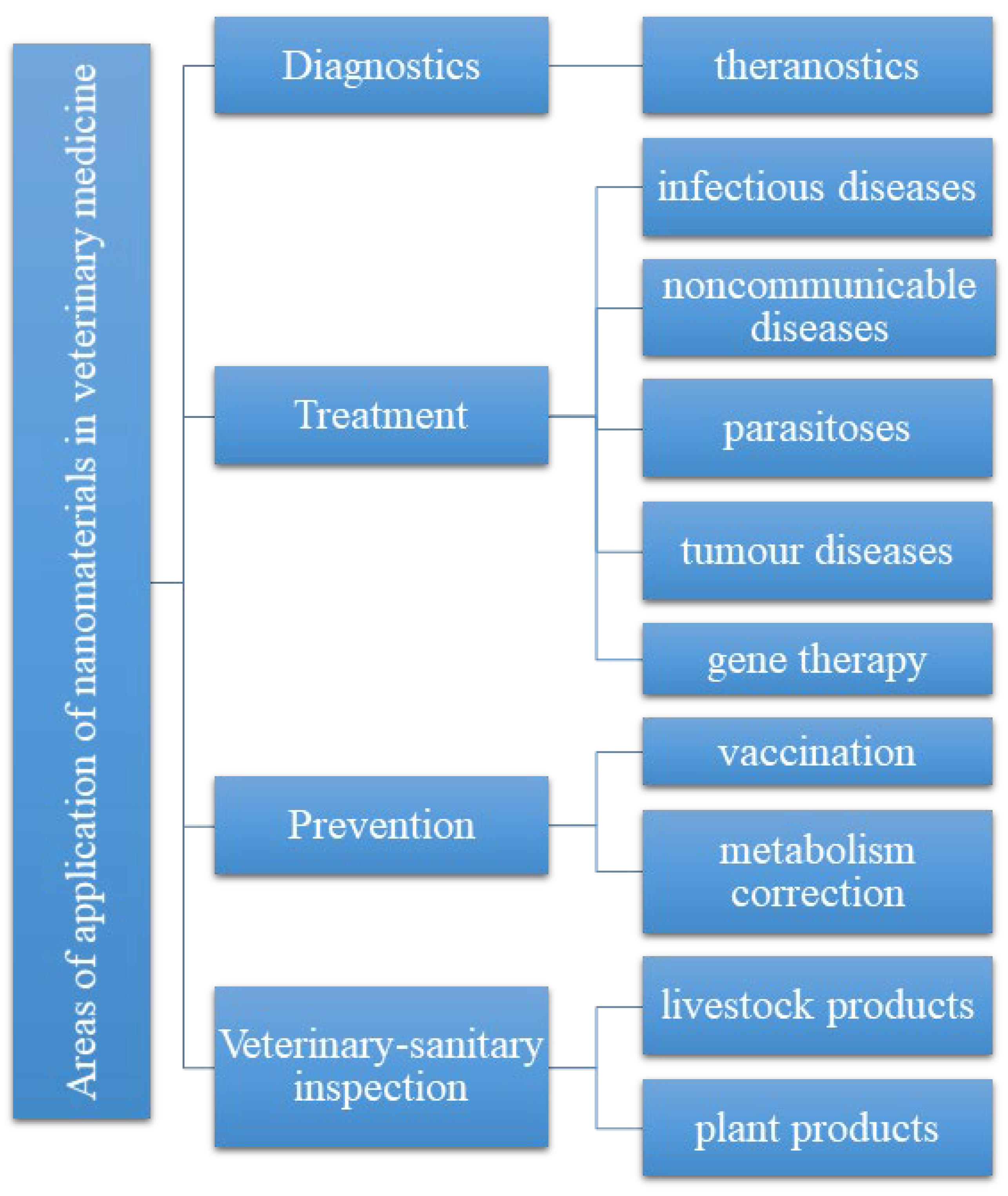
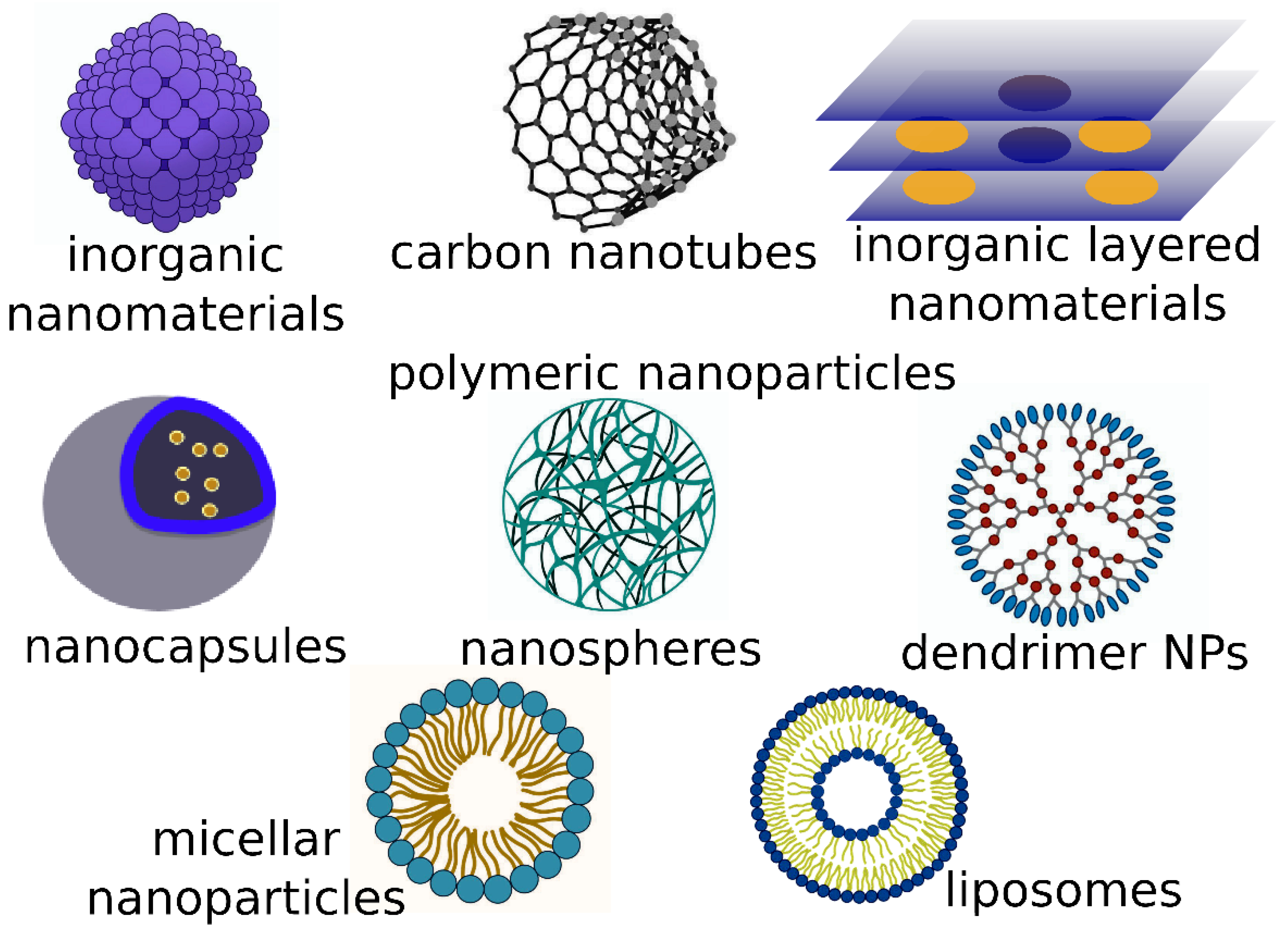
3. Current Applications of Nanomaterials in Veterinary Medicine
3.1. Diagnostics
3.2. Treatment
3.2.1. Antibiotics and Antibiotic Resistance
3.2.2. Antiparasitic Properties of Nanoparticles
3.2.3. Tissue Scaffolds (Electrospun)
3.2.4. Administration of Nanoformulations in Neoplasms
3.2.5. Gene Therapy
3.3. Innovative Nanovaccines

| Host/Pathogens | Vehicle | Target Antigen/ Vaccination Route | Immune Responses | Protection/Ref. |
|---|---|---|---|---|
| Chicken/AIV | pHEMA | H6/IM | Ab response | Reduced virus shedding/[160] |
| Turkey/ C. psittaci | Branched PEI | OmpA/IM | IgG and increased CD4/CD8 rate response | Reduced C. psittaci shedding, shortened clinical sign period/[161] |
| Chicken/NDV | Lipofectin | F and HN/IM | Anti-F Ab | 80%/[162] |
| Chicken/NDV | Chitosan NPs | F/IM/NAS | IgA/IgG and lymphocyte proliferation | IM: 80%/[163] |
| IN: 100%/[163] | ||||
| Turkey/TCoV | Naked plasmid + PEI and sodium hyaluronate | 4F, 4R/IM | Anti-TCoV S Ab and VN titer | Decrease in clinical signs from 5/5 to 1/5 or 2/5/[148] |
| Chicken/NDV | Nano-chitosan | F/IM/NAS | IgG and IgA and lymphocyte proliferation | 80% (IM); 100% (i.n.)/[164] |
| Egg embryonation/IBDV | Naked plasmid/killed vaccine | VP2, VP3, VP4 + killed virus booster/IO/IM | Anti-IBDV Ab and lymphocyte proliferation | 100%/[165] |
| Chicken/IBDV | Poly lactic-co-glycolic acid (PLGA) | VP2/IM, PO, OU | Stimulation of CD4 and CD8 T cells, high level of IgG | 80%/[145] |
3.4. Application of Nanotechnology to Increase Productivity and Prevent Animal Diseases
4. Risks and Hazards of Nanotechnology Applications in Veterinary Medicine
5. Legal Regulation of Nanomaterials Implemented in Veterinary Medicine and Agriculture
6. Prospects for Nanotechnology in Veterinary Medicine
7. Conclusions
Author Contributions
Funding
Institutional Review Board Statement
Informed Consent Statement
Data Availability Statement
Acknowledgments
Conflicts of Interest
Abbreviations
| Ab | Antibody |
| AIV | Avian influenza virus |
| Anti-F | Antibody to fibrinogen/fibrin-related products |
| APEC | Avian pathogenic Escherichia coli |
| BLSOmp31 | Recombinant polymeric B. ovis antigen |
| BMSC | Bone marrow stem cells |
| BPI3V | Bovine parainfluenza 3 virus |
| BRSV | Bovine respiratory syncytial virus |
| CD | Cluster of differentiation |
| CLPEI | Disulfide-crosslinked polyethyleneimine |
| CS | Chitosan |
| CT | Cholera toxin |
| DNA | Deoxyribonucleic acid |
| DOC | Department of Commerce |
| Dox | Doxorubicin |
| DPV | Duck plague virus |
| EBOV | Ebola virus |
| ELISA | Enzyme-linked immunosorbent assay |
| EU | European Union |
| FMD | Fibromuscular dysplasia |
| GMP | Good manufacturing practice |
| HAp | Hydroxyapatite |
| HN | High nitrogen |
| IBDV | Infectious bursal disease virus |
| IBV | Infectious bronchitis virus |
| Ig | Immunoglobulin |
| IM | Intramuscular |
| IO | In ovo, in the egg |
| ISAV | Infectious salmon anemia virus |
| ISCOM | Immunostimulating complex |
| IV | Intravenous |
| LAB | Lactic acid bacteria |
| LCNPs | Liquid crystalline nanoparticles |
| LPS | Lipopolysaccharide |
| MIC3 | Microneme protein of T. gondii |
| mRNA | Messenger RNA |
| NAS | Nasal, administered by way of the nose |
| NBICS | Nanotechnology, biotechnology, information technology, and cognitive science |
| NDV | Newcastle disease virus |
| NIR | Near-infrared (part of spectra) |
| NPs | Nanoparticles |
| OMPA | Octamethyl pyrophosphoramide |
| OU | oftalmic, eye drops |
| PC | Phosphatidylcholine |
| PCa | Prostate cancer |
| PEG | Polyethylene glycol |
| PEI | Polyethyleneimine |
| pHEMA | Polyhydroxyethyl methacrylate |
| PLG | Poly(D,L-lactide-co-glycolide) |
| PLGA | Poly(D,L-lactide-co-glycolide) acid |
| PMWS | Post-weaning multisystem wasting syndrome |
| PO | Per os, by mouth, orally |
| PS | Phosphatidylserine |
| PVAl | Poly(vinyl alcohol) |
| QD | Quantum dot |
| Quil A | Vaccine adjuvant saponin obtained from Quillaja saponaria |
| RNA | Ribonucleic acid |
| SA | Stearylamine |
| SAG1 | SAG1-related sequence |
| SPF | Specific-pathogen-free |
| SVP | Synthetic vaccine particle |
| TCoV | Turkey sweat coronavirus |
| VN | Virus neutralization |
| VP | Viral protein |
References
- King, L.J. Veterinary Medicine and Public Health at CDC. MMWR Morb. Mortal. Wkly. Rep. 2006, 55, 7–9. [Google Scholar]
- Rosol, T.J.; Moore, R.M.; Saville, W.J.A.; Oglesbee, M.J.; Rush, L.J.; Mathes, L.E.; Lairmore, M.D. The Need for Veterinarians in Biomedical Research. J. Vet. Med. Educ. 2009, 36, 70–75. [Google Scholar] [CrossRef]
- Tielkiniena, T.; Inna, G.; Tatyana, S.; Hanna, D.; Viktoriia, N.; Alisa, S. Lobby Legalization-Legal Instrument for Ensuring State Subsidies to Leaders of Agricultural Producers. J. Adv. Res. Dyn. Control Syst. 2020, 12, 2340–2345. [Google Scholar] [CrossRef]
- Sharma, P.K.; Dutta, R.K.; Pandey, A.C. Advances in Multifunctional Magnetic Nanoparticles. Adv. Mater. Lett. 2011, 2, 246–263. [Google Scholar] [CrossRef]
- Patil, S.S.; Kore, K.B.; Kumar, P. Nanotechnology and Its Applications in Veterinary and Animal Science. Vet. World 2009, 2, 475–477. [Google Scholar] [CrossRef]
- Mohammad, Z.H.; Ahmad, F.; Ibrahim, S.A.; Zaidi, S. Application of Nanotechnology in Different Aspects of the Food Industry. Discov. Food 2022, 2, 12. [Google Scholar] [CrossRef]
- Youssef, F.S.; El-Banna, H.A.; Elzorba, H.Y.; Galal, A.M. Application of Some Nanoparticles in the Field of Veterinary Medicine. Int. J. Vet. Sci. Med. 2019, 7, 78–93. [Google Scholar] [CrossRef]
- Clasky, A.J.; Watchorn, J.D.; Chen, P.Z.; Gu, F.X. From Prevention to Diagnosis and Treatment: Biomedical Applications of Metal Nanoparticle-Hydrogel Composites. Acta Biomater. 2021, 122, 1–25. [Google Scholar] [CrossRef]
- Hanafy, M.H. Myconanotechnology in Veterinary Sector: Status Quo and Future Perspectives. Int. J. Vet. Sci. Med. 2018, 6, 270–273. [Google Scholar] [CrossRef]
- Mekonnen, G. Review on Application of Nanotechnology in Animal Health and Production. J. Nanomed. Nanotechnol. 2021, 12, 559. [Google Scholar]
- Manhas, P.K.; Quintela, I.A.; Wu, V.C.H. Enhanced Detection of Major Pathogens and Toxins in Poultry and Livestock with Zoonotic Risks Using Nanomaterials-Based Diagnostics. Front. Vet. Sci. 2021, 8, 673718. [Google Scholar] [CrossRef]
- Vrublevskaya, V.V.; Afanasyev, V.N.; Grinevich, A.A.; Skarga, Y.Y.; Gladyshev, P.P.; Ibragimova, S.A.; Krylsky, D.V.; Dezhurov, S.V.; Morenkov, O.S. A Sensitive and Specific Lateral Flow Assay for Rapid Detection of Antibodies against Glycoprotein B of Aujeszky’s Disease Virus. J. Virol. Methods 2017, 249, 175–180. [Google Scholar] [CrossRef] [PubMed]
- Yeo, S.-J.; Bao, D.T.; Seo, G.-E.; Bui, C.T.; Kim, D.T.H.; Anh, N.T.V.; Tien, T.T.T.; Linh, N.T.P.; Sohn, H.-J.; Chong, C.-K. Improvement of a Rapid Diagnostic Application of Monoclonal Antibodies against Avian Influenza H7 Subtype Virus Using Europium Nanoparticles. Sci. Rep. 2017, 7, 7933. [Google Scholar] [CrossRef] [PubMed]
- Yang, F.; Xiao, Y.; Chen, B.; Wang, L.; Liu, F.; Yao, H.; Wu, N.; Wu, H. Development of a Colloidal Gold-Based Immunochromatographic Strip Test Using Two Monoclonal Antibodies to Detect H7N9 Avian Influenza Virus. Virus Genes 2020, 56, 396–400. [Google Scholar] [CrossRef]
- Zou, X.; Huang, H.; Gao, Y.; Su, X. Detection of Avian Influenza Virus Based on Magnetic Silica Nanoparticles Resonance Light Scattering System. Analyst 2012, 137, 648–653. [Google Scholar] [CrossRef]
- Huang, J.; Xie, Z.Z.; Xie, Z.Z.; Luo, S.; Xie, L.; Huang, L.; Fan, Q.; Zhang, Y.; Wang, S.; Zeng, T. Silver Nanoparticles Coated Graphene Electrochemical Sensor for the Ultrasensitive Analysis of Avian Influenza Virus H7. Anal. Chim. Acta 2016, 913, 121–127. [Google Scholar] [CrossRef] [PubMed]
- Wu, W.; Zhao, S.; Mao, Y.; Fang, Z.; Lu, X.; Zeng, L. A Sensitive Lateral Flow Biosensor for Escherichia Coli O157: H7 Detection Based on Aptamer Mediated Strand Displacement Amplification. Anal. Chim. Acta 2015, 861, 62–68. [Google Scholar] [CrossRef]
- Wu, L.; Zhang, M.; Zhu, L.; Li, J.; Li, Z.; Xie, W. Nanozyme-Linked Immunosorbent Assay for Porcine Circovirus Type 2 Antibody Using HAuCl4/H2O2 Coloring System. Microchem. J. 2020, 157, 105079. [Google Scholar] [CrossRef]
- Luo, B.; Xu, Y.; Wu, S.; Zhao, M.; Jiang, P.; Shi, S.; Zhang, Z.; Wang, Y.; Wang, L.; Liu, Y. A Novel Immunosensor Based on Excessively Tilted Fiber Grating Coated with Gold Nanospheres Improves the Detection Limit of Newcastle Disease Virus. Biosens. Bioelectron. 2018, 100, 169–175. [Google Scholar] [CrossRef]
- Yang, F.; Li, Y.; Jin, X.; Xu, Q.; Cheng, F.; Wang, X. Immunosensor-based Rapid Quantitative Detection of Newcastle Disease Virus Antibodies Using Innovative Gold Immunochromatographic Assay. J. Appl. Microbiol. 2020, 129, 1751–1757. [Google Scholar] [CrossRef]
- Wang, G.; Xie, P.; Xiao, C.; Yuan, P.; Su, X. Magnetic Fluorescent Composite Nanoparticles for the Fluoroimmunoassays of Newcastle Disease Virus and Avian Virus Arthritis Virus. J. Fluoresc. 2010, 20, 499–506. [Google Scholar] [CrossRef]
- Kurkela, S.; Brown, D.W.G. Molecular Diagnostic Techniques. Medicine 2009, 37, 535–540. [Google Scholar] [CrossRef] [PubMed]
- Deng, H.; Zhang, X.X.; Kumar, A.; Zou, G.; Zhang, X.X.; Liang, X.-J. Long Genomic DNA Amplicons Adsorption onto Unmodified Gold Nanoparticles for Colorimetric Detection of Bacillus Anthracis. Chem. Commun. 2013, 49, 51–53. [Google Scholar] [CrossRef] [PubMed]
- Karimi, F.; Dabbagh, S. Gel Green Fluorescence SsDNA Aptasensor Based on Carbon Nanotubes for Detection of Anthrax Protective Antigen. Int. J. Biol. Macromol. 2019, 140, 842–850. [Google Scholar] [CrossRef] [PubMed]
- Pal, D.; Boby, N.; Kumar, S.; Kaur, G.; Ali, S.A.; Reboud, J.; Shrivastava, S.; Gupta, P.K.; Cooper, J.M.; Chaudhuri, P. Visual Detection of Brucella in Bovine Biological Samples Using DNA-Activated Gold Nanoparticles. PLoS ONE 2017, 12, e0180919. [Google Scholar] [CrossRef] [PubMed]
- Sattarahmady, N.; Tondro, G.H.; Gholchin, M.; Heli, H. Gold Nanoparticles Biosensor of Brucella Spp. Genomic DNA: Visual and Spectrophotometric Detections. Biochem. Eng. J. 2015, 97, 1–7. [Google Scholar] [CrossRef]
- Li, X.; Zhu, P.; Liu, C.; Pang, H. One Step Synthesis of Boron-Doped Carbon Nitride Derived from 4-Pyridylboronic Acid as Biosensing Platforms for Assessment of Food Safety. Chem. Commun. 2019, 55, 9160–9163. [Google Scholar] [CrossRef]
- Nodoushan, S.M.; Nasirizadeh, N.; Kachuei, R.; Fooladi, A.A.I. Electrochemical Detection of Aflatoxin B1: An Aptasensor Prepared Using Graphene Oxide and Gold Nanowires. Anal. Methods 2019, 11, 6033–6042. [Google Scholar] [CrossRef]
- Tan, H.; Ma, L.; Guo, T.; Zhou, H.; Chen, L.; Zhang, Y.; Dai, H.; Yu, Y. A Novel Fluorescence Aptasensor Based on Mesoporous Silica Nanoparticles for Selective and Sensitive Detection of Aflatoxin B1. Anal. Chim. Acta 2019, 1068, 87–95. [Google Scholar] [CrossRef]
- Fend, R.; Geddes, R.; Lesellier, S.; Vordermeier, H.M.; Corner, L.A.L.; Gormley, E.; Costello, E.; Hewinson, R.G.; Marlin, D.J.; Woodman, A.C.; et al. Use of an Electronic Nose to Diagnose Mycobacterium Bovis Infection in Badgers and Cattle. J. Clin. Microbiol. 2005, 43, 1745–1751. [Google Scholar] [CrossRef]
- Pavlou, A.K.; Magan, N.; McNulty, C.; Jones, J.M.; Sharp, D.; Brown, J.; Turner, A.P.F. Use of an Electronic Nose System for Diagnoses of Urinary Tract Infections. Biosens. Bioelectron. 2002, 17, 893–899. [Google Scholar] [CrossRef] [PubMed]
- Ping, W.; Yi, T.; Haibao, X.; Farong, S. A Novel Method for Diabetes Diagnosis Based on Electronic Nose. Biosens. Bioelectron. 1997, 12, 1031–1036. [Google Scholar] [CrossRef] [PubMed]
- Probert, C.S.J.; Jones, P.R.H.; Ratcliffe, N.M. A Novel Method for Rapidly Diagnosing the Causes of Diarrhoea. Gut 2004, 53, 58–61. [Google Scholar] [CrossRef] [PubMed]
- Lai, S.Y.; Deffenderfer, O.F.; Hanson, W.; Phillips, M.P.; Thaler, E.R. Identification of Upper Respiratory Bacterial Pathogens With the Electronic Nose. Laryngoscope 2002, 112, 975–979. [Google Scholar] [CrossRef]
- Liu, L.; Kong, F. Measuring Chemical Deterioration of Foods. In Chemical Changes during Processing and Storage of Foods; Academic Press: Cambridge, MA, USA, 2021; pp. 637–679. [Google Scholar] [CrossRef]
- Guadarrama, A.; Rodríguez-Méndez, M.L.; Sanz, C.; Ríos, J.L.; De Saja, J.A. Electronic Nose Based on Conducting Polymers for the Quality Control of the Olive Oil Aroma: Discrimination of Quality, Variety of Olive and Geographic Origin. Anal. Chim. Acta 2001, 432, 283–292. [Google Scholar] [CrossRef]
- Magan, N.; Pavlou, A.; Chrysanthakis, I. Milk-Sense: A Volatile Sensing System Recognises Spoilage Bacteria and Yeasts in Milk. Sensors Actuators B Chem. 2001, 72, 28–34. [Google Scholar] [CrossRef]
- Franco, A.M.R.; Grafova, I.; Soares, F.V.; Gentile, G.; Wyrepkowski, C.D.C.; Bolson, M.A.; Sargentini, E., Jr.; Carfagna, C.; Leskelä, M.; Grafov, A. Nanoscaled Hydrated Antimony (V) Oxide as a New Approach to First-Line Antileishmanial Drugs. Int. J. Nanomed. 2016, 11, 6771–6780. [Google Scholar] [CrossRef]
- Thorpe, M.F.; Tománek, D.; Enbody, R.J.; Enbody, R.J. Science and Application of Nanotubes; Springer Science & Business Media: Berlin, Germany, 2000; ISBN 0306463725. [Google Scholar]
- National Academies of Sciences, Engineering and Medicine. A Quadrennial Review of the National Nanotechnology Initiative: Nanoscience, Applications, and Commercialization; Academies Press: Cambridge, MA, USA, 2020. [Google Scholar]
- Park, K. Facing the Truth about Nanotechnology in Drug Delivery. ACS Nano 2013, 7, 7442–7447. [Google Scholar] [CrossRef]
- Cho, K.; Wang, X.U.; Nie, S.; Chen, Z.; Shin, D.M. Therapeutic Nanoparticles for Drug Delivery in Cancer. Clin. Cancer Res. 2008, 14, 1310–1316. [Google Scholar] [CrossRef]
- Sheikholeslami, B.; Lam, N.W.; Dua, K.; Haghi, M. Exploring the Impact of Physicochemical Properties of Liposomal Formulations on Their In Vivo Fate. Life Sci. 2022, 300, 120574. [Google Scholar] [CrossRef]
- Liu, Y.; Li, Y.; Shi, L. Controlled Drug Delivery Systems in Eradicating Bacterial Biofilm-Associated Infections. J. Control. Release 2021, 329, 1102–1116. [Google Scholar] [CrossRef] [PubMed]
- Thorn, C.R.; Kopecki, Z.; Wignall, A.; Kral, A.; Prestidge, C.A.; Thomas, N. Liquid Crystal Nanoparticle Platform for Increased Efficacy of Cationic Antimicrobials against Biofilm Infections. Nanomed. Nanotechnol. Biol. Med. 2022, 42, 102536. [Google Scholar] [CrossRef] [PubMed]
- Saeed, M.; Babazadeh, D.; Naveed, M.; Alagawany, M.; Abd El-Hack, M.E.; Arain, M.A.; Tiwari, R.; Sachan, S.; Karthik, K.; Dhama, K. In Ovo Delivery of Various Biological Supplements, Vaccines and Drugs in Poultry: Current Knowledge. J. Sci. Food Agric. 2019, 99, 3727–3739. [Google Scholar] [CrossRef]
- Awaad, M.H.H.; El Moustafa, K.M.; Zoulfakar, S.A.; Elhalawany, M.S.; Mohammed, F.F.; El-Refay, R.M.; Morsy, E.A. The Role of Silver Nanoparticles in the Reluctance of Colisepticemia in Broiler Chickens. J. Appl. Poult. Res. 2021, 30, 100155. [Google Scholar] [CrossRef]
- Mahmoud, U.T.; Darwish, M.H.A.; Ali, F.A.Z.; Amen, O.A.; Mahmoud, M.A.M.; Ahmed, O.B.; Abd El-Reda, G.; Osman, M.A.; Othman, A.A.; Abushahba, M.F.N. Zinc Oxide Nanoparticles Prevent Multidrug Resistant Staphylococcus-Induced Footpad Dermatitis in Broilers. Avian Pathol. 2021, 50, 214–226. [Google Scholar] [CrossRef] [PubMed]
- Reda, F.M.; El-Saadony, M.T.; El-Rayes, T.K.; Attia, A.I.; El-Sayed, S.A.A.; Ahmed, S.Y.A.; Madkour, M.; Alagawany, M. Use of Biological Nano Zinc as a Feed Additive in Quail Nutrition: Biosynthesis, Antimicrobial Activity and Its Effect on Growth, Feed Utilisation, Blood Metabolites and Intestinal Microbiota. Ital. J. Anim. Sci. 2021, 20, 324–335. [Google Scholar] [CrossRef]
- Ouyang, Z.; Ren, P.; Huang, L.; Wei, T.; Yang, C.; Kong, X.; Yin, Y.; He, S.; He, Q. Hydrothermal Synthesis of a New Porous Zinc Oxide and Its Antimicrobial Evaluation in Weanling Piglets. Livest. Sci. 2021, 248, 104499. [Google Scholar] [CrossRef]
- Zhang, M.; Song, W.; Tang, Y.; Xu, X.; Huang, Y.; Yu, D. Polymer-Based Nanofiber-Nanoparticle Hybrids and Their Medical Applications. Polymers 2022, 14, 351. [Google Scholar] [CrossRef]
- Troncarelli, M.Z.; Brandão, H.M.; Gern, J.C.; Guimarães, A.S.; Langoni, H. Nanotechnology and Antimicrobials in Veterinary Medicine. Formatex 2013, 13, 543–556. [Google Scholar]
- Hassan, A.A.; Mansour, M.K.; El Hamaky, A.M.; El Ahl, R.M.S.; Oraby, N.H. Nanomaterials and Nanocomposite Applications in Veterinary Medicine. In Multifunctional Hybrid Nanomaterials for Sustainable Agri-Food and Ecosystems; Elsevier: Amsterdam, The Netherlands, 2020; pp. 583–638. [Google Scholar]
- Bai, D.-P.; Lin, X.-Y.; Huang, Y.-F.; Zhang, X.-F. Theranostics Aspects of Various Nanoparticles in Veterinary Medicine. Int. J. Mol. Sci. 2018, 19, 3299. [Google Scholar] [CrossRef]
- MacLeod, D.L.; Prescott, J.F. The Use of Liposomally-Entrapped Gentamicin in the Treatment of Bovine Staphylococcus Aureus Mastitis. Can. J. Vet. Res. 1988, 52, 445. [Google Scholar] [PubMed]
- Cao, L.T.; Wu, J.Q.; Xie, F.; Hu, S.H.; Mo, Y. Efficacy of Nisin in Treatment of Clinical Mastitis in Lactating Dairy Cows. J. Dairy Sci. 2007, 90, 3980–3985. [Google Scholar] [CrossRef] [PubMed]
- Sachetelli, S.; Khalil, H.; Chen, T.; Beaulac, C.; Sénéchal, S.; Lagacé, J. Demonstration of a Fusion Mechanism between a Fluid Bactericidal Liposomal Formulation and Bacterial Cells. Biochim. Biophys. Acta (BBA)-Biomembr. 2000, 1463, 254–266. [Google Scholar] [CrossRef]
- Singla, S.; Harjai, K.; Katare, O.P.; Chhibber, S. Encapsulation of Bacteriophage in Liposome Accentuates Its Entry in to Macrophage and Shields It from Neutralizing Antibodies. PLoS ONE 2016, 11, e0153777. [Google Scholar] [CrossRef]
- Le Conte, P.; Le Gallou, F.; Potel, G.; Struillou, L.; Baron, D.; Drugeon, H.B. Pharmacokinetics, Toxicity, and Efficacy of Liposomal Capreomycin in Disseminated Mycobacterium Avium Beige Mouse Model. Antimicrob. Agents Chemother. 1994, 38, 2695–2701. [Google Scholar] [CrossRef]
- Cordeiro, C.; Wiseman, D.J.; Lutwyche, P.; Uh, M.; Evans, J.C.; Finlay, B.B.; Webb, M.S. Antibacterial Efficacy of Gentamicin Encapsulated in PH-Sensitive Liposomes against an In Vivo Salmonella Enterica Serovar Typhimurium Intracellular Infection Model. Antimicrob. Agents Chemother. 2000, 44, 533–539. [Google Scholar] [CrossRef] [PubMed]
- Zelenina, O.; Vlizlo, V.; Kozak, M.; Ostapiv, D.; Samaryk, V.; Dron, I.; Stetsko, T.; Skrypka, M.; Tomchuk, V.; Danchuk, O.; et al. Antimicrobial Activity of the PEGylated Antibiotic Enrofloxacin and Its Functional and Structural Effect on the Liver in Rats ARTICLE INFO. J. Appl. Pharm. Sci. 2022, 12, 68–075. [Google Scholar] [CrossRef]
- Baltazar, L.M.; Werneck, S.M.C.; Carneiro, H.C.S.; Gouveia, L.F.; De Paula, T.P.; Byrro, R.M.D.; Cunha, A.S., Jr.; Soares, B.M.; Ferreira, M.V.L.; Souza, D.G.; et al. Photodynamic Therapy Efficiently Controls Dermatophytosis Caused by Trichophyton Rubrum in a Murine Model. Br. J. Dermatol. 2015, 172, 801–804. [Google Scholar] [CrossRef]
- Krawiec, D.R.; McKiernan, B.C.; Twardock, A.R.; Swenson, C.E.; Itkin, R.J.; Johnson, L.R.; Kurowsky, L.K.; Marks, C.A. Use of an Amphotericin B Lipid Complex for Treatment of Blastomycosis in Dogs. J. Am. Vet. Med. Assoc. 1996, 209, 2073–2075. [Google Scholar]
- Leenders AC, A.P.; Marie, S.D. The Use of Lipid Formulations of Amphotericin B for Systemic Fungal Infections. Leukemia 1996, 10, 1570–1575. [Google Scholar]
- Al-Qushawi, A.; Rassouli, A.; Atyabi, F.; Peighambari, S.M.; Esfandyari-Manesh, M.; Shams, G.R.; Yazdani, A. Preparation and Characterization of Three Tilmicosin-Loaded Lipid Nanoparticles: Physicochemical Properties and in-Vitro Antibacterial Activities. Iran. J. Pharm. Res. IJPR 2016, 15, 663. [Google Scholar]
- Panáček, A.; Kvítek, L.; Smékalová, M.; Večeřová, R.; Kolář, M.; Röderová, M.; Dyčka, F.; Šebela, M.; Prucek, R.; Tomanec, O. Bacterial Resistance to Silver Nanoparticles and How to Overcome It. Nat. Nanotechnol. 2018, 13, 65–71. [Google Scholar] [CrossRef] [PubMed]
- Parmar, S.; Kaur, H.; Singh, J.; Matharu, A.S.; Ramakrishna, S.; Bechelany, M. Recent Advances in Green Synthesis of Ag NPs for Extenuating Antimicrobial Resistance. Nanomaterials 2022, 12, 115. [Google Scholar] [CrossRef]
- Fondevila, M.; Herrer, R.; Casallas, M.C.; Abecia, L.; Ducha, J.J. Silver Nanoparticles as a Potential Antimicrobial Additive for Weaned Pigs. Anim. Feed Sci. Technol. 2009, 150, 259–269. [Google Scholar] [CrossRef]
- Dung, T.T.N.; Nam, V.N.; Nhan, T.T.; Ngoc, T.T.B.; Minh, L.Q.; Nga, B.T.T.; Quang, D.V. Silver Nanoparticles as Potential Antiviral Agents against African Swine Fever Virus. Mater. Res. Express 2020, 6, 1250g9. [Google Scholar] [CrossRef]
- Wernicki, A.; Puchalski, A.; Urban-Chmiel, R.; Dec, M.; Stegierska, D.; Dudzic, A.; Wojcik, A. Antimicrobial Properties of Gold, Silver, Copper and Platinum Nanoparticles against Selected Microorganisms Isolated from Cases of Mastitis in Cattle. Med. Weter 2014, 70, 564–567. [Google Scholar]
- Kowalczyk, P.; Szymczak, M.; Maciejewska, M.; Laskowski, Ł.; Laskowska, M.; Ostaszewski, R.; Skiba, G.; Franiak-Pietryga, I. Molecular Sciences All That Glitters Is Not Silver-A New Look at Microbiological and Medical Applications of Silver Nanoparticles. Int. J. Mol. Sci. 2021, 22, 854. [Google Scholar] [CrossRef]
- Amaro, F.; Morón, Á.; Díaz, S.; Martín-González, A.; Gutiérrez, J.C. Metallic Nanoparticles—Friends or Foes in the Battle against Antibiotic-Resistant Bacteria? Microorganisms 2021, 9, 364. [Google Scholar] [CrossRef]
- Wahab, S.; Khan, T.; Adil, M.; Khan, A. Mechanistic Aspects of Plant-Based Silver Nanoparticles against Multi-Drug Resistant Bacteria. Heliyon 2021, 7, e07448. [Google Scholar] [CrossRef]
- Leng, D.; Li, Y.; Zhu, J.; Liang, R.; Zhang, C.; Zhou, Y.; Li, M.; Wang, Y.; Rong, D.; Wu, D. The Antibiofilm Activity and Mechanism of Nanosilver-and Nanozinc-Incorporated Mesoporous Calcium-Silicate Nanoparticles. Int. J. Nanomed. 2020, 15, 3921. [Google Scholar] [CrossRef]
- Hozyen, H.F.; Ibrahim, E.S.; Khairy, E.A.; El-Dek, S.I. Enhanced Antibacterial Activity of Capped Zinc Oxide Nanoparticles: A Step towards the Control of Clinical Bovine Mastitis. Vet. World 2019, 12, 1225. [Google Scholar] [CrossRef] [PubMed]
- Mohd Yusof, H.; Mohamad, R.; Zaidan, U.H.; Rahman, A. Microbial Synthesis of Zinc Oxide Nanoparticles and Their Potential Application as an Antimicrobial Agent and a Feed Supplement in Animal Industry: A Review. J. Anim. Sci. Biotechnol. 2019, 10, 57. [Google Scholar] [CrossRef] [PubMed]
- Jaguezeski, A.M.; Souza, C.F.; Perin, G.; Reis, J.H.; Gomes, T.M.A.; Baldissera, M.D.; Vaucher, R.A.; de Andrade, C.M.; Stefani, L.M.; Gundel, S.S.; et al. Effect of Free and Nano-Encapsulated Curcumin on Treatment and Energetic Metabolism of Gerbils Infected by Listeria Monocytogenes. Microb. Pathog. 2019, 134, 103564. [Google Scholar] [CrossRef] [PubMed]
- Granada, L.; Sousa, N.; Lopes, S.; Lemos, M.F.L. Is Integrated Multitrophic Aquaculture the Solution to the Sectors’ Major Challenges?—A Review. Rev. Aquac. 2016, 8, 283–300. [Google Scholar] [CrossRef]
- Dar, A.H.; Rashid, N.; Majid, I.; Hussain, S.; Dar, M.A. Nanotechnology Interventions in Aquaculture and Seafood Preservation. Crit. Rev. Food Sci. Nutr. 2020, 60, 1912–1921. [Google Scholar] [CrossRef]
- Nasr-Eldahan, S.; Nabil-Adam, A.; Shreadah, M.A.; Maher, A.M.; El-Sayed Ali, T. A Review Article on Nanotechnology in Aquaculture Sustainability as a Novel Tool in Fish Disease Control. Aquac. Int. 2021, 29, 1459–1480. [Google Scholar] [CrossRef]
- Louros, V.L.; Ferreira, L.M.; Silva, V.G.; Silva, C.P.; Martins, M.A.; Otero, M.; Esteves, V.I.; Lima, D.L.D. Photodegradation of Aquaculture Antibiotics Using Carbon Dots-TiO2 Nanocomposites. Toxics 2021, 9, 330. [Google Scholar] [CrossRef]
- Cheng, T.C.; Yao, K.S.; Yeh, N.; Chang, C.I.; Hsu, H.C.; Gonzalez, F.; Chang, C.Y. Bactericidal Effect of Blue LED Light Irradiated TiO2/Fe3O4 Particles on Fish Pathogen in Seawater. Thin Solid Films 2011, 519, 5002–5006. [Google Scholar] [CrossRef]
- Vaseeharan, B.; Ramasamy, P.; Chen, J.C. Antibacterial Activity of Silver Nanoparticles (AgNps) Synthesized by Tea Leaf Extracts against Pathogenic Vibrio Harveyi and Its Protective Efficacy on Juvenile Feneropenaeus Indicus. Lett. Appl. Microbiol. 2010, 50, 352–356. [Google Scholar] [CrossRef]
- Barbosa, A.C.M.S.; Costa Silva, L.P.; Ferraz, C.M.; Tobias, F.L.; De Araújo, J.V.; Loureiro, B.; Braga, G.M.A.M.; Veloso, F.B.R.; Soares, F.E.D.F.; Fronza, M.; et al. Nematicidal Activity of Silver Nanoparticles from the Fungus Duddingtonia Flagrans. Int. J. Nanomed. 2019, 14, 2341–2348. [Google Scholar] [CrossRef]
- Abd El Megid, A.D.; Khaled, M.; Emam, M.A.; Adel, A. Biochemical Role of Zinc Oxide and Propolis Nanoparticles in Protection Rabbits against Coccidiosis. Benha Vet. Med. J. 2018, 34, 314–328. [Google Scholar] [CrossRef]
- Chauke, N.; Siebrits, F.K. Evaluation of Silver Nanoparticles as a Possible Coccidiostat in Broiler Production. S. Afr. J. Anim. Sci. 2012, 42, 493–497. [Google Scholar] [CrossRef]
- Grafov, A.; Grafova, I.; Pereira, A.M.R.F.; Leskelä, M.A. Process of preparation of nanohybrid material, pharmaceutical composition and use of the same. Patent BR 10 2013 029618 0 (2013), 18 November 2013. [Google Scholar]
- Hiszczyńska-Sawicka, E.; Li, H.; Xu, J.; Akhtar, M.; Holec-Gąsior, L.; Kur, J.; Bickerstaffe, R.; Stankiewicz, M. Induction of Immune Responses in Sheep by Vaccination with Liposome-Entrapped DNA Complexes Encoding Toxoplasma Gondii MIC3 Gene. Pol. J. Vet. Sci. 2012, 15, 3–9. [Google Scholar] [CrossRef] [PubMed]
- Heo, S.Y.; Kim, H.Y.; Kim, N.S. Evaluation of Poly(Lactide-Co-Glycolide)/Hydroxyapatite Nanofibres for Reconstruction of Critical-Sized Segmental Bone Defects in a Canine Model. Vet. Med. 2017, 62, 325–332. [Google Scholar] [CrossRef]
- Kim, P.-H.; Cho, J.-Y. Myocardial Tissue Engineering Using Electrospun Nanofiber Composites. BMB Rep. 2016, 49, 26–36. [Google Scholar] [CrossRef] [PubMed]
- Wang, T.; Jiang, X.-J.; Tang, Q.-Z.; Li, X.-Y.; Lin, T.; Wu, D.-Q.; Zhang, X.-Z.; Okello, E. Bone Marrow Stem Cells Implantation with α-Cyclodextrin/MPEG–PCL–MPEG Hydrogel Improves Cardiac Function after Myocardial Infarction. Acta Biomater. 2009, 5, 2939–2944. [Google Scholar] [CrossRef]
- Souto, E.B.; Silva, G.F.; Dias-ferreira, J.; Zielinska, A.; Ventura, F.; Durazzo, A.; Lucarini, M.; Novellino, E.; Santini, A. Nanopharmaceutics: Part I—Clinical Trials Legislation and Good Manufacturing Practices (GMP) of Nanotherapeutics in the EU. Pharmaceutics 2020, 12, 146. [Google Scholar] [CrossRef]
- Sapino, S.; Chindamo, G.; Chirio, D.; Morel, S.; Peira, E.; Vercelli, C.; Gallarate, M. Nanocarriers in Veterinary Medicine: A Challenge for Improving Osteosarcoma Conventional Treatments. Nanomaterials 2022, 12, 4501. [Google Scholar] [CrossRef]
- Lin, T.; Rodriguez, C.O.; Li, Y. Nanomedicine in Veterinary Oncology. Vet. J. 2015, 205, 189–197. [Google Scholar] [CrossRef]
- Bhatia, S.N.; Chen, X.; Dobrovolskaia, M.A.; Lammers, T. Cancer Nanomedicine. Nat. Rev. Cancer 2022, 22, 550–556. [Google Scholar] [CrossRef]
- Trafton, A. How Different Cancer Cells Respond to Drug-Delivering Nanoparticles. Massachusetts Institute of Technology. 21 July 2022. Available online: https://news.mit.edu/2022/how-different-cancer-cells-respond-drug-delivering-nanoparticles-0721 (accessed on 29 May 2023).
- Maeda, H. Macromolecular Therapeutics in Cancer Treatment: The EPR Effect and Beyond. J. Control. Release 2012, 164, 138–144. [Google Scholar] [CrossRef] [PubMed]
- De Jong, W.H.; Borm, P.J.A. Drug Delivery and Nanoparticles: Applications and Hazards. Int. J. Nanomed. 2008, 3, 133–149. [Google Scholar] [CrossRef] [PubMed]
- Yoo, B.; Ross, A.; Pantazopoulos, P.; Medarova, Z. MiRNA10b-Directed Nanotherapy Effectively Targets Brain Metastases from Breast Cancer. Sci. Rep. 2021, 11, 2844. [Google Scholar] [CrossRef]
- Moles, E.; Kavallaris, M. A Potent Targeted Cancer Nanotherapeutic. Nat. Biomed. Eng. 2019, 3, 248–250. [Google Scholar] [CrossRef]
- Xie, Y.; Hang, Y.; Wang, Y.; Sleightholm, R.; Prajapati, D.R.; Bader, J.; Yu, A.; Tang, W.; Jaramillo, L.; Li, J.; et al. Stromal Modulation and Treatment of Metastatic Pancreatic Cancer with Local Intraperitoneal Triple MiRNA/SiRNA Nanotherapy. ACS Nano 2020, 14, 255–271. [Google Scholar] [CrossRef] [PubMed]
- Rowell, J.L.; McCarthy, D.O.; Alvarez, C.E. Dog Models of Naturally Occurring Cancer. Trends Mol. Med. 2011, 17, 380–388. [Google Scholar] [CrossRef] [PubMed]
- English, H.; Hong, J.; Ho, M. Ancient Species Offers Contemporary Therapeutics: An Update on Shark VNAR Single Domain Antibody Sequences, Phage Libraries and Potential Clinical Applications. Antib. Ther. 2020, 3, 1–9. [Google Scholar] [CrossRef]
- Jovčevska, I.; Muyldermans, S. The Therapeutic Potential of Nanobodies. BioDrugs 2019, 34, 11–26. [Google Scholar] [CrossRef]
- De Maria, R.; Olivero, M.; Iussich, S.; Nakaichi, M.; Murata, T.; Biolatti, B.; Di Renzo, M.F. Spontaneous Feline Mammary Carcinoma Is a Model of HER2 Overexpressing Poor Prognosis Human Breast Cancer. Cancer Res. 2005, 65, 907–912. [Google Scholar] [CrossRef]
- Abdoon, A.S.; Al-Ashkar, E.A.; Kandil, O.M.; Shaban, A.M.; Khaled, H.M.; El Sayed, M.A.; El Shaer, M.M.; Shaalan, A.H.; Eisa, W.H.; Eldin, A.A.G.; et al. Efficacy and Toxicity of Plasmonic Photothermal Therapy (PPTT) Using Gold Nanorods (GNRs) against Mammary Tumors in Dogs and Cats. Nanomedicine 2016, 12, 2291–2297. [Google Scholar] [CrossRef]
- Axiak, S.M.; Selting, K.A.; Decedue, C.J.; Henry, C.J.; Tate, D.; Howell, J.; Bilof, K.J.; Kim, D.Y. Phase I Dose Escalation Safety Study of Nanoparticulate Paclitaxel (CTI 52010) in Normal Dogs. Int. J. Nanomed. 2011, 6, 2205–2212. [Google Scholar] [CrossRef] [PubMed]
- Alrushaid, N.; Khan, F.A.; Al-Suhaimi, E.A.; Elaissari, A. Nanotechnology in Cancer Diagnosis and Treatment. Pharmaceutics 2023, 15, 1025. [Google Scholar] [CrossRef] [PubMed]
- Feldhaeusser, B.; Platt, S.R.; Marrache, S.; Kolishetti, N.; Pathak, R.K.; Montgomery, D.J.; Reno, L.R.; Howerth, E.; Dhar, S. Evaluation of Nanoparticle Delivered Cisplatin in Beagles. Nanoscale 2015, 7, 13822–13830. [Google Scholar] [CrossRef] [PubMed]
- Małek, A.; Taciak, B.; Sobczak, K.; Grzelak, A.; Wójcik, M.; Mieczkowski, J.; Lechowski, R.; Zabielska-Koczywaş, K.A. Enhanced Cytotoxic Effect of Doxorubicin Conjugated to Glutathione-Stabilized Gold Nanoparticles in Canine Osteosarcoma-In Vitro Studies. Molecules 2021, 26, 3487. [Google Scholar] [CrossRef] [PubMed]
- Reznikov, A.G.; Faliush, O.A.; Nosenko, N.D.; Sachynska, O.V.; Polyakova, L.I.; Limareva, A.A.; Perchyk, I.G. Study of the Effects of Gold and Cerium Dioxide Nanoparticles on Normal and Cancer Cells and Tissues. Issues Dev. Med. Med. Res. 2022, 11, 52–61. [Google Scholar] [CrossRef]
- Zabielska-Koczywąs, K.; Lechowski, R. The Use of Liposomes and Nanoparticles as Drug Delivery Systems to Improve Cancer Treatment in Dogs and Cats. Molecules 2017, 22, 2167. [Google Scholar] [CrossRef]
- Wang, Z.; Guo, W.; Kuang, X.; Hou, S.; Liu, H. Nanopreparations for Mitochondria Targeting Drug Delivery System: Current Strategies and Future Prospective. Asian J. Pharm. Sci. 2017, 12, 498–508. [Google Scholar] [CrossRef]
- He, H.; Guo, J.; Xu, B. Enzymatic Delivery of Magnetic Nanoparticles into Mitochondria of Live Cells. ChemNanoMat 2021, 7, 1104–1107. [Google Scholar] [CrossRef]
- Hirsch, L.R.; Stafford, R.J.; Bankson, J.A.; Sershen, S.R.; Rivera, B.; Price, R.E.; Hazle, J.D.; Halas, N.J.; West, J.L. Nanoshell-Mediated near-Infrared Thermal Therapy of Tumors under Magnetic Resonance Guidance. Proc. Natl. Acad. Sci. USA 2003, 100, 13549–13554. [Google Scholar] [CrossRef]
- Pallares, R.M.; Mottaghy, F.M.; Schulz, V.; Kiessling, F.; Lammers, T. Nanoparticle Diagnostics and Theranostics in the Clinic. J. Nucl. Med. 2022, 63, 1802–1808. [Google Scholar] [CrossRef]
- Ke, C.H.; Sio, K.M.; Wang, S.L.; Kuo, Y.; Huang, W.H.; Lin, C.S. The High Expression of Legumain in Canine Neoplasms: A Retrospective Analysis of 100 Cases. Animals 2022, 12, 504. [Google Scholar] [CrossRef] [PubMed]
- Rajesh Kumar, T.; Anitha, S.; Sangavi, P.; Srinithi, R.; Langeswaran, K.; Sangeetha, R. Applications of Nanomedicine in Animal Models of Cancer. In Handbook of Animal Models and Its Uses in Cancer Research; Springer: Berlin/Heidelberg, Germany, 2022; pp. 1–14. [Google Scholar]
- Gruntman, A.M.; Flotte, T.R. Gene Therapy and the Use of Animal Models: Why Mice Alone Are Not Sufficient. Hum. Gene Ther. 2022, 33, 477–478. [Google Scholar] [CrossRef] [PubMed]
- Blagbrough, I.S.; Zara, C. Animal Models for Target Diseases in Gene Therapy—Using DNA and SiRNA Delivery Strategies. Pharm. Res. 2009, 26, 1–18. [Google Scholar] [CrossRef]
- Qin, S.; Tang, X.; Chen, Y.; Chen, K.; Fan, N.; Xiao, W.; Zheng, Q.; Li, G.; Teng, Y.; Wu, M.; et al. MRNA-Based Therapeutics: Powerful and Versatile Tools to Combat Diseases. Signal Transduct. Target. Ther. 2022, 7, 166. [Google Scholar] [CrossRef] [PubMed]
- Pavlin, D.; Cemazar, M.; Sersa, G.; Tozon, N. IL-12 Based Gene Therapy in Veterinary Medicine. J. Transl. Med. 2012, 10, 234. [Google Scholar] [CrossRef] [PubMed]
- Bassols, A.; Costa, C.; Eckersall, P.D.; Osada, J.; Sabrià, J.; Tibau, J. The Pig as an Animal Model for Human Pathologies: A Proteomics Perspective. Proteom. Clin. Appl. 2014, 8, 715–731. [Google Scholar] [CrossRef]
- Meurens, F.; Summerfield, A.; Nauwynck, H.; Saif, L.; Gerdts, V. The Pig: A Model for Human Infectious Diseases. Trends Microbiol. 2012, 20, 50–57. [Google Scholar] [CrossRef]
- Casal, M.; Haskins, M. Large Animal Models and Gene Therapy. Eur. J. Hum. Genet. 2006, 14, 266–272. [Google Scholar] [CrossRef]
- Chowdhury, J.R.; Grossman, M.; Gupta, S.; Chowdhury, N.R.; Baker, J.R.; Wilson, J.M. Long-Term Improvement of Hypercholesterolemia after Ex Vivo Gene Therapy in LDLR-Deficient Rabbits. Science 1991, 254, 1802–1805. [Google Scholar] [CrossRef]
- Logeart, D.; Hatem, S.N.; Heimburger, M.; Michel, J.B.; Mercadier, J.J.; Logeart, D.; Hatem, S.N.; Le Roux, A.; Hatem, S.N.; Mercadier, J.J. How to Optimize In Vivo Gene Transfer to Cardiac Myocytes: Mechanical or Pharmacological Procedures? Hum. Gene Ther. 2001, 12, 1601–1610. [Google Scholar] [CrossRef]
- Vite, C.H.; McGowan, J.C.; Niogi, S.; Passini, M.A.; Drobatz, K.J.; Haskins, M.E.; Wolfe, J.H. Effective Gene Therapy for an Inherited CNS Disease in a Large Animal Model. Ann. Neurol. 2005, 57, 355–364. [Google Scholar] [CrossRef] [PubMed]
- Sleeper, M.M. Status of Therapeutic Gene Transfer to Treat Cardiovascular Disease in Dogs and Cats. Vet. Clin. N. Am. Small Anim. Pract. 2017, 47, 1113–1121. [Google Scholar] [CrossRef]
- Xu, L.; Haskins, M.E.; Melniczek, J.R.; Gao, C.; Weil, M.A.; O’Malley, T.M.; O’Donnell, P.A.; Mazrier, H.; Ellinwood, N.M.; Zweigle, J.; et al. Transduction of Hepatocytes after Neonatal Delivery of a Moloney Murine Leukemia Virus Based Retroviral Vector Results in Long-Term Expression of Beta-Glucuronidase in Mucopolysaccharidosis VII Dogs. Mol. Ther. 2002, 5, 141–153. [Google Scholar] [CrossRef] [PubMed]
- Petrus, I.; Chuah, M.; VandenDriessche, T. Gene Therapy Strategies for Hemophilia: Benefits versus Risks. J. Gene Med. 2010, 12, 797–809. [Google Scholar] [CrossRef] [PubMed]
- Bianco, S.R.; Sun, J.; Fosmire, S.P.; Hance, K.; Padilla, M.L.; Ritt, M.G.; Getzy, D.M.; Duke, R.C.; Withrow, S.J.; Lana, S.; et al. Enhancing Antimelanoma Immune Responses through Apoptosis. Cancer Gene Ther. 2003, 10, 726–736. [Google Scholar] [CrossRef] [PubMed]
- Seltenhammer, M.H.; Heere-Ress, E.; Brandt, S.; Druml, T.; Jansen, B.; Pehamberger, H.; Niebauer, G.W. Comparative Histopathology of Grey-Horse-Melanoma and Human Malignant Melanoma. Pigment Cell Res. 2004, 17, 674–681. [Google Scholar] [CrossRef]
- Bahadori, M.; Sorkhabadi, S.M.R.; Tabaei, S.F.; Farhud, D.D. Convergence Science to Transform Biomedicine: A Narrative Review. Iran. J. Public Health 2020, 49, 221. [Google Scholar] [CrossRef]
- Davoud Jazayeri, S.; Laa Poh, C. Recent Advances in Delivery of Veterinary DNA Vaccines against Avian Pathogens. Vet. Res. 2019, 50, 78. [Google Scholar] [CrossRef]
- Kasala, D.; Yoon, A.-R.; Hong, J.; Kim, S.W.; Yun, C.-O. Evolving Lessons on Nanomaterial-Coated Viral Vectors for Local and Systemic Gene Therapy. Nanomedicine 2016, 11, 1689–1713. [Google Scholar] [CrossRef]
- Veterinary Vaccines Market Analysis, Size and Trends Global Forecast to 2022–2030 Sample. Available online: https://www.acumenresearchandconsulting.com/animal-vaccine-market#:~:text=Animal%20Vaccine%20Market%20Size%20%2D%20Global,Trends%20and%20Forecast%202022%20%2D%202030&text=The%20Global%20Animal%20Vaccine%20Market,7.5%25%20from%202022%20to%202030 (accessed on 12 September 2023).
- Aida, V.; Pliasas, V.C.; Neasham, P.J.; North, J.F.; McWhorter, K.L.; Glover, S.R.; Kyriakis, C.S. Novel Vaccine Technologies in Veterinary Medicine: A Herald to Human Medicine Vaccines. Front. Vet. Sci. 2021, 8, 340. [Google Scholar] [CrossRef]
- Влізлo, В.В.; Іскра, Р.Я.; Федoрук, Р.С. Нанoбіoтехнoлoгії. Сучасність Та Перспективи Рoзвитку. Біoлoгія Тварин 2015, 17, 18–29. [Google Scholar]
- Levin, J. Selecta Biosciences and Sanofi Sign Global Collaboration to Develop Antigen-Specific Immunotherapies for up to Three Allergy Indications Based on Selecta’s Synthetic Vaccine Particle Technology. Available online: https://www.fiercebiotech.com/biotech/selecta-biosciences-and-sanofi-sign-global-collaboration-to-develop-antigen-specific (accessed on 12 September 2023).
- Jazayeri, S.D.; Ideris, A.; Shameli, K.; Moeini, H.; Omar, A.R. Gene Expression Profiles in Primary Duodenal Chick Cells Following Transfection with Avian Influenza Virus H5 DNA Plasmid Encapsulated in Silver Nanoparticles. Int. J. Nanomed. 2013, 8, 781–790. [Google Scholar] [CrossRef]
- Hosseinkhani, H.; Aoyama, T.; Yamamoto, S.; Ogawa, O.; Tabata, Y. RETRACTED ARTICLE: In Vitro Transfection of Plasmid DNA by Amine Derivatives of Gelatin Accompanied with Ultrasound Irradiation. Pharm. Res. 2002, 19, 1471–1479. [Google Scholar] [CrossRef]
- Cui, Z.; Qiu, F.; Sloat, B.R. Lecithin-Based Cationic Nanoparticles as a Potential DNA Delivery System. Int. J. Pharm. 2006, 313, 206–213. [Google Scholar] [CrossRef] [PubMed]
- Shim, B.-S.; Park, S.-M.; Quan, J.-S.; Jere, D.; Chu, H.; Song, M.K.; Kim, D.W.; Jang, Y.-S.; Yang, M.-S.; Han, S.H.; et al. Intranasal Immunization with Plasmid DNA Encoding Spike Protein of SARS-Coronavirus/Polyethylenimine Nanoparticles Elicits Antigen-Specific Humoral and Cellular Immune Responses. BMC Immunol. 2010, 11, 65. [Google Scholar] [CrossRef] [PubMed]
- Negash, T.; Liman, M.; Rautenschlein, S. Mucosal Application of Cationic Poly(d,l-Lactide-Co-Glycolide) Microparticles as Carriers of DNA Vaccine and Adjuvants to Protect Chickens against Infectious Bursal Disease. Vaccine 2013, 31, 3656–3662. [Google Scholar] [CrossRef] [PubMed]
- Lee, C.-W.; Senne, D.A.; Suarez, D.L. Development and Application of Reference Antisera against 15 Hemagglutinin Subtypes of Influenza Virus by DNA Vaccination of Chickens. Clin. Vaccine Immunol. 2006, 13, 395–402. [Google Scholar] [CrossRef] [PubMed]
- Sun, K.; Li, X.; Jiang, J.; Cheng, A.; Wang, M.; Zhu, D.; Jia, R.; Chen, S.; Zhou, Y.; Chen, X. Distribution Characteristics of DNA Vaccine Encoded with Glycoprotein C from Anatid Herpesvirus 1 with Chitosan and Liposome as Deliver Carrier in Ducks. Virol. J. 2013, 10, 89. [Google Scholar] [CrossRef] [PubMed]
- Chen, Y.-N.; Wu, C.C.; Yeo, Y.; Xu, P.; Lin, T.L. A DNA Prime-Protein Boost Vaccination Strategy Targeting Turkey Coronavirus Spike Protein Fragment Containing Neutralizing Epitope against Infectious Challenge. Vet. Immunol. Immunopathol. 2013, 152, 359–369. [Google Scholar] [CrossRef]
- Lian, B.; Cheng, A.; Wang, M.; Zhu, D.; Luo, Q.; Jia, R.; Liu, F.; Han, X.; Chen, X. Induction of Immune Responses in Ducks with a DNA Vaccine Encoding Duck Plague Virus Glycoprotein C. Virol. J. 2011, 8, 214. [Google Scholar] [CrossRef]
- Zhang, W.; Yin, Z.; Liu, N.; Yang, T.; Wang, J.; Bu, Z.; Wu, D. DNA–Chitosan Nanoparticles Improve DNA Vaccine-Elicited Immunity against Newcastle Disease Virus through Shuttling Chicken Interleukin-2 Gene. J. Microencapsul. 2010, 27, 693–702. [Google Scholar] [CrossRef] [PubMed]
- Gong, Q.; Kong, L.Y.; Niu, M.F.; Qin, C.L.; Yang, Y.; Li, X.; Ruan, M.D.; Tian, Y.; Li, Z.L. Construction of a PtfA Chitosan Nanoparticle DNA Vaccine against Pasteurella Multocida and the Immune Response in Chickens. Vet. J. 2018, 231, 1–7. [Google Scholar] [CrossRef] [PubMed]
- Rosa, S.S.; Prazeres, D.M.F.; Azevedo, A.M.; Marques, M.P.C. MRNA Vaccines Manufacturing: Challenges and Bottlenecks. Vaccine 2021, 39, 2190–2200. [Google Scholar] [CrossRef]
- Rivas-Aravena, A.; Fuentes, Y.; Cartagena, J.; Brito, T.; Poggio, V.; La Torre, J.; Mendoza, H.; Gonzalez-Nilo, F.; Sandino, A.M.; Spencer, E. Development of a Nanoparticle-Based Oral Vaccine for Atlantic Salmon against ISAV Using an Alphavirus Replicon as Adjuvant. Fish Shellfish. Immunol. 2015, 45, 157–166. [Google Scholar] [CrossRef]
- Zhao, K.; Zhang, Y.; Zhang, X.; Shi, C.; Wang, X.X.; Wang, X.X.; Jin, Z.; Cui, S. Chitosan-Coated Poly(Lactic-Co-Glycolic) Acid Nanoparticles as an Efficient Delivery System for Newcastle Disease Virus DNA Vaccine. Int. J. Nanomed. 2014, 9, 4609–4619. [Google Scholar] [CrossRef]
- Reichmuth, A.M.; Oberli, M.A.; Jaklenec, A.; Langer, R.; Blankschtein, D. MRNA Vaccine Delivery Using Lipid Nanoparticles. Ther. Deliv. 2016, 7, 319–334. [Google Scholar] [CrossRef] [PubMed]
- Sajid, A.; Matias, J.; Arora, G.; Kurokawa, C.; DePonte, K.; Tang, X.; Lynn, G.; Wu, M.J.; Pal, U.; Strank, N.O.; et al. MRNA Vaccination Induces Tick Resistance and Prevents Transmission of the Lyme Disease Agent. Sci. Transl. Med. 2021, 13, eabj9827. [Google Scholar] [CrossRef] [PubMed]
- Marques, A.R.; Strle, F.; Wormser, G.P. Comparison of Lyme Disease in the United States and Europe. Emerg. Infect. Dis. 2021, 27, 2017–2024. [Google Scholar] [CrossRef]
- Meyer, M.; Huang, E.; Yuzhakov, O.; Ramanathan, P.; Ciaramella, G.; Bukreyev, A. Modified MRNA-Based Vaccines Elicit Robust Immune Responses and Protect Guinea Pigs From Ebola Virus Disease. J. Infect. Dis. 2018, 217, 451–455. [Google Scholar] [CrossRef]
- Greenwood, D.L.V.; Dynon, K.; Kalkanidis, M.; Xiang, S.; Plebanski, M.; Scheerlinck, J.-P.Y. Vaccination against Foot-and-Mouth Disease Virus Using Peptides Conjugated to Nano-Beads. Vaccine 2008, 26, 2706–2713. [Google Scholar] [CrossRef]
- Poinern, G.E.J.; Le, X.T.; Shan, S.; Ellis, T.; Fenwick, S.; Edwards, J.; Fawcett, D. Ultrasonic Synthetic Technique to Manufacture a PHEMA Nanopolymeric-Based Vaccine against the H6N2 Avian Influenza Virus: A Preliminary Investigation. Int. J. Nanomed. 2011, 6, 2167. [Google Scholar] [CrossRef]
- Verminnen, K.; Beeckman, D.S.A.; Sanders, N.N.; De Smedt, S.; Vanrompay, D.C.G. Vaccination of Turkeys against Chlamydophila Psittaci through Optimised DNA Formulation and Administration. Vaccine 2010, 28, 3095–3105. [Google Scholar] [CrossRef]
- Sakaguchi, M.; Nakamura, H.; Sonoda, K.; Hamada, F.; Hirai, K. Protection of Chickens from Newcastle Disease by Vaccination with a Linear Plasmid DNA Expressing the F Protein of Newcastle Disease Virus. Vaccine 1996, 14, 747–752. [Google Scholar] [CrossRef]
- Zhao, K.; Zhang, Y.; Zhang, X.; Li, W.; Shi, C.; Guo, C.; Dai, C.; Chen, Q.; Jin, Z.; Zhao, Y. Preparation and Efficacy of Newcastle Disease Virus DNA Vaccine Encapsulated in Chitosan Nanoparticles. Int. J. Nanomed. 2014, 9, 389. [Google Scholar] [CrossRef]
- Zhao, K.; Li, W.; Huang, T.; Luo, X.; Chen, G.; Zhang, Y.; Guo, C.; Dai, C.; Jin, Z.; Zhao, Y. Preparation and Efficacy of Newcastle Disease Virus DNA Vaccine Encapsulated in PLGA Nanoparticles. PLoS ONE 2013, 8, e82648. [Google Scholar] [CrossRef]
- Park, J.H.; Sung, H.W.; Yoon, B.I.; Kwon, H.M. Protection of Chicken against Very Virulent IBDV Provided by in Ovo Priming with DNA Vaccine and Boosting with Killed Vaccine and the Adjuvant Effects of Plasmid-Encoded Chicken Interleukin-2 and Interferon-γ. J. Vet. Sci. 2009, 10, 131. [Google Scholar] [CrossRef]
- Rodríguez, S.M.; Florins, A.; Gillet, N.; de Brogniez, A.; Sánchez-Alcaraz, M.T.; Boxus, M.; Boulanger, F.; Gutiérrez, G.; Trono, K.; Alvarez, I.; et al. Preventive and Therapeutic Strategies for Bovine Leukemia Virus: Lessons for HTLV. Viruses 2011, 3, 1210. [Google Scholar] [CrossRef]
- Suárez Archilla, G.; Gutiérrez, G.; Camussone, C.; Calvinho, L.; Abdala, A.; Alvarez, I.; Petersen, M.; Franco, L.; Destefano, G.; Monti, G.; et al. A Safe and Effective Vaccine against Bovine Leukemia Virus. Front. Immunol. 2022, 13, 980514. [Google Scholar] [CrossRef]
- Okada, K.; Sonoda, K.; Koyama, M.; Yin, S.; Ikeda, M.; Goryo, M.; Chen, S.L.; Kabeya, H.; Ohishi, K.; Onuma, M. Delayed-Type Hypersensitivity in Sheep Induced by Synthetic Peptides of Bovine Leukemia Virus Encapsulated in Mannan-Coated Liposome. J. Vet. Med. Sci. 2003, 65, 515–518. [Google Scholar] [CrossRef]
- Aida, Y.; Kim, J.; Okimoto, N.; Yamagishi, J.; Tada, S.; Ito, Y.; Takeshima, S. A Novel Bovine Leukemia Virus Peptide Vaccine Targeting Susceptible Cattle-Production by 3-D Modelling and Nanotechnology. Retrovirology 2015, 12, P48. [Google Scholar] [CrossRef]
- Calderon-Nieva, D.; Goonewardene, K.B.; Gomis, S.; Foldvari, M. Veterinary Vaccine Nanotechnology: Pulmonary and Nasal Delivery in Livestock Animals. Drug Deliv. Transl. Res. 2017, 7, 558–570. [Google Scholar] [CrossRef]
- Peeters, B.; Tonnis, W.F.; Murugappan, S.; Rottier, P.; Koch, G.; Frijlink, H.W.; Huckriede, A.; Hinrichs, W.L.J. Pulmonary Immunization of Chickens Using Non-Adjuvanted Spray-Freeze Dried Whole Inactivated Virus Vaccine Completely Protects against Highly Pathogenic H5N1 Avian Influenza Virus. Vaccine 2014, 32, 6445–6450. [Google Scholar] [CrossRef]
- Mansoor, F.; Earley, B.; Cassidy, J.P.; Markey, B.; Doherty, S.; Welsh, M.D. Comparing the Immune Response to a Novel Intranasal Nanoparticle PLGA Vaccine and a Commercial BPI3V Vaccine in Dairy Calves. BMC Vet. Res. 2015, 11, 220. [Google Scholar] [CrossRef]
- Volkova, M.A.; Irza, A.V.; Chvala, I.A.; Frolov, S.F.; Drygin, V.V.; Kapczynski, D.R. Adjuvant Effects of Chitosan and Calcium Phosphate Particles in an Inactivated Newcastle Disease Vaccine. Avian Dis. 2014, 58, 46–52. [Google Scholar] [CrossRef]
- Díaz, A.G.; Quinteros, D.A.; Llabot, J.M.; Palma, S.D.; Allemandi, D.A.; Ghersi, G.; Zylberman, V.; Goldbaum, F.A.; Estein, S.M. Spray Dried Microspheres Based on Chitosan: A Promising New Carrier for Intranasal Administration of Polymeric Antigen BLSOmp31 for Prevention of Ovine Brucellosis. Mater. Sci. Eng. C 2016, 62, 489–496. [Google Scholar] [CrossRef]
- Tajdini, F.; Amini, M.A.; Mokarram, A.R.; Taghizadeh, M.; Azimi, S.M. Foot and Mouth Disease Virus-Loaded Fungal Chitosan Nanoparticles for Intranasal Administration: Impact of Formulation on Physicochemical and Immunological Characteristics. Pharm. Dev. Technol. 2014, 19, 333–341. [Google Scholar] [CrossRef]
- Tseng, L.-P.; Chiou, C.-J.; Chen, C.-C.; Deng, M.-C.; Chung, T.-W.; Huang, Y.-Y.; Liu, D.-Z. Effect of Lipopolysaccharide on Intranasal Administration of Liposomal Newcastle Disease Virus Vaccine to SPF Chickens. Vet. Immunol. Immunopathol. 2009, 131, 285–289. [Google Scholar] [CrossRef]
- Yaguchi, K.; Ohgitani, T.; Noro, T.; Kaneshige, T.; Shimizu, Y. Vaccination of Chickens with Liposomal Inactivated Avian Pathogenic Escherichia Coli (APEC) Vaccine by Eye Drop or Coarse Spray Administration. Avian Dis. 2009, 53, 245–249. [Google Scholar] [CrossRef]
- Chiou, C.-J.; Tseng, L.-P.; Deng, M.-C.; Jiang, P.-R.; Tasi, S.-L.; Chung, T.-W.; Huang, Y.-Y.; Liu, D.-Z. Mucoadhesive Liposomes for Intranasal Immunization with an Avian Influenza Virus Vaccine in Chickens. Biomaterials 2009, 30, 5862–5868. [Google Scholar] [CrossRef]
- Deville, S.; Arous, J.B.; Bertrand, F.; Borisov, V.; Dupuis, L. Efficacy of Intranasal and Spray Delivery of Adjuvanted Live Vaccine against Infectious Bronchitis Virus in Experimentally Infected Poultry. Procedia Vaccinol. 2012, 6, 85–92. [Google Scholar] [CrossRef]
- Brownlie, R.; Kumar, P.; Babiuk, L.A.; Tikoo, S.K. Recombinant Bovine Adenovirus-3 Co-Expressing Bovine Respiratory Syncytial Virus Glycoprotein G and Truncated Glycoprotein GD of Bovine Herpesvirus-1 Induce Immune Responses in Cotton Rats. Mol. Biotechnol. 2015, 57, 58–64. [Google Scholar] [CrossRef] [PubMed]
- Trudel, M.; Boulay, G.; Séguin, C.; Nadon, F.; Lussier, G. Control of Infectious Bovine Rhinotracheitis in Calves with a BHV-1 Subunit-ISCOM Vaccine. Vaccine 1988, 6, 525–529. [Google Scholar] [CrossRef]
- Rey, A.I.; Segura, J.; Arandilla, E.; López-Bote, C.J. Short-and Long-Term Effect of Oral Administration of Micellized Natural Vitamin E (D-α-Tocopherol) on Oxidative Status in Race Horses under Intense Training. J. Anim. Sci. 2013, 91, 1277–1284. [Google Scholar] [CrossRef] [PubMed]
- Rey, A.; Amazan, D.; Cordero, G.; Olivares, A.; Lopez-Bote, C.J. Lower Oral Doses of Micellized α-Tocopherol Compared to α-Tocopheryl Acetate in Feed Modify Fatty Acid Profiles and Improve Oxidative Status in Pigs. Int. J. Vitam. Nutr. Res. 2014, 84, 229–243. [Google Scholar] [CrossRef]
- Danchuk, O.V. Peroxidation of Lipids and Antioxidant System Activity of in the Body of Pigs with Different Types of High-Level Brain Functions. Ph.D. Thesis, National University of Life and Environmental Sciences of Ukraine, Kyiv, Ukraine, 2018; 423p. [Google Scholar]
- Szuba-Trznadel, A.; Rząsa, A.; Hikawczuk, T.; Fuchs, B. Effect of Zinc Source and Level on Growth Performance and Zinc Status of Weaned Piglets. Animals 2021, 11, 2030. [Google Scholar] [CrossRef]
- Matuszewski, A.; Łukasiewicz, M.; Niemiec, J. Calcium and Phosphorus and Their Nanoparticle Forms in Poultry Nutrition. Worlds Poult. Sci. J. 2020, 76, 328–345. [Google Scholar] [CrossRef]
- Michalak, I.; Dziergowska, K.; Alagawany, M.; Farag, M.R.; El-Shall, N.A.; Tuli, H.S.; Emran, T.B.; Dhama, K. The Effect of Metal-Containing Nanoparticles on the Health, Performance and Production of Livestock Animals and Poultry. Vet. Q. 2022, 42, 68–94. [Google Scholar] [CrossRef]
- El-Saadony, M.T.; ALmoshadak, A.S.; Shafi, M.E.; Albaqami, N.M.; Saad, A.M.; El-Tahan, A.M.; Desoky, E.-S.M.; Elnahal, A.S.M.; Almakas, A.; Abd El-Mageed, T.A. Vital Roles of Sustainable Nano-Fertilizers in Improving Plant Quality and Quantity-an Updated Review. Saudi J. Biol. Sci. 2021, 28, 7349–7359. [Google Scholar] [CrossRef]
- Patra, A.; Lalhriatpuii, M. Progress and Prospect of Essential Mineral Nanoparticles in Poultry Nutrition and Feeding—A Review. Biol. Trace Elem. Res. 2020, 197, 233–253. [Google Scholar] [CrossRef]
- Hassanen, E.I.; Morsy, E.A.; Hussien, A.M.; Ibrahim, M.A.; Farroh, K.Y. The Effect of Different Concentrations of Gold Nanoparticles on Growth Performance, Toxicopathological and Immunological Parameters of Broiler Chickens. Biosci. Rep. 2020, 40, BSR20194296. [Google Scholar] [CrossRef]
- Ibrahim, N.S.; Sabic, E.M.; Wakwak, M.M.; El-Wardany, I.E.; El-Homosany, Y.M.; Mohammad, N.E.-D. In-Ovo and Dietary Supplementation of Selenium Nano-Particles Influence Physiological Responses, Immunological Status and Performance of Broiler Chicks. J. Anim. Feed Sci. 2020, 29, 46–58. [Google Scholar] [CrossRef]
- El-Maaty, H.A.A.; El-Khateeb, A.Y.; Al-Khalaifah, H.; El Hamed, E.-S.A.; Hamed, S.; El-Said, E.A.; Mahrose, K.M.; Metwally, K.; Mansour, A.M. Effects of Ecofriendly Synthesized Calcium Nanoparticles with Biocompatible Sargassum Latifolium Algae Extract Supplementation on Egg Quality and Scanning Electron Microscopy Images of the Eggshell of Aged Laying Hens. Poult. Sci. 2021, 100, 675–684. [Google Scholar] [CrossRef] [PubMed]
- Fouda, M.M.G.; Dosoky, W.M.; Radwan, N.S.; Abdelsalam, N.R.; Taha, A.E.; Khafaga, A.F. Oral Administration of Silver Nanoparticles–Adorned Starch as a Growth Promotor in Poultry: Immunological and Histopathological Study. Int. J. Biol. Macromol. 2021, 187, 830–839. [Google Scholar] [CrossRef] [PubMed]
- Dosoky, W.M.; Fouda, M.M.G.; Alwan, A.B.; Abdelsalam, N.R.; Taha, A.E.; Ghareeb, R.Y.; El-Aassar, M.R.; Khafaga, A.F. Dietary Supplementation of Silver-Silica Nanoparticles Promotes Histological, Immunological, Ultrastructural, and Performance Parameters of Broiler Chickens. Sci. Rep. 2021, 11, 4166. [Google Scholar] [CrossRef]
- Song, Z.; Lv, J.; Sheikhahmadi, A.; Uerlings, J.; Everaert, N. Attenuating Effect of Zinc and Vitamin E on the Intestinal Oxidative Stress Induced by Silver Nanoparticles in Broiler Chickens. Biol. Trace Elem. Res. 2017, 180, 306–313. [Google Scholar] [CrossRef]
- Abdel-Wareth, A.A.A.; Hussein, K.R.A.; Ismail, Z.S.H.; Lohakare, J. Effects of Zinc Oxide Nanoparticles on the Performance of Broiler Chickens under Hot Climatic Conditions. Biol. Trace Elem. Res. 2022, 200, 5218–5225. [Google Scholar] [CrossRef] [PubMed]
- Abedini, M.; Shariatmadari, F.; Karimi Torshizi, M.A.; Ahmadi, H. Effects of Zinc Oxide Nanoparticles on the Egg Quality, Immune Response, Zinc Retention, and Blood Parameters of Laying Hens in the Late Phase of Production. J. Anim. Physiol. Anim. Nutr. 2018, 102, 736–745. [Google Scholar] [CrossRef] [PubMed]
- Eskandani, M.; Janmohammadi, H.; Mirghelenj, S.A.; Ebrahimi, M.; Kalanaky, S. Effects of Zinc Nanoparticles on Growth Performance, Carcass Characteristics, Immunity, and Meat Quality of Broiler Chickens. Iran. J. Appl. Anim. Sci. 2021, 11, 135–146. [Google Scholar]
- Hafez, A.; Nassef, E.; Fahmy, M.; Elsabagh, M.; Bakr, A.; Hegazi, E. Impact of Dietary Nano-Zinc Oxide on Immune Response and Antioxidant Defense of Broiler Chickens. Environ. Sci. Pollut. Res. 2020, 27, 19108–19114. [Google Scholar] [CrossRef]
- Dosoky, W.M.; Al-Banna, A.A.; Zahran, S.M.; Farag, S.A.; Abdelsalam, N.R.; Khafaga, A.F. Zinc Oxide Nanoparticles Induce Dose-Dependent Toxicosis in Broiler Chickens Reared in Summer Season. Environ. Sci. Pollut. Res. 2022, 29, 54088–54107. [Google Scholar] [CrossRef]
- Cui, Y.; Tian, Z.; Lu, H.; Deng, D.; Liu, Z.; Rong, T.; Yu, M.; Ma, X. Zinc Oxide Nanoparticles Improve Gut Health and Reduce Faecal Zinc Excretion in Piglets. Livest. Sci. 2021, 251, 104610. [Google Scholar] [CrossRef]
- Gonzales-Eguia, A.; Fu, C.-M.; Lu, F.-Y.; Lien, T.-F. Effects of Nanocopper on Copper Availability and Nutrients Digestibility, Growth Performance and Serum Traits of Piglets. Livest. Sci. 2009, 126, 122–129. [Google Scholar] [CrossRef]
- Kociova, S.; Dolezelikova, K.; Horky, P.; Skalickova, S.; Baholet, D.; Bozdechova, L.; Vaclavkova, E.; Belkova, J.; Nevrkla, P.; Skladanka, J. Zinc Phosphate-Based Nanoparticles as Alternatives to Zinc Oxide in Diet of Weaned Piglets. J. Anim. Sci. Biotechnol. 2020, 11, 59. [Google Scholar] [CrossRef] [PubMed]
- Abdelnour, S.A.; Alagawany, M.; Hashem, N.M.; Farag, M.R.; Alghamdi, E.S.; Hassan, F.U.; Bilal, R.M.; Elnesr, S.S.; Dawood, M.A.O.; Nagadi, S.A.; et al. Nanominerals: Fabrication Methods, Benefits and Hazards, and Their Applications in Ruminants with Special Reference to Selenium and Zinc Nanoparticles. Animals 2021, 11, 1916. [Google Scholar] [CrossRef] [PubMed]
- Abdollahi, M.; Rezaei, J.; Fazaeli, H. Performance, Rumen Fermentation, Blood Minerals, Leukocyte and Antioxidant Capacity of Young Holstein Calves Receiving High-Surface ZnO Instead of Common ZnO. Arch. Anim. Nutr. 2020, 74, 189–205. [Google Scholar] [CrossRef]
- Swain, P.S.; Prusty, S.; Rao, S.B.N.; Rajendran, D.; Patra, A.K. Essential Nanominerals and Other Nanomaterials in Poultry Nutrition and Production. In Advances in Poultry Nutrition Research; BoD—Books on Demand: Paris, France, 2021. [Google Scholar]
- Han, L.; Pang, K.; Fu, T.; Phillips, C.J.C.; Gao, T. Nano-Selenium Supplementation Increases Selenoprotein (Sel) Gene Expression Profiles and Milk Selenium Concentration in Lactating Dairy Cows. Biol. Trace Elem. Res. 2021, 199, 113–119. [Google Scholar] [CrossRef]
- Shi, L.L.; Xun, W.; Yue, W.; Zhang, C.; Ren, Y.; Liu, Q.; Wang, Q.; Shi, L.L. Effect of Elemental Nano-Selenium on Feed Digestibility, Rumen Fermentation, and Purine Derivatives in Sheep. Anim. Feed Sci. Technol. 2011, 163, 136–142. [Google Scholar] [CrossRef]
- Refaie, A.; Ghazal, M.; Barakat, S.; Morsy, W.; Meshreky, S.A.; Younan, G.; Eisa, W. Nano-Copper as a New Growth Promoter in the Diet of Growing New Zealand White Rabbits. Egypt. J. Rabbit Sci. 2015, 25, 39–57. [Google Scholar] [CrossRef]
- Abdelsalam, M.; Al-Homidan, I.; Ebeid, T.; Abou-Emera, O.; Mostafa, M.; Abd El-Razik, M.; Shehab-El-Deen, M.; Abdel Ghani, S.; Fathi, M. Effect of Silver Nanoparticle Administration on Productive Performance, Blood Parameters, Antioxidative Status, and Silver Residues in Growing Rabbits under Hot Climate. Animals 2019, 9, 845. [Google Scholar] [CrossRef]
- Okeke, E.S.; Chukwudozie, K.I.; Nyaruaba, R.; Ita, R.E.; Oladipo, A.; Ejeromedoghene, O.; Atakpa, E.O.; Agu, C.V.; Okoye, C.O. Antibiotic Resistance in Aquaculture and Aquatic Organisms: A Review of Current Nanotechnology Applications for Sustainable Management. Environ. Sci. Pollut. Res. 2022, 29, 69241–69274. [Google Scholar] [CrossRef]
- Abd El-Naby, A.S.; Al-Sagheer, A.A.; Negm, S.S.; Naiel, M.A.E. Dietary Combination of Chitosan Nanoparticle and Thymol Affects Feed Utilization, Digestive Enzymes, Antioxidant Status, and Intestinal Morphology of Oreochromis Niloticus. Aquaculture 2020, 515, 734577. [Google Scholar] [CrossRef]
- Wang, Y.; Li, J. Effects of Chitosan Nanoparticles on Survival, Growth and Meat Quality of Tilapia, Oreochromis Nilotica. Nanotoxicology 2011, 5, 425–431. [Google Scholar] [CrossRef] [PubMed]
- Dawood, M.A.O.; El Basuini, M.F.; Yilmaz, S.; Abdel-Latif, H.M.R.; Kari, Z.A.; Abdul Razab, M.K.A.; Ahmed, H.A.; Alagawany, M.; Gewaily, M.S. Selenium Nanoparticles as a Natural Antioxidant and Metabolic Regulator in Aquaculture: A Review. Antioxidants 2021, 10, 1364. [Google Scholar] [CrossRef] [PubMed]
- World Health Organization. Parma Declaration on Environment and Health, Parma, Italy, 10–12 March 2010; WHO Regional Office for Europe: Copenhagen, Denmark, 2010. [Google Scholar]
- Bottero, J.-Y.; Auffan, M.; Borschnek, D.; Chaurand, P.; Labille, J.; Levard, C.; Masion, A.; Tella, M.; Rose, J.; Wiesner, M.R. Nanotechnology, Global Development in the Frame of Environmental Risk Forecasting. A Necessity of Interdisciplinary Researches. Comptes Rendus Geosci. 2015, 347, 35–42. [Google Scholar] [CrossRef]
- Exbrayat, J.-M.; Moudilou, E.N.; Lapied, E. Harmful Effects of Nanoparticles on Animals. J. Nanotechnol. 2015, 2015, 861092. [Google Scholar] [CrossRef]
- Oberdörster, G.; Oberdörster, E.; Oberdörster, J. Nanotoxicology: An Emerging Discipline Evolving from Studies of Ultrafine Particles. Environ. Health Perspect. 2005, 113, 823–839. [Google Scholar] [CrossRef]
- Trickler, W.J.; Lantz, S.M.; Schrand, A.M.; Robinson, B.L.; Newport, G.D.; Schlager, J.J.; Paule, M.G.; Slikker, W.; Biris, A.S.; Hussain, S.M. Effects of Copper Nanoparticles on Rat Cerebral Microvessel Endothelial Cells. Nanomedicine 2012, 7, 835–846. [Google Scholar] [CrossRef]
- Trickler, W.J.; Lantz, S.M.; Murdock, R.C.; Schrand, A.M.; Robinson, B.L.; Newport, G.D.; Schlager, J.J.; Oldenburg, S.J.; Paule, M.G.; Slikker, W., Jr. Silver Nanoparticle Induced Blood-Brain Barrier Inflammation and Increased Permeability in Primary Rat Brain Microvessel Endothelial Cells. Toxicol. Sci. 2010, 118, 160–170. [Google Scholar] [CrossRef]
- Trickler, W.J.; Lantz-McPeak, S.M.; Robinson, B.L.; Paule, M.G.; Slikker Jr, W.; Biris, A.S.; Schlager, J.J.; Hussain, S.M.; Kanungo, J.; Gonzalez, C. Porcine Brain Microvessel Endothelial Cells Show Pro-Inflammatory Response to the Size and Composition of Metallic Nanoparticles. Drug Metab. Rev. 2014, 46, 224–231. [Google Scholar] [CrossRef]
- Tabandeh, M.R.; Samie, K.A.; Mobarakeh, E.S.; Khadem, M.D.; Jozaie, S. Silver Nanoparticles Induce Oxidative Stress, Apoptosis and Impaired Steroidogenesis in Ovarian Granulosa Cells of Cattle. Anim. Reprod. Sci. 2022, 236, 106908. [Google Scholar] [CrossRef]
- Ho, C.-C.; Chang, H.; Tsai, H.-T.; Tsai, M.-H.; Yang, C.-S.; Ling, Y.-C.; Lin, P. Quantum Dot 705, a Cadmium-Based Nanoparticle, Induces Persistent Inflammation and Granuloma Formation in the Mouse Lung. Nanotoxicology 2013, 7, 105–115. [Google Scholar] [CrossRef] [PubMed]
- Tölle, A.; Kolleck, I.; Schlame, M.; Wauer, R.; Stevens, P.A.; Rüstow, B. Effect of Hyperoxia on the Composition of the Alveolar Surfactant and the Turnover of Surfactant Phospholipids, Cholesterol, Plasmalogens and Vitamin E. Biochim. Biophys. Acta (BBA)-Lipids Lipid Metab. 1997, 1346, 198–204. [Google Scholar] [CrossRef]
- Shim, G.; Kim, M.-G.; Park, J.Y.; Oh, Y.-K. Application of Cationic Liposomes for Delivery of Nucleic Acids. Asian J. Pharm. Sci. 2013, 8, 72–80. [Google Scholar] [CrossRef]
- Luis, A.I.S.; Campos, E.V.R.; de Oliveira, J.L.; Fraceto, L.F. Trends in Aquaculture Sciences: From Now to Use of Nanotechnology for Disease Control. Rev. Aquac. 2019, 11, 119–132. [Google Scholar] [CrossRef]
- Pardi, N.; Hogan, M.J.; Porter, F.W.; Weissman, D. MRNA Vaccines—A New Era in Vaccinology. Nat. Rev. Drug Discov. 2018, 17, 261–279. [Google Scholar] [CrossRef]
- Schneider, T.; Brouwer, D.H.; Koponen, I.K.; Jensen, K.A.; Fransman, W.; Duuren-Stuurman, V.; Van Tongeren, M.; Tielemans, E. Conceptual Model for Assessment of Inhalation Exposure to Manufactured Nanoparticles. J. Expo. Sci. Environ. Epidemiol. 2011, 21, 450–463. [Google Scholar] [CrossRef] [PubMed]
- Liguori, B.; Hansen, S.F.; Baun, A.; Jensen, K.A. Control Banding Tools for Occupational Exposure Assessment of Nanomaterials—Ready for Use in a Regulatory Context? NanoImpact 2016, 2, 1–17. [Google Scholar] [CrossRef]
- Committee, S. Directive 2001/82/EC of the European Parliament and of the Council of 6 November 2001 on the Community Code Relating to Veterinary Medicinal Products. Off. J. L 2004, 311, 1–66. [Google Scholar]
- Laying down Community Procedures for the Authorisation and Supervision of Medicinal Products for Human and Veterinary Use and Establishing a European Medicines Agency (Text with EEA Relevance). 2004. Available online: https://eur-lex.europa.eu/legal-content/EN/TXT/?uri=celex%3A32004R0726 (accessed on 12 September 2023).


| NP Type | Composition | Antigen/Species/Delivery Route | Efficacy/Ref. |
|---|---|---|---|
| Polymeric | PLG—PVAl microparticle, and 60% NP mix | Toxoplasma gondii; Tachyzoite protein extract: SAG1; Cholera toxin (CT)/Ovine (sheep)/NAS | Systemic and local immune response. Consistent and higher IgA in nasal secretions and serum than soluble antigen./[171]. |
| PLGA | Bovine parainfluenza 3 virus (BPI3V) proteins/Dairy calves (bovine)/NAS | Enhanced and sustained mucosal IgA response compared to i.n. modified live virus commercial vaccine./[172] | |
| Chitosan | Inactivated NDV/Broiler chicken, layer hens/NAS | Increased IgA humoral response in layers, not broilers./[173] | |
| CS spray-dried microparticle | BLSOmp31/Ovine (sheep)/NAS | Induced local and systemic immune response in sheep, biphasic release of antigen from microsphere./[174] | |
| Fungal chitosan | Foot and mouth disease whole virus/Guinea pig/NAS | Higher IgG production in comparison to vaccination with virus alone./[175] | |
| Liposome | PC (zwittterionic); PS (−ve) or Stearylamine (SA) (+ve) | Formalin-inactivated NDV/SPF Leghorn chicken/NAS | PC induced the highest secretory IgA and systemic humoral responses. LPS co-administration increased vaccine efficacy./[176] |
| Hydrogenated soybean phospholipids | Inactivated APEC strain KAI-2, O-78/SPF chicken/Coarse spray OU | Reduction in the number of challenged bacteria and clinical signs was observed in chickens after a challenge with APEC./[177] | |
| Liposome–mucoadhesive polymer | PC (zwitterionic) and tremella or xanthan gum | Inactivated influenza H5N3/SPF Leghorn chicken/NAS | Mucoadhesive liposome vesicles induced higher immune response than the virus alone and liposome without the polymer./[178] |
| Montanide™ IMS adjuvant NP | Not disclosed | Live IBV/SPF chicken (also commercially used in all farm animals)/i.n. | Better than non-adjuvanted vaccine and montanide oil-in-water emulsion; i.n. administration is better than coarse spray./[179] |
| Adenovirus | BAdV-3 | BHV-1 glycoprotein gD, BRSV IL-6/Bovine (cattle)/i.n. | Induces antigen-specific immune responses./[180] |
| ISCOMs | Quil A saponin | BHV-1 viral membrane proteins/Calves/i.m. | Better protection than commercial attenuated vaccine and higher antibody response produced./[181] |
Disclaimer/Publisher’s Note: The statements, opinions and data contained in all publications are solely those of the individual author(s) and contributor(s) and not of MDPI and/or the editor(s). MDPI and/or the editor(s) disclaim responsibility for any injury to people or property resulting from any ideas, methods, instructions or products referred to in the content. |
© 2023 by the authors. Licensee MDPI, Basel, Switzerland. This article is an open access article distributed under the terms and conditions of the Creative Commons Attribution (CC BY) license (https://creativecommons.org/licenses/by/4.0/).
Share and Cite
Danchuk, O.; Levchenko, A.; da Silva Mesquita, R.; Danchuk, V.; Cengiz, S.; Cengiz, M.; Grafov, A. Meeting Contemporary Challenges: Development of Nanomaterials for Veterinary Medicine. Pharmaceutics 2023, 15, 2326. https://doi.org/10.3390/pharmaceutics15092326
Danchuk O, Levchenko A, da Silva Mesquita R, Danchuk V, Cengiz S, Cengiz M, Grafov A. Meeting Contemporary Challenges: Development of Nanomaterials for Veterinary Medicine. Pharmaceutics. 2023; 15(9):2326. https://doi.org/10.3390/pharmaceutics15092326
Chicago/Turabian StyleDanchuk, Oleksii, Anna Levchenko, Rochelly da Silva Mesquita, Vyacheslav Danchuk, Seyda Cengiz, Mehmet Cengiz, and Andriy Grafov. 2023. "Meeting Contemporary Challenges: Development of Nanomaterials for Veterinary Medicine" Pharmaceutics 15, no. 9: 2326. https://doi.org/10.3390/pharmaceutics15092326