
Synthesis, Characterization, Theoretical and Experimental Anticancer Evaluation of Novel Cocrystals of 5-Fluorouracil and Schiff Bases against SW480 Colorectal Carcinoma
 ,
,  , and
, and 
Abstract
:1. Introduction
2. Materials and Methods
2.1. Chemicals
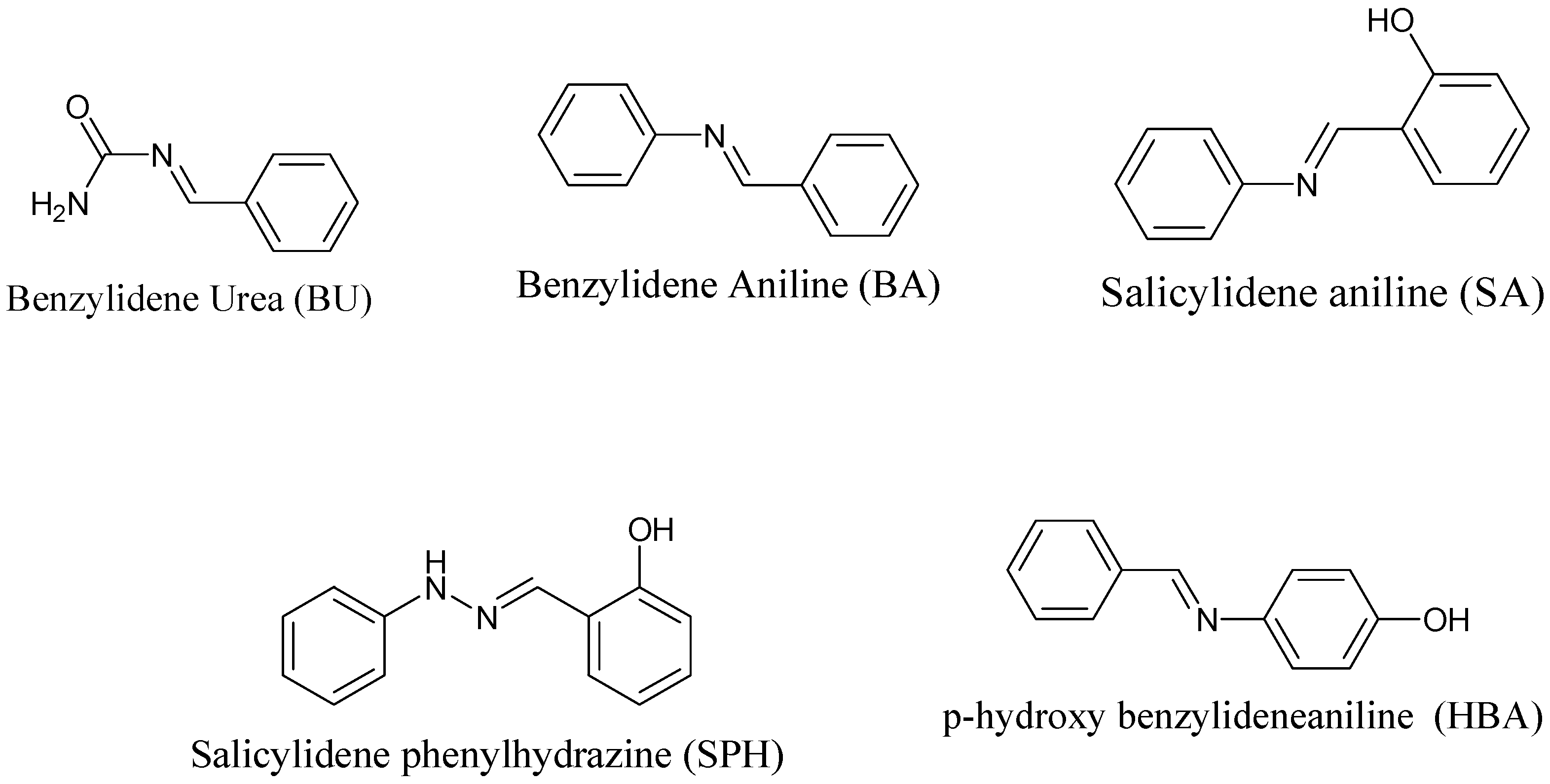
2.2. Synthesis of Schiff Bases
2.3. Synthesis of 5-FU-Containing Cocrystals
2.4. FTIR and PXRD Analyses
2.5. In Vitro MTT Antitumor Bioassay
2.6. Protein Structure Selection and Preparation
2.7. Ligand Data Collection
2.8. Molecular Docking
2.9. Computational Studies
3. Results and Discussion
3.1. Fourier Transform Infrared (FTIR) Analysis
3.2. FTIR Analysis of Schiff Bases
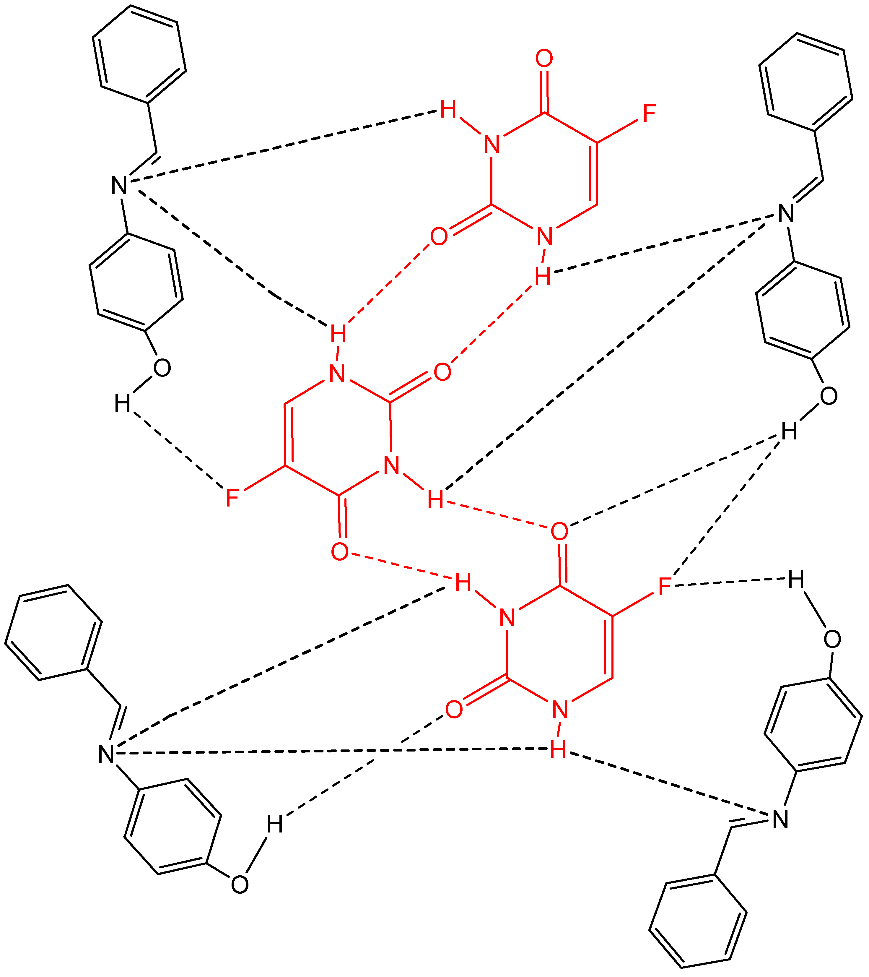
3.3. FTIR Analysis via Supramolecular Interactions of 5-FU Cocrystals
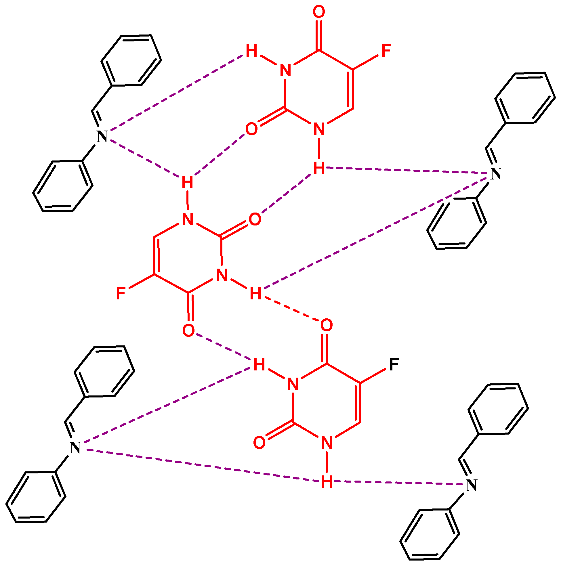
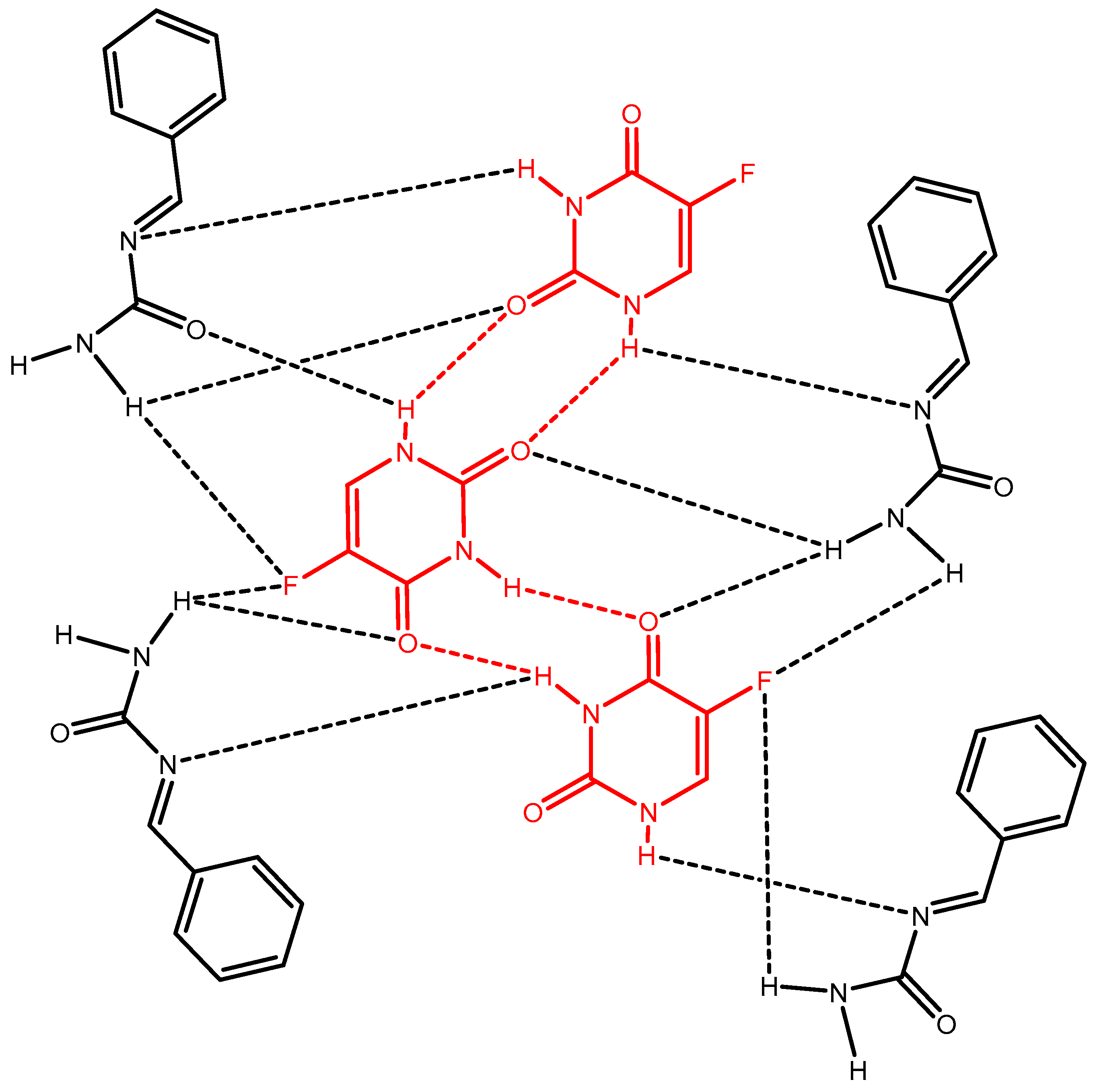
3.4. 5-FU-BA
3.5. 5-FU-BU
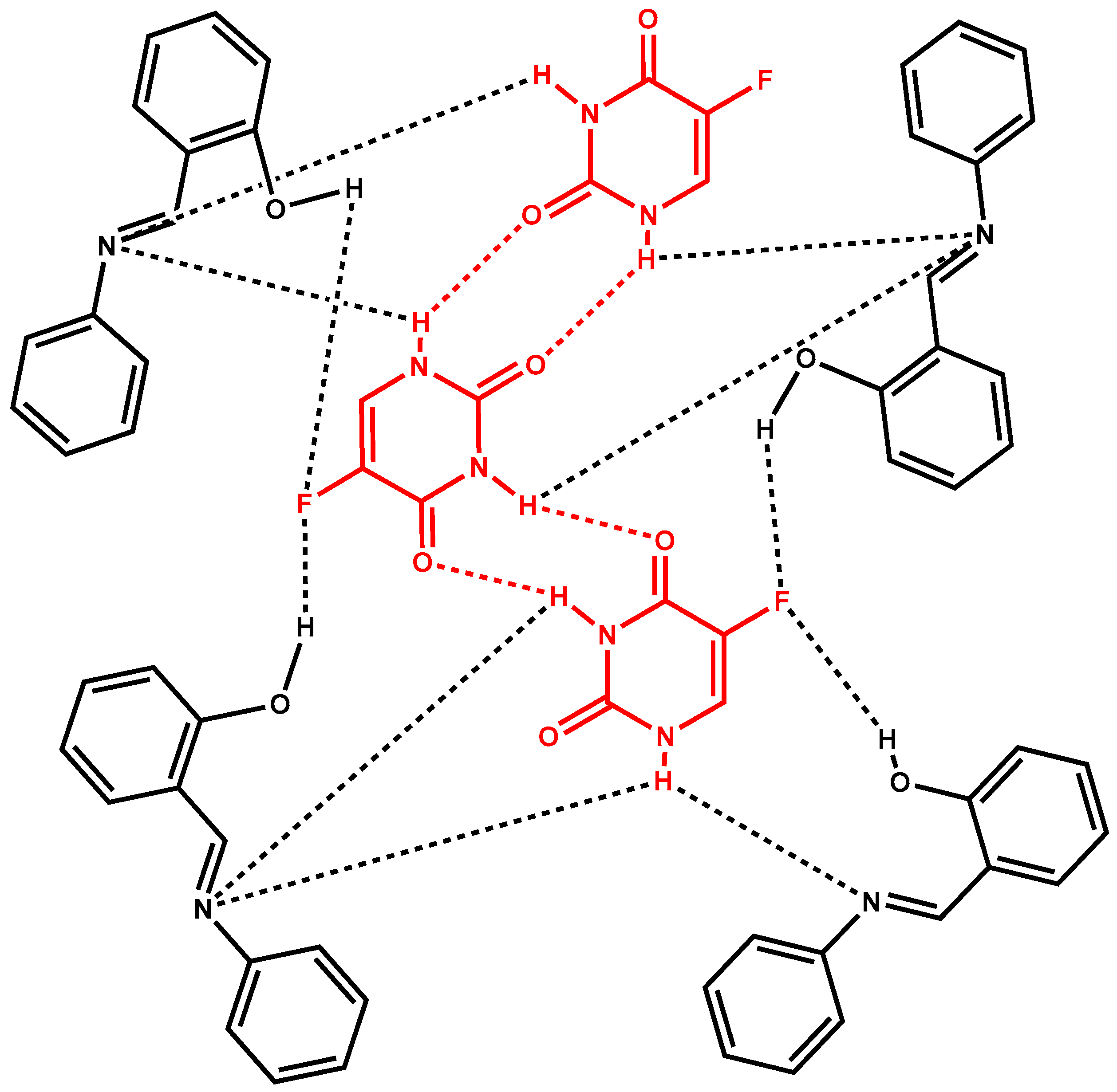
3.6. 5-FU-SA
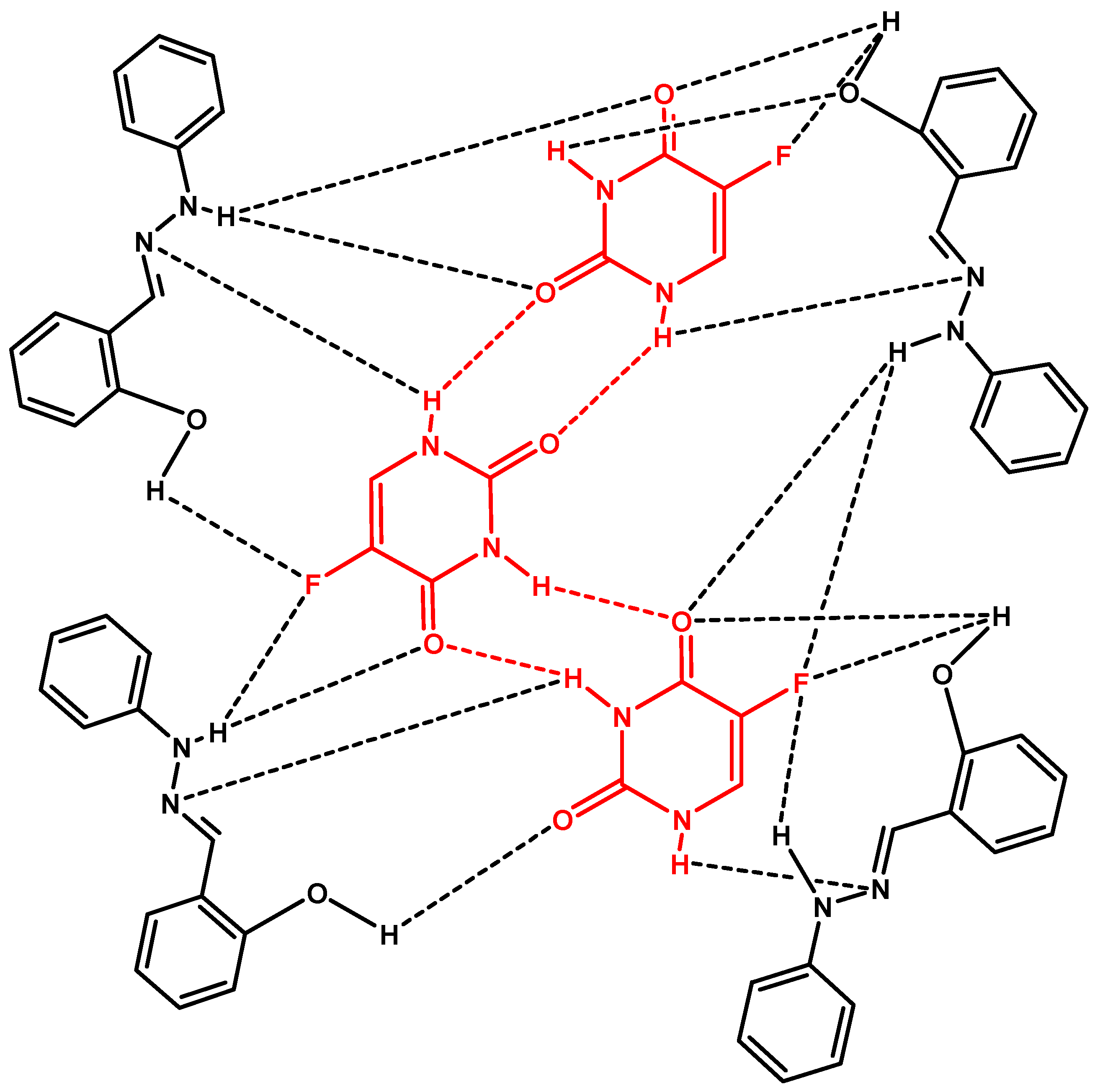
3.7. 5-FU-SPH
3.8. 5-FU-HBA
3.9. Structural Analysis
3.10. In Vitro Anticancer Activity
3.11. Computational Studies
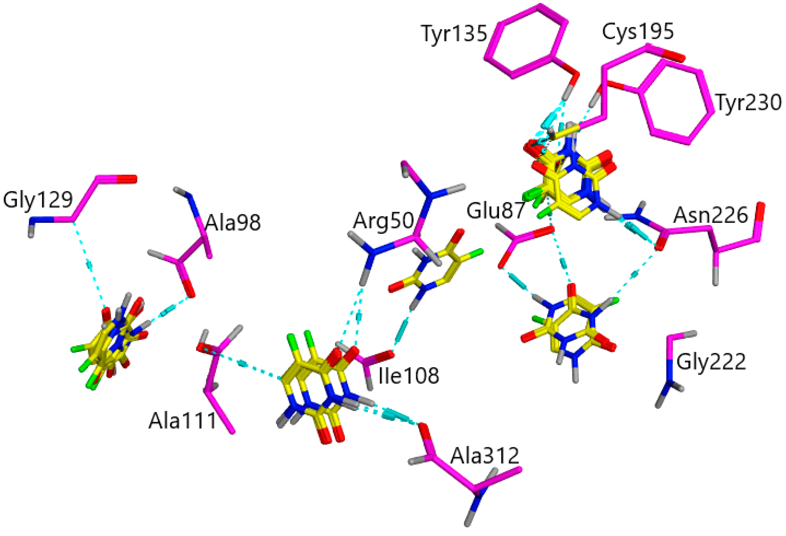
3.11.1. TS (1HVY) Docked with 5-FU
3.11.2. TS (1HVY) Docked with 5-Fluorouracil and Benzylidene-Urea (BU)
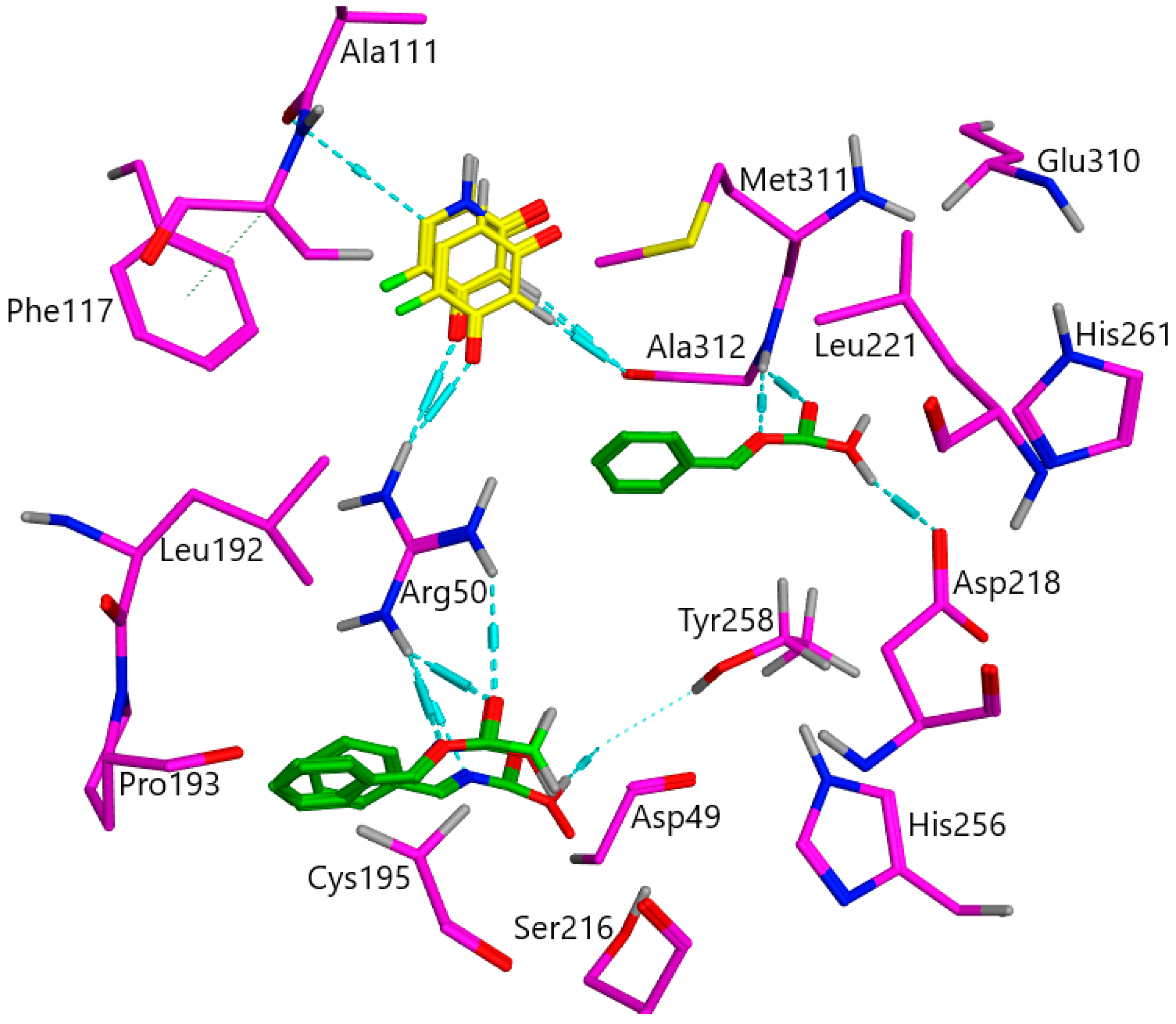
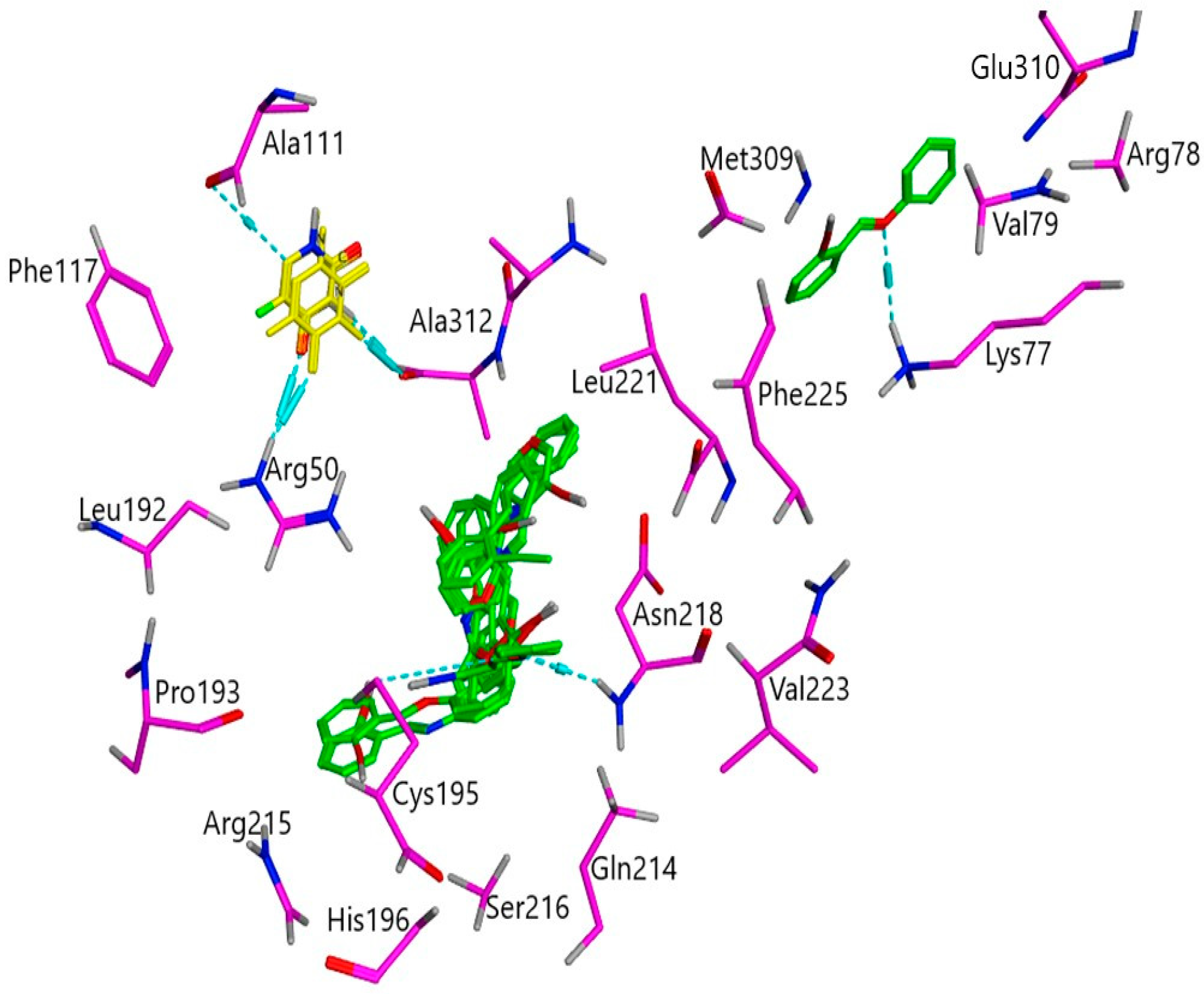
3.11.3. TS (1HVY) Docked with 5-Fluorouracil and Salicylidene-Aniline (SA)
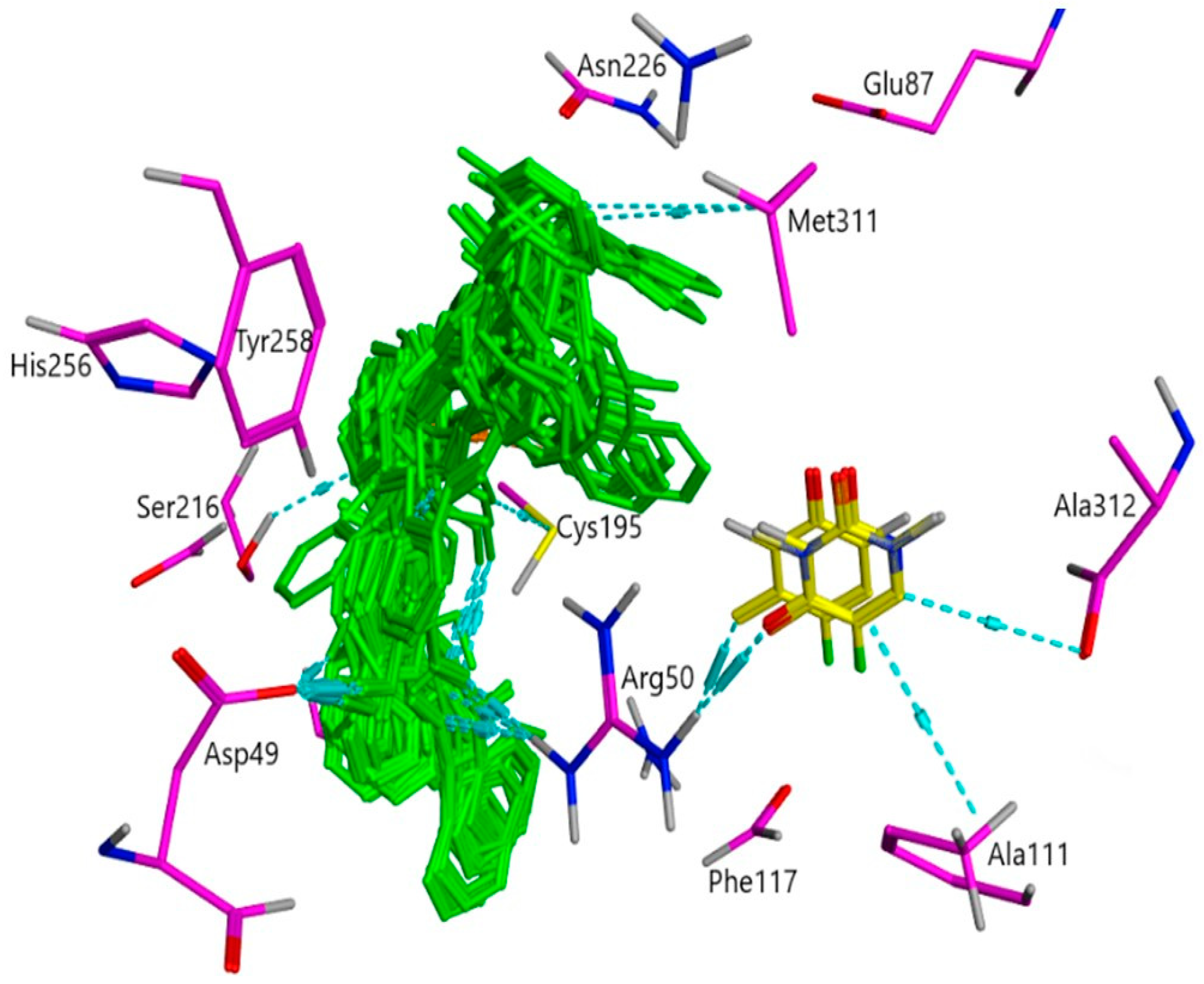
3.11.4. TS (1HVY) Docked with 5-Fluorouracil and Salicylidene-Phenylhydrazine (SPH)
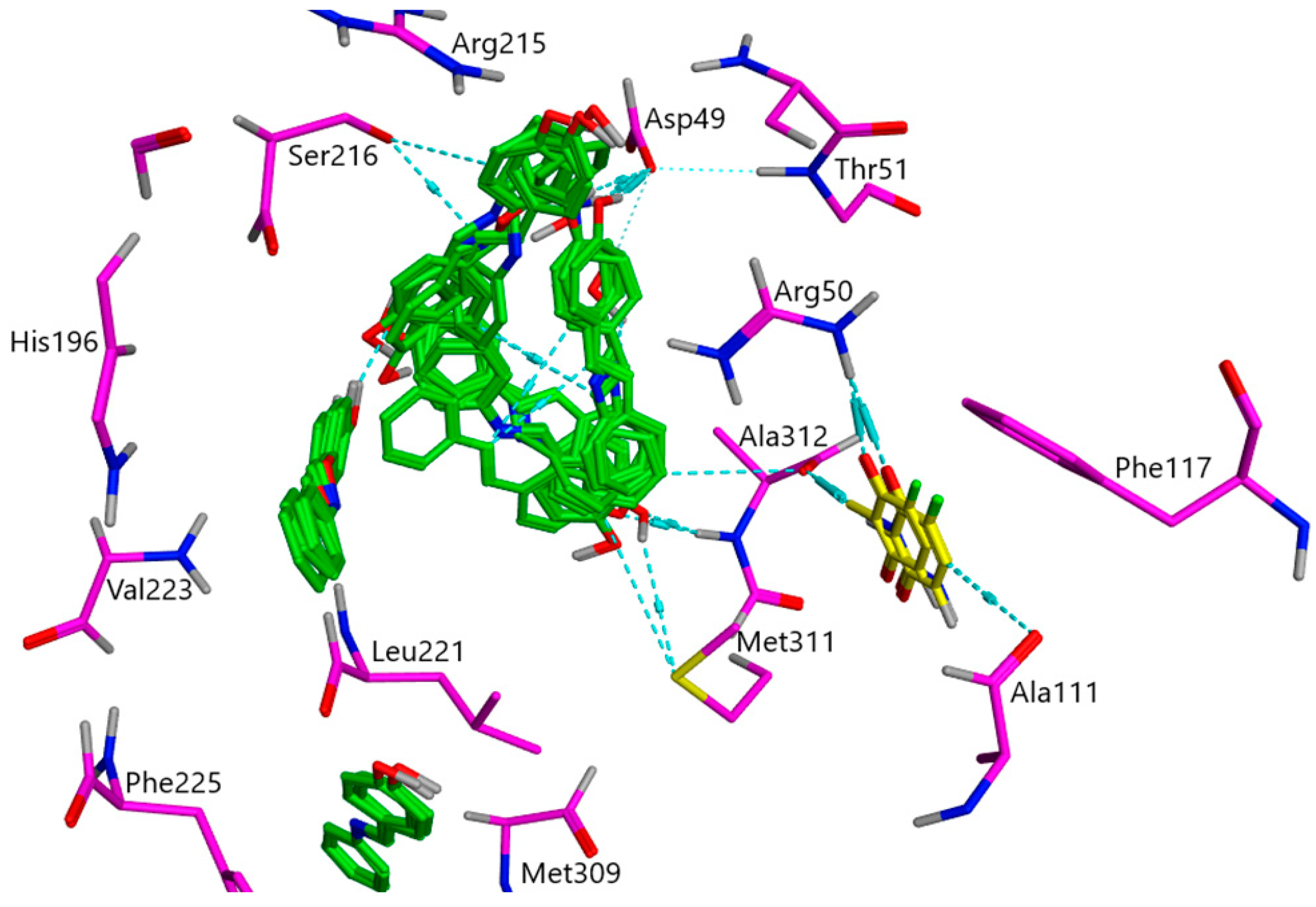
3.11.5. TS (1HVY) Docked with 5-Fluorouracil and Para-hydroxy Benzylideneaniline (HBA)
4. Conclusions
Supplementary Materials
Author Contributions
Funding
Institutional Review Board Statement
Informed Consent Statement
Data Availability Statement
Acknowledgments
Conflicts of Interest
References
- Heidelberger, C.; Chaudhuri, N.K.; Danneberg, P.; Mooren, D.; Griesbach, L.; Duschinsky, R.; Schnitzer, R.J.; Pleven, E.; Scheiner, J. Fluorinated Pyrimidines, A New Class of Tumour-Inhibitory Compounds. Nature 1957, 179, 663–666. [Google Scholar] [CrossRef]
- Sher, F.; Iqbal, S.Z.; Jubeen, F. Future of 5-fluorouracil in cancer therapeutics, current pharmacokinetics issues and a way forward. J. Cancer Res. Pract. 2019, 6, 155. [Google Scholar] [CrossRef]
- Alter, P.; Herzum, M.; Soufi, M.; Schaefer, J.R.; Maisch, B. Cardiotoxicity of 5-fluorouracil. Cardiovasc. Hematol. Agents Med. Chem. 2006, 4, 1–5. [Google Scholar] [CrossRef] [PubMed]
- Latchman, J.; Guastella, A.; Tofthagen, C. 5-Fluorouracil Toxicity and Dihydropyrimidine Dehydrogenase Enzyme: Implications for Practice. Clin. J. Oncol. Nurs. 2014, 18, 581–585. [Google Scholar] [CrossRef] [PubMed] [Green Version]
- Entezar-Almahdi, E.; Mohammadi-Samani, S.; Tayebi, L.; Farjadian, F. Recent Advances in Designing 5-Fluorouracil Delivery Systems: A Stepping Stone in the Safe Treatment of Colorectal Cancer. Int. J. Nanomed. 2020, 15, 5445–5458. [Google Scholar] [CrossRef]
- Macdonald, J.S. Toxicity of 5-fluorouracil. Oncology 1999, 13 (Suppl. S3), 33–34. [Google Scholar]
- Vodenkova, S.; Buchler, T.; Cervena, K.; Veskrnova, V.; Vodicka, P.; Vymetalkova, V. 5-fluorouracil and other fluoropyrimidines in colorectal cancer: Past, present and future. Pharmacol. Ther. 2020, 206, 107447–107511. [Google Scholar] [CrossRef]
- Longley, D.B.; Harkin, D.P.; Johnston, P.G. 5-Fluorouracil: Mechanisms of action and clinical strategies. Nat. Rev. Cancer 2003, 3, 330–338. [Google Scholar] [CrossRef]
- Moreno-Quintero, G.; Castrillón-Lopez, W.; Herrera-Ramirez, A.; Yepes-Pérez, A.F.; Quintero-Saumeth, J.; Cardona-Galeano, W. Synthesis and Chemopreventive Potential of 5-FU/Genistein Hybrids on Colorectal Cancer Cells. Pharmaceuticals 2022, 15, 1299. [Google Scholar] [CrossRef] [PubMed]
- Ajalli, N.; Pourmadadi, M.; Yazdian, F.; Rashedi, H.; Navaei-Nigjeh, M.; Díez-Pascual, A.M. Chitosan/Gamma-Alumina/Fe3O4@5-FU Nanostructures as Promising Nanocarriers: Physiochemical Characterization and Toxicity Activity. Molecules 2022, 27, 5369. [Google Scholar] [CrossRef]
- Mustafa, Y.F.; Mohammed, N. A promising oral 5-fluorouracil prodrug for lung tumor: Synthesis, characterization and releas. Biochem. Cell. Arch. 2021, 21, 1991–1999. [Google Scholar]
- Yusefi, M.; Shameli, K.; Hedayatnasab, Z.; Teow, S.-Y.; Ismail, U.N.; Azlan, C.A.; Ali, R.R. Green synthesis of Fe3O4 nanoparticles for hyperthermia, magnetic resonance imaging and 5-fluorouracil carrier in potential colorectal cancer treatment. Res. Chem. Intermed. 2021, 47, 1789–1808. [Google Scholar] [CrossRef]
- Qin, W.; Long, S.; Panunzio, M.; Biondi, S. Schiff Bases: A Short Survey on an Evergreen Chemistry Tool. Molecules 2013, 18, 12264–12289. [Google Scholar] [CrossRef] [PubMed]
- Iftikhar, B.; Javed, K.; Khan, M.S.U.; Akhter, Z.; Mirza, B.; Mckee, V. Synthesis, characterization and biological assay of Salicylaldehyde Schiff base Cu(II) complexes and their precursors. J. Mol. Struct. 2018, 1155, 337–348. [Google Scholar] [CrossRef]
- Temel, H.; Ziyadanoğullari, B.; Aydin, I.; Aydin, F. Synthesis, spectroscopic and thermodynamic studies of new transition metal complexes with N,N′-bis(2-hydroxynaphthalin-1-carbaldehydene)-1,2-bis(m-aminophenoxy)ethane and their determination by spectrophotometric methods. J. Coord. Chem. 2005, 58, 1177–1185. [Google Scholar] [CrossRef]
- Tümer, M.; Akgün, E.; Toroğlu, S.; Kayraldiz, A.; Dönbak, L. Synthesis and characterization of Schiff base metal complexes: Their antimicrobial, genotoxicity and electrochemical properties. J. Coord. Chem. 2008, 61, 2935–2949. [Google Scholar] [CrossRef]
- Laidler, D.A.; Milner, D.J. Asymmetric synthesis of cyclopropane carboxylates: Catalysis of diazoacetate reactions by copper(II) Schiff base complexes derived from α-amino acids. J. Organomet. Chem. 1984, 270, 121–129. [Google Scholar] [CrossRef]
- Young, R.J.; Cooper, G.W. Dissociation of intermolecular linkages of the sperm head and tail by primary amines, aldehydes, sulphydryl reagents and detergents. Reproduction 1983, 69, 1–10. [Google Scholar] [CrossRef] [Green Version]
- Ibrahim, M.N.; Sharif, S.E.A. Synthesis, Characterization and Use of Schiff Bases as Fluorimetric Analytical Reagents. E-J. Chem. 2007, 4, 531–535. [Google Scholar] [CrossRef]
- Ebosie, N.P.; Ogwuegbu, M.O.C.; Onyedika, G.O.; Onwumere, F.C. Biological and analytical applications of Schiff base metal complexes derived from salicylidene-4-aminoantipyrine and its derivatives: A review. J. Iran. Chem. Soc. 2021, 18, 3145–3175. [Google Scholar] [CrossRef]
- Ceramella, J.; Iacopetta, D.; Catalano, A.; Cirillo, F.; Lappano, R.; Sinicropi, M.S. A Review on the Antimicrobial Activity of Schiff Bases: Data Collection and Recent Studies. Antibiotics 2022, 11, 191. [Google Scholar] [CrossRef] [PubMed]
- Yousif, E.; Majeed, A.; Al-Sammarrae, K.; Salih, N.; Salimon, J.; Abdullah, B. Metal complexes of Schiff base: Preparation, characterization and antibacterial activity. Arab. J. Chem. 2017, 10, S1639–S1644. [Google Scholar] [CrossRef] [Green Version]
- Tadavi, S.K.; Yadav, A.A.; Bendre, R.S. Synthesis and characterization of a novel schiff base of 1,2-diaminopropane with substituted salicyaldehyde and its transition metal complexes: Single crystal structures and biological activities. J. Mol. Struct. 2018, 1152, 223–231. [Google Scholar] [CrossRef]
- Rakesh, K.; Manukumar, H.; Gowda, D.C. Schiff’s bases of quinazolinone derivatives: Synthesis and SAR studies of a novel series of potential anti-inflammatory and antioxidants. Bioorganic Med. Chem. Lett. 2015, 25, 1072–1077. [Google Scholar] [CrossRef] [PubMed] [Green Version]
- Tople, M.S.; Patel, N.B.; Patel, P.P.; Purohit, A.C.; Ahmad, I.; Patel, H. An in silico-in vitro antimalarial and antimicrobial investigation of newer 7-chloroquinoline based Schiff-bases. J. Mol. Struct. 2023, 1271, 134016. [Google Scholar] [CrossRef]
- Jarrahpour, A.; Khalili, D.; De Clercq, E.; Salmi, C.; Brunel, J.M. Synthesis, Antibacterial, Antifungal and Antiviral Activity Evaluation of Some New bis-Schiff Bases of Isatin and Their Derivatives. Molecules 2007, 12, 1720–1730. [Google Scholar] [CrossRef] [Green Version]
- Cheng, L.-X.; Tang, J.-J.; Luo, H.; Jin, X.-L.; Dai, F.; Yang, J.; Qian, Y.-P.; Li, X.-Z.; Zhou, B. Antioxidant and antiproliferative activities of hydroxyl-substituted Schiff bases. Bioorganic Med. Chem. Lett. 2010, 20, 2417–2420. [Google Scholar] [CrossRef]
- Uddin, M.N.; Ahmed, S.S.; Alam, S.M.R. REVIEW: Biomedical applications of Schiff base metal complexes. J. Coord. Chem. 2020, 73, 3109–3149. [Google Scholar] [CrossRef]
- Savcı, A.; Buldurun, K.; Kırkpantur, G. A new Schiff base containing 5-FU and its metal Complexes: Synthesis, Characterization, and biological activities. Inorg. Chem. Commun. 2021, 134, 109060. [Google Scholar] [CrossRef]
- Wu, X.; He, C.; Wu, Y.; Chen, X. Synergistic therapeutic effects of Schiff’s base cross-linked injectable hydrogels for local co-delivery of metformin and 5-fluorouracil in a mouse colon carcinoma model. Biomaterials 2016, 75, 148–162. [Google Scholar] [CrossRef]
- Li, H.; Zhao, Y.; Jia, Y.; Chen, G.; Peng, J.; Li, J. pH-responsive dopamine-based nanoparticles assembled via Schiff base bonds for synergistic anticancer therapy. Chem. Commun. 2020, 56, 13347–13350. [Google Scholar] [CrossRef] [PubMed]
- Lemilemu, F.; Bitew, M.; Demissie, T.B.; Eswaramoorthy, R.; Endale, M. Synthesis, antibacterial and antioxidant activities of Thiazole-based Schiff base derivatives: A combined experimental and computational study. BMC Chem. 2021, 15, 67. [Google Scholar] [CrossRef] [PubMed]
- Thayyil, A.R.; Juturu, T.; Nayak, S.; Kamath, S. Pharmaceutical Co-Crystallization: Regulatory Aspects, Design, Characterization, and Applications. Adv. Pharm. Bull. 2020, 10, 203–212. [Google Scholar] [CrossRef] [PubMed]
- Nadzri, N.I.; Sabri, N.H.; Lee, V.S.; Halim, S.N.A. 5-Fluorouracil Co-crystals and Their Potential Anti-cancer Activities Calculated by Molecular Docking Studies. J. Chem. Crystallogr. 2016, 46, 144–154. [Google Scholar] [CrossRef]
- Jubeen, F.; Liaqat, A.; Sultan, M.; Iqbal, S.Z.; Sajid, I.; Sher, F. Green synthesis and biological evaluation of novel 5-fluorouracil derivatives as potent anticancer agents. Saudi Pharm. J. 2019, 27, 1164–1173. [Google Scholar] [CrossRef] [PubMed]
- Yu, Y.-M.; Liu, L.; Bu, F.-Z.; Li, Y.-T.; Yan, C.-W.; Wu, Z.-Y. A novice cocrystal nanomicelle formulation of 5-fluorouracil with proline: The design, self-assembly and in vitro/vivo biopharmaceutical characteristics. Int. J. Pharm. 2022, 617, 121635. [Google Scholar] [CrossRef] [PubMed]
- Dai, X.-L.; Voronin, A.P.; Gao, W.; Perlovich, G.L.; Lu, T.-B.; Chen, J.-M. Intermolecular interactions and permeability of 5-fluorouracil cocrystals with a series of isomeric hydroxybenzoic acids: A combined theoretical and experimental study. Crystengcomm 2019, 21, 5095–5105. [Google Scholar] [CrossRef]
- Jubeen, F.; Ijaz, S.; Jabeen, I.; Aftab, U.; Mehdi, W.; Altaf, A.; Alissa, S.A.; Al-Ghulikah, H.A.; Ezzine, S.; Bejaoui, I.; et al. Anticancer potential of novel 5-Fluorouracil co-crystals against MCF7 breast and SW480 colon cancer cell lines along with docking studies. Arab. J. Chem. 2022, 15, 104299–104316. [Google Scholar] [CrossRef]
- Jubeen, F.; Liaqat, A.; Amjad, F.; Sultan, M.; Iqbal, S.Z.; Sajid, I.; Niazi, M.B.K.; Sher, F. Synthesis of 5-Fluorouracil Cocrystals with Novel Organic Acids as Coformers and Anticancer Evaluation against HCT-116 Colorectal Cell Lines. Cryst. Growth Des. 2020, 20, 2406–2414. [Google Scholar] [CrossRef]
- Fallon, L. The Crystal and Molecular Structure of 5-Fluorouracil. Acta Crystalographia 1973, 29, 2549–2556. [Google Scholar] [CrossRef]
- Delori, A.; Eddleston, M.D.; Jones, W. Cocrystals of 5-fluorouracil. Cystal Eng. Commun. 2013, 15, 73–77. [Google Scholar] [CrossRef]
- Kim, S.H.; Rich, A. Crystal Structure of the 1: 1 Complex of 5-Fluorouracil and 9-Ethylhypoxanthine. Science 1967, 158, 1046–1048. [Google Scholar] [CrossRef]
- Wang, L.L.; Wang, L.Y.; Yu, Y.M.; Li, Y.T.; Wu, Z.Y.; Yan, C.W. Cocrystallization of 5-fluorouracil and L-phenylalanine: The first zwitterionic cocrystal of 5-fluorouracil with amino acid exhibiting perfect in vitro/vivo pharmaceutical properties. Cryst. Eng. Commun. 2020, 22, 5010–5021. [Google Scholar] [CrossRef]
- Hussain, Z.; Yousif, E.; Ahmed, A.; Altaie, A. Synthesis and characterization of Schiff’s bases of sulfamethoxazole. Org. Med. Chem. Lett. 2014, 4, 1. [Google Scholar] [CrossRef] [Green Version]
- Mosmann, T. Rapid colorimetric assay for cellular growth and survival: Application to proliferation and cytotoxicity assays. J. Immunol. Methods 1983, 65, 55–63. [Google Scholar] [CrossRef] [PubMed]
- Aftab, U.; Sajid, I. Antitumor Peptides from Streptomyces sp. SSA 13, Isolated from Arabian Sea. Int. J. Pept. Res. Ther. 2017, 23, 199–211. [Google Scholar] [CrossRef]
- Phan, J.; Koli, S.; Minor, W.; Dunlap, R.B.; Berger, S.H.; Lebioda, L. Human Thymidylate Synthase Is in the Closed Conformation When Complexed with dUMP and Raltitrexed, an Antifolate Drug. Biochemistry 2001, 40, 1897–1902. [Google Scholar] [CrossRef]
- Jones, G.; Willett, P.; Glen, R.C.; Leach, A.R.; Taylor, R. Development and validation of a genetic algorithm for flexible docking. J. Mol. Biol. 1997, 267, 727–748. [Google Scholar] [CrossRef] [PubMed] [Green Version]
- Srivastava, V.; Gupta, S.P.; Siddiqi, M.I.; Mishra, B.N. Molecular docking studies on quinazoline antifolate derivatives as human thymidylate synthase inhibitors. Bioinformation 2010, 4, 357–365. [Google Scholar] [CrossRef]
- El-Naggar, A.M.; Abou-El-Regal, M.M.; El-Metwally, S.A.; Sherbiny, F.F.; Eissa, I.H. Synthesis, characterization and molecular docking studies of thiouracil derivatives as potent thymidylate synthase inhibitors and potential anticancer agents. Mol. Divers. 2017, 21, 967–983. [Google Scholar] [CrossRef]
- Kawatsuki, N.; Washio, T.; Kozuki, J.; Kondo, M.; Sasaki, T.; Ono, H. Photoinduced orientation of photoresponsive copolymers with N-benzylideneaniline and nonphotoreactive mesogenic side groups. Polymer 2015, 56, 318–326. [Google Scholar] [CrossRef]
- Moisescu-Goia, C.; Muresan-Pop, M.; Simon, V. New solid state forms of antineoplastic 5-fluorouracil with anthelmintic piperazine. J. Mol. Struct. 2017, 1150, 37–43. [Google Scholar] [CrossRef]
- Gromiuk, S.J.; Antonik, A.D. The First Noncovalent-Bonded Supramolecular Frameworks of (Benzylthio)Acetic Acid with Proline Compounds, Isonicotinamide and Tryptamine. Molecules 2022, 27, 8203. [Google Scholar] [CrossRef] [PubMed]
- Ashwanikumar, N.; Kumar, N.A.; Nair, S.A.; Kumar, G.V. Dual drug delivery of 5-fluorouracil (5-FU) and methotrexate (MTX) through random copolymeric nanomicelles of PLGA and polyethylenimine demonstrating enhanced cell uptake and cytotoxicity. Colloids Surf. B Biointerfaces 2014, 122, 520–528. [Google Scholar] [CrossRef]


| Sr. No. | Schiff Base | C=N Stretch (cm−1) | Physical Appearance of Schiff Base Crystals |
|---|---|---|---|
| 1 | Benzylideneaniline (BA) | 1607 | White |
| 2 | Benzylideneurea (BU) | 1613 | Yellowish white |
| 3 | Salicylideneaniline (SA) | 1593 | Yellow |
| 4 | Salicylidenephenyl-hydrazine (SPH) | 1614 | Yellow |
| 5 | Parahydroxy Benzylidene-aniline (HBA) | 1621 | White |
| Sr. No. | Sample ID | V(N-H) cm−1 | V(C=O) cm−1 |
|---|---|---|---|
| 1 | 5-FU | 3220 | 1662 |
| 2 | 5-FU-BA | 3248 | 1690 |
| 3 | 5-FU-BU | 3366 | 1704 |
| 4 | 5-FU-SA | 3324 | 1795 |
| 5 | 5-FU-SPH | 3304 | 1746 |
| 6 | 5-FU-HBA | 3311 | 1753 |
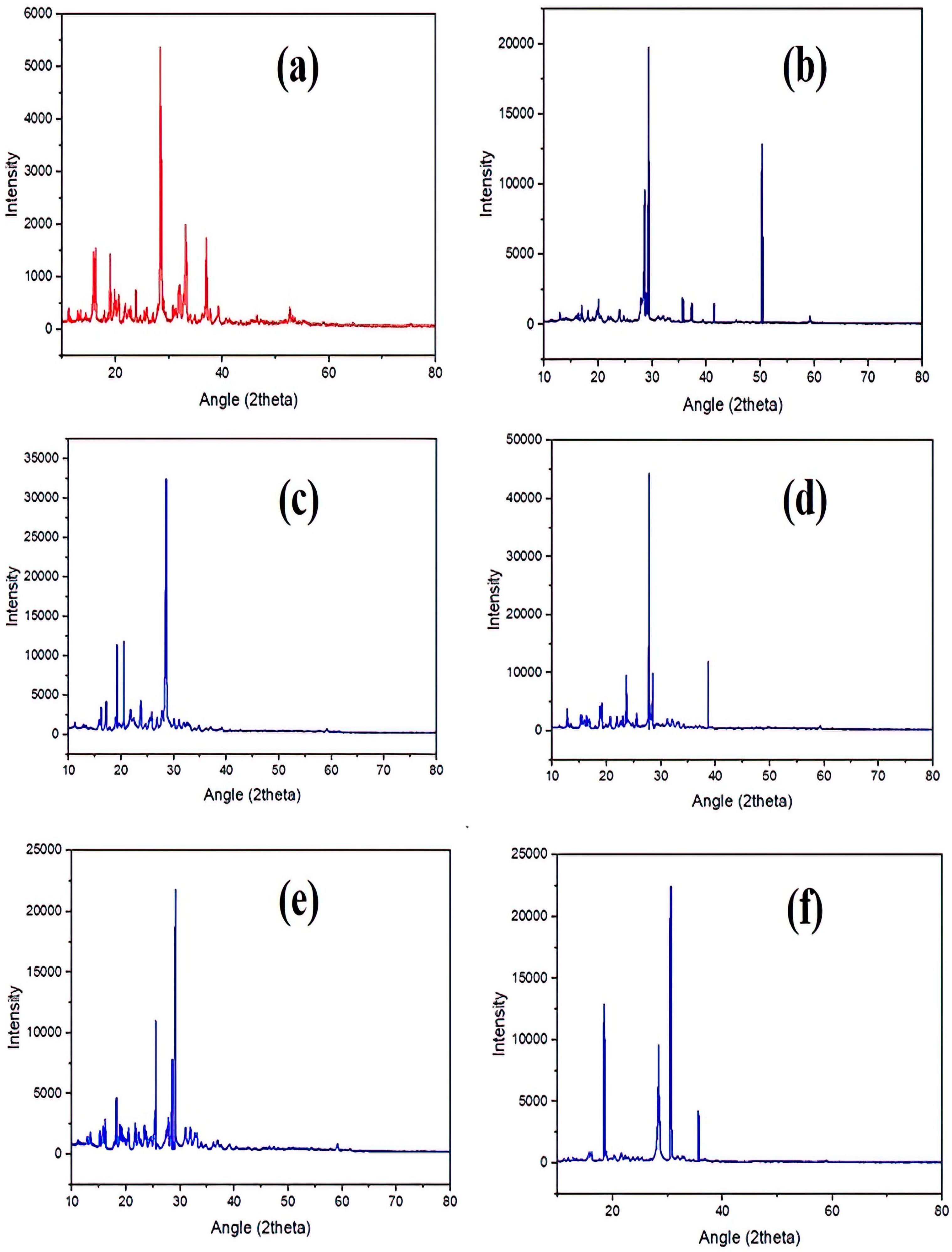
| Sr. No. | Sample | 2 θ | Intensity | FWHM | Crystalline Size (nm) |
|---|---|---|---|---|---|
| 1 | 5-FU | 28.37 | 5367 | 0.28 | 30.58 |
| 2 | 5-FU-BA | 29.34 | 19,748 | 0.15 | 57.20 |
| 3 | 5-FU-BU | 28.56 | 32,440 | 0.25 | 34.26 |
| 4 | 5-FU-SA | 27.82 | 44,325 | 0.09 | 95.02 |
| 5 | 5-FU-SPH | 29.15 | 21,804 | 0.12 | 71.47 |
| 6 | 5-FU-HBA | 30.59 | 22,432 | 0.21 | 40.98 |
| IC50 (µg/mL) against Cell Lines | ||
|---|---|---|
| Sample | Compounds | SW-480 |
| 1 | 5-FU | 12.1166 |
| 2 | 5-FU-BA * | 6.4731 |
| 3 | 5-FU-BU | 13.5591 |
| 4 | 5-FU-SA | 97.1558 |
| 5 | 5-FU-SPH | 11.6904 |
| 6 | 5-FU-HBA * | 10.2174 |
| 7 | Standard Drug (Doxorubicin) | 2.3159 |
| Inhibition Rate (%) against SW480 Cell Line | |||||||
|---|---|---|---|---|---|---|---|
| Conc. (µg/mL) | 1 | 2 | 3 | 4 | 5 | 6 | Doxorubicin |
| 200 | 97.4047 | 99.8492 | 88.5714 | 60.7540 | 95.6905 | 99.3650 | 97.1032 |
| 100 | 94.0476 | 95.6587 | 87.5000 | 57.1587 | 95.1111 | 95.7936 | 93.8889 |
| 50 | 80.1984 | 93.2540 | 87.8968 | 46.0079 | 94.7064 | 82.14286 | 92.2222 |
| 25 | 70.4365 | 81.7857 | 87.6191 | 39.4762 | 92.5555 | 65.5555 | 90.9524 |
| 12.5 | 52.5794 | 76.2302 | 13.0476 | 30.3968 | 77.0238 | 52.7381 | 84.9603 |
| 6.25 | 7.3413 | 28.3333 | 25.9524 | 17.6587 | 37.8730 | 21.58730 | 38.7698 |
Disclaimer/Publisher’s Note: The statements, opinions and data contained in all publications are solely those of the individual author(s) and contributor(s) and not of MDPI and/or the editor(s). MDPI and/or the editor(s) disclaim responsibility for any injury to people or property resulting from any ideas, methods, instructions or products referred to in the content. |
© 2023 by the authors. Licensee MDPI, Basel, Switzerland. This article is an open access article distributed under the terms and conditions of the Creative Commons Attribution (CC BY) license (https://creativecommons.org/licenses/by/4.0/).
Share and Cite
Jubeen, F.; Jabeen, I.; Aftab, U.; Noor, S.; Hareem, M.e.; Sultan, M.; Kazi, M. Synthesis, Characterization, Theoretical and Experimental Anticancer Evaluation of Novel Cocrystals of 5-Fluorouracil and Schiff Bases against SW480 Colorectal Carcinoma. Pharmaceutics 2023, 15, 1929. https://doi.org/10.3390/pharmaceutics15071929
Jubeen F, Jabeen I, Aftab U, Noor S, Hareem Me, Sultan M, Kazi M. Synthesis, Characterization, Theoretical and Experimental Anticancer Evaluation of Novel Cocrystals of 5-Fluorouracil and Schiff Bases against SW480 Colorectal Carcinoma. Pharmaceutics. 2023; 15(7):1929. https://doi.org/10.3390/pharmaceutics15071929
Chicago/Turabian StyleJubeen, Farhat, Ishrat Jabeen, Usman Aftab, Sadia Noor, Mah e Hareem, Misbah Sultan, and Mohsin Kazi. 2023. "Synthesis, Characterization, Theoretical and Experimental Anticancer Evaluation of Novel Cocrystals of 5-Fluorouracil and Schiff Bases against SW480 Colorectal Carcinoma" Pharmaceutics 15, no. 7: 1929. https://doi.org/10.3390/pharmaceutics15071929