

Updates on Biogenic Metallic and Metal Oxide Nanoparticles: Therapy, Drug Delivery and Cytotoxicity


Abstract
:1. Introduction
2. Characteristics of Some Biosynthesized NPs
2.1. Gold NPs
2.2. Silver NPs
2.3. Platinum and Palladium NPs
2.4. Copper and Copper Oxide NPs
2.5. Zinc Oxide NPs
2.6. Other Metal Oxide NPs
3. Biogenic Metallic NPs Production
3.1. Biosynthesized NPs by Using Plants and Their Extracts
3.2. Biosynthesized NPs by Using Microorganisms
3.2.1. Bacterial Synthesis
3.2.2. Fungal Synthesis
3.2.3. Yeast Synthesis
3.3. Virus Synthesis
3.4. Algae Synthesis
3.5. Biosynthesized NPs by Using Enzymes and Biomolecules
4. Therapy and Drug Delivery of Biosynthesized NPs
4.1. Antitumor Activity
4.1.1. Biosynthesized Metal NPs in Cancer Therapy and Delivery of Antitumor Drugs
4.1.2. Biogenic Metal Oxide NPs in Cancer Therapy and Delivery of Antitumor Drugs
4.2. Biogenic NPs for Antimicrobial/Antifungal Therapy
4.3. Anti-Inflammatory Activity
4.4. Wound Healing Properties
4.5. Osteoinductive and Angiogenetic Activities
4.6. Anti-Viral Activity
4.7. Antiparasitic Activity
5. Cytotoxicity of Biosynthesized NPs
6. Future Perspectives and Outlook
Author Contributions
Funding
Institutional Review Board Statement
Informed Consent Statement
Data Availability Statement
Conflicts of Interest
References
- Singh, J.; Dutta, T.; Kim, K.-H.; Rawat, M.; Samddar, P.; Kumar, P. ‘Green’ synthesis of metals and their oxide nanoparticles: Applications for environmental remediation. J. Nanobiotechnol. 2018, 16, 84. [Google Scholar] [CrossRef] [PubMed]
- Ahmed, S.; Saifullah, A.M.; Swami, B.L.; Ikram, S. Green synthesis of silver nanoparticles using Azadirachta indica aqueous leaf extract. J. Radiat. Res. Appl. Sci. 2019, 9, 1–7. [Google Scholar] [CrossRef] [Green Version]
- Hua, S.; de Matos, M.B.C.; Metselaar, J.M.; Storm, G. Current trends and challenges in the clinical translation of nanoparticulate nanomedicines: Pathways for translational development and commercialization. Front. Pharmacol. 2018, 9, 790. [Google Scholar] [CrossRef] [Green Version]
- Zhang, D.; Ma, X.-L.; Gu, Y.; Huang, H.; Zhang, G.-W. Green Synthesis of Metallic Nanoparticles and Their Potential Applications to Treat Cancer. Front. Chem. 2020, 8, 799. [Google Scholar] [CrossRef] [PubMed]
- Shah, M.; Fawcett, D.; Sharma, S.; Tripathy, S.K.; Jai Poinern, G.E. Green synthesis of metallic nanoparticles via biological entities. Materials 2015, 8, 7278–7308. [Google Scholar] [CrossRef] [Green Version]
- El Shafey, A.M. Green synthesis of metal and metal oxide nanoparticles from plant leaf extracts and their applications: A review. Green Process. Synth. 2020, 9, 304–339. [Google Scholar] [CrossRef]
- Phan, H.; Haes, A.J. What Does Nanoparticle Stability Mean? J. Phys. Chem. C 2019, 123, 16495–16507. [Google Scholar] [CrossRef]
- Chung, I.M.; Rahuman, A.A.; Marimuthu, S.; Kirthi, A.V.; Anbarasan, K.; Padmini, P.; Rajakumar, G. Green synthesis of copper nanoparticles using Eclipta prostrata leaves extract and their antioxidant and cytotoxic activities. Nanoscale Res. Lett. 2017, 11, 18–24. [Google Scholar] [CrossRef] [Green Version]
- Bonifacio, B.; da Silva, P.; Ramos, M.; Negri, K.; Maria Bauab, T.; Chorilli, M. Nanotechnology-based drug delivery systems and herbal medicines: A review. Int. J. Nanomed. 2014, 9, 1–5. [Google Scholar] [CrossRef] [Green Version]
- Mody, V.V.; Nounou, M.I.; Bikram, M. Novel nanomedicine-based MRI contrast agents for gynecological malignancies. Adv. Drug Deliv. Rev. 2009, 61, 795–807. [Google Scholar] [CrossRef]
- Tinajero-Díaz, E.; Salado-Leza, D.; Gonzalez, C.; Velázquez, M.M.; López, Z.; Bravo-Madriga, J.; Knauth, P.; Flores-Hernández, F.Y.; Herrera-Rodríguez, S.E.; Navarro, R.E.; et al. Green Metallic Nanoparticles for Cancer Therapy: Evaluation Models and Cancer Applications. Pharmaceutics 2021, 13, 1719. [Google Scholar] [CrossRef] [PubMed]
- Beveridge, T.; Murray, R. Sites of metal deposition in the cell wall of Bacillus subtilis. J. Bacteriol. 1980, 141, 876–887. [Google Scholar] [CrossRef] [PubMed] [Green Version]
- Gahlawat, G.; Choudhury, R. A review on the biosynthesis of metal and metal salt nanoparticles by microbes. RSC Adv. 2019, 9, 12944. [Google Scholar] [CrossRef] [Green Version]
- Deshmukh, S.P.; Patil, S.M.; Mullani, S.B.; Delekar, S.D. Silver nanoparticles as an effective disinfectant: A review. Mater. Sci. Eng. C 2019, 97, 954–965. [Google Scholar] [CrossRef] [PubMed]
- Siddiqi, K.S.; Husen, A. Recent advances in plant-mediated engineered gold nanoparticles and their application in biological system. J. Trace Elem. Med. Biol. 2017, 40, 10–23. [Google Scholar] [CrossRef]
- Patil, M.P.; Bayaraa, E.; Subedi, P.; Piad, L.L.A.; Tarte, N.H.; Kim, G.D. Biogenic synthesis, characterization of gold nanoparticles using Lonicera japonica and their anticancer activity on HeLa cells. J. Drug Deliv. Sci. Technol. 2019, 51, 83–90. [Google Scholar] [CrossRef]
- Mathivanan, K.; Selva, R.; Chandirika, J.U.; Govindarajan, R.K.; Srinivasan, R.; Annadurai, G.; Duc, P.A. Biologically synthesized silver nanoparticles against pathogenic bacteria: Synthesis, calcination and characterization. Biocatal. Agric. Biotechnol. 2019, 22, 101373. [Google Scholar] [CrossRef]
- Qu, Y.; Pei, X.; Shen, W.; Shen, W.; Zhang, X.; Wang, J.; Zhang, Z.; Li, S.; You, S.; Ma, F.; et al. Biosynthesis of gold nanoparticles by Aspergillum sp. WL-Au for degradation of aromatic pollutants. Physica E Low-Dimens. Syst. Nanostruct. 2017, 88, 133–141. [Google Scholar] [CrossRef]
- Senapati, S.; Syed, A.; Moeez, S.; Kumar, A.; Ahmad, A. Intracellular synthesis of gold nanoparticles using alga Tetraselmis kochinensis. Mater. Lett. 2012, 79, 116–118. [Google Scholar] [CrossRef]
- Liua, Q.; Aouidat, F.; Sacco, P.; Marsich, E.; Djaker, N.; Spadavecchia, J. Galectin-1 protein modified gold (III)-PEGylated complex-nanoparticles: Proof of concept of an alternative probe in colorimetric glucose detection. Colliods Surf. B 2020, 185, 110588. [Google Scholar] [CrossRef]
- Busch, R.T.; Karim, F.; Weis, J.; Sun, Y.; Zhao, C.; Vasquez, E.S. Optimization and Structural Stability of Gold Nanoparticle–Antibody Bioconjugates. ACS Omega 2019, 4, 15269–15279. [Google Scholar] [CrossRef] [PubMed] [Green Version]
- Ameen, F.; AlYahya, S.; Govarthanan, M.; Aljahdali, N.; Al-Enazi, N.; Alsamhary, K.; Alshehri, W.A.; Alwakeel, S.S.; Alharbi, S.A. Soil bacteria Cupriavidus sp. mediates the extracellular synthesis of antibacterial silver nanoparticles. J. Mol. Struct. 2020, 1202, 127233. [Google Scholar] [CrossRef]
- Hooda, H.; Singh, P.; Bajpai, S. Effect of quercitin impregnated silver nanoparticle on growth of some clinical pathogens. Mater. Today Proc. 2020, 31, 625–630. [Google Scholar] [CrossRef]
- Vance, M.E.; Kuiken, T.; Vejerano, E.P.; McGinnis, S.P.; Hochella Jr, M.F.; Rejeski, D.; Hull, M.S. Nanotechnology in the real world: Redeveloping the nanomaterial consumer products inventory. J. Nanotechnol. 2015, 6, 1769–1780. [Google Scholar] [CrossRef] [Green Version]
- Husain, S.; Verma, S.K.; Yasin, D.; Hemlata, F.; Rizvi, M.M.A.; Fatma, T. Facile green Bio-Fabricated Silver Nanoparticles from Microchaete Infer Dose-dependent Antioxidant and Anti-proliferative Activity to Mediate Cellular Apoptosis. Bioorg. Chem. 2021, 107, 104535. [Google Scholar] [CrossRef] [PubMed]
- Zhao, G.J.; Stevens, S.E. Multiple parameters for the comprehensive evaluation of the susceptibility of Escherichia coli to the silver ion. Biometals 1998, 11, 27–32. [Google Scholar] [CrossRef]
- Vaidyanathan, R.; Gopalram, S.; Kalishwaralal, K.; Deepak, V.; Pandian, S.R.; Gurunathan, S. Enhanced silver nanoparticle synthesis by optimization of nitrate reductase activity. Colloids Surf. B 2010, 75, 335–341. [Google Scholar] [CrossRef]
- Patil, M.P.; Singh, R.D.; Koli, P.B.; Patil, K.T.; Jagadale, B.S.; Tipare, A.R.; Kim, G.D. Antibacterial potential of silver nanoparticles synthesized using Madhuca longifolia flower extract as a green resource. Microb. Pathog. 2018, 121, 184–189. [Google Scholar] [CrossRef]
- Alsamhary, K.I. Eco-friendly synthesis of silver nanoparticles by Bacillus subtilis and their antibacterial activity. Saudi J. Biol. Sci. 2020, 27, 2185–2191. [Google Scholar] [CrossRef]
- Tyagi, S.; Tyagi, P.K.; Gola, D.; Chauhan, N.; Bharti, R.K. Extracellular synthesis of silver nanoparticles using entomopathogenic fungus: Characterization and antibacterial potential. SN Appl. Sci. 2019, 1, 1545. [Google Scholar] [CrossRef] [Green Version]
- Arya, A.; Mishra, V.; Chundawat, T.S. Green synthesis of silver nanoparticles from green algae (Botryococcus braunii) and its catalytic behavior for the synthesis of benzimidazoles. Chem. Data Collect. 2019, 20, 100190. [Google Scholar] [CrossRef]
- West, P.R.; Ishii, S.; Naik, G.V.; Emani, N.K.; Shalaev, V.M.; Boltasseva, A. Searching for Better Plasmonic Materials Paul. Laser Photonics Rev. 2010, 4, 795–808. [Google Scholar] [CrossRef] [Green Version]
- Brayner, R.; Hélène, B.; Miryana, H.; Chakib, D.; Claude, Y.; Thibaud, C.; Jacques, L.; Fernand, F.; Alain, C. Cyanobacteria as bioreactors for the synthesis of Au, Ag, Pd, and Pt nanoparticles via an enzyme-mediated route. J. Nanosci. Nanotechnol. 2007, 7, 2696–2708. [Google Scholar] [CrossRef]
- Shiny, P.; Mukherjee, A.; Chandrasekaran, N. DNA damage and mitochondria-mediated apoptosis of A549 lung carcinoma cells induced by rnatechzati silver and platinum nanoparticles. RSC Adv. 2016, 6, 27775–27787. [Google Scholar] [CrossRef]
- Alshatwi, A.A.; Athinarayanan, J.; Subbarayan, P.V. Green synthesis of platinum nanoparticles that induce cell death and G2/M-phase cell cycle arrest in human cervical cancer cells. J. Mater. Sci. Mater. Med. 2015, 26, 7. [Google Scholar] [CrossRef] [PubMed]
- Leo, A.J.; Oluwafemi, O.S. Plant-mediated synthesis of platinum nanoparticles using water hyacinth as an efficient biomatrix source—An eco-friendly development. Mater. Lett. 2017, 196, 141–144. [Google Scholar] [CrossRef]
- Nadaroglu, H.; Gungor, A.A.; Ince, S.; Babagil, A. Green synthesis and rnatechzation of platinum nanoparticles using quail egg yolk. Spectrochim. Acta Part A Mol. Biomol. Spectrosc. 2017, 172, 43–47. [Google Scholar] [CrossRef] [PubMed]
- Mikhailova, E.O. Green Synthesis of Platinum Nanoparticles for Biomedical Applications. J. Funct. Biomater. 2022, 13, 260. [Google Scholar] [CrossRef] [PubMed]
- Jameel, M.S.; Aziz, A.A.; Dheyab, M.A. Green synthesis: Proposed mechanism and factors influencing the synthesis of platinum nanoparticles. Green Process. Synth. 2020, 9, 386–398. [Google Scholar] [CrossRef]
- Fahmy, S.A.; Preis, E.; Bakowsky, U.; El-Said Azzazy, H.M. Palladium Nanoparticles Fabricated by Green Chemistry: Promising Chemotherapeutic, Antioxidant and Antimicrobial Agents. Materials 2020, 13, 3661. [Google Scholar] [CrossRef] [PubMed]
- Nasrollahzadeh, M.; Sajadi, S.M.; Maham, M. Green synthesis of palladium nanoparticles using Hippophae rhamnoides Linn leaf extract and their catalytic activity for the Suzuki-Miyaura coupling in water. J. Mol. Catal. A Chem. 2015, 396, 297–303. [Google Scholar] [CrossRef]
- Bankar, A.; Joshi, B.; Kumar, A.R.; Zinjarde, S. Banana peel extract mediated novel route for the synthesis of palladium nanoparticles. Mater. Lett. 2010, 64, 1951–1953. [Google Scholar] [CrossRef]
- Khazaei, A.; Rahmati, S.; Hekmatian, Z.; Saeednia, S. A green approach for the synthesis of palladium nanoparticles supported on pectin: Application as a catalyst for solvent-free Mizoroki-Heck reaction. J. Mol. Catal. A Chem. 2013, 372, 160–166. [Google Scholar] [CrossRef]
- Hemmati, S.; Mehrazin, L.; Ghorban, H.; Garakani, S.H.; Mobaraki, T.H.; Mohammadia, P.; Veisi, H. Green synthesis of Pd nanoparticles supported on reduced graphene oxide, using the extract of Rosa canina fruit, and their use as recyclable and heterogeneous nanocatalysts for the degradation of dye pollutants in water. RSC Adv. 2018, 8, 21020. [Google Scholar] [CrossRef] [PubMed] [Green Version]
- Khan, M.; Albalawi, G.H.; Shaik, M.R.; Khan, M.; Adil, S.F.; Kuniyil, M.; Alkhathlan, H.Z.; Al-Warthan, A.; Siddiqui, M.R.H. Miswak mediated green synthesized palladium nanoparticles as effective catalysts for the Suzuki coupling reactions in aqueous media. J. Saudi Chem. Soc. 2017, 21, 450–457. [Google Scholar] [CrossRef] [Green Version]
- Kora, A.J.; Rastogi, L. Green synthesis of palladium nanoparticles using gum ghatti (Anogeissus latifolia) and its application as an antioxidant and catalyst. Arab. J. Chem. 2018, 11, 1097–1106. [Google Scholar] [CrossRef] [Green Version]
- Ko, Y.L.; Krishnamurthy, S.; Yun, Y.S. Facile synthesis of monodisperse Pt and Pd nanoparticles using antioxidants. J. Nanosci. Nanotechnol. 2015, 15, 412–417. [Google Scholar] [CrossRef]
- Lee, H.-J.; Lee, G.; Jang, N.R.; Yun, J.H.; Song, J.Y.; Kim, B.S. Biological Synthesis of Copper Nanoparticles Using Plant Extract. Nanotechnology 2011, 1, 371–374. [Google Scholar]
- Amjad, R.; Mubeen, B.; Ali, S.S.; Imam, S.S.; Alshehri, S.; Ghoneim, M.M.; Alzarea, S.I.; Rasool, R.; Ullah, I.; Nadeem, M.S.; et al. Green Synthesis and Characterization of Copper Nanoparticles Using Fortunella margarita Leaves. Polymers 2021, 13, 4364. [Google Scholar] [CrossRef]
- Jayandran, M.; Haneefa, M.M.; Balasubramanian, V. Green synthesis of copper nanoparticles using natural reducer and stabilizer and an evaluation of antimicrobial activity. J. Chem. Pharm. Res. 2015, 7, 251–259. [Google Scholar]
- Koopi, H.; Buazar, F. A novel one-pot biosynthesis of pure alpha aluminum oxide nanoparticles using the macroalgae Sargassum ilicifolium: A green marine approach. Ceram. Int. 2018, 44, 8940–8945. [Google Scholar] [CrossRef]
- Sepahvand, M.; Buazar, F.; Sayahi, M.H. Novel marine-based gold nanocatalyst in solvent-free synthesis of polyhydroquinoline derivatives: Green and sustainable protocol. Appl. Organomet. Chem. 2020, 34, e6000. [Google Scholar] [CrossRef]
- Rezazadeh, N.H.; Buazar, F.; Matroodi, S. Synergistic effects of combinatorial chitosan and polyphenol biomolecules on enhanced antibacterial activity of biofunctionalized silver nanoparticles. Sci. Rep. 2020, 10, 19615. [Google Scholar] [CrossRef] [PubMed]
- Buazar, F.; Sweidi, S.; Badri, M.; Kroushawi, F. Biofabrication of highly pure copper oxide nanoparticles using wheat seed extract and their catalytic activity: A mechanistic approach. Green Process. Synth. 2019, 8, 691–702. [Google Scholar] [CrossRef]
- Ying, S.; Guan, Z.; Ofoegbu, P.C.; Clubb, P.; Rico, C.; He, F.; Hong, J. Green synthesis of nanoparticles: Current developments and limitations. Environ. Technol. Innov. 2022, 26, 102336. [Google Scholar] [CrossRef]
- Rajesh, K.M.; Ajitha, B.; Reddy, Y.A.K.; Suneetha, Y.; Reddy, P.S. Assisted green synthesis of copper nanoparticles using Syzygium aromaticum bud extract: Physical, optical and antimicrobial properties. Optik 2018, 154, 593–600. [Google Scholar] [CrossRef]
- Perreault, F.; Melegari, S.P.; da Costa, C.H.; de Oliveira Franco Rossetto, A.L.; Popovic, R.; Matias, W.G. Genotoxic effects of copper oxide nanoparticles in Neuro 2A cell cultures. Sci. Total Environ. 2012, 441, 117–124. [Google Scholar] [CrossRef]
- Alhalili, Z. Green synthesis of copper oxide nanoparticles CuO NPs from Eucalyptus globoulus leaf extract: Adsorption and design of experiments. Arab. J. Chem. 2022, 15, 103739. [Google Scholar] [CrossRef]
- El Din Mahmoud, A.; Al-Qahtani, K.M.; Alflaij, S.O.; Al-Qahtani, S.F.; Alsamhan, F.A. Green copper oxide nanoparticles for lead, nickel, and cadmium removal from contaminated water. Sci. Rep. 2021, 11, 12547. [Google Scholar] [CrossRef]
- Bhattacharya, P.; Swarnakar, S.; Ghosh, S.; Majumdar, S.; Banerjee, S. Disinfection of drinking water via algae mediated green synthesized copper oxide nanoparticles and its toxicity evaluation. J. Environ. Chem. Eng. 2019, 7, 102867. [Google Scholar] [CrossRef]
- Singh, D.; Jain, D.; Rajpurohit, D.; Jat, G.; Kushwaha, H.S.; Singh, A.; Mohanty, S.R.; Al-Sadoon, M.K.; Zaman, W.; Upadhyay, S.K. Bacteria assisted green synthesis of copper oxide nanoparticles and their potential applications as antimicrobial agents and plant growth stimulants. Front. Chem. 2023, 11, 1154128. [Google Scholar] [CrossRef] [PubMed]
- Wang, Y.; Aker, W.G.; Hwang, H.-M.; Yedjou, C.G.; Yu, H.; Tchounwou, P.B. A study of the mechanism of in vitro cytotoxicity of metal oxide nanoparticles using catfish primary hepatocytes and human HepG2 cells. Sci. Total Environ. 2011, 409, 4753–4762. [Google Scholar] [CrossRef] [Green Version]
- Kim, I.; Viswanathan, K.; Kasi, G.; Thanakkasaranee, S.; Sadeghi, K.; Seo, J. ZnO Nanostructures in Active Antibacterial Food Packaging: Preparation Methods, Antimicrobial Mechanisms, Safety Issues, Future Prospects, and Challenges. Food Rev. Int. 2020, 38, 537–565. [Google Scholar] [CrossRef] [Green Version]
- Sonia, S.; Linda Jeeva Kumari, H.; Ruckmanib, K.; Sivakumara, M. Antimicrobial and antioxidant potentials of biosynthesized colloidal zinc oxide nanoparticles for a fortified cold cream formulation: A potent nanocosmeceutical application. Mater. Sci. Eng. C Mater. Biol. Appl. 2017, 79, 581–589. [Google Scholar] [CrossRef]
- Basnet, P.; Inakhunbi Chanu, T.; Samanta, D.; Chatterjee, S. A Review on Bio-Synthesized Zinc Oxide Nanoparticles Using Plant Extracts as Reductants and Stabilizing Agents. J. Photochem. Photobiol. B 2018, 183, 201–221. [Google Scholar] [CrossRef] [PubMed]
- Nguyen, V.T.; Vu, V.T.; Nguyen, T.H.; Nguyen, T.A.; Tran, V.K.; Nguyen-Tri, P. Antibacterial Activity of TiO2- and ZnO-Decorated with Silver Nanoparticles. J. Compos. Sci. 2019, 3, 61. [Google Scholar] [CrossRef] [Green Version]
- Yusof, N.A.A.; Zain, N.M.; Pauzi, N. Synthesis of ZnO nanoparticles with chitosan as stabilizing agent and their antibacterial properties against Gram-positive and Gram-negative bacteria. Int. J. Biol. Macromol. 2019, 124, 1132–1136. [Google Scholar] [CrossRef]
- Rad, S.S.; Sani, A.M.; Mohseni, S. Biosynthesis, characterization and antimicrobial activities of zinc oxide nanoparticles from leaf extract of Mentha pulegium (L.). Microb. Pathog. 2019, 131, 239–245. [Google Scholar] [CrossRef]
- Rauf, M.A.; Owais, M.; Rajpoot, R.; Ahmad, F.; Khan, N.; Zubair, S. Biomimetically synthesized ZnO nanoparticles attain potent antibacterial activity against less susceptible: S. aureus skin infection in experimental animals. RSC Adv. 2017, 7, 36361–36373. [Google Scholar] [CrossRef] [Green Version]
- Sumanth, B.; Lakshmeesha, T.R.; Ansari, M.A.; Alzohairy, M.A.; Udayashankar, A.C.; Shobha, B.; Niranjana, S.R.; Srinivas, C.; Almatroudi, A. Mycogenic synthesis of extracellular zinc oxide nanoparticles from Xylaria acuta and its nanoantibiotic potential. Int. J. Nanomed. 2020, 15, 8519. [Google Scholar] [CrossRef]
- Sanaeimehr, Z.; Javadi, I.; Namvar, F. Antiangiogenic and antiapoptotic effects of green-synthesized zinc oxide nanoparticles using Sargassum muticum algae extraction. Cancer Nanotechnol. 2018, 9, 3. [Google Scholar] [CrossRef] [PubMed]
- Naseer, M.; Aslam, U.; Khalid, B.; Chen, B. Green route to synthesize Zinc Oxide Nanoparticles using leaf extracts of Cassia fistula and Melia rnatech and their antibacterial potential. Sci. Rep. 2020, 10, 9055. [Google Scholar] [CrossRef] [PubMed]
- Moreira, A.F.; Rodrigues, C.F.; Reis, C.A.; Costa, E.C.; Correia, I.J. Gold-core silica shell nanoparticles application in imaging and therapy: A review. Microporous Mesoporous Mater. 2018, 270, 168–179. [Google Scholar] [CrossRef]
- Tong, S.; Zhu, H.; Bao, G. Magnetic iron oxide nanoparticles for disease detection and therapy. Mater. Today 2019, 31, 86–99. [Google Scholar] [CrossRef]
- Gokila, V.; Perarasu, V.; Rufina, R.D.J. Qualitative comparison of chemical and green synthesized Fe3O4 nanoparticles. Adv. Nano Res. 2021, 10, 71–76. [Google Scholar] [CrossRef]
- Senarathna, U.; Fernando, S.S.; Gunasekara, T.D.; Weerasekera, M.M.; Hewageegana, H.G.; Arachchi, N.D.; Siriwardena, H.D.; Jayaweera, P.M. Enhanced antibacterial activity of TiO2 nanoparticle surface modified with Garcinia zeylanica extract. Chem. Cent. J. 2017, 11, 7. [Google Scholar] [CrossRef] [Green Version]
- Verma, V.; Al-Dossari, M.; Singh, J.; Rawat, M.; Kordy, M.G.M.; Shaban, M. A Review on Green Synthesis of TiO2 NPs: Photocatalysis and Antimicrobial Applications. Polymers 2022, 14, 1444. [Google Scholar] [CrossRef]
- Maheswari, P. Bio-modified TiO2 nanoparticles with Withania somnifera, Eclipta prostrata and Glycyrrhiza glabra for anticancer and antibacterial applications. Mater. Sci. Eng. C Mater. Biol. Appl. 2020, 108, 110457. [Google Scholar] [CrossRef]
- Ikram, M.; Javed, B.; Hassan, S.W.U.; Satti, S.H.; Sarwer, A.; Raja, N.I.; Mashwani, Z.-R. Therapeutic potential of biogenic titanium dioxide nanoparticles: A review on mechanistic approaches. Nanomedicine 2021, 16, 1429–1446. [Google Scholar] [CrossRef]
- Charbgoo, F.; Ahmad, M.B.; Darroudi, M. Cerium oxide nanoparticles: Green synthesis and biological applications. Int. J. Nanomed. 2017, 12, 1401–1413. [Google Scholar] [CrossRef] [Green Version]
- Nadeem, M.; Khan, R.; Afridi, K.; Nadhman, A.; Ullah, S.; Faisal, S.; Ul Mabood, Z.; Hano, C.; Abbasi, B.H. Green Synthesis of Cerium Oxide Nanoparticles (CeO2 NPs) and Their Antimicrobial Applications: A Review. Int. J. Nanomed. 2020, 15, 5951–5961. [Google Scholar] [CrossRef] [PubMed]
- Nezhad, S.; Haghi, A.; Homayouni, M. Green synthesis of cerium oxide nanoparticle using Origanum majorana L. leaf extract, its characterization and biological activities: Green synthesis of nanoparticle. Appl. Organomet. Chem. 2019, 34, e5314. [Google Scholar] [CrossRef]
- Krishnaveni, M.P.; Priya, M.L.; Annadurai, G. Biosynthesis of nanoceria from bacillus subtilis: Characterization and antioxidant potential. Res. J. Life Sci. Bioinform. Pharmac. Chem. Sci. 2019, 5, 632–644. [Google Scholar] [CrossRef]
- Sugumaran, V.K.; Gopinath, K.; Palani, N.S.; Arumugam, A.; Jose, S.P.; Bahadura, S.A.; Ilangovan, R. Plant pathogenic fungus F. solani mediated biosynthesis of Nanoceria: Antibacterial and antibiofilm activity. RSC Adv. 2016, 6, 42720–42729. [Google Scholar] [CrossRef]
- Rosi, H.; Ethrajavalli, R.; Janci, M.I. Synthesis of Cerium Oxide Nanoparticles Using Marine Algae Sargassum wightii Greville Extract: Implications for Antioxidant Applications. In Proceedings of the 2020 International Conference on System, Computation, Automation and Networking (ICSCAN), Pondicherry, India, 3–4 July 2020; pp. 1–3. [Google Scholar] [CrossRef]
- Patil, S.N.; Paradeshi, J.S.; Chaudhari, P.B.; Mishra, S.J.; Chaudhari, B.L. Bio-therapeutic potential and cytotoxicity assessment of pectin-mediated synthesized nanostructured cerium oxide. Appl. Biochem. Biotechnol. 2016, 180, 638–654. [Google Scholar] [CrossRef] [PubMed]
- Mughal, B.; Zohaib, S.; Zaidi, J.; Zhang, X.; Ul Hassan, S. Biogenic Nanoparticles: Synthesis, Characterisation and Applications. Appl. Sci. 2021, 11, 2598. [Google Scholar] [CrossRef]
- Abdel-Raouf, N.; Al-Enazi, N.M.; Ibraheem, I.B.M.; Alharbi, R.M.; Alkhulaifi, M.M. Biosynthesis of silver nanoparticles by using of the marine brown alga Padina pavonia and their characterization. Saudi J. Biol. Sci. 2019, 26, 1207–1215. [Google Scholar] [CrossRef]
- Nasrollahzadeh, M.; Atarod, M.; Sajjadi, M.; Sajadi, S.M.; Issaabadi, Z. Plant mediated green synthesis of nanostructures: Mechanisms, characterization, and applications. Interface Sci. Technol. 2019, 28, 199–322. [Google Scholar] [CrossRef]
- Karunakaran, G.; Jagathambal, M.; Gusev, A.; Torres, J.A.L.; Kolesnikov, E.; Kuznetsov, D. Rapid Biosynthesis of AgNPs Using Soil Bacterium Azotobacter vinelandii With Promising Antioxidant and Antibacterial Activities for Biomedical Applications. JOM 2017, 69, 1206–1212. [Google Scholar] [CrossRef]
- Singh, A.; Gautam, P.K.; Verma, A.; Singh, V.; Shivapriya, P.M.; Shivalkar, S.; Sahoo, A.K.; Samanta, S.K. Green synthesis of metallic nanoparticles as effective alternatives to treat antibiotics resistant bacterial infections: A review. Biotechnol. Rep. 2020, 25, e00427. [Google Scholar] [CrossRef]
- Ghosh, S.; Ahmad, R.; Banerjee, K.; AlAjmi, M.F.; Rahman, S. Mechanistic Aspects of Microbe-Mediated Nanoparticle Synthesis. Front. Microbiol. 2021, 12, 638068. [Google Scholar] [CrossRef] [PubMed]
- Küünal, S.; Rauwel, P.; Rauwel, E. Plant extract mediated synthesis of nanoparticles. In Emerging Applications of Nanoparticles and Architecture Nanostructures; Barhoum, A., Makhlouf, A.S.H., Eds.; Elsevier: Amsterdam, The Netherlands, 2018; pp. 411–446. [Google Scholar] [CrossRef]
- Nasrollahzadeh, M.; Mahmoudi-Gom Yek, S.; Motahharifar, N.; Ghafori Gorab, M. Recent developments in the plant-mediated green synthesis of Ag-Based nanoparticles for environmental and catalytic applications. Chem. Rec. 2019, 19, 2436–2479. [Google Scholar] [CrossRef] [PubMed]
- Nasrollahzadeh, M.; Sajjadi, M.; Dadashi, J.; Ghafuri, H. Pd-based nanoparticles: Plant-assisted biosynthesis, characterization, mechanism, stability, catalytic and antimicrobial activities. Adv. Colloid Interface Sci. 2020, 276, 102103. [Google Scholar] [CrossRef] [PubMed]
- Dwivedi, A.D.; Gopal, K. Biosynthesis of silver and gold nanoparticles using Chenopodium album leaf extract. Colloids Surf. A Physicochem. Eng. Asp. 2010, 369, 27–33. [Google Scholar] [CrossRef]
- Rokade, S.S.; Joshi, K.A.; Mahajan, K.; Tomar, G.; Dubal, D.S.; Singh, V.; Kitture, R.; Bellare, J.; Ghosh, S. Novel anticancer platinum and palladium nanoparticles from Barleria prionitis. Micro Nano Lett. 2017, 2, 555600. [Google Scholar] [CrossRef]
- Yang, N.; Li, W. Mango peel extract mediated novel route for synthesis of silver nanoparticles and antibacterial application of silver nanoparticles loaded onto non-woven fabrics. Ind. Crops Prod. 2013, 48, 81–88. [Google Scholar] [CrossRef]
- Ndikau, M.; Noah, N.M.; Andala, D.M.; Masika, E. Green synthesis and characterization of silver nanoparticles using Citrullus lanatus fruit rind extract. Int. J. Anal. Chem. 2017, 2017, 8108504. [Google Scholar] [CrossRef] [Green Version]
- Patil, S.; Chandrasekaran, R. Biogenic nanoparticles: A comprehensive perspective in synthesis, characterization, application and its challenges. J. Genet. Eng. Biotechnol. 2020, 18, 67. [Google Scholar] [CrossRef]
- Jacob, J.; Mukherjee, T.; Kapoor, S. A simple approach for facile synthesis of Ag, anisotropic Au and bimetallic (Ag/Au) nanoparticles using cruciferous vegetable extracts. Mater. Sci. Eng. C 2012, 32, 1827–1834. [Google Scholar] [CrossRef]
- Das, D.R.K.; Pachapur, V.; Lonappan, L.; Naghdi, M.; Rama, P.; Maiti, S.; Cledón, M.; Larios, A.; Sarma, S.; Brar, S. Biological synthesis of metallic nanoparticles: Plants, animals and microbial aspects Nanotechnol. Environ. Eng. 2017, 2, 18. [Google Scholar] [CrossRef] [Green Version]
- Ahmad, T.; Iqbal, J.; Bustam, M.A.; Irfan, M.; Asghar, H.M.A. A critical review on phytosynthesis of gold nanoparticles: Issues, challenges and future perspectives. J. Clean. Prod. 2021, 309, 127460. [Google Scholar] [CrossRef]
- Netai, M.M.; Stephen, N.; Musekiwa, C. Synthesis of silver nanoparticles using wild Cucumis anguria: Characterization and antibacterial activity. Afr. J. Biotechnol. 2017, 16, 1911–1921. [Google Scholar] [CrossRef] [Green Version]
- El-Desouky, N.; Shoueir, K.; El-Mehasseb, I.; El-Kemary, M. Bio-Inspired Green Manufacturing of Plasmonic Silver Nanoparticles/Degussa using Banana Waste Peduncles: Photocatalytic, Antimicrobial, and Cytotoxicity Evaluation. J. Mater. Res. Technol. 2021, 10, 671–686. [Google Scholar] [CrossRef]
- Perveen, K.; Husain, F.M.; Qais, F.A.; Khan, A.; Razak, S.; Afsar, T.; Alam, P.; Almajwal, A.M.; Abulmeaty, M. Microwave-assisted rapid green Synthesis of gold nanoparticles using seed extract of Trachyspermum ammi: ROS mediated biofilm inhibition and anticancer activity. Biomolecules 2021, 11, 197. [Google Scholar] [CrossRef] [PubMed]
- Elumalai Abel, E.; Poonga, P.R.J.; Panicker, S.G. Characterization and in vitro studies on anticancer, antioxidant activity against colon cancer cell line of gold nanoparticles capped with Cassia tora SM leaf extract. Appl. Nanosci. 2016, 6, 121–129. [Google Scholar] [CrossRef] [Green Version]
- Amina, M.; Al Musayeib, N.M.; Alarfaj, N.A.; El-Tohamy, M.F.; Al-Hamoud, G.A. Antibacterial and immunomodulatory potentials of biosynthesized Ag, Au, AgAu bimetallic alloy nanoparticles using the Asparagus racemosus root extract. Nanomaterials 2020, 10, 2453. [Google Scholar] [CrossRef]
- Al Qhtani, M.S.; El-Debaiky, S.A.; Sayed, M. Antifungal and cytotoxic activities of biosynthesized silver, zinc and gold nanoparticles by flower extract of Rhanterium epapposum. Open J. Appl. Sci. 2020, 10, 663. [Google Scholar] [CrossRef]
- Rabiee, N.; Bagherzadeh, M.; Kiani, M.; Ghadiri, A.M. Rosmarinus officinalis directed palladium nanoparticle synthesis: Investigation of potential antibacterial, antifungal and Mizoroki-Heck catalytic activities. Adv. Powder Technol. 2020, 31, 1402–1411. [Google Scholar] [CrossRef]
- Vinodhini, S.; Vithiya, B.S.M.; Prasad, T.A.A. Green synthesis of palladium nanoparticles using aqueous plant extracts and its biomedical applications. J. King Saud Univ. Sci. 2022, 34, 102017. [Google Scholar] [CrossRef]
- Al-Radadi, N.S. Green synthesis of platinum nanoparticles using Saudi’s dates extract and their sage on the cancer cell treatment. Arab. J. Chem. 2019, 12, 330–349. [Google Scholar] [CrossRef]
- Mali, S.C.; Dhaka, A.; Githala, C.K.; Trivedi, R. Green synthesis of copper nanoparticles using Celastrus paniculatus Willd. Leaf extract and their photocatalytic and antifungal properties. Biotechnol. Rep. 2020, 27, e00518. [Google Scholar] [CrossRef] [PubMed]
- Wu, S.; Rajeshkumar, S.; Madasamy, M.; Mahendran, V. Green synthesis of copper nanoparticles using Cissus vitiginea and its antioxidant and antibacterial activity against urinary tract infection pathogens. Artif. Cells Nanomed. Biotechnol. 2020, 48, 1153–1158. [Google Scholar] [CrossRef] [PubMed]
- Dey, A.; Manna, S.; Chattopadhyay, S.; Mondal, D.; Chattopadhyay, D.; Raj, A.; Das, S.; Bag, B.G.; Roy, S. Azadirachta indica leaves mediated green synthesized copper oxide nanoparticles induce apoptosis through activation of TNF-α and caspases signaling pathway against cancer cells. J. Saudi Chem. Soc. 2019, 23, 222–238. [Google Scholar] [CrossRef]
- Rabiee, N.; Bagherzadeh, M.; Kiani, M.; Ghadiri, A.M.; Etessamifar, F.; Jaberizadeh, A.H.; Shakeri, A. Biosynthesis of Copper Oxide Nanoparticles with Potential Biomedical Applications. Int. J. Nanomed. 2020, 15, 3983–3999. [Google Scholar] [CrossRef] [PubMed]
- Saranya, S. Green synthesis of iron nanoparticles using aqueous extract of Musa rnate flower sheath against pathogenic bacteria. Indian J. Pharm. Sci. 2017, 79, 688–694. [Google Scholar] [CrossRef]
- Üstün, E.; Önbas, S.C.; Çelik, S.K.; Ayvaz, M.Ç.; Sahin, N. Green Synthesis of Iron Oxide Nanoparticles by Using Ficus Carica Leaf Extract and Its Antioxidant Activity. Biointerface Res. Appl. Chem. 2022, 2022, 2108–2116. [Google Scholar] [CrossRef]
- Devi, H.S.; Boda, M.A.; Shah, M.A.; Parveen, S. Green synthesis of iron oxide nanoparticles using Platanus orientalis leaf extract for antifungal activity. Green Process. Synth. 2019, 8, 38–45. [Google Scholar] [CrossRef]
- Ali, J.; Irshad, R.; Li, B.; Tahir, K. Synthesis and characterization of phytochemical fabricated zinc oxide nanoparticles with enhanced antibacterial and catalytic applications. J. Photochem. Photobiol. B Biol. 2018, 183, 349–356. [Google Scholar] [CrossRef]
- Suresh, J.; Pradheesh, G.; Alexramani, V. Green synthesis and characterization of zinc oxide nanoparticle using insulin plant (Costus pictus D. Don) and investigation of its antimicrobial as well as anticancer activities. Adv. Nat. Sci. Nanosci. Nanotechnol. 2018, 9, 015008. [Google Scholar] [CrossRef]
- Umar, H.; Kavaz, D.; Rizaner, N. Biosynthesisof zinc oxide nanoparticles using Albizia lebbeck stem bark, and evaluation of its antimicrobial, antioxidant, and cytotoxic activities on human breast cancer cell lines. Int. J. Nanomed. 2019, 14, 87–100. [Google Scholar] [CrossRef] [Green Version]
- Awan, S.S.; Khan, R.T.; Mehmood, A.; Hafeez, M.; Abass, S.R.; Nazir, M.; Raffi, M. Ailanthus altissima leaf extract mediated green production of zinc oxide (ZnO) nanoparticles for antibacterial and antioxidant activity. Saudi J. Biol. Sci. 2023, 30, 103487. [Google Scholar] [CrossRef]
- Abdelmigid, H.M.; Hussien, N.A.; Alyamani, A.A.; Morsi, M.M.; AlSufyani, N.M.; Kadi, H.A. Green Synthesis of Zinc Oxide Nanoparticles Using Pomegranate Fruit Peel and Solid Coffee Grounds vs. Chemical Method of Synthesis, with Their Biocompatibility and Antibacterial Properties Investigation. Molecules 2022, 27, 1236. [Google Scholar] [CrossRef]
- Vergheese, M.; Vishal, S.K. Green synthesis of magnesium oxide nanoparticles using Trigonella foenum-graecum leaf extract and its antibacterial activity. J. Pharmacogn. Phytochem. 2018, 7, 1193–1200. [Google Scholar]
- Javadi, F.; Yazdi, M.E.T.; Baghani, M.; Es-haghi, A. Biosynthesis, characterization of cerium oxide nanoparticles using Ceratonia siliqua and evaluation of antioxidant and cytotoxicity activities. Mater. Res. Express 2019, 6, 065408. [Google Scholar] [CrossRef]
- Thakkar, K.N.; Mhatre, S.S.; Parikh, R.Y. Biological synthesis of metallic nanoparticles. Nanomed. Nanotechnol. Biol. Med. 2010, 6, 257–262. [Google Scholar] [CrossRef]
- Nasrollahzadeh, M.; Sajjadi, M.; Iravani, S.; Varmac, R.S. Green-synthesized nanocatalysts and nanomaterials for water treatment: Current challenges and future perspectives. J. Hazard. Mater. 2021, 401, 123401. [Google Scholar] [CrossRef] [PubMed]
- Banerjee, A.; Halder, U.; Bandopadhyay, R. Preparations and applications of polysaccharide based green synthesized metal nanoparticles: A state-of-the-art. J. Clust. Sci. 2017, 28, 1803–1813. [Google Scholar] [CrossRef]
- Parandhaman, T.; Dey, M.D.; Das, S.K. Biofabrication of supported metal nanoparticles: Exploring the bioinspiration strategy to mitigate the environmental challenges. Green Chem. 2019, 21, 5469–5500. [Google Scholar] [CrossRef]
- Durán, M.; Silveira, C.P.; Durán, N. Catalytic role of traditional enzymes for biosynthesis of biogenic metallic nanoparticles: A mini-review. IET Nanobiotechnol. 2015, 9, 314–323. [Google Scholar] [CrossRef] [PubMed]
- Shi, L.; Rosso, K.M.; Clarke, T.A.; Richardson, D.J.; Zachara, J.M.; Fredrickson, J.K. Molecular underpinnings of Fe(III) oxide reduction by Shewanella oneidensis MR-1. Front. Microbiol. 2012, 3, 50. [Google Scholar] [CrossRef] [Green Version]
- Kang, F.; Alvarez, P.J.; Zhu, D. Microbial Extracellular Polymeric Substances Reduce Ag+ to Silver Nanoparticles and Antagonize Bactericidal Activity. Environ. Sci. Technol. 2013, 48, 316–322. [Google Scholar] [CrossRef]
- Yan, L.; Da, H.; Zhang, S.; López, V.M.; Wang, W. Bacterial magnetosome and its potential application. Microbiol. Res. 2017, 203, 19–28. [Google Scholar] [CrossRef]
- Boucher, M.; Geffroy, F.; Prévéral, S.; Bellanger, L.; Selingue, E.; Adryanczyk-Perrier, G.; Péan, M.; Lefèvre, C.T.; Pignol, D.; Ginet, N.; et al. Genetically tailored magnetosomes used as MRI probe for molecular imaging of brain tumor. Biomaterials 2017, 121, 167–178. [Google Scholar] [CrossRef]
- Vargas, G.; Cypriano, J.; Correa, T.; Leao, P.; Bazylinski, D.A.; Abreu, F. Applications of magnetotactic bacteria, magnetosomes and magnetosome crystals in biotechnology and nanotechnology: Mini-review. Molecules 2018, 23, 2438. [Google Scholar] [CrossRef] [Green Version]
- Vaseghi, Z.; Nematollahzadeh, A.; Tavakoli, O. Green methods for the synthesis of metal nanoparticles using biogenic reducing agents: A review. Rev. Chem. Eng. 2018, 34, 529. [Google Scholar] [CrossRef]
- Mukherjee, P.; Senapati, S.; Mandal, D.; Ahmad, A.; Khan, M.I.; Kumar, R.; Sastry, M. Extracellular synthesis of gold nanoparticles by the fungus Fusarium oxysporum. ChemBioChem 2002, 3, 461–463. [Google Scholar] [CrossRef] [PubMed]
- Lee, K.X.; Shameli, K.; Yew, Y.P.; Teow, S.Y.; Jahangirian, H.; Rafiee-Moghaddam, R.; Webster, T.J. Recent Developments in the Facile Bio-Synthesis of Gold Nanoparticles (AuNPs) and Their Biomedical Applications. Int. J. Nanomed. 2020, 15, 275–300. [Google Scholar] [CrossRef] [PubMed]
- Chen, Y.-L.; Tuan, H.-Y.; Tien, C.-W.; Lo, W.-H.; Liang, H.-C.; Hu, Y.-C. Augmented biosynthesis of cadmium sulfide nanoparticles by genetically engineered Escherichia coli. Biotechnol. Prog. 2009, 25, 1260–1266. [Google Scholar] [CrossRef] [PubMed]
- Singh, P.; Kim, Y.-J.; Zhang, D.; Yang, D.-C. Biological Synthesis of Nanoparticles from Plants and Microorganisms. Trends Biotechnol. 2016, 34, 588–599. [Google Scholar] [CrossRef]
- Narayanan, K.B.; Sakthivel, N. Synthesis and characterization of nano-gold composite using Cylindrocladium floridanum and its heterogeneous catalysis in the degradation of 4-nitrophenol. J. Hazard. Mater. 2011, 189, 519–525. [Google Scholar] [CrossRef]
- Wang, L.; Liu, X.; Lee, D.-J.; Tay, J.-H.; Zhang, Y.; Wan, C.-L.; Chen, X.-F. Recent advances on biosorption by aerobic granular sludge. J. Hazard. Mater. 2018, 357, 253–270. [Google Scholar] [CrossRef]
- Durán, N.; Marcato, P.D.; Alves, O.L.; De Souza, G.I.H.; Esposito, E. Mechanistic aspects of biosynthesis of silver nanoparticles by several Fusarium oxysporum strains. J. Nanobiotechnol. 2005, 3, 8. [Google Scholar] [CrossRef] [PubMed] [Green Version]
- Prabhu, S.; Poulose, E.K. Silver nanoparticles: Mechanism of antimicrobial action, synthesis, medical applications, and toxicity effects. Int. Nano Lett. 2012, 2, 32. [Google Scholar] [CrossRef] [Green Version]
- Rajput, S.; Werezuk, R.; Lange, R.M.; McDermott, M.T. Fungal isolate optimized for biogenesis of silver nanoparticles with enhanced colloidal stability. Langmuir 2016, 32, 8688–8697. [Google Scholar] [CrossRef] [PubMed]
- El Domany, E.B.; Essam, T.M.; Ahmed, A.E.; Farghali, A.A. Biosynthesis physico-chemical optimization of gold nanoparticles as anti-cancer and synergetic antimicrobial activity using pleurotus ostreatus fungus. J. Appl. Pharm. Sci. 2018, 8, 119–128. [Google Scholar] [CrossRef] [Green Version]
- Šebesta, M.; Vojtková, H.; Cyprichová, V.; Ingle, A.P.; Urík, M.; Kolenčík, M. Mycosynthesis of Metal-Containing Nanoparticles—Fungal Metal Resistance and Mechanisms of Synthesis. Int. J. Mol. Sci. 2022, 23, 14084. [Google Scholar] [CrossRef]
- Fawcett, D.; Verduin, J.J.; Shah, M.; Sharma, S.B.; Poinern, G.E.J. A Review of Current Research into the Biogenic Synthesis of Metal and Metal Oxide Nanoparticles via Marine Algae and Seagrasses. J. Nanosci. 2017, 2017, 8013850. [Google Scholar] [CrossRef] [Green Version]
- Roychoudhury, A. Yeast-mediated Green Synthesis of Nanoparticles for Biological Applications. Indian J. Pharm. Biol. Res. 2020, 8, 26–31. [Google Scholar]
- Stegenga, R.W.; Al-Azawi, S.; Bandyopadhyay, D.; Bandyopadhyay, K. Biosynthesis of Gold Nanoparticles by Saccharomyces cerevisiae. FASEB J. 2011, 25, 726.8. [Google Scholar]
- Niknejad, F.; Nabili, M.; Daie Ghazvini, R.; Moazeni, M. Green synthesis of silver nanoparticles: Advantages of the yeast Saccharomyces cerevisiae model. Curr. Med. Mycol. 2015, 1, 17–24. [Google Scholar] [CrossRef] [Green Version]
- Zhang, X.; Qu, Y.; Shen, W.; Wang, J.; Li, H.; Zhang, Z.; Li, S.; Zhou, J. Biogenic synthesis of gold nanoparticles by yeast Magnusiomyces ingens lh-f1 for catalytic reduction of nitrophenols. Colloids Surf. A 2016, 497, 280–285. [Google Scholar] [CrossRef]
- Elahian, F.; Reiisi, S.; Shahidi, A.; Mirzaei, S.A. High-throughput bioaccumulation, biotransformation, and production of silver and selenium nanoparticles using genetically engineered Pichia pastoris. Nanomed. Nanotechnol. Biol. Med. 2017, 13, 853–861. [Google Scholar] [CrossRef] [PubMed]
- Sriramulu, M.; Sumathi, S. Biosynthesis of palladium nanoparticles using Saccharomyces cerevisiae extract and its photocatalytic degradation behaviour. Adv. Nat. Sci. Nanosci. Nanotechnol. 2018, 9, 025018. [Google Scholar] [CrossRef]
- Zhang, W.; Bao, S.; Fang, T. The neglected nano-specific toxicity of ZnO nanoparticles in the yeast Saccharomyces cerevisiae. Sci. Rep. 2016, 6, 24839. [Google Scholar] [CrossRef] [PubMed] [Green Version]
- Peiris, M.; Gunasekara, T.; Jayaweera, P.M.; Fernando, S. TiO2 nanoparticles from baker’s yeast: A potent antimicrobial. J. Microbiol. Biotechnol. 2018, 28, 1664–1670. [Google Scholar] [CrossRef] [Green Version]
- Velusamy, P.; Venkat Kumar, G.; Jeyanthi, V.; Das, J.; Pachaiappan, R. Bio-Inspired Green Nanoparticles: Synthesis, Mechanism, and Antibacterial Application. Toxicol. Res. 2016, 32, 95–102. [Google Scholar] [CrossRef] [Green Version]
- Shedbalkar, U.; Singh, R.; Wadhwani, S.; Gaidhani, S.; Chopade, B. Microbial synthesis of gold nanoparticles: Current status and future prospects. Adv. Colloid Interface Sci. 2014, 209, 40–48. [Google Scholar] [CrossRef]
- Love, A.J.; Makarov, V.V.; Sinitsyna, O.V.; Shaw, J.; Yaminsky, I.V.; Kalinina, N.O.; Taliansky, M.E. A Genetically Modified Tobacco Mosaic Virus that can Produce Gold Nanoparticles from a Metal Salt Precursor. Front. Plant Sci. 2015, 6, 984. [Google Scholar] [CrossRef] [Green Version]
- Love, A.; Makarov, V.; Yaminsky, I.; Kalinina, N.; Taliansky, M. The use of tobacco mosaic virus and cowpea mosaic virus for the production of novel metal nanomaterials. Virology 2014, 449, 133–139. [Google Scholar] [CrossRef] [Green Version]
- Thangavelu, R.M.; Ganapathy, R.; Ramasamy, P.; Krishnan, K. Fabrication of virus metal hybrid nanomaterials: An ideal reference for bio semiconductor. Arab. J. Chem. 2020, 13, 2750–2765. [Google Scholar] [CrossRef]
- Ahiwale, S.S.; Bankar, A.V.; Tagunde, S.; Kapadnis, B.P. A Bacteriophage Mediated Gold Nanoparticles Synthesis and Their Anti-biofilm Activity. Indian J. Microbiol. 2017, 57, 188–194. [Google Scholar] [CrossRef] [PubMed]
- Zeng, Q.; Wen, H.; Wen, Q.; Chen, X.; Wang, Y.; Xuan, W.; Liang, J.; Wan, S. Cucumber mosaic virus as drug delivery vehicle for doxorubicin. Biomaterials 2013, 34, 4632–4642. [Google Scholar] [CrossRef] [PubMed]
- Everts, M.; Saini, V.; Leddon, J.L.; Kok, R.J.; Stoff-Khalili, M.; Preuss, M.A.; Millican, C.L.; Perlins, G.; Brown, J.M.; Bagaria, H.; et al. Covalently linked Au nanoparticles to a viral vector: Potential for combined photothermal and gene cancer therapy. Nano Lett. 2006, 6, 587–591. [Google Scholar] [CrossRef] [PubMed]
- Sharma, A.; Sharma, S.; Sharma, K.; Chetri, S.P.K.; Vashishtha, A.; Singh, P.; Kumar, R.; Rathi, B.; Agrawal, V. Algae as crucial organisms in advancing nanotechnology: A systematic review. J. Appl. Phycol. 2016, 28, 1759–1774. [Google Scholar] [CrossRef]
- Hamouda, R.; Hussein, M.; Abo-elmagd, R.; Bawazir, S. Synthesis and biological characterization of silver nanoparticles derived from the cyanobacterium Oscillatoria limnetica. Sci. Rep. 2019, 9, 13071. [Google Scholar] [CrossRef] [Green Version]
- Jacob, J.M.; Ravindran, R.; Narayanan, M.; Samuel, S.M.; Pugazhendhi, A.; Kumar, G. Microalgae: A prospective low cost green alternative for nanoparticle synthesis. Curr. Opin. Environ. Sci. Health 2021, 20, 100163. [Google Scholar] [CrossRef]
- Patel, V.; Berthold, D.; Puranik, P.; Gantar, M. Screening of cyanobacteria and microalgae for their ability to synthesize silver nanoparticles with antibacterial activity. Biotechnol. Rep. 2015, 5, 112–119. [Google Scholar] [CrossRef] [Green Version]
- Al-Katib, M.; Al-Shahri, Y.; Al-Niemi, A. Biosynthesis of silver nanoparticles by cyanobacterium Gloeocapsa sp. Int. J. Enhanc. Res. Sci. Technol. Eng. 2015, 4, 60–73. [Google Scholar]
- Rajeshkumar, S. Synthesis of Zinc oxide nanoparticles using algal formulation (Padina tetrastromatica and Turbinaria conoides) and their antibacterial activity against fish pathogens. Res. J. Biotechnol. 2018, 13, 15–19. [Google Scholar]
- Mubarak Ali, D.; Sasikala, M.; Gunasekaran, M.; Thajuddin, N. Biosynthesis and characterization of silver nanoparticles using marine cyanobacterium, Oscillatoria willei NTDM01. Dig. J. Nanomater. Biostruct. 2011, 6, 385–390. [Google Scholar]
- Khan, F.; Shahid, A.; Zhu, H.; Wang, N.; Javed, M.R.; Ahmad, N.; Xu, J.; Alam, M.A.; Mehmood, M.A. Prospects of algae-based green synthesis of nanoparticles for environmental applications. Chemosphere 2022, 293, 133571. [Google Scholar] [CrossRef] [PubMed]
- Jamkhande, P.G.; Ghule, N.W.; Bamer, A.H.; Kalaskar, M.G. Metal nanoparticles synthesis: An overview on methods of preparation, advantages and disadvantages, and applications. J. Drug Deliv. Sci. Technol. 2019, 53, 101174. [Google Scholar] [CrossRef]
- Li, S.N.; Wang, R.; Ho, S.H. Algae-mediated biosystems for metallic nanoparticle production: From synthetic mechanisms to aquatic environmental applications. J. Hazard. Mater. 2021, 420, 126625. [Google Scholar] [CrossRef] [PubMed]
- Fakayode, O.J.; Oladipo, A.O.; Oluwafemi, O.S.; Songca, S.P. Biopolymer-mediated Green Synthesis of Noble Metal Nanostructures. In Recent Advances in Biopolymers; Perveen, F.K., Ed.; InTech: Houston, TX, USA, 2016. [Google Scholar] [CrossRef] [Green Version]
- Rie, V.J.; Thielemans, W. Cellulose–gold nanoparticle hybrid materials. Nanoscale 2017, 9, 8525–8554. [Google Scholar] [CrossRef]
- González, P.A.; Zamora-Justo, J.A.; Sotelo-López, A.; Vázquez-Martínez, G.R.; Balderas-López, J.A.; Muñoz-Diosdado, A.; Hernández, M.I. Gold nanoparticles with chitosan, N-acylated chitosan, and chitosanoligosaccharide as DNA carriers. Nanoscale Res. Lett. 2019, 14, 258. [Google Scholar] [CrossRef]
- Singh, A.; Hede, S.; Sastry, M. Spider Silk as an Active Scaffold in the Assembly of Gold Nanoparticles and Application of the Gold–Silk Bioconjugate in Vapor Sensing. Small 2007, 3, 466–473. [Google Scholar] [CrossRef]
- Muniandy, S.S.; Mohd Kaus, N.H.; Jiang, Z.T.; Altarawneh, M.; Lee, H.L. Green synthesis of mesoporous anatase TiO2 nanoparticles and their photocatalytic activities. RSC Adv. 2017, 7, 48083–48094. [Google Scholar] [CrossRef] [Green Version]
- Venkataramanan, N.S.; Matsui, K.; Kawanami, H.; Ikushima, Y. Green synthesis of titania nanowire composites on natural cellulose fibers. Green Chem. 2007, 9, 18–19. [Google Scholar] [CrossRef]
- Yan, J.; Wu, G.; Li, L.; Yu, A.; Sun, X.; Guan, N. Synthesis of uniform TiO2 nanoparticles with egg albumen proteins as novel biotemplate. J. Nanosci. Nanotechnol. 2010, 10, 5767–5775. [Google Scholar] [CrossRef]
- Mulmi, D.D.; Dahal, B.; Kim, H.-Y.; Nakarmi, M.L.; Panthi, G. Optical and photocatalytic properties of lysozyme mediated titanium dioxide nanoparticles. Optik 2018, 154, 769–776. [Google Scholar] [CrossRef]
- Choudhury, A.R.; Malhotra, A.; Bhattacharjee, P.; Prasad, G. Facile and rapid errula-regulated biomineralization of gold by pullulan and study of its thermodynamic parameters. Carbohydr. Polym. 2014, 106, 154–159. [Google Scholar] [CrossRef]
- Safat, S.; Buazar, F.; Albukhaty, S.; Matroodi, S. Enhanced sunlight photocatalytic activity and biosafety of marine-driven synthesized cerium oxide nanoparticles. Sci. Rep. 2021, 11, 14734. [Google Scholar] [CrossRef] [PubMed]
- Mukherjee, S.; Patra, C.R. Biologically synthesized metal nanoparticles: Recent advancement and future perspectives in cancer theranostics. Future Sci. OA 2017, 3, FSO203. [Google Scholar] [CrossRef] [PubMed] [Green Version]
- Maddinedi, S.B.; Mandal, B.K.; Ranjan, S.; Dasgupta, N. Diastase assisted green synthesis of size-controllable gold nanoparticles. RSC Adv. 2015, 5, 26727–26733. [Google Scholar] [CrossRef]
- Otari, S.V.; Kumar, M.; Kim, I.-W.; Lee, J.H.; Lee, J.-K. Rapid, thermostable antimicrobial peptide-mediated synthesis gold nanoparticles as highly efficient charge trapping medium for sol-gel-derived thin film. Mater. Lett. 2017, 188, 375–378. [Google Scholar] [CrossRef]
- Arib, C.; Spadavecchia, J.; Chapelle, M.L. Enzyme mediated synthesis of hybrid polyedric gold nanoparticles. Sci. Rep. 2021, 11, 3208. [Google Scholar] [CrossRef]
- Patil, M.; Kang, M.-J.; Niyonizigiye, I.; Singh, A.; Kim, J.-O.; Seo, Y.B.; Kim, G.-D. Extracellular synthesis of gold nanoparticles using the marine bacterium Paracoccus haeundaensis BC74171T and evaluation of their antioxidant activity and antiproliferative effect on normal and cancer cell lines. Colloids Surf. B 2019, 183, 110455. [Google Scholar] [CrossRef]
- Markus, J.; Mathiyalagan, R.; Kim, Y.J.; Abbai, R.; Singh, P.; Ahn, S.; Perez, Z.E.J.; Hurh, J.; Yang, D.C. Intracellular synthesis of gold nanoparticles with antioxidant activity by probiotic Lactobacillus kimchicus DCY51T isolated from Korean kimchi. Enzym. Microb. Technol. 2016, 95, 85–93. [Google Scholar] [CrossRef]
- Yuan, Q.; Bomma, M.; Xiao, Z. Enhanced silver nanoparticle synthesis by Escherichia coli transformed with Candida albicans metallothionein gene. Materials 2019, 12, 4180. [Google Scholar] [CrossRef] [Green Version]
- Saravanan, M.; Barik, S.K.; MubarakAli, D.; Prakash, P.; Pugazhendhi, A. Synthesis of silver nanoparticles from Bacillus brevis (NCIM 2533) and their antibacterial activity against pathogenic bacteria. Microb. Pathog. 2018, 116, 221–226. [Google Scholar] [CrossRef]
- Gaidhani, S.V.; Yeshvekar, R.K.; Shedbalkar, U.U.; Bellare, J.H.; Chopade, B.A. Bio-reduction of hexachloroplatinic acid to platinum nanoparticles employing Acinetobacter calcoaceticus. Process Biochem. 2014, 49, 2313–2319. [Google Scholar] [CrossRef]
- Hassan, S.E.L.D.; Salem, S.S.; Fouda, A.; Awad, M.A.; El-Gamal, M.S.; Abdo, A.M. New approach for antimicrobial activity and bio-control of various pathogens by biosynthesized copper nanoparticles using endophytic actinomycetes. J. Radiat. Res. Appl. Sci. 2018, 11, 262–270. [Google Scholar] [CrossRef] [Green Version]
- Jayabalan, J.; Mani, G.; Krishnan, N.; Pernabas, J.; Devadoss, J.M.; Jang, H.T. Green biogenic synthesis of zinc oxide nanoparticles using Pseudomonas putida culture and its In vitro antibacterial and anti-biofilm activity. Biocatal. Agric. Biotechnol. 2019, 21, 101327. [Google Scholar] [CrossRef]
- Jain, D.; Shivani, A.A.B.; Singh, H.; Daima, H.K.; Singh, M.; Mohanty, S.R.; Stephen, B.J.; Singh, A. Microbial fabrication of zinc oxide nanoparticles and evaluation of their antimicrobial and photocatalytic properties. Front. Chem. 2020, 8, 778. [Google Scholar] [CrossRef] [PubMed]
- Taran, M.; Rad, M.; Alavi, M. Biosynthesis of TiO2 and ZnO nanoparticles by Halomonas elongata ibrc-m 10214 in different conditions of medium. BioImpacts 2018, 8, 81–89. [Google Scholar] [CrossRef] [PubMed] [Green Version]
- Ağçeli, G.K.; Hammachi, H.; Kodal, S.P.; Cihangir, N.; Aksu, Z. A novel approach to synthesize tio 2 nanoparticles: Biosynthesis by using Streptomyces sp. Hc1. J. Inorg. Organomet. Polym. Mater. 2020, 30, 3221–3229. [Google Scholar] [CrossRef]
- Hassan, S.E.-D.; Fouda, A.; Radwan, A.A.; Salem, S.S.; Barghoth, M.G.; Awad, M.A.; Abdo, A.M.; El-Gamal, M.S. Endophytic actinomycetes Streptomyces spp mediated biosynthesis of copper oxide nanoparticles as a promising tool for biotechnological applications. JBIC J. Biol. Inorg. Chem. 2019, 24, 377–393. [Google Scholar] [CrossRef] [PubMed]
- Bukhari, S.I.; Hamed, M.M.; Al-Agamy, M.H.; Gazwi, H.S.; Radwan, H.H.; Youssif, A.M. Biosynthesis of copper oxide nanoparticles using Streptomyces MHM38 and its biological applications. J. Nanomater. 2021, 2021, 6693302. [Google Scholar] [CrossRef]
- Majeed, S.; Danish, M.; Ibrahim, M.N.M.; Sekeri, S.H.; Ansari, M.T.; Nanda, A.; Ahmad, G. Bacteria Mediated Synthesis of Iron Oxide Nanoparticles and Their Antibacterial, Antioxidant, Cytocompatibility Properties. J. Clust. Sci. 2021, 32, 1083–1094. [Google Scholar] [CrossRef]
- Hulikere, M.M.; Joshi, C.G.; Danagoudar, A.; Poyya, J.; Kudva, A.K.; Dhananjaya, B.L. Biogenic synthesis of gold nanoparticles by marine endophytic fungus-Cladosporium cladosporioides isolated from seaweed and evaluation of their antioxidant and antimicrobial properties. Process Biochem. 2017, 63, 137–144. [Google Scholar] [CrossRef]
- Hulikere, M.M.; Joshi, C.G. Characterization, antioxidant and antimicrobial activity of silver nanoparticles synthesized using marine endophytic fungus-Cladosporium cladosporioides. Process Biochem. 2019, 82, 199–204. [Google Scholar] [CrossRef]
- Gupta, K.; Chundawat, T.S. Bio-inspired synthesis of platinum nanoparticles from fungus Fusarium oxysporum: Its characteristics, potential antimicrobial, antioxidant and photocatalytic activities. Mater. Res. Express 2019, 6, 1050d6. [Google Scholar] [CrossRef]
- Mohana, S.; Sumathi, S. Multi-Functional Biological Effects of Palladium Nanoparticles Synthesized Using Agaricus bisporus. J. Clust. Sci. 2020, 31, 391–400. [Google Scholar] [CrossRef]
- Noor, S.; Shah, Z.; Javed, A.; Ali, A.; Bilal Hussain, S.; Zafar, S.; Ali, H.; Muhammad, S.A. A fungal based synthesis method for copper nanoparticles with the determination of anticancer, antidiabetic and antibacterial activities. J. Microbiol. Methods 2020, 174, 105966. [Google Scholar] [CrossRef]
- Es-Haghi, A.; Taghavizadeh Yazdi, M.E.; Sharifalhoseini, M.; Baghani, M.; Yousefi, E.; Rahdar, A.; Baino, F. Application of response surface methodology for optimizing the therapeutic activity of ZnO nanoparticles biosynthesized from Aspergillus niger. Biomimetics 2021, 6, 34. [Google Scholar] [CrossRef]
- Mohamed, A.A.; Fouda, A.; Abdel-Rahman, M.A.; Hassan, S.E.-D.; El-Gamal, M.S.; Salem, S.S.; Shaheen, T.I. Fungal strain impacts the shape, bioactivity and multifunctional properties of green synthesized zinc oxide nanoparticles. Biocatal. Agric. Biotechnol. 2019, 19, 101103. [Google Scholar] [CrossRef]
- Mohamed, A.A.; Abu-Elghait, M.; Ahmed, N.E.; Salem, S.S. Eco-friendly mycogenic synthesis of ZnO and CuO nanoparticles for in vitro antibacterial, antibiofilm, and antifungal applications. Biol. Trace Elem. Res. 2021, 199, 2788–2799. [Google Scholar] [CrossRef] [PubMed]
- Manimaran, K.; Murugesan, S.; Ragavendran, C.; Balasubramani, G.; Natarajan, D.; Ganesan, A.; Seedevi, P. Biosynthesis of TiO2 nanoparticles using edible mushroom (Pleurotus djamor) extract: Mosquito larvicidal, histopathological, antibacterial and anticancer effect. J. Clust. Sci. 2021, 32, 1229–1240. [Google Scholar] [CrossRef]
- Bhuyar, P.; Rahim, M.H.A.; Sundararaju, S.; Ramaraj, R.; Maniam, G.P.; Govindan, N. Synthesis of silver nanoparticles using marine macroalgae Padina sp. And its antibacterial activity towards pathogenic bacteria. Beni-Suef Univ. J. Basic Appl. Sci. 2020, 9, 3. [Google Scholar] [CrossRef] [Green Version]
- Ozturk, B.Y.; Gursu, B.Y.; Dag, I. Antibiofilm and antimicrobial activities of green synthesized silver nanoparticles using marine red algae Gelidium corneum. Process Biochem. 2020, 89, 208–219. [Google Scholar] [CrossRef]
- González-Ballesteros, N.; Prado-López, S.; Rodríguez-González, J.B.; Lastra, M.; Rodríguez-Argüelles, M.C. Green synthesis of gold nanoparticles using brown algae Cystoseira baccata: Its activity in colon cancer cells. Colloids Surf. B 2017, 153, 190–198. [Google Scholar] [CrossRef]
- Sayadi, M.H.; Salmani, N.; Heidari, A.; Rezaei, M.R. Bio-synthesis of palladium nanoparticle using Spirulina platensis alga extract and its application as adsorbent. Surf. Interfaces 2018, 10, 136–143. [Google Scholar] [CrossRef]
- Arsiya, F.; Sayadi, M.H.; Sobhani, S. Green synthesis of palladium nanoparticles using Chlorella vulgaris. Mater. Lett. 2017, 186, 113–115. [Google Scholar] [CrossRef]
- Ramkumar, V.S.; Pugazhendhi, A.; Prakash, S.; Ahila, N.K.; Vinoj, G.; Selvam, S.; Kumar, G.; Kannapiran, E.; Rajendran, R.B. Synthesis of platinum nanoparticles using seaweed Padina gymnospora and their catalytic activity as PVP/PtNPs nanocomposite towards biological applications. Biomed. Pharmacother. 2017, 92, 479–490. [Google Scholar] [CrossRef]
- Arya, A.; Gupta, K.; Chundawat, T.S.; Vaya, D. Biogenic synthesis of copper and silver nanoparticles using green alga Botryococcus braunii and its antimicrobial activity. Bioinorg. Chem. Appl. 2018, 2018, 7879403. [Google Scholar] [CrossRef] [PubMed] [Green Version]
- Salem, D.M.S.A.; Ismail, M.M.; Aly-Eldeen, M.A. Biogenic synthesis and antimicrobial potency of iron oxide (Fe3O4) nanoparticles using algae harvested from the Mediterranean Sea. Egypt. J. Aquat. Res. 2019, 45, 197–204. [Google Scholar] [CrossRef]
- El-Belely, E.F.; Farag, M.; Said, H.A.; Amin, A.S.; Azab, E.; Gobouri, A.A.; Fouda, A. Green synthesis of zinc oxide nanoparticles (ZnO-NPs) using Arthrospira platensis (class: Cyanophyceae) and evaluation of their biomedical activities. Nanomaterials 2021, 11, 95. [Google Scholar] [CrossRef]
- Kobayashi, M.; Tomita, S.; Sawada, K.; Shiba, K.; Yanagi, H.; Yamashita, I.; Uraoka, Y. Chiral meta-molecules consisting of gold nanoparticles and genetically engineered tobacco mosaic virus. Optics Express 2012, 20, 24856–24863. [Google Scholar] [CrossRef] [PubMed]
- Pan, Y.; Blum, A.S.; Simine, L.; Mauzeroll, J. Nanometals templated by tobacco mosaic virus coat protein with enhanced catalytic activity. Appl. Catal. B 2021, 298, 120540. [Google Scholar] [CrossRef]
- Singh, A.; Dar, M.Y.; Joshi, B.; Sharma, B.; Shrivastava, S.; Shukla, S. Phytofabrication of silver nanoparticles: Novel drug to overcome hepatocellular ailments. Toxicol. Rep. 2018, 5, 333–342. [Google Scholar] [CrossRef]
- Kanwar, R.; Rathee, J.; Salunke, D.B.; Mehta, S.K. Green Nanotechnology-Driven Drug Delivery Assemblies. ACS Omega 2019, 4, 8804–8815. [Google Scholar] [CrossRef] [Green Version]
- Namdari, M.; Eatemadi, A.; Soleimaninejad, M.; Hammed, A.T. A brief review on the application of nanoparticle enclosed herbal medicine for the treatment of infective endocarditis. Biomed. Pharm. 2017, 87, 321–331. [Google Scholar] [CrossRef]
- Patra, J.K.; Das, G.; Fraceto, L.F.; Campos, E.V.R.; del, P. Rodriguez-Torres, M.; Acosta-Torres, L.S.; Diaz-Torres, L.A.; Grillo, R.; Swamy, M.K.; Sharma, S.; et al. Nano-based drug delivery systems: Recent developments and future prospects. J. Nanobiotech. 2018, 16, 71. [Google Scholar] [CrossRef] [PubMed] [Green Version]
- Perez-Herrero, E.; Fernandez-Medarde, A. Advanced targeted therapies in cancer: Drug nanocarriers, the future of chemotherapy. Eur. J. Pharm. Biopharm. 2015, 93, 52–79. [Google Scholar] [CrossRef] [PubMed] [Green Version]
- Ibrahim, A.Y.; Mahmoud, K.; El-Hallouty, S.M. Screening of antioxidant and cytotoxicity activities of some plant extracts from Egyptian flora. J. Appl. Sci. Res. 2011, 7, 1246–1258. [Google Scholar]
- Pradhan, D. Pharmacological effect of some fractions obtained from Sapindus trifoliatus acting as an antioxidant and against mammary cell proliferation. Afr. J. Pharm. Pharmacol. 2014, 8, 455–463. [Google Scholar] [CrossRef] [Green Version]
- Yang, G.; Chen, C.; Zhu, Y.; Liu, Z.; Xue, Y.; Zhong, S.; Wang, C.; Gao, Y.; Zhang, W. GSH-Activatable NIR Nanoplatform with Mitochondria Targeting for Enhancing Tumor-Specific Therapy. ACS Appl. Mater. Interfaces 2019, 11, 44961–44969. [Google Scholar] [CrossRef] [PubMed]
- Bruins, M.R.; Kapil, S.; Oehme, F.W. Microbial resistance to metals in the environment. Ecotoxicol. Environ. Saf. 2000, 45, 198–207. [Google Scholar] [CrossRef]
- Zhang, P.; Wang, P.; Yan, L.; Liu, L. Synthesis of gold nanoparticles with Solanum xanthocarpum extract and their in vitro anticancer potential on nasopharyngeal carcinoma cells. Int. J. Nanomed. 2018, 13, 7047–7059. [Google Scholar] [CrossRef] [Green Version]
- Wang, L.; Xu, J.; Yan, Y.; Liu, H.; Karunakaran, T.; Li, F. Green synthesis of gold nanoparticles from Scutellaria barbata and its anticancer activity in pancreatic cancer cell (PANC-1) activity in pancreatic cancer cell (PANC-1). Artif. Cells Nanomed. Biotechnol. 2019, 47, 1617–1627. [Google Scholar] [CrossRef] [Green Version]
- Zhang, Y.; Liu, B.; Wu, H.; Li, B.; Xu, J.; Duan, L.; Jiang, C.; Zhao, X.; Yuan, Y.; Zhang, G.; et al. Anti-tumor activity of verbascoside loaded gold nanoparticles. J. Biomed. Nanotechnol. 2014, 10, 3638–3646. [Google Scholar] [CrossRef] [PubMed]
- Todescato, F.; Fortunati, I.; Minotto, A.; Signorini, R.; Jasieniak, J.J.; Bozio, R. Engineering of semiconductor nanocrystals for light emitting applications. Materials 2016, 9, 672. [Google Scholar] [CrossRef] [PubMed] [Green Version]
- Martinez-Carmona, M.; Gunko, Y.; Vallet-Regi, M. ZnO Nanostructures for Drug Delivery and Theranostic Applications. Nanomaterials 2018, 8, 268. [Google Scholar] [CrossRef] [Green Version]
- Thambiraj, S.; Hema, S.; Shankaran, D.R. Functionalized Gold Nanoparticles for Drug Delivery Applications. Mater. Today Proc. 2018, 5, 16763–16773. [Google Scholar] [CrossRef]
- Malathi, S.; Balakumaran, M.D.; Kalaichelvan, P.T.; Balasubramanian, S. Green synthesis of gold nanoparticles for controlled delivery. Adv. Mater. Lett. 2013, 4, 933–940. [Google Scholar] [CrossRef]
- Vijayashree, I.; Niranjana, P.; Prabhu, G.; Sureshbabu, V.; Manjanna, J. Conjugation of Au nanoparticles with chlorambucil for improved anticancer activity. J. Clust. Sci. 2017, 28, 133–148. [Google Scholar] [CrossRef] [Green Version]
- Alqahtani, M.A.; Al Othman, M.R.; Mohammed, A.E. Bio fabrication of silver nanoparticles with antibacterial and cytotoxic abilities using lichens. Sci. Rep. 2020, 10, 16781. [Google Scholar] [CrossRef]
- Thirumurugan, A.; Blessy, V.; Karthikeyan, M. Comparative Study on Doxorubicin Loaded Metallic Nanoparticles in Drug Delivery Against MCF-7 Cell Line. In Applications of Nanomaterials; Bhagyaraj, S.M., Oluwafemi, O.S., Kalarikkal, N., Thomas, S., Eds.; Elsevier Ltd.: Amsterdam, The Netherlands, 2018; pp. 303–313. [Google Scholar] [CrossRef]
- Rokade, S.S.; Joshi, K.A.; Mahajan, K.; Patil, S.; Tomar, G.; Dubal, D.S.; Parihar, V.S.; Kitture, R.; Bellare, J.R. Gloriosa superba mediated synthesis of platinum and palladium nanoparticles for induction of apoptosis in breast cancer. Bioinorg. Chem. Appl. 2018, 2018, 4924186. [Google Scholar] [CrossRef] [Green Version]
- Azizi, S.; Shahri, M.M.; Rahman, H.S.; Rahim, R.A.; Rasedee, A.; Mohamad, R. Green synthesis palladium nanoparticles mediated by white tea (Camellia sinensis) extract with antioxidant, antibacterial, and antiproliferative activities toward the human leukemia (MOLT-4) cell line. Int. J. Nanomed. 2017, 12, 8841–8853. [Google Scholar] [CrossRef] [Green Version]
- Prakashkumar, N.; Vignesh, M.; Brindhadevi, K.; Phuong, N.-T.; Pugazhendhi, A.; Suganthy, N. Enhanced antimicrobial, antibiofilm and anticancer activities of biocompatible neem gum coated palladium nanoparticles. Prog. Org. Coat. 2021, 151, 106098. [Google Scholar] [CrossRef]
- Vimala, D.K.; Sundarraj, S.; Paulpandi, M.; Srinivasan, V.; Kannan, S. Green synthesized doxorubicin-loaded zinc oxide nanoparticles regulate the bax and Bcl-2 expression in breast and colon carcinoma. Process Biochem. 2013, 49, 160–172. [Google Scholar] [CrossRef]
- Jacob, S.J.P.; Bharathkumar, R.; Ashwathram, G. Aspergillus niger mediated synthesis of ZnO Nanoparticles and their antimicrobial and in vitro Anticancerous activity. World J. Pharm. Res. 2014, 3, 3044–3054. [Google Scholar]
- Fadeel, D.A.A.; Hanafy, M.S.; Kelany, N.A.; Elywa, M.A. Novel greenly synthesized titanium dioxide nanoparticles compared to liposomes in drug delivery: In vivo investigation on Ehrlich solid tumor model. Heliyon 2021, 7, 6. [Google Scholar] [CrossRef] [PubMed]
- Rao, T.N.; Riyazuddin, B.P.; Ahmad, N.; Khan, R.A.; Hassan, I.; Shahzad, S.A.; Husain, F.M. Green synthesis and structural classification of Acacia nilotica mediated-silver doped titanium oxide (Ag/TiO2) spherical nanoparticles: Assessment of its antimicrobial and anticancer activity. Saudi J. Biol. Sci. 2019, 26, 1385–1391. [Google Scholar] [CrossRef] [PubMed]
- Sankar, R.; Maheswari, R.; Karthik, S.; Shivashangari, K.S.; Ravikumar, V. Anticancer activity of Ficus religiosa engineered copper oxide nanoparticles. Mater. Sci. Eng. C 2014, 44, 234–239. [Google Scholar] [CrossRef]
- Siddiqi, K.S.; Rahman, A.U.; Tajuddin, H.A. Biogenic Fabrication of Iron/Iron Oxide Nanoparticles and Their Application. Nanoscale Res. Lett. 2016, 11, 498. [Google Scholar] [CrossRef] [Green Version]
- Tyagi, N.; Gupta, P.; Khan, Z.; Neupane, Y.R.; Mangla, B.; Mehra, N.; Ralli, T.; Alhalmi, A.; Ali, A.; Al Kamaly, O.; et al. Superparamagnetic Iron-Oxide Nanoparticles Synthesized via Green Chemistry for the Potential Treatment of Breast Cancer. Molecules 2023, 28, 2343. [Google Scholar] [CrossRef]
- Fakhri, A.; Tahami, S.; Nejad, P.A. Preparation and Characterization of Fe3O4-Ag2O Quantum Dots Decorated Cellulose Nanofibers as a Carrier of Anticancer Drugs for Skin Cancer. J. Photochem. Photobiol. B 2017, 175, 83–88. [Google Scholar] [CrossRef]
- Mollick, M.M.R.; Rana, D.; Dash, S.K.; Chattopadhyay, S.; Bhowmick, B.; Maity, D.; Mondal, D.; Pattanayak, S.; Roy, S.; Chakraborty, M.; et al. Studies on green synthesized silver nanoparticles using Abelmoschus esculentus (L.) pulp extract having anticancer (in vitro) and antimicrobial applications. Arab. J. Chem. 2019, 12, 2572–2584. [Google Scholar] [CrossRef] [Green Version]
- Sarkar, S.; Kotteeswaran, V. Green synthesis of silver nanoparticles from aqueous leaf extract of Pomegranate (Punica granatum) and their anticancer activity on human cervical cancer cells. Adv. Nat. Sci. Nanosci. Nanotechnol. 2018, 9, 025014. [Google Scholar] [CrossRef]
- Saratale, R.G.; Shin, H.S.; Kumar, G.; Benelli, G.; Kim, D.-S.; Saratale, G.D. Exploiting antidiabetic activity of silver nanoparticles synthesized using Punica granatum leaves and anticancer potential against human liver cancer cells (HepG2). Artif. Cells Nanomed. Biotechnol. 2017, 46, 211–222. [Google Scholar] [CrossRef] [Green Version]
- Saratale, R.G.; Benelli, G.; Kumar, G.; Su Kim, D.; Saratale, G.D. Bio-fabrication of silver nanoparticles using the leaf extract of an ancient herbal medicine, dandelion (Taraxacum officinale), evaluation of their antioxidant, anticancer potential, and antimicrobial activity against phytopathogens. Environ. Sci. Pollut. Res. Int. 2018, 25, 10392–10406. [Google Scholar] [CrossRef]
- Mukherjee, S.; Chowdhury, D.; Kotcherlakota, R.; Patra, S.; Vinothkumar, B.; Bhadra, M.P.; Sreedhar, B.; Patra, C.R. Potential Theranostics Application of Bio-Synthesized Silver Nanoparticles (4-in-1 System). Theranostics 2014, 4, 316–335. [Google Scholar] [CrossRef] [Green Version]
- Mukherjee, S.; Sau, S.; Madhuri, D.; Bollu, V.S.; Madhusudana, K.; Sreedhar, B.; Banerjee, R.; Patra, C.R. Green synthesis and characterization of monodispersed gold nanoparticles: Toxicity study, delivery of doxorubicin and its bio-distribution in mouse model. J. Biomed. Nanotechnol. 2016, 12, 165–181. [Google Scholar] [CrossRef]
- Shivashankarappa, A.; Sanjay, K.R. Photodynamic therapy on skin melanoma and epidermoid carcinoma cells using conjugated 5-aminolevulinic acid with microbial errulatem silver nanoparticles. J. Drug Target. 2019, 27, 434–441. [Google Scholar] [CrossRef]
- Parida, U.K.; Biswal, S.K.; Bindhani, B.K. Green Synthesis and Characterization of Gold Nanoparticles: Study of Its Biological Mechanism in Human SUDHL-4 Cell Line. Adv. Biol. Chem. 2014, 04, 360–375. [Google Scholar] [CrossRef] [Green Version]
- Gul, A.R.; Shaheen, F.; Rafique, R.; Bal, J.; Waseem, S.; Park, T.J. Grass-mediated biogenic synthesis of silver nanoparticles and their drug delivery evaluation: A biocompatible anti-cancer therapy. Chem. Eng. J. 2021, 407, 127202. [Google Scholar] [CrossRef]
- Rajan, A.; Rajan, A.R.; Philip, D. Elettaria cardamomum seed mediated rapid synthesis of gold nanoparticles and its biological activities. OpenNano 2017, 2, 1–8. [Google Scholar] [CrossRef]
- Devi, P.R.; Kumar, C.S.; Selvamani, P.; Subramanian, N.; Ruckmani, K. Synthesis and characterization of Arabic gum capped gold nanoparticles for tumor-targeted drug delivery. Mater. Lett. 2015, 139, 241–244. [Google Scholar] [CrossRef]
- Khoobchandani, M.; Katti, K.K.; Karikachery, A.R.; Thipe, V.C.; Srisrimal, D.; Mohandoss, D.K.D.; Darshakumar, R.D.; Joshi, C.M.; Katti, K.V. New Approaches in Breast Cancer Therapy Through Green Nanotechnology and Nano-Ayurvedic Medicine–Pre-Clinical and Pilot Human Clinical Investigations. Int. J. Nanomed. 2020, 15, 181–197. [Google Scholar] [CrossRef] [PubMed] [Green Version]
- Yin, H.Q.; Shao, G.; Gan, F.; Ye, G. One-step, Rapid and Green Synthesis of Multifunctional Gold Nanoparticles for Tumor-Targeted Imaging and Therapy. Nanoscale Res. Lett. 2020, 15, 15–29. [Google Scholar] [CrossRef]
- Aboyewa, J.A.; Sibuyi, N.R.S.; Meyer, M.; Oguntibeju, O.O. Gold Nanoparticles Synthesized Using Extracts of Cyclopia intermedia, Commonly Known as Honeybush, Amplify the Cytotoxic Effects of Doxorubicin. Nanomaterials 2021, 11, 132. [Google Scholar] [CrossRef]
- Oladipo, A.O.; Iku, S.I.; Ntwasa, M.; Nkambule, T.T.; Mamba, B.B.; Msagati, T.A. Doxorubicin conjugated hydrophilic AuPt bimetallic nanoparticles fabricated from Phragmites australis: Characterization and cytotoxic activity against human cancer cells. J. Drug Deliv. Sci. Technol. 2020, 57, 101749. [Google Scholar] [CrossRef]
- Nagajyothi, P.C.; Pandurangan, M.; Kim, D.H.; Sreekanth, T.V.M.; Shim, J. Green synthesis of iron oxide nanoparticles and their catalytic and in vitro anticancer activities. J. Clust. Sci. 2016, 28, 245–257. [Google Scholar] [CrossRef]
- Zadeh, F.A.; Jasim, S.A.; Atakhanova, N.E.; Majdi, H.S.; Jawad, M.A.; Hasan, M.K.; Borhani, F.; Khatami, M. Drug delivery and anticancer activity of errulatemmat mesoporous Fe2O3 nanoparticles. IET Nanobiotechnol. 2022, 16, 85. [Google Scholar] [CrossRef] [PubMed]
- Naz, S.; Islam, M.; Tabassum, S.; Fernandes, N.F.; De Blanco, E.J.C.; Zia, M. Green synthesis of hematite (α-Fe2O3) nanoparticles using Rhus punjabensis extract and their biomedical prospect in pathogenic diseases and cancer. J. Mol. Struct. 2019, 1185, 1–7. [Google Scholar] [CrossRef]
- Rajeswaran, S.; Thirugnanasambandan, S.; Dewangan, N.K.; Moorthy, R.K.; Kandasamy, S.; Vilwanathan, R. Multifarious Pharmacological Applications of Green Routed Eco-Friendly Iron Nanoparticles Synthesized by Streptomyces sp. (SRT12). Biol. Trace Elem. Res. 2019, 194, 273–283. [Google Scholar] [CrossRef] [PubMed]
- Nagayothi, P.C.; Muthuraman, P.; Sreekanth, T.V.M.; Kim, D.H.; Shim, J. Green synthesis: In-vitro anticancer activity of copper oxide nanoparticles against human cervical carcinoma cells. Arab. J. Chem. 2017, 10, 215–225. [Google Scholar] [CrossRef] [Green Version]
- Kanagamani, K.; Muthukrishnan, P.; Saravanakumar, K.; Shankar, K.; Kathiresan, A. Photocatalytic degradation of environmental perilous gentian violet dye using errulat-mediated zinc oxide nanoparticle and its anticancer activity. Rare Met. 2019, 38, 277–286. [Google Scholar] [CrossRef]
- He, F.; Yu, W.; Fan, X.; Jin, B. In vitro cytotoxicity of biosynthesized titanium dioxide nanoparticles in human prostate cancer cell lines. Trop. J. Pharm. Res. 2018, 16, 2793. [Google Scholar] [CrossRef] [Green Version]
- Dobrucka, R.; Romaniuk-Drapała, A.; Kaczmarek, M. Anti-Leukemia Activity of Au/CuO/ZnO Nanoparticles Synthesized used Verbena officinalis Extract. J. Inorg. Organomet. Polym. Mater. 2021, 31, 191–202. [Google Scholar] [CrossRef]
- Verma, A.; Mehata, M.S. Controllable synthesis of silver nanoparticles using neem leaves and their antimicrobial activity. J. Radiat. Res. Appl. Sci. 2016, 9, 109–115. [Google Scholar] [CrossRef] [Green Version]
- Wang, L.; Hu, C.; Shao, L. The-antimicrobial-activity-of-nanoparticles-present-situati. Int. J. Nanomed. 2017, 12, 1227–1249. [Google Scholar] [CrossRef] [Green Version]
- Hamed, A.A.; Kabary, H.; Khedr, M.; Emam, A.N. Antibiofilm, antimicrobial and cytotoxic activity of extracellular green-synthesized silver nanoparticles by two marine-derived actinomycete. RSC Adv. 2020, 10, 10361–10367. [Google Scholar] [CrossRef]
- Shaaban, M.; El-Mahdy, A.M. Biosynthesis of Ag, Se, and ZnO nanoparticles with antimicrobial activities against resistant pathogens using waste isolate Streptomyces enissocaesilis. IET Nanobiotechnol. 2018, 12, 741–747. [Google Scholar] [CrossRef]
- Tahir, K.; Nazir, S.; Ahmad, A.; Li, B.; Ullah, A.; Ul, Z.; Khan, H.; Ullah, F.; Ullah, Q.; Khan, A.; et al. Facile and green synthesis of phytochemicals capped platinum nanoparticles and in vitro their superior antibacterial activity. J. Photochem. Photobiol. B Biol. 2017, 166, 246–251. [Google Scholar] [CrossRef]
- Haider, A.; Ijaz, M.; Ali, S.; Haider, J.; Imran, M.; Majeed, H.; Shahzadi, I.; Ali, M.M.; Khan, J.A.; Ikramet, M. Green synthesized phytochemically (Zingiber officinale and Allium sativum) reduced nickel oxide nanoparticles and confirmed bactericidal and catalytic potential. Nanoscale Res. Lett. 2020, 15, 50. [Google Scholar] [CrossRef]
- Ashwini, J.; Aswathy, T.R.; Rahul, A.B.; Thara, G.M.; Nair, A.S. Synthesis and Characterization of Zinc Oxide Nanoparticles Using Acacia caesia Bark Extract and Its Photocatalytic and Antimicrobial Activities. Catalysts 2021, 11, 1507. [Google Scholar] [CrossRef]
- Da, E.; Taha, A.; Afkar, E. Green synthesis of iron nanoparticles by Acacia nilotica pods extract and its catalytic, adsorption, and antibacterial activities. Appl. Sci. 2018, 8, 1922. [Google Scholar] [CrossRef] [Green Version]
- Yugandhar, P.; Vasavi, T.; Uma, P.; Devi, M. Bioinspired green synthesis of copper oxide nanoparticles from Syzygium alternifolium (Wt.) Walp: Characterization and evaluation of its synergistic antimicrobial and anticancer activity. Appl. Nanosci. 2017, 7, 417–427. [Google Scholar] [CrossRef] [Green Version]
- Rajkuberan, C.; Sudha, K.; Sathishkumar, G.; Sivaramakrishnan, S. Antibacterial and cytotoxic potential of silver nanoparticles synthesized using latex of Calotropis gigantea L. Spectrochim. Acta A Mol. Biomol. Spectrosc. 2015, 136, 924–930. [Google Scholar] [CrossRef] [PubMed]
- Rajkuberan, C.; Prabukumar, S.; Sathishkumar, G.; Wilson, A.; Ravindran, K.; Sivaramakrishnan, S. Facile synthesis of silver nanoparticles using Euphorbia antiquorum L. latex extract and evaluation of their biomedical perspectives as anticancer agents. J. Saudi Chem. Soc. 2017, 21, 911–919. [Google Scholar] [CrossRef] [Green Version]
- Karakoti, A.S.; Hench, L.L.; Seal, S. The potential toxicity of nanomaterials—The role of surfaces. JOM 2006, 58, 77–82. [Google Scholar] [CrossRef]
- Romero, M.; Cantón, E.; Pemán, J.; Gobernado, M. Antifúngicos inhibidores de la síntesis del glucano. Rev. Esp. Quimioter. 2005, 18, 281–299. [Google Scholar] [PubMed]
- Arciniegas-Grijalba, P.A.; Patiño-Portela, M.C.; Mosquera-Sánchez, L.P.; Guerrero-Vargas, J.A.; Rodríguez-Páez, J.E. ZnO nanoparticles (ZnONPs) and their antifungal activity against coffee fungus Erythricium salmonicolor. Appl. Nanosci. 2017, 7, 225–241. [Google Scholar] [CrossRef] [Green Version]
- Kumar, C.G.; Poornachandra, Y. Biodirected synthesis of Miconazole-conjugated bacterial silver nanoparticles and their application as antifungal agents and drug delivery vehicles. Colloids Surf. B Biointerfaces 2015, 125, 110–119. [Google Scholar] [CrossRef] [PubMed]
- Kahzad, N.; Salehzadeh, A. Green synthesis of CuFe2O4@Ag nanocomposite using the Chlorella vulgaris and evaluation of its effect on the expression of norA efflux pump gene among Staphylococcus aureus strains. Biol. Trace Elem. Res. 2020, 198, 359–370. [Google Scholar] [CrossRef]
- Emmanuel, R.; Palanisamy, S.; Chen, S.-M.; Chelladurai, K.; Padmavathy, S.; Saravanan, M.; Prakash, P.; Ajmal Ali, M.; Al-Hemaid, F.M.A. Antimicrobial efficacy of green synthesized drug blended silver nanoparticles against dental caries and periodontal disease causing microorganisms. Mater. Sci. Eng. C 2015, 56, 374–379. [Google Scholar] [CrossRef]
- Agarwal, H.; Nakara, A.; Shanmugam, V.K. Anti-inflammatory mechanism of various metal and metal oxide nanoparticles synthesized using plant extracts: A review. Biomed. Pharmacother. 2019, 109, 2561–2572. [Google Scholar] [CrossRef]
- Agarwal, H.; Shanmugam, V. A review on anti-inflammatory activity of green synthesized zinc oxide nanoparticle: Mechanism-based approach. Bioorg. Chem. 2020, 94, 103423. [Google Scholar] [CrossRef]
- Kedi, P.B.E.; Meva, F.E.; Kotsedi, L.; Nguemfo, E.L.; Zangueu, C.B.; Ntoumba, A.A.; Mohamed, H.E.A.; Dongmo, A.B.; Maaza, M. Eco-friendly synthesis, characterization, in vitro and in vivo anti-inflammatory activity of silver nanoparticle-mediated Selaginella myosurus aqueous extract. Int. J. Nanomed. 2018, 13, 8537–8548. [Google Scholar] [CrossRef] [PubMed] [Green Version]
- Singh, P.; Ahn, S.; Kang, J.P.; Veronika, S.; Huo, Y.; Singh, H.; Chokkaligam, M.; ElAgamy Farh, M.; Aceituno, V.C.; Kim, Y.J.; et al. In vitro errulatemmatory activity of spherical silver nanoparticles and monodisperse hexagonal gold nanoparticles by fruit extract of Prunus errulate: A green synthetic approach. Artif Cells. Nanomed. Biotechnol. 2018, 46, 2022–2032. [Google Scholar] [CrossRef] [Green Version]
- Swamy, M.M.; Surendra, B.S.; Mallikarjunaswamy, C.; Pramila, S.; Rekha, N.D. Bio-mediated synthesis of ZnO nanoparticles using Lantana camara flower extract: Its characterizations, photocatalytic, electrochemical and anti-inflammatory applications. Environ. Nanotechnol. Monit. Manag. 2021, 15, 100442. [Google Scholar] [CrossRef]
- Abdelbaky, A.S.; Abd El-Mageed, T.A.; Babalghith, A.O.; Selim, S.; Mohamed, A. Green Synthesis and Characterization of ZnO Nanoparticles Using Pelargonium odoratissimum (L.) Aqueous Leaf Extract and Their Antioxidant, Antibacterial and Anti-inflammatory Activities. Antioxidants 2022, 11, 1444. [Google Scholar] [CrossRef]
- Eming, S.A.; Martin, P.; Tomic-Canic, M. Wound repair and regeneration mechanisms. Sci. Transl. Med. 2014, 6, 265sr6. [Google Scholar] [CrossRef] [Green Version]
- Nosrati, H.; Heydari, M.; Tootiaei, Z.; Ganjbar, S.; Khodaei, M. Delivery of antibacterial agents for wound healing applications using polysaccharide-based scaffolds. J. Drug Deliv. Sci. Technol. 2023, 84, 104516. [Google Scholar] [CrossRef]
- Ahn, E.Y.; Jin, H.; Park, Y. Assessing the antioxidant, cytotoxic, apoptotic and wound healing properties of silver nanoparticles green-synthesized by plant extracts. Mater. Sci. Eng. C 2019, 101, 204–216. [Google Scholar] [CrossRef]
- Moniri, M.; Moghaddam, A.B.; Azizi, S.; Rahim, R.A.; Saad, W.Z.; Navaderi, M.; Arulselvan, P.; Mohamad, R. Molecular study of wound healing after using biosynthesized BNC/Fe3O4 nanocomposites assisted with a bioinformatics approach. Int. J. Nanomed. 2018, 13, 2955–2971. [Google Scholar] [CrossRef] [Green Version]
- Shao, F.; Yang, A.J.; Yu, D.M.; Wang, J.; Gong, X.; Tian, H.X. Bio-synthesis of Barleria gibsoni leaf extract mediated zinc oxide nanoparticles and their formulation gel for wound therapy in nursing care of infants and children. J. Photochem. Photobiol. B 2018, 189, 267–273. [Google Scholar] [CrossRef]
- Sankar, R.; Baskaran, A.; Subramanian, K. Inhibition of pathogenic bacterial growth on excision wound by green synthesized copper oxide nanoparticles leads to accelerated wound healing activity in Wistar albino rats. J. Mater. Sci. Mater. Med. 2015, 26, 214. [Google Scholar] [CrossRef]
- Augustine, R. Skin bioprinting: A novel approach for creating artificial skin from synthetic and natural building blocks. Prog. Biomater. 2018, 7, 77–92. [Google Scholar] [CrossRef] [PubMed] [Green Version]
- Augustine, R.; Hasan, A. Emerging applications of biocompatible phytosynthesized metal/metal oxide nanoparticles in healthcare. J. Drug Deliv. Sci. Technol. 2020, 56, 101516. [Google Scholar] [CrossRef]
- Joseph, B.; Augustine, R.; Kalarikkal, N.; Thomas, S.; Seantier, B.; Grohens, Y. Recent advances in electrospun polycaprolactone based scaffolds for wound healing and skin bioengineering applications. Mater. Today Commun. 2019, 19, 319–335. [Google Scholar] [CrossRef]
- Augustine, R.; Kalarikkal, N.; Thomas, S. Electrospun PCL membranes incorporated with biosynthesized silver nanoparticles as antibacterial wound dressings. Appl. Nanosci. 2016, 6, 337–344. [Google Scholar] [CrossRef] [Green Version]
- Augustine, R.; Hasan, A.; Yadu Nath, V.K.; Thomas, J.; Augustine, A.; Kalarikkal, N.; Moustafa, A.-E.A.; Thomas, S. Electrospun polyvinyl alcohol membranes incorporated with green synthesized silver nanoparticles for wound dressing applications. J. Mater. Sci. Mater. Med. 2018, 29, 205–212. [Google Scholar] [CrossRef]
- Jadhav, K.; Rajeshwari, H.; Deshpande, S.; Jagwani, S.; Dhamecha, D.; Jalalpure, S.; Subburayan, K.; Baheti, D. Phytosynthesis of gold nanoparticles: Characterization, biocompatibility, and evaluation of its osteoinductive potential for application in implant dentistry. Mater. Sci. Eng. C 2018, 93, 664–670. [Google Scholar] [CrossRef]
- Wang, M.; Wang, L. Plant polyphenols mediated synthesis of gold nanoparticles for pain management in nursing care for dental tissue implantation applications. J. Drug Deliv. Sci. Technol. 2020, 58, 101753. [Google Scholar] [CrossRef]
- Tang, Y.; Rajendran, P.; Veeraraghavan, V.P.; Hussain, S.; Balakrishna, J.P.; Chinnathambi, A.; Alharbi, S.A.; Alahmadi, T.A.; Rengarajan, T.; Mohan, S.K. Osteogenic differentiation and mineralization potential of zinc oxide nanoparticles from Scutellaria baicalensis on human osteoblast-like MG-63 cells. Mater. Sci. Eng. C 2021, 119, 111656. [Google Scholar] [CrossRef]
- Naito, H.; Iba, T.; Takakura, N. Mechanisms of new blood-vessel formation and proliferative heterogeneity of endothelial cells. Int. Immunol. 2020, 32, 295–305. [Google Scholar] [CrossRef] [Green Version]
- Park, M.H.; kyung Kim, A.; Manandhar, S.; Oh, S.Y.; Jang, G.H.; Kang, L.; Lee, D.W.; Lee, S.H.; Lee, H.E.; Huh, T.L.; et al. CCN1 interlinks integrin and hippo pathway to autoregulate tip cell activity. Elife 2019, 8, e46012. [Google Scholar] [CrossRef]
- Johnson, K.E.; Wilgus, T.A. Vascular endothelial growth factor and angiogenesis in the regulation of cutaneous wound repair. Adv. Wound Care 2014, 3, 647–661. [Google Scholar] [CrossRef] [Green Version]
- Gacche, R.N.; Meshram, R.J. Angiogenic factors as potential drug target: Efficacy and limitations of anti-angiogenic therapy. Biochim. Biophys. Acta Rev. Cancer 2014, 1846, 161–179. [Google Scholar] [CrossRef]
- Marew, T.; Birhanu, G. Three dimensional printed nanostructure biomaterials for bone tissue engineering. Regener. Ther. 2021, 18, 102–111. [Google Scholar] [CrossRef] [PubMed]
- Xia, Y.; Sun, J.; Zhao, L.; Zhang, F.; Liang, X.-J.; Guo, Y.; Weir, M.D.; Reynolds, M.A.; Gu, N.; Xu, H.H.K. Magnetic field and nano-scaffolds with stem cells to enhance bone regeneration. Biomaterials 2018, 183, 151–170. [Google Scholar] [CrossRef] [PubMed]
- Moise, S.; Céspedes, E.; Soukup, D.; Byrne, J.M.; El Haj, A.J.; Telling, N.D. The cellular magnetic response and biocompatibility of biogenic zinc- and cobalt-doped magnetite nanoparticles. Sci. Rep. 2017, 7, 39922. [Google Scholar] [CrossRef] [PubMed] [Green Version]
- Atanasov, A.G.; Waltenberger, B.; Pferschy-Wenzig, E.M.; Linder, T.; Wawrosch, C.; Uhrin, P.; Temml, V.; Wang, L.; Schwaiger, S.; Heiss, E.H.; et al. Discovery and resupply of pharmacologically active plant-derived natural products: A review. Biotechnol. Adv. 2015, 33, 1582–1614. [Google Scholar] [CrossRef] [Green Version]
- Zhou, J.; Hu, Z.; Zabihi, F.; Chen, Z.; Zhu, M. Progress and perspective of antiviral protective material. Adv. Fiber Mater. 2020, 2, 123–139. [Google Scholar] [CrossRef]
- Lysenko, V.; Lozovski, V.; Lokshyn, M.; Gomeniuk, Y.V.; Dorovskih, A.; Rusinchuk, N.; Pankivska, Y.; Povnitsa, O.; Zagorodnya, S.; Tertykh, V. Nanoparticles as antiviral agents against adenoviruses. Adv. Nat. Sci. Nanosci. 2018, 9, 025021. [Google Scholar] [CrossRef]
- Fouad, G.I. A proposed insight into the anti-viral potential of metallic nanoparticles against novel coronavirus disease-19 (COVID-19). Bull. Natl. Res. Cent. 2021, 45, 36. [Google Scholar] [CrossRef]
- Murugan, K.; Dinesh, D.; Paulpandi, M.; Althbyani, A.D.M.; Subramaniam, J.; Madhiyazhagan, P.; Wang, L.; Suresh, U.; Kumar, P.M.; Mohan, J.; et al. Nanoparticles in the fight against mosquito-borne diseases: Bioactivity of Bruguiera cylindrical-synthesized nanoparticles against dengue virus DEN-2 (in vitro) and its mosquito vector aedes aegypti (Diptera: Culicidae). Parasitol. Res. 2015, 114, 4349–4361. [Google Scholar] [CrossRef]
- Sharma, V.; Kaushik, S.; Pandit, P.; Dhull, D.; Yadav, J.P.; Kaushik, S. Green synthesis of silver nanoparticles from medicinal plants and evaluation of their antiviral potential against chikungunya virus. Appl. Microbiol. Biotechnol. 2019, 103, 881–891. [Google Scholar] [CrossRef] [PubMed]
- Al-Sanea, M.M.; Abelyan, N.; Abdelgawad, M.A.; Musa, A.; Ghoneim, M.M.; Al-Warhi, T.; Aljaeed, N.; Alotaibi, O.J.; Alnusaire, T.S.; Abdelwahab, S.F.; et al. Strawberry and ginger silver nanoparticles as potential inhibitors for SARS-CoV-2 assisted by in silico modeling and metabolic profiling. Antibiotics 2021, 10, 824. [Google Scholar] [CrossRef] [PubMed]
- Fatima, M.; Sadaf Zaidi, N.-u.S.; Amraiz, D.; Afzal, F. In vitro antiviral activity of Cinnamomum cassia and its nanoparticles against H7N3 influenza A virus. J. Microbiol. Biotechnol. 2016, 26, 151–159. [Google Scholar] [CrossRef]
- Kumar, S.D.; Singaravelu, G.; Ajithkumar, S.; Murugan, K.; Nicoletti, M.; Benelli, G. Mangrove-Mediated Green Synthesis of Silver Nanoparticles with High HIV-1 Reverse Transcriptase Inhibitory Potential. J. Clust. Sci. 2017, 28, 359–367. [Google Scholar] [CrossRef]
- Dev, A.; Binulal, N.S.; Anitha, A.; Nair, S.V.; Furuike, T.; Tamura, H.; Jayakumar, R. Preparation of Poly (Lactic Acid)/Chitosan Nanoparticles for Anti-HIV Drug Delivery Applications. Carbohydr. Polym. 2010, 80, 833–838. [Google Scholar] [CrossRef] [Green Version]
- Dhanasezhian, A.; Srivani, S.; Govindaraju, K.; Parija, P.; Sasikala, S.; Kumar, M. Anti-herpes simplex virus (HSV-1 and HSV-2) activity of biogenic gold and silver nanoparticles using seaweed Sargassum wightii. Indian J. Geo-Mar. Sci. 2019, 48, 1252–1257. [Google Scholar]
- Yugandhar, P.; Vasavi, T.; Rao, Y.J.; Devi, P.U.M.; Narasimha, G.; Savithramma, N. Cost effective, green synthesis of copper oxide nanoparticles using fruit extract of Syzygium alternifolium (Wt.) Walp: Characterization and evaluation of antiviral activity. J. Clust. Sci. 2018, 29, 743–755. [Google Scholar] [CrossRef]
- Srivastava, S.; Usmani, Z.; Atanasov, A.G.; Singh, V.K.; Singh, N.P.; Abdel-Azeem, A.M.; Prasad, R.; Gupta, G.; Sharma, M.; Bhargava, A. Biological nanofactories: Using living forms for metal nanoparticle synthesis. Mini Rev. Med. Chem. 2021, 21, 245–265. [Google Scholar] [CrossRef]
- Raj, S.; Sasidharan, S.; Balaji, S.N.; Saudagar, P. An overview of biochemically characterized drug targets in metabolic pathways of Leishmania parasite. Parasitol. Res. 2020, 119, 2025–2037. [Google Scholar] [CrossRef]
- Bajwa, H.U.R.; Khan, M.K.; Abbas, Z.; Riaz, R.; ur Rehman, T.; Abbas, R.Z.; Aleem, M.T.; Abbas, A.; Almutairi, M.M.; Alshammari, F.A.; et al. Nanoparticles: Synthesis and Their Role as Potential Drug Candidates for the Treatment of Parasitic Diseases. Life 2022, 12, 750. [Google Scholar] [CrossRef]
- Karthik, L.; Kumar, G.; Vishnu Kirthi, A.; Rahuman, A.A.; Rao, K.V.B. Streptomyces sp. LK3 mediated synthesis of silver nanoparticles and its biomedical application. Bioprocess Biosyst. Eng. 2014, 37, 261–267. [Google Scholar] [CrossRef]
- Alajmi, R.A.; AL-Megrin, W.A.; Metwally, D.; AL-Subaie, H.; Altamrah, N.; Barakat, A.M.; Moneim, A.E.A.; Al-Otaibi, T.T.; El-Khadragy, M. Anti-Toxoplasma activity of silver nanoparticles green synthesized with Phoenix dactylifera and Ziziphus spina-christi extracts which inhibits inflammation through liver regulation of cytokines in Balb/c mice. Biosci. Rep. 2019, 39, BSR20190379. [Google Scholar] [CrossRef] [Green Version]
- Ullah, I.; Cosar, G.; Abamor, E.S.; Bagirova, M.; Shinwari, Z.K.; Allahverdiyev, A.M. Comparative study on the antileishmanial activities of chemically and biologically synthesized silver nanoparticles (AgNPs). Biotech 2018, 8, 98. [Google Scholar] [CrossRef] [PubMed]
- Kalangi, S.K.; Dayakar, A.; Gangappa, D.; Sathyavathi, R.; Maurya, R.S.; Narayana, D. Biocompatible silver nanoparticles reduced from Anethum graveolens leaf extract augments the antileishmanial efficacy of miltefosine. Experim. Parasitol. 2016, 170, 184–192. [Google Scholar] [CrossRef]
- Jan, H.; Shah, M.; Usman, H.; Khan, M.A.; Zia, M.; Hano, C.; Abbasi, B.H. Biogenic Synthesis and Characterization of Antimicrobial and Antiparasitic Zinc Oxide (ZnO) Nanoparticles Using Aqueous Extracts of the Himalayan Columbine (Aquilegia pubiflora). Front. Mater. 2020, 7, 2020. [Google Scholar] [CrossRef]
- Sumbal, A.; Nadeem, S.; Naz, J.S.; Ali, A.; Mannan, M.; Zia, M. Synthesis, characterization and biological activities of monometallic and bimetallic nanoparticles using Mirabilis jalapa leaf extract. Biotechnol. Rep. 2019, 22, e00338. [Google Scholar] [CrossRef]
- Ott, M.; Norberg, E.; Zhivotovsky, B.; Orrenius, S. Mitochondrial targeting of tBid/Bax: A role for the TOM complex? Cell Death Differ. 2009, 16, 1075–1082. [Google Scholar] [CrossRef] [Green Version]
- Leonard, K.; Ahmad, B.; Okamura, H.; Kurawaki, J. In situ green synthesis of biocompatible ginseng capped gold nanoparticles with remarkable stability. Colloids Surf. B Biointerfaces 2011, 82, 391–396. [Google Scholar] [CrossRef]
- Docea, A.O.; Calina, D.; Buga, A.M.; Zlatian, O.; Paoliello, M.M.B.; Mogosanu, G.D.; Streba, C.T.; Popescu, E.L.; Stoica, A.E.; Bîrcă, A.C.; et al. The effect of silver nanoparticles on antioxidant/pro-oxidant balance in a murine model. Int. J. Mol. Sci. 2020, 21, 1233. [Google Scholar] [CrossRef] [PubMed] [Green Version]
- Virkutyte, J.; Varma, R.S. Green synthesis of metal nanoparticles: Biodegradable polymers and enzymes in stabilization and surface functionalization. Chem. Sci. 2011, 2, 837. [Google Scholar] [CrossRef]
- Sharma, V.K.; Siskova, K.M.; Zboril, R.; Gardea-Torresdey, J.L. Organic-coated silver nanoparticles in biological and environmental conditions: Fate, stability and toxicity. Adv. Colloid Interface Sci. 2014, 204, 15–34. [Google Scholar] [CrossRef]
- Liu, W.; Wu, Y.; Wang, C.; Li, H.C.; Wang, T.; Liao, C.Y.; Cui, L.; Zhou, Q.F.; Yan, B.; Jiang, G.B. Impact of silver nanoparticles on human cells: Effect of particle size. Nanotoxicology 2010, 4, 319–330. [Google Scholar] [CrossRef]
- Rheder, D.T.; Guilger, M.; Bilesky-José, N.; Germano-Costa, T.; Pasquoto-Stigliani, T.; Gallep, T.B.B.; Grillo, R.; Carvalho, C.; Dos, S.; Fraceto, L.F.; et al. Synthesisof biogenic silver nanoparticles using Althaea officinalis as reducing agent: Evaluation of toxicity and ecotoxicity. Sci. Rep. 2018, 8, 12397. [Google Scholar] [CrossRef] [Green Version]
- Pattanayak, S.; Rahaman, M.; Maity, D.; Chakraborty, S.; Kumar, S.; Chattopadhyay, S. Butea monosperma bark extract mediated green synthesis of silver nanoparticles: Characterization and biomedical applications. J. Saudi Chem. Soc. 2017, 21, 673–684. [Google Scholar] [CrossRef] [Green Version]
- Murugesan, K.; Koroth, J.; Srinivasan, P.P.; Singh, A.; Mukundan, S.; Karki, S.S.; Choudhary, B.; Gupta, C.M. Effects of green errulate m silver nanoparticles (ST06-AgNPs) using curcumin derivative (ST06) on human cervical cancer cells (HeLa) in vitro and EAC tumor bearing mice models. Int. J. Nanomed. 2019, 14, 5257–5270. [Google Scholar] [CrossRef] [Green Version]
- Moldovan, B.; David, L.; Vulcu, A.; Olenic, L.; Perde-Schrepler, M.; Fischer-Fodor, E.; Baldea, I.; Clichici, S.; Filip, G.A. In vitro and in vivo anti-inflammatory properties of green synthesized silver nanoparticles using Viburnum opulus L. fruits extract. Mater. Sci. Eng. C 2017, 79, 720–727. [Google Scholar] [CrossRef]
- Yang, L.; Kuang, H.; Zhang, W.; Aguilar, Z.P.; Wei, H.; Xu, H. Comparisons of the Biodistribution and Toxicological Examinations after Repeated Intravenous Administration of Silver and Gold Nanoparticles in Mice. Sci. Rep. 2017, 7, 3303. [Google Scholar] [CrossRef] [PubMed] [Green Version]
- Gupta, M.; Mazumder, U.K.; Kumar, R.S.; Kumar, T.S. Antitumor activity and antioxidant role of Bauhinia errulat against Ehrlich ascites carcinoma in Swiss albino mice. Acta Pharmacol. Sin. 2004, 25, 1070–1076. [Google Scholar] [PubMed]
- Antony, J.J.; Sithika, M.A.A.; Joseph, T.A.; Suriyakalaa, U.; Sankarganesh, A.; Siva, D.; Kalaiselvi, S.; Achiraman, S. In vivo antitumor activity of biosynthesized silver nanoparticles using Ficus religiosa as a nanofactory in DAL induced mice model. Colloids Surf. B 2013, 108, 185–190. [Google Scholar] [CrossRef]
- Vasanth, S.B.; Kurian, G.A. Toxicity evaluation of silver nanoparticles synthesized by chemical and green route in different experimental models. Artif. Cells Nanomed. Biotechnol. 2017, 45, 1720–1726. [Google Scholar] [CrossRef] [PubMed] [Green Version]
- Vasquez, R.D.; Apostol, J.G.; de Leon, J.D.; Mariano, J.D.; Mirhan, C.M.C.; Pangan, S.S.; Reyes, A.G.M.; Zamora, E.T. Polysaccharide-mediated green synthesis of silver nanoparticles from Sargassum siliquosum J.G. Agardh: Assessment of toxicity and hepatoprotective activity. OpenNano 2016, 1, 16–24. [Google Scholar] [CrossRef] [Green Version]
- Hanan, N.A.; Chiu, H.I.; Ramachandran, M.R.; Tung, W.H.; Nadhirah, N.; Zain, M.; Yahaya, N.; Lim, V. Cytotoxicity of plant-mediated synthesis of metallic nanoparticles: A systematic review. Int. J. Mol. Sci. 2018, 19, 1725. [Google Scholar] [CrossRef] [Green Version]
- Foo, Y.Y.; Periasamy, V.; Kiew, L.V.; Kumar, G.G.; Malek, S.N.A. Curcuma mangga-mediated synthesis of gold nanoparticles: Characterization, stability, cytotoxicity, and blood compatibility. Nanomaterials 2017, 7, 123. [Google Scholar] [CrossRef] [PubMed] [Green Version]
- Nadhini, J.T.; Ezhilarasan, D.; Rajeshkumar, S. An eco-friendly synthesized gold nanoparticles induces cytotoxicity via apoptosis in HepG2 cells. Environ. Toxicol. 2021, 36, 24–32. [Google Scholar] [CrossRef] [PubMed]
- Elia, P.; Zach, R.; Hazan, S.; Kolusheva, S.; Porat, Z.E.; Zeiri, Y. Green synthesis of gold nanoparticles using plant extracts as reducing agents. Int. J. Nanomed. 2014, 9, 4007. [Google Scholar] [CrossRef] [Green Version]
- Ganeshkumar, M.; Ponrasu, T.; Raja, M.D.; Subamekala, M.K.; Suguna, L. Green synthesis of pullulan stabilized gold nanoparticles for cancer targeted drug delivery. Spectrochim. Acta Mol. Biomol. Spectrosc. 2014, 130, 64–67. [Google Scholar] [CrossRef]
- Lee, Y.J.; Ahn, E.Y.; Park, Y. Shape-dependent cytotoxicity and cellular uptake of gold nanoparticles synthesized using green tea extract. Nanoscale Res. Lett. 2019, 14, 129. [Google Scholar] [CrossRef] [Green Version]
- Mironava, T.; Hadjiargyrou, M.; Simon, M.; Jurukovski, V.; Rafailovich, M.H. Gold nanoparticles cellular toxicity and recovery: Effect of size, concentration and exposure time. Nanotoxicology 2010, 4, 120–137. [Google Scholar] [CrossRef]
- Eisa, N.; Almansour, S.; Alnaim, I.; Ali, A.; Algrafy, E.; Ortashi, K.; Awad, M.; Virk, P.; Hendi, A.; Eissa, F. Eco-synthesis and characterization of titanium nanoparticles: Testing its cytotoxicity and antibacterial effects. Green Process. Synth. 2020, 9, 462–468. [Google Scholar] [CrossRef]
- Selim, Y.A.; Azb, M.A.; Ragab, I.; Abd El-Azim, M.H.M. Green Synthesis of Zinc Oxide Nanoparticles Using Aqueous Extract of Deverra tortuosa and their Cytotoxic Activities. Sci. Rep. 2020, 10, 3445. [Google Scholar] [CrossRef] [Green Version]


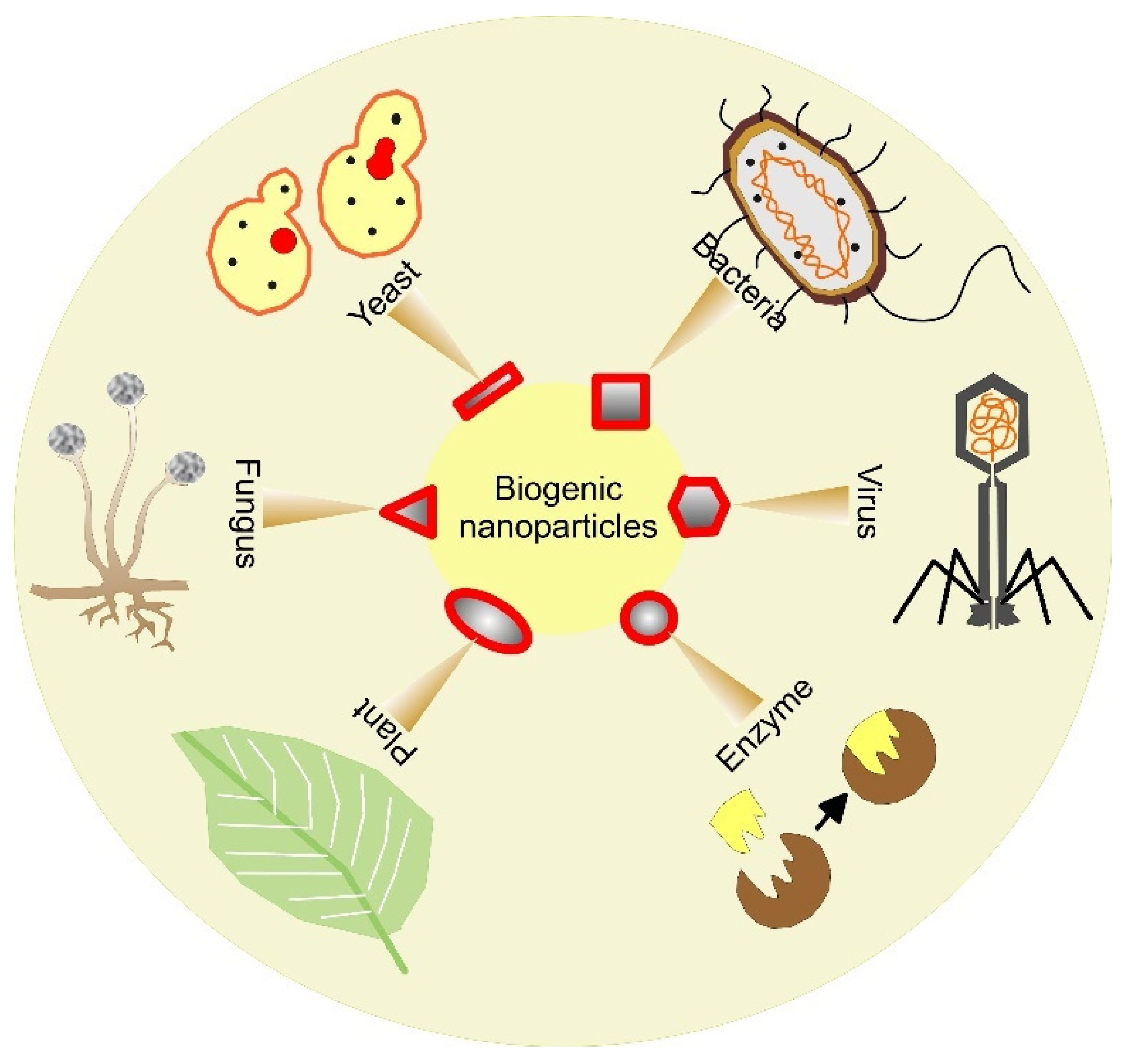
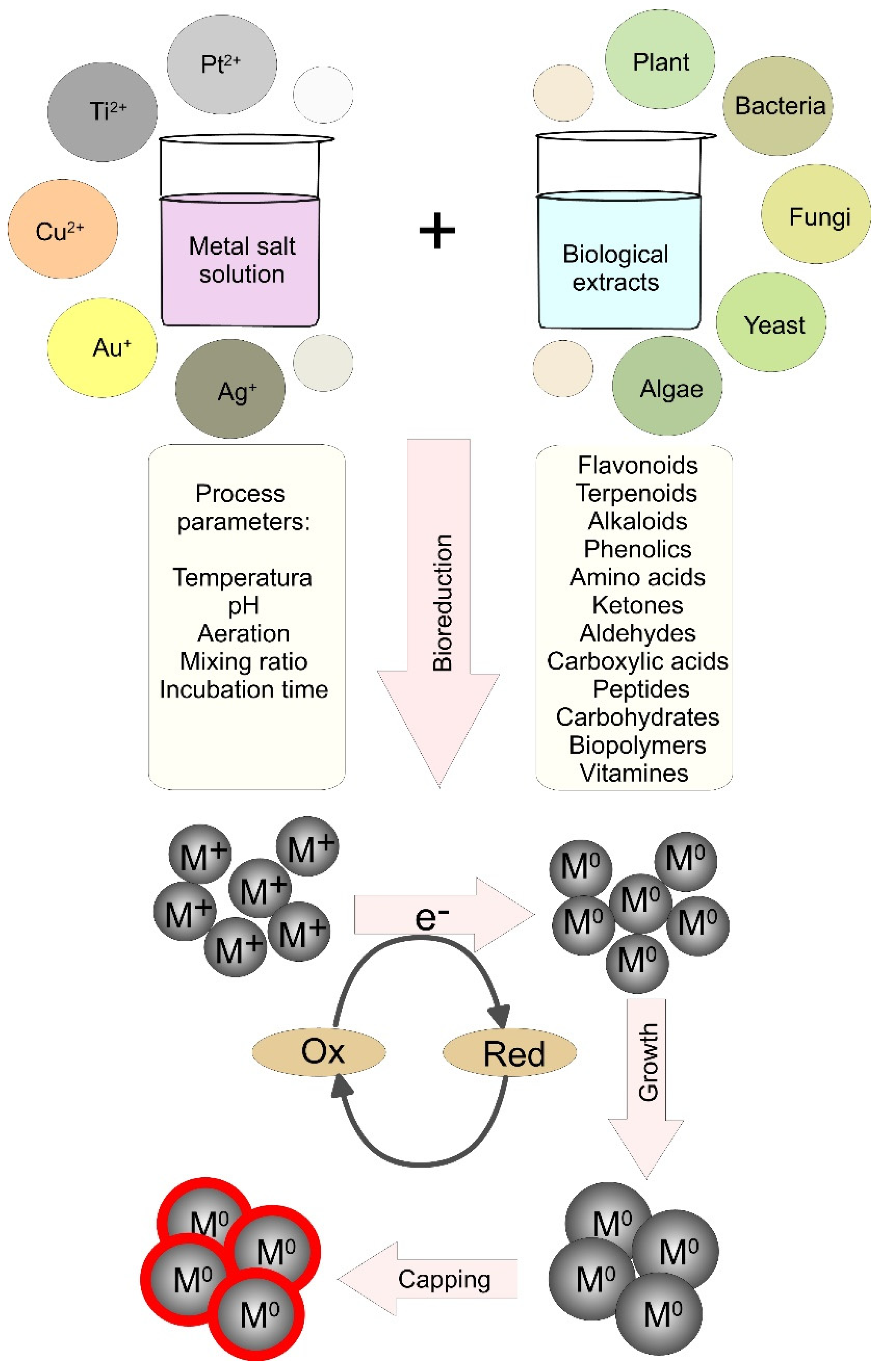
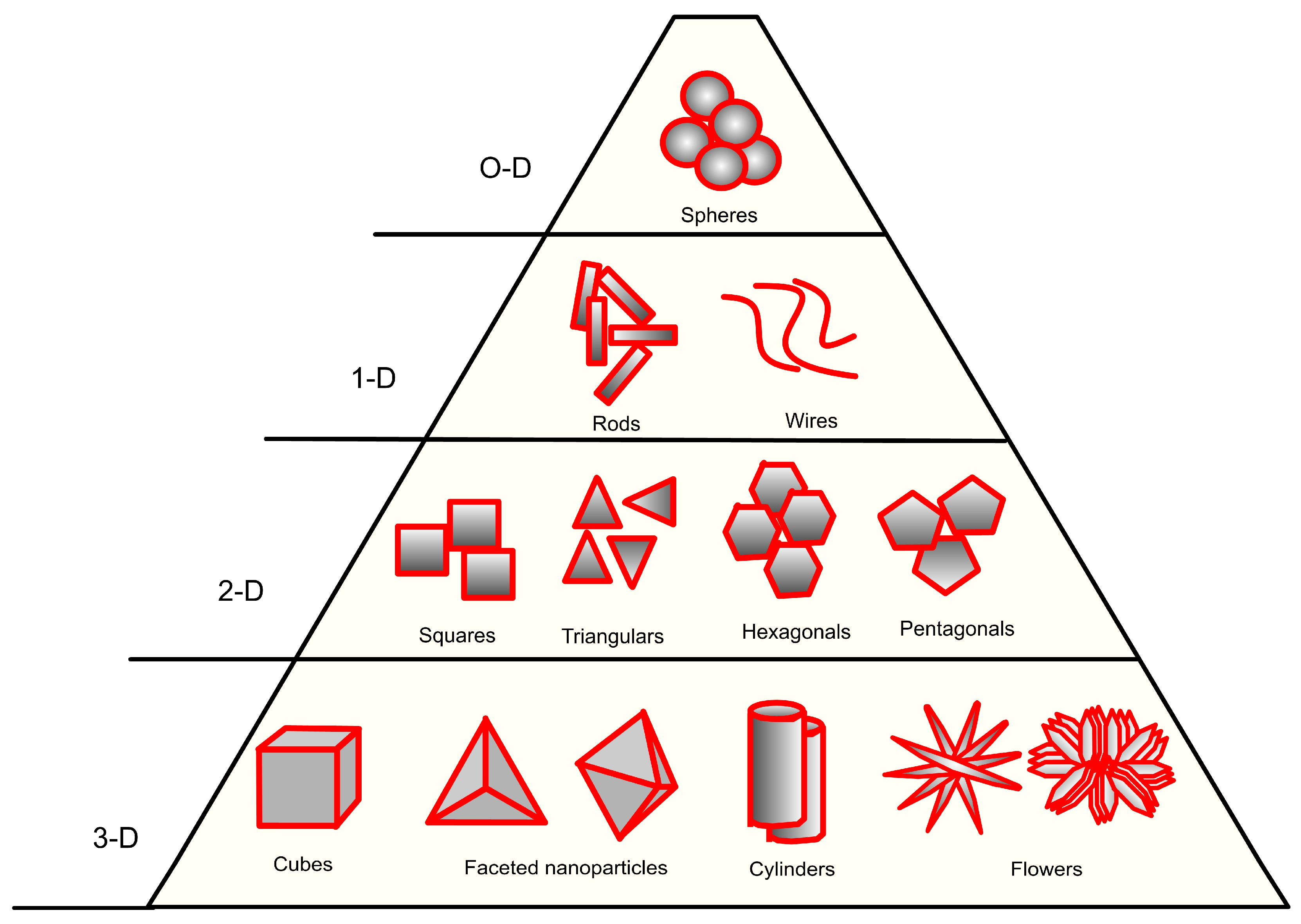
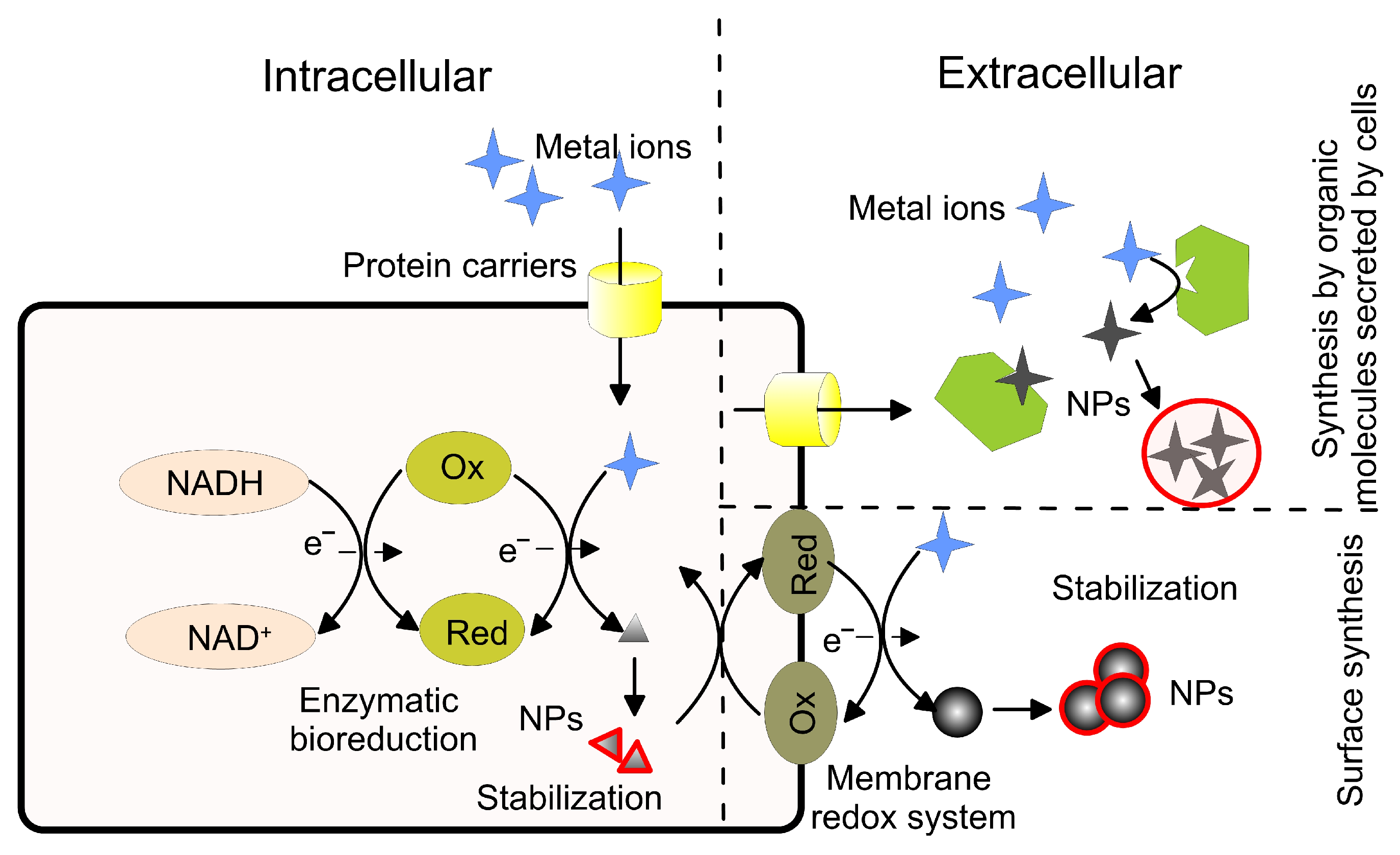
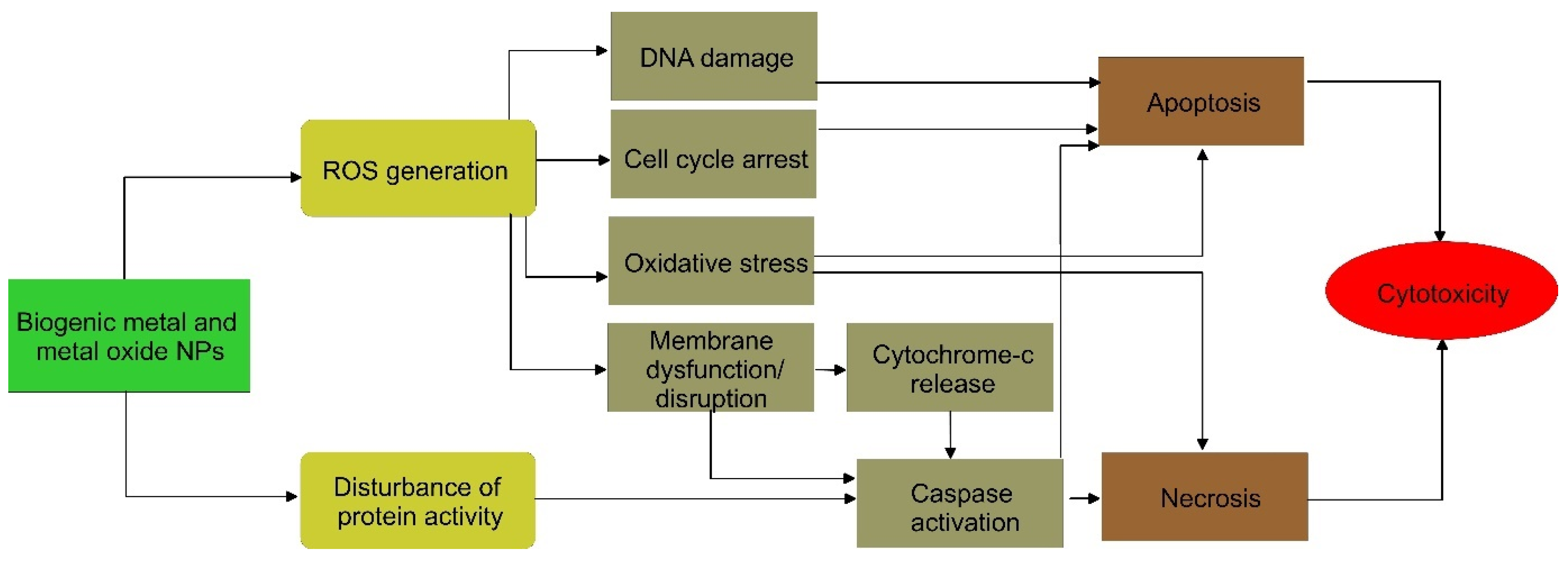
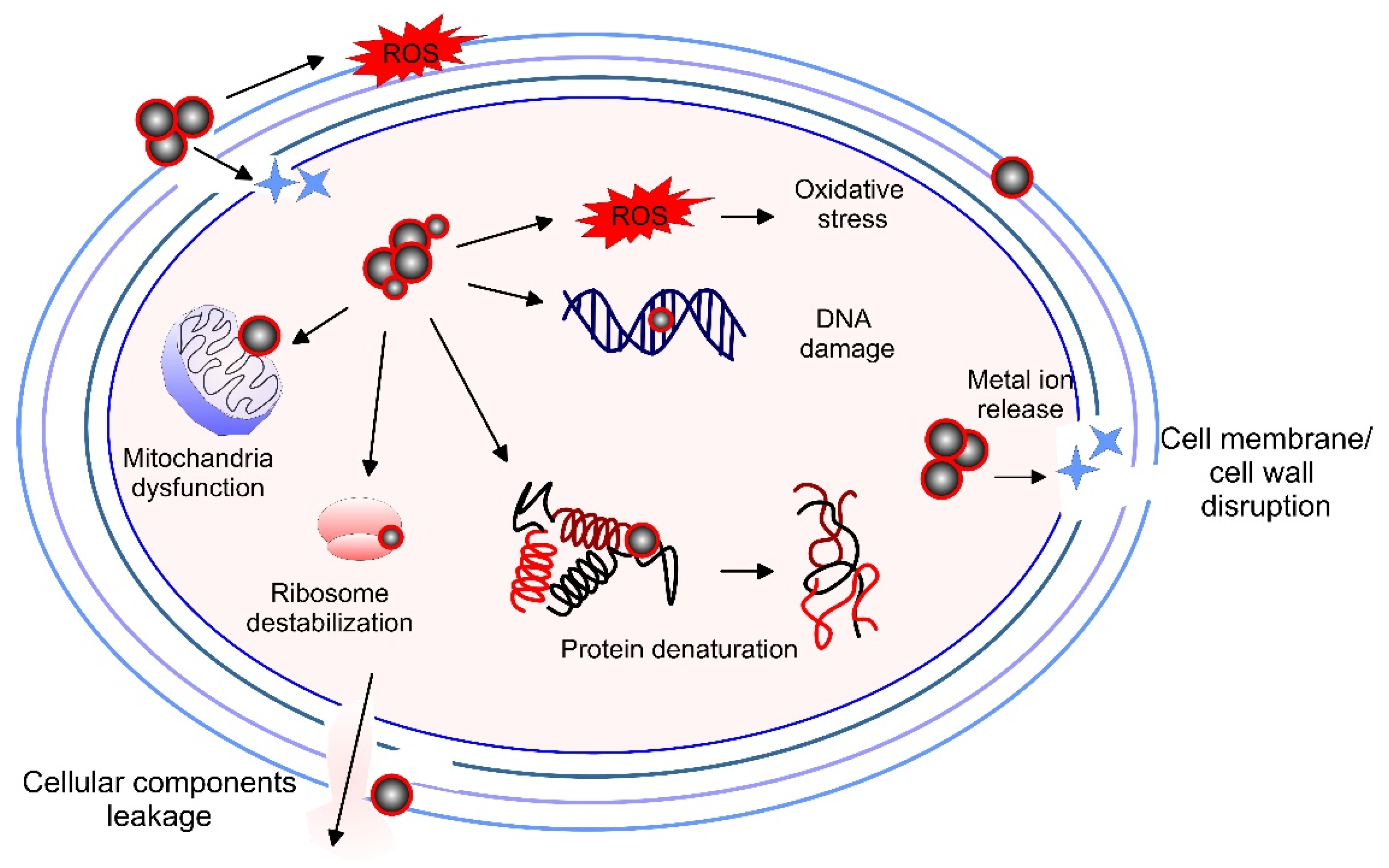
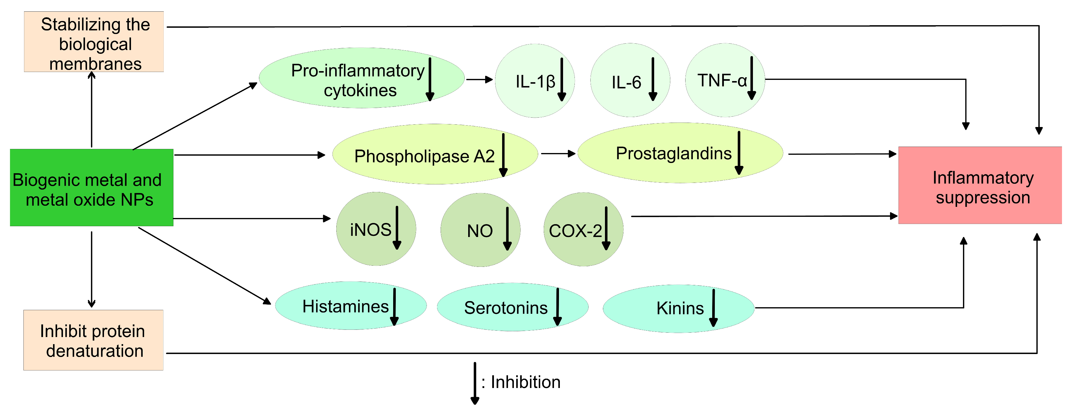
| Plant | Source | NPs | Size, nm | Morphology | Application/ Tested Objects | Ref |
|---|---|---|---|---|---|---|
| Cucumis anguria | Leaf | Ag | 11–27 | Spherical | Antibacterial against S. aureus, E. coli | [104] |
| Madhuca longifolia | Flower | Ag | 30–50 | Spherical and oval | Antibacterial against E. coli, S. typhimurium, B. cereus, S. saprophyticus | [28] |
| Banana waste | Peduncle | Ag | 14.1 | Spherical | Antimicrobial against S. aureus, E. coli, C. albicans, A. niger | [105] |
| Trachyspermum ammi | Seed | Au | 16.6 | Spheroidal | Anti-biofilm against L. monocytogenes and S. marcescens; anticancer against HepG2 cell lines | [106] |
| Garcinia mangostana | Pericarp | Au, Ag | 13.7 ± 5.1 31.1 ± 4 | Nanodumbbell shapes | Anticancer, antioxidant against colon cancer Col320 cell line | [107] |
| Asparagus racemosus | Root | Ag, Au, Ag-Au | 10–50 | Spherical | Antibacterial against P. aeurgnosia and S. aureus, anti-inflammatory in NK92 cells | [108] |
| Rhanterium epapposum | Flower | Ag Au Zn | 16.1 17.9 23.5 | Spherical, triangular, hexagonal | Antifungal against C. albicans and A. melleus, cytotoxic analysis against breast adenocarcinoma MCF-7, hepatocellular carcinoma HepG-2 and colorectal carcinoma HCT 116 human cell lines | [109] |
| Rosmarinus officinalis | Leaf | Pd | 15–90 | Semi-spherical | Antibacterial against E. coli, S. aureus, S. epidermidis and M. lutens; antifungal against C. albicans, C. parapsilolis, C. glabrata and C. krusei | [110] |
| Allium fistulosum, Tabernaemontanadivaricata, and Basella alba | Leaf | Pd | 20–50 | Spherical | Antibacterial against Basella alba, Allium fistulosum; antifungal against C. albicans, A. flavus, and Penicillinium sp.; antioxidant (free radical scavenging), antidiabetic (α-amylase inhibition) | [111] |
| Saudi’s dates | Fruit | Pt | 1.3–6.6 | Spherical | Anticancer against colon cancer HCT-116, breast cancer MCF-7 and hepatocellular carcinoma HePG-2 cell line | [112] |
| Celastrus paniculatus Willd. | Leaf | Cu | 2–10 | Spherical | Antifungal against F. oxysporum | [113] |
| Cissus vitiginea | Leaf | Cu | 2–20 | Spherical | Antioxidant, antibacterial against E. coli, Enterococcus sp., Proteus sp. Klebsiella sp. | [114] |
| Syzygium aromaticum | Bud | Cu | 12–15 | Spherical | Antimicrobial against Staphylococcus spp., Bacillus spp., Pseudomonas spp., E. coli, A. niger, A. flavus and Penicillium spp. | [56] |
| Azadirachta indica | Leaf | CuO | NA | Spherical | Anticancer against breast cancer MCF-7 and Hela cell lines | [115] |
| Achillea millefolium | Leaf | CuO | 28 | Semi-spherical | Antibacterial against S. aureus, M. tuberculosis, E. coli, K. pneumoniae, P. mirabili, C. diphtheriae, S. pyogenens; antifungal against C. albicans, A. flavus, M. canis and G. glabrata | [116] |
| Musa ornate | Flower | Fe | 43.7 | NA | Antibacterial against S. aureus, S. agalactiae, S. enterica, E. coli | [117] |
| Ficus carica | Leaf | Fe3O4 | 43–57 | Multiform | Antioxidant (DPPH free radical scavenging) | [118] |
| Platanus orientalis | Leaf | Iron oxide | 30–40 | Spherical | Antifungal against A. niger and M. piriformis | [119] |
| Mentha pulegium (L.) | Leaf | ZnO | 38–49 | Semi-spherical | Antimicrobial against E. coli and S. aureus | [68] |
| Conyza canadensis | Leaf | ZnO | - | Spherical | Antibacterial against E. coli and S. aureus | [120] |
| Costus pictus D. Don | Leaf | ZnO | 40 | Elongated, hexagonal, and rod shape | Antimicrobial against S. aureus, B. subtilis, E. coli, S. parathyphi, A. niger, C. albicans anticancer against Dalton’s ascites cells | [121] |
| Albizia lebbeck | Stem bark | ZnO | 66.6 | Irregular spherical | Antimicrobial against B. cereus, S. aureus, E. coli, K. pneumoniae and S. typhi, antioxidant (hydrogen peroxide free radical scavenging), anticancer against breast cancer MDA-MB 231 and MCF-7 cell lines | [122] |
| Ailanthus altissima | Plant extract | ZnO | 13.3 | Spherical | Antibacterial against S. aureus, K. pneumonia, E. coli, S. pyogenes; antioxidant (DPPH free radical scavenging) | [123] |
| Punica granatum | Peel | ZnO | 118.6 | Irregular nanorods | Antibacterial against S. aureus, E. aerogenes, P. aeruginosa, and K. pneumoniae | [124] |
| Trigonella foenum-graecum | Leaf | MgO | 13 | Spherical | Antibacterial against E. coli, Bacillus spp., and S. aureus | [125] |
| Ceratonia siliqua | Leaf | CeO2 | 22 | NA | Antioxidant (DPPH free radical scavenging), cytotoxic against breast cancer MCF-7 cell lines | [126] |
| Class Organism | Metallic NPs | Intra/Extracellular Synthesis | Species | Morphology | Size, nm | Application/ Tested Objects | Ref. |
|---|---|---|---|---|---|---|---|
| Bacteria | Au | Extracellular | Paracoccus haeundaensis | spherical | 20.9 ± 3.5 | Antioxidant (DPPH free radical scavenging), cytotoxicity against normal kidney cells HEK 293, anticancer against lung carcinoma A549 and gastric adenocarcinoma AGS cell lines | [190] |
| Au | Intracellular | Lactobacillus kimchikus | spherical | 5–30 | Antioxidant (DPPH free radical scavenging) | [191] | |
| Ag | Intracellular | Transformed E. coli | spherical | 20 | NA | [192] | |
| Ag | Extracellular | Bacillus brevis | spherical | 41–68 | Antibacterial against S. aureus and S. typhi | [193] | |
| Ag | Extracellular | Bacillus subtilis | spherical | 3–20 | Antimicrobial against S. aureus, S. epidermidis, K. pneumoniae, E. coli and C. albicans | [29] | |
| Pt | Intracellular | Acinetobacter calcoaceticus | cubic | 2–3 | NA | [194] | |
| Cu | Intercellular | Streptomyces capillispiralis Ca-1 | spherical | 3.6–59 | Antimicrobial against S. aureus, B. subtilis, B. dimenuta, P. aeruginosa, E. coli; Antifungal against C. albicans and A. brasiliensis | [195] | |
| ZnO | Extracellular | Pseudomonas putida | spherical | 44.5 | Anti-biofilm against B. cereus and E. faecalis, antibacterial against P. otitidis, P. oleovorans, A. baumannii, B. cereus, E. faecalis | [196] | |
| ZnO | Extracellular | Staphilococcus aureus | irregular | 10–50 | Antibacterial against E. coli, S. aureus, S. epidermis, E. faecalis, K. pneumonia, P. aeruginosa | [69] | |
| ZnO | Intercellular | Serratia nematodipila | spherical | 15–30 | Antibacterial against X. oryzae and antifungal against Alternaria sp. | [197] | |
| TiO2 ZnO | Extracellular | Halomonas elongata | spherical | 104.6 18.1 | Antibacterial against E. coli and S. aureus | [198] | |
| TiO2 | Extracellular | Streptomyces sp. | spherical | 30–70 | Antimicrobial activity against E. coli, S. aureus, C. albicans, A. niger; antibiofilm against P. aeruginosa | [199] | |
| CuO | Extracellular | Streptomyces sp. | spherical | 78–80 | Antifungal against F. oxysporum, P. ultimum, A. niger, A. alternata cytotoxicity against Vero and Caco-2 cell lines | [200] | |
| CuO | Extracellular | Streptomyces sp. | spherical | 1.7–13.5 | Antibacterial against E. faecalis, S. typhimurium, P. aeruginosa, E. coli; Antifungal against C. albicans, R. solani, A. niger | [201] | |
| Iron oxide | Extracellular | Proteus vulgaris ATCC-29905 | spherical | 19.2–30.5 | Antibacterial against methicillin resistant S. aureus; anticancer against U87MG-glioblastoma cancer and HT-29 cancer cell line | [202] | |
| Fungi and Yeast | Au | Extracellular | Cladosporium cladosporioides | quasi-spherical | 60 | Antioxidant (DPPH and FRAP assay), antimicrobial against S. aureus, B. subtilis, P. aeruninosa, A. niger | [203] |
| Ag | Extracellular | Cladosporium cladosporioides | spherical | 30–60 | Antimicrobial against S. aureus, B. subtilis, S. E. coli, S. epidermidis, and C. albicans; antioxidant (DPPH assay) | [204] | |
| Ag | Extracellular | Beauveria bassiana | triangular, circular, hexagonal | 10–50 | Antibacterial against E. coli, P. aeruginosa, S. aureus | [30] | |
| Pt | Extracellular | Fusarium oxysporum | spherical | 25 | Antibacterial against E. coli Antioxidant (DPPH assay) | [205] | |
| Pd | Intracellular | Agaricus bisporus | triangular spherical | 13–18 | Antibacterial against S. aureus, S. pyrogens, B. subtilis, E. aerogenes, K. pneomoniae, P. vulgaris, anticancer against PK13 cell lines, anti-inflammatory with RBC cells Antioxidant (DPPH method) | [206] | |
| Cu | Extracellular | Aspergillus niger | round | 500–800 | Antidiabetic (α-glucosidase assay), anticancer against human hepatocellular carcinoma Huh-7 cell lines; antibacterial against E. coli, S. aureus, K. pneumoniae, M. luteus, B. subtilis | [207] | |
| ZnO | Extracellular | Aspergillus niger | spherical | 35 | Antibacterial against E. coli; cytotoxicity against breast cancer MCF-7 cell lines | [208] | |
| ZnO | Extracellular | Xylaria acuta | hexagonal | 34–55 | Antimicrobial against S. aureus, B. subtilis, P. aeruginosa, E. coli | [70] | |
| ZnO | Extracellular | Aspergillus niger and F. keratoplasticum | nanorods hexagonal | 8–32 10–42 | Antimicrobial against B. subtilis, S. aureus, P. aeruginosa, E. coli | [209] | |
| CuO ZnO | Extracellular | Penicillium chryogenum | spherical hexagonal | 10.5–59.7 9–35 | Antibiofilm against S. aureus and P. aeruginosa Antimicrobial against S. aureus, B. subtilis, P. aeruginosa, E. coli, S. typhimurium and fungi F. solani, F. oxysporum, S. sclerotia, A. terreus | [210] | |
| TiO2 | Extracellular | Pleurotus djamor | spherical | 31 | Anticancer against A549 cell lines, Antibacterial against P. fluorescens, S. aureus, C. diphtheriae | [211] | |
| Algae | Ag | Intracellular | Padina sp. | spherical | 25–60 | Antimicrobial against S. aureus, B. subtilis, E. coli, S. typhi, P. aeruginosa | [212] |
| Ag | Extracellular | Gelidium corneum | spherical | 20–50 | Anti-biofilm and antibacterial against C. albicans and E. coli | [213] | |
| Au | Extracellular | Cystosuira baccata | spherical | 8.4 | Anticancer against colon cancer cell lines HT-29 and Caco-2 | [214] | |
| Pd | Extracellular | Spirulina patensis | spherical | 10–20 | - | [215] | |
| Pd | Extracellular | Chlorella vulgaris | spherical | 5–20 | - | [216] | |
| Pt | Extracellular | Padina gymnospora | spherical, octahedral | 10–60 | Antimicrobial against E. coli, L. lactis, K. pneumoniae with no hemolytic activity in vitro | [217] | |
| Cu | Extracellular | Botryococcus braunii | cubic, spherical, triangular | 10–70 | Anti-microbial against P. aeruginosa, E. coli, K. pneumoniae, S. aureus and F. oxysporum | [218] | |
| Fe3O4 | Extracellular | Colpomenia sinuosa and Pterocladia capillacea | nanospheres | 11.2–33.7 16.9–22.5 | Antibacterial against E. coli, P. aeruginosa, S. typhi, V. cholera, B. subtilis, S. aureus Antifungal against A. flavus and F. oxysporum | [219] | |
| ZnO | Extracellular | Sargassium muticum | hexagonal | 30–57 | Anti-angiogenetic and apoptotic effect on human liver cancer cell line HepG2 | [71] | |
| ZnO | Extracellular | Arthrospira platensis | spherical | 30–55 | Antimicrobial against B. subtilis, S. aureus, P. aeruginosa, E. coli and C. albicans | [220] | |
| Viruses | Au | intracellular | Tobacco mosaic virus (TMV) | spherical | 5 | - | [221] |
| Au | intracellular | Genetically modified Tobacco mosaic virus (TMV) | spherical | 9–33 | - | [160] | |
| Au Ag | intracellular | Plant pathogenic virus of Squash leaf curl China virus | spherical | 5–12 5–20 | Cytotoxicity against A549 lung cancer cell line | [162] | |
| Pd Pt Au | Intra- and extracellular | Tobacco mosaic virus (TMV) | spherical | 3.4 ± 0.5 3.1 ± 0.5 2.9 ± 0.5 | Catalytic (allyl alcohol hydrogenation) | [222] |
| NPs | Source | Size, nm | Shape | Drug | Tested Object | Main Outcome | Ref. |
|---|---|---|---|---|---|---|---|
| Ag | Abelmoschus esculentus (L.) | 21.3 | Spherical | - | Jurkat cells (human T-cell lymphoma) | -Antiproliferative effect in a dose-dependent manner with an IC50 value of 16.15 μg/mL -Triggered high levels of ROS; -Loss of integrity of mitochondrial membrane; | [253] |
| Ag | Punica granatul leaf extract | 41.7–69.6 | spherical | - | Cervical cancer HeLa cell line | -IC50 for inhibiting 50% of the Hela cell line was 100 μg/mL; -at a concentration of 100 μg/mL Ag NPs induced apoptosis by fragmentation of DNA | [254] |
| Ag | Punica granulatum leaf extract | 35–60 | Spherical | - | Liver cancer cell line HepG2 | -IC50 for the HepG2 cell line was 70 μg/mL; -In vitro free radical scavenging activity. | [255] |
| Ag | Taraxacum officinale | 5–30 | Spherical | - | Liver cancer cell line HepG2 | -High cytotoxic effect against HepG2 | [256] |
| Ag | Olax scandens leaf extract | 20–60 | Spherical | - | B16 mouse melanoma cell line; A549 human lung cancer cell line; MCF7 human breast cancer cells | -Inhibition of proliferation of both A549 and B16 in a dose-dependent manner; -Compared to chemically synthesized Ag NPs, the biogenic showed biocompatible nature with normal cells; | [257] |
| Au | Peltophorumpterocarpum | 65–149 | Spherical | Dox | Lung (A549) and melanoma (B16F10) cancer cell line, C57BL6/J mice | -Nano-conjugate cellular uptake and drug release were faster than pure Dox; -No significant changes in hematology, serum clinical biochemistry, or histopathology in C57BL6/J mice were observed after 7 days | [258] |
| Ag | Bacillus licheniformis | 20–80 | Triangular | 5-aminoevulinic acid (ALA) | Skin melanoma (B16F10), epidermoid carcinoma (A431) | -Higher cytotoxicity on both cell lines than the pure ALA and Ag NPs; | [259] |
| Au | Syzygium aromaticum | 12–20 | Spherical | - | Human lymphoma SUDHL-4 cell line | -The IC50 of Au NPs after 48 h was 30 μM; -Au NPs induced ROS accumulation and dose and time-dependent manner; -Apoptosis mechanisms involved caspase activation, formation of pores in the mitochondrial membrane, and release of cytochrome c; -The NPs decrease the growth and increase apoptosis of cells | [260] |
| Ag (Starch coated) | Meadow grass (Poa anua) | 36.7 ± 7.9 | Spherical | Euphorbia dracunculoides Lam. Plant extract | Human embryonic kidney (HEK293), Hep3B liver cancer, SCC-7 murine cancer cell line, Sprague Dawley rats | -Up to 96% drug loading efficacy was observed; -Good biocompatibility (90–100%) with HE293 at high concentrations (up to 1000 μg/mL) -at concentration 400 μg/mL of Hep3B and SCC-7 decreased to 20 and 30%, respectively after 72 h; -IC50 of Au NPs was 113.3 μg/mL; -Significantly low cytotoxicity in vivo of the bio-drug NP conjugates | [261] |
| Au | Elettaria cardamomum seeds | 15.2 | Spherical | - | Cervical cancer (HeLa) cell line | -the IC50 of Au NPs was 42.6 μL; -Penetration of Au NPs through the cell membrane; -Shrinking, rounding granulation morphological changes | [262] |
| Au | Vitex negundo (reducing agents) and Arabic gum (capping agent) | 98.7 ± 1.9 | Spherical | Epirubicin | Lung adenocarcinoma A549 cell line | -The amount of epirubicin bound was 85%; -The IC50 for value NP conjugate was around 4 μg/mL whereas for the drug alone was 24 μg/mL -The drug release from the conjugate was slow, thus the cytotoxicity and therapeutic efficacy were higher | [263] |
| Au | Paracoccus haeundaensis BC74171I bacterium | 20.9 ± 3.5 | Spherical | - | A549 and AGS cancer cell lines | -Au NPs exert an antiproliferative effect on cancer cells and did not affect the normal cells HEK293 and HaCaT up to a concentration of 200 μg/mL; -The percentage of cell growth inhibition of AGS cells was higher than that of A549 indicating selective cytotoxicity; | [190] |
| Au | Magnifera indica | 55.5–65.5 | Spherical | - | MDA-MB-231 and breast cancer mice | -Highly effective in controlling the growth of breast cancer in a dose-dependent fashion; | [264] |
| Au | Peptides | 100–150 | Spherical | Dox | Hela and nude mice | -Tumor inhibition in nude mice via tumor and tail vain was 66.7 and 57.7%, respectively; -Efficient Dox transport vector for cervical cancer therapy. | [265] |
| Au | Cyclopia intemedia and magniferin | 20 | Spherical and Triangular | Dox | Human colon cancer (Caco-2), prostate cancer (PC-3) and glioblastoma (U87) cell lines; normal breast epithelial cell lines | -In combination with Dox, antitumor effects are augmented. -At the same time, relatively low cytotoxicity to normal cells was observed | [266] |
| Au/Pt | Phragmites australis leaves | 35.1 ± 2.7 | Flower-like shape | Dox | Breast (MCF-7) and lung (A459) cancer cell line | -pH-controlled dependency response under acidic tumor conditions; -A three-fold cell death to cancer cells compared to Dox alone; -Dox-conjugated Au/Pt NPs exhibited time-release phosphatidylserine exposure; -Cell-specific response against MCF-7 compared to A459 | [267] |
| Fe2O3 | Psoralea corylifolia | 39 | Spherical | - | Renal tumor MDCK and Caki-2 cell lines | -Strong cancer cell growth inhibiting in a dose-dependent manner | [268] |
| Fe2O3 | Mentha piperita leaves | 17.9 | Polygonal (rhombic and hexagonal) | Dox | Breast MCF-7 cancer cell line | -The IC50 of Fe2O3 NPs against cancer cells was 0.5 μg/mL while at 1 μg/mL their toxicity to normal was negligible; -71% Dox loading efficacy; -NPs-Dox conjugates significantly inhibited the tested cancer cells compared to free drug; | [269] |
| Fe2O3 | Rhus punjabensis extract | 41.5 ± 5 | Rhombohedral | HL-60 leukemia and DU-145 prostate cancer cell line | -The NPs were active against both cancer cells and leaved health control cell unaffected; | [270] | |
| Fe | Streptomyces sp. | 65–87 | Spherical | DU145 and prostate cancer (PC3) cell line | -The IC50 for value NPs was around 65 μg/mL; -The color staining revealed an antiproliferative effect of the NPs on tumor cells; | [271] | |
| CuO | Bean extract | 26.6 | Spherical, hexagonal, uneven shape | - | Human cervical carcinoma (HeLa) cell line | -The IC50 for value NPs was around 13 μg/mL increased ROS and lipid peroxidation of liposomal membrane; -Alteration in the mitochondrial structure; -Inability of cells to proliferate. | [272] |
| ZnO | Leucaena leucocephala | 50–200 | Hexagonal | - | Breast cancer (MCF-7) and prostate cancer (PC3) cell line; Dalton lymphoma ascites (DLA) cell line | -The IC50 for value NPs was around 103 μg/mL -Better cytotoxic activity on PC-3 than MCF-7 cell line; -Against DLA cells the biosynthesized ZnO NPs revealed 92% inhibition with a concentration of 200 μg/mL; | [273] |
| TiO2 | Cinnamomum tamala | 23 | Irregular | - | Human prostate cancer (D145) cell line | -TiO2 NPs showed a dose-dependent anticancer effect | [274] |
| Au/ CuO/ ZnO | Verbena officinalis L. extract | 35 | Spherical | - | Jurkat cell line | -The IC50 for value NPs was around 0.64 μmol; -Within 24 h over 80% of the cells cultured in the presence of Au/CuO/ZnO NPs at a concentration of 10 μmol exhibited sighs of late apoptosis; -about 60% of the cells at a concentration of 100 μmol underwent necrosis. | [275] |
Disclaimer/Publisher’s Note: The statements, opinions and data contained in all publications are solely those of the individual author(s) and contributor(s) and not of MDPI and/or the editor(s). MDPI and/or the editor(s) disclaim responsibility for any injury to people or property resulting from any ideas, methods, instructions or products referred to in the content. |
© 2023 by the authors. Licensee MDPI, Basel, Switzerland. This article is an open access article distributed under the terms and conditions of the Creative Commons Attribution (CC BY) license (https://creativecommons.org/licenses/by/4.0/).
Share and Cite
Nikolova, M.P.; Joshi, P.B.; Chavali, M.S. Updates on Biogenic Metallic and Metal Oxide Nanoparticles: Therapy, Drug Delivery and Cytotoxicity. Pharmaceutics 2023, 15, 1650. https://doi.org/10.3390/pharmaceutics15061650
Nikolova MP, Joshi PB, Chavali MS. Updates on Biogenic Metallic and Metal Oxide Nanoparticles: Therapy, Drug Delivery and Cytotoxicity. Pharmaceutics. 2023; 15(6):1650. https://doi.org/10.3390/pharmaceutics15061650
Chicago/Turabian StyleNikolova, Maria P., Payal B. Joshi, and Murthy S. Chavali. 2023. "Updates on Biogenic Metallic and Metal Oxide Nanoparticles: Therapy, Drug Delivery and Cytotoxicity" Pharmaceutics 15, no. 6: 1650. https://doi.org/10.3390/pharmaceutics15061650