Article

Insightful Improvement in the Design of Potent Uropathogenic E. coli FimH Antagonists

1
Glycosciences and Nanomaterial Laboratory, Université du Québec à Montréal, Succ. Centre-Ville, P.O. Box 8888, Montréal, QC H3C 3P8, Canada
2
Research Laboratories, Bambino Gesù Children’s Hospital, Istituto di Ricovero e Cura a Carattere Scientifico, 00146 Rome, Italy
3
Unité de Glycobiologie Structurale et Fonctionnelle (UGSF), UMR8576 du CNRS, Univ. Lille, 59000 Lille, France
4
Department of Public Health and Infectious Diseases, Sapienza University of Rome, 00185 Rome, Italy
5
Department of Human Sciences and Promotion of the Quality of Life, San Raffaele Roma Open University, 00166 Rome, Italy
6
Istituto di Ricovero e Cura a Carattere Scientifico, San Raffaele Roma, 00166 Rome, Italy
7
Department of Infectious Diseases, National Institute of Health, 00161 Rome, Italy
8
Institut de Chimie Pharmaceutique Albert Lespagnol (ICPAL), Faculté de Pharmacie, University of Lille, 3 Rue du Professeur Laguesse, BP-83, 59006 Lille, France
9
GLYcoDiag, 2 Rue du Cristal, 45100 Orléans, France
*
Authors to whom correspondence should be addressed.
Pharmaceutics 2023, 15(2), 527; https://doi.org/10.3390/pharmaceutics15020527
Submission received: 10 January 2023 / Revised: 25 January 2023 / Accepted: 2 February 2023 / Published: 4 February 2023

Abstract

:
Selective antiadhesion antagonists of Uropathogenic Escherichia coli (UPEC) type-1 Fimbrial adhesin (FimH) are attractive alternatives for antibiotic therapies and prophylaxes against acute or recurrent urinary tract infections (UTIs) caused by UPECs. A rational small library of FimH antagonists based on previously described C-linked allyl α-D-mannopyranoside was synthesized using Heck cross-coupling reaction using a series of iodoaryl derivatives. This work reports two new members of FimH antagonist amongst the above family with sub nanomolar affinity. The resulting hydrophobic aglycones, including constrained alkene and aryl groups, were designed to provide additional favorable binding interactions with the so-called FimH “tyrosine gate”. The newly synthesized C-linked glycomimetic antagonists, having a hydrolytically stable anomeric linkage, exhibited improved binding when compared to previously published analogs, as demonstrated by affinity measurement through interactions by FimH lectin. The crystal structure of FimH co-crystallized with one of the nanomolar antagonists revealed the binding mode of this inhibitor into the active site of the tyrosine gate. In addition, selected mannopyranoside constructs neither affected bacterial growth or cell viability nor interfered with antibiotic activity. C-linked mannoside antagonists were effective in decreasing bacterial adhesion to human bladder epithelial cells (HTB-9). Therefore, these molecules constituted additional therapeutic candidates’ worth further development in the search for potent anti-adhesive drugs against infections caused by UPEC.

1. Introduction

One of the major pathogens responsible for urinary tract infections (UTIs) is uropathogenic Escherichia coli (E. coli) (UPEC), leading to a major burden in public health [1]. As part of the normal microbiota, E. coli exhibits diverse species with a wide spectrum of phenotypes that reside in the large intestine of humans and many animals [2]. Several virulence factors are responsible for the establishment of current infections [2,3]. While E. coli mainly live harmlessly in the gut as their primary niche by establishment of a symbiotic relationship, some strains act as pathogens and cause a variety of enteric and extra-enteric diseases [4,5,6]. Among these, the adhesion of UPECs to the host uroepithelial tissues is due to the E. coli type 1 pili, called FimH, which mediate their attachment in a mannose-dependent interaction [4,5]. FimH adhesin of type 1 fimbriae promotes intestinal colonization by binding to colonic crypts of epithelial cells, facilitating E. coli adhesion and invasion to the urinary epithelium, a key player in UPEC pathogenicity [6]. Preventing bacterial FimH adhesion to highly mannosylated glycoprotein uroplakin Ia from bladder cells represents an appealing strategy to control UTIs [4]. Since bacterial adhesion to host cells is a critical and initial step in most infectious diseases, anti-adhesion therapy can effectively compete against antibiotic therapy and is seriously considered for the treatment of acute uncomplicated lower UTIs (AUC) [7,8,9].
Importantly, the mannose-specific adhesin FimH has an essential hydrophobic binding pocket of non-polar amino acids that surrounds the aglycon portion of α-D-mannopyranoside glycomimetics. These findings have been fully exploited in the design of potent FimH anti-adhesins [10,11,12,13]. Glycomimetics are simplified representatives of more complex glycoconjugate structures found in nature. They have been designed to improve carbohydrate-protein binding interactions and to provide better pharmacokinetic and pharmacodynamic properties [14,15].
As alternative therapeutic strategies against UTIs, monomeric mannoside antagonists [15,16,17,18] and clusters [19,20,21,22,23,24,25] with varied hetero-anomeric linkages O-[26,27,28,29], S-[30,31,32], or N-[33,34] have been developed as appealing candidates. Using a plethora of available crystallographic data, detailed structure-activity relationships (SARs) have been established to provide further insight into the binding mode of E. coli FimH. This approach has greatly helped in improving the conception of potent antagonists [4,35,36,37,38,39]. Amid the various α-D-mannopyranoside inhibitors described thus far, C-linked glycomimetics residues harboring hydrophobic aglycons are still considered worthy candidates as potent FimH inhibitors. Several of these synthetic candidates showed improved binding affinities and increased hydrolytic stability [30,40,41].
This work describes the synthesis, relative binding affinities, crystallographic data, and anti-adhesive properties of a small library of C-linked mannopyranoside inhibitors. The data provide further pivotal insights into the potential of these glycomimetics against UPEC strains and give rise to a solid basis toward the development of new and effective FimH antagonists.

2. Results and Discussion

2.1. Synthesis and Structural Characterization

In previous studies, several aryl C-mannopyranosides carrying alkenyl aglycons were shown as promising candidates with excellent binding affinities against E. coli FimH [40,41]. However, their poor water solubility has restricted their further development as drug candidates. To address this issue, we propose herein the use of heteroaryl-substituted mannopyranosides based upon numerous available crystallographic data.
The first step toward this next generation of antagonists was the improved preparation of C-allyl α-D-mannopyranoside 1 as a key starting material. Compound 1 was obtained by per-O-benzoylation of commercially available methyl α-D-mannopyranoside (MeMan) followed by a Hosomi–Sakurai reaction to afford the pure α-anomer 1 [40]. Key precursor 1 was next treated under palladium-catalyzed Heck conditions in the presence of aryl iodides (DMF, TBAB, NaHCO3, Pd(OAc)2, 90 °C, o.n.) to afford a family of (E)-linked aryl derivatives (210) in good yields (Scheme 1).
As depicted in Figures S1, S2 and S20, 1H-NMR spectra (600 MHz) of both compounds 2 and 11, together with detailed 2D NMR spectrum of compound 2, unambiguously confirmed that all the major compounds were the trans stereoisomers. Reliable evidence of this claim was the signal of H-3’ of the alkene that appeared as a doublet at δ 6.67 ppm with a J2’,3’ = 15.8 Hz and 16 Hz for both compounds 2 and 11, respectively. Moreover, the coupling constants for the H4 signal at δ 6.00 ppm (dd, 1H, J3,4 = J4,5 = 8.5 Hz, H-4) unambiguously indicated a trans-diaxial relationship between H3-H4 and H4-H5, thus confirming this series of compounds to be in the proper 4C1 chair conformation.
Because C-linked mannopyranosides do not exhibit an anomeric effect in comparison to their O-linked analogs, there was a risk that the analogs had undergone conformational changes from the desired 4C1 chair to skew boats (0S2 or 0S2) [4]. The X-ray crystal structure of perbenzoylated compound 2 showed it to be in the desired chair conformation as previously observed with several related analogs (Figure S4). Since the conformation of 2 in the solid-state could be different than that in solution, an in-depth NOESY analysis was also performed to further confirm its conformation (Figure S3). The nOe between the geminal H1’a, H1’a’ with the axial H3 and H5 unmistakably implied the 4C1 conformation. Analysis of the 1H-NMR spectrum of C-linked mannosides (1118, and 20) also showed values of ca. 1.63 Hz for their 3J1,2 coupling constants of their vicinal equatorial/axial arrangements (Figure S2).
Finally, benzoyl ester deprotection of C-linked mannopyranosides (29) was performed using ammonia in methanol (1M, r.t., o.n.) to provide unprotected derivatives (1118 and 20), essentially quantitatively.

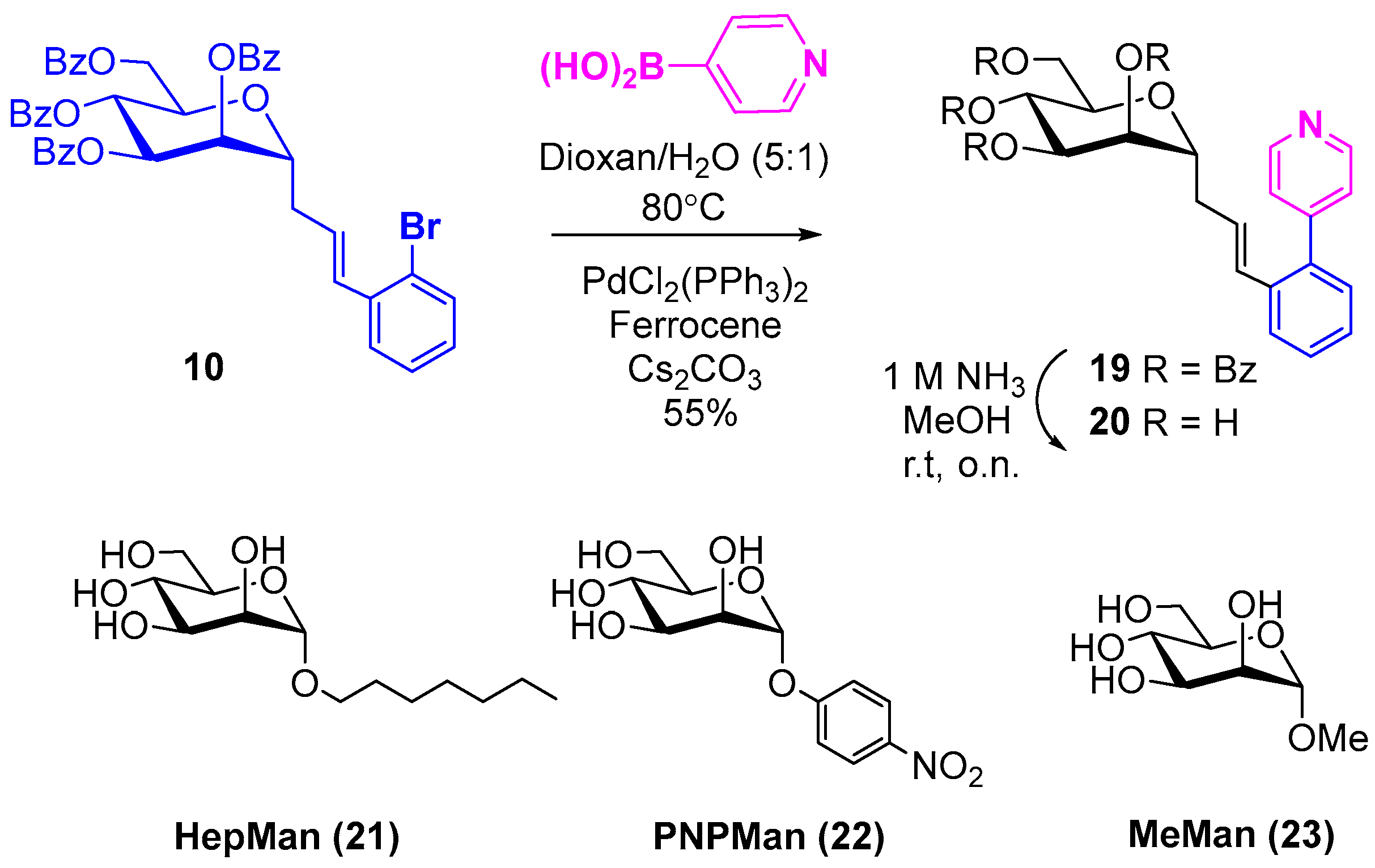
2.2. Synthesis of a Key Ortho-Substituted Biphenyl Derivative

In our previous work, an ortho-substituted C-linked biphenyl mannopyranoside candidate was considered as a promising lead with a KD of 6.9 nM [41]. Unfortunately, this compound had poor water solubility. To further exploit this promising series of analogs, we envisaged the insertion of heteroaryl moieties at the ortho position in order to improve their water-solubility. Hence, compound 10 was synthesized under the above Heck reaction conditions using 1-bromo-2-iodobenzene (DMF, TBAB, NaHCO3, Pd(OAc)2, 90 °C, o.n.). This intermediate was then treated under Suzuki–Miyaura [42,43] coupling conditions using 4-pyridylboronic acid (cesium carbonate, [1,1’-bis(diphenylphosphino)ferrocene] dichloropalladium(II), 80 °C, dioxane/water, 5:1, o.n.) to give compound 19 in 55% yield. Compound 19 was deprotected using a solution of ammonia (1M) in methanol (r.t., o.n.) to afford compound 20 in 80% yield (Scheme 2).

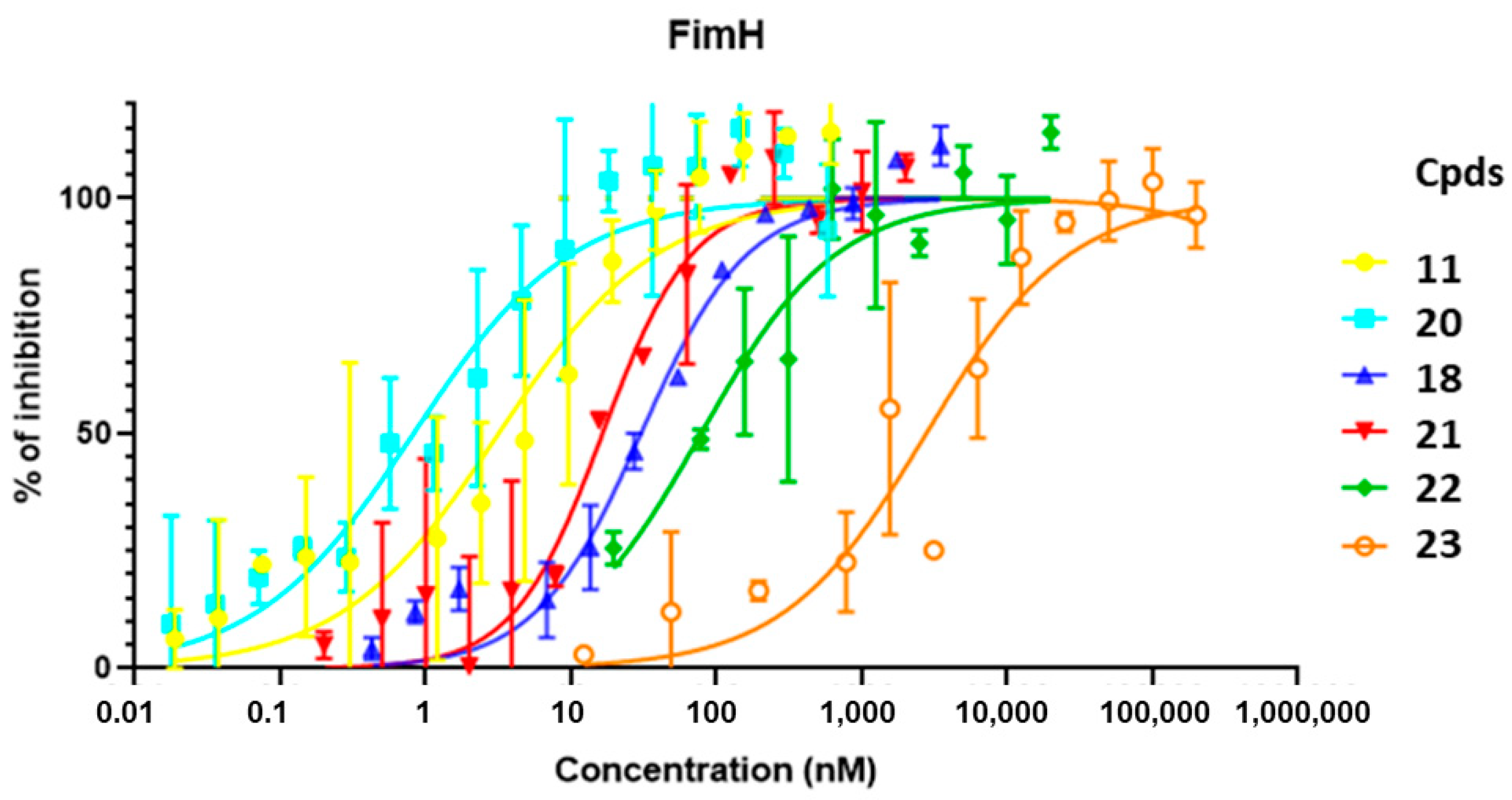
2.3. Affinity Evaluation of Mannosides through FimH LEctPROFILE Kit

To measure the relative binding affinity of the newly synthesized compounds 11, 20, and 18 against FimH, the usual referenced standards were used for comparison with previously published data: heptyl α-D-mannopyranoside (HepMan, 21), para-nitrophenyl α-D-mannopyranoside (PNPMan, 22), and methyl α-D-mannopyranoside (MeMan, 23) [4]. The relative inhibitory potential of the above compounds was determined against a biotinylated Man-BSA conjugate using a FimH lectin domain (amino acids 1-158) LEctPROFILE kit (provided by GLYcoDiag) (Figure 1, Table 1). In this assay, IC50s of 19.4 nM, 74.1 nM, and 2810.7 nM, were respectively obtained for 21, 22, and 23. As Figure 1 shows, the best inhibition was obtained from the inhibitor 20, followed by 11, and finally the C-linked mannopyranoside 18 with IC50s of 0.82 nM, 3.17 nM, and 30.28 nM, respectively. Undoubtably, the ortho-substituted pyridyl derivative 20 is the most efficient monomeric antagonist identified thus far. Interestingly, and as anticipated, compound 20 showed a calculated LogP (cLogP) improved (1.66) in comparison to its fully aromatic counterparts 18 (3.16).

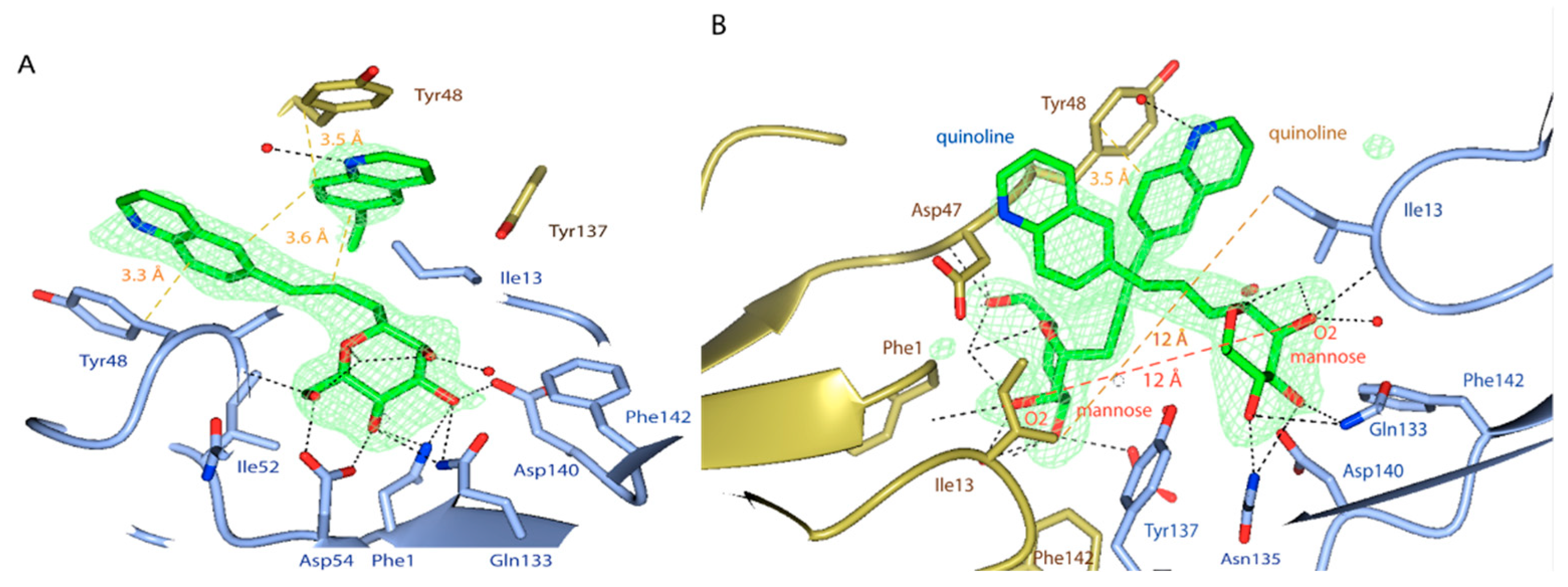
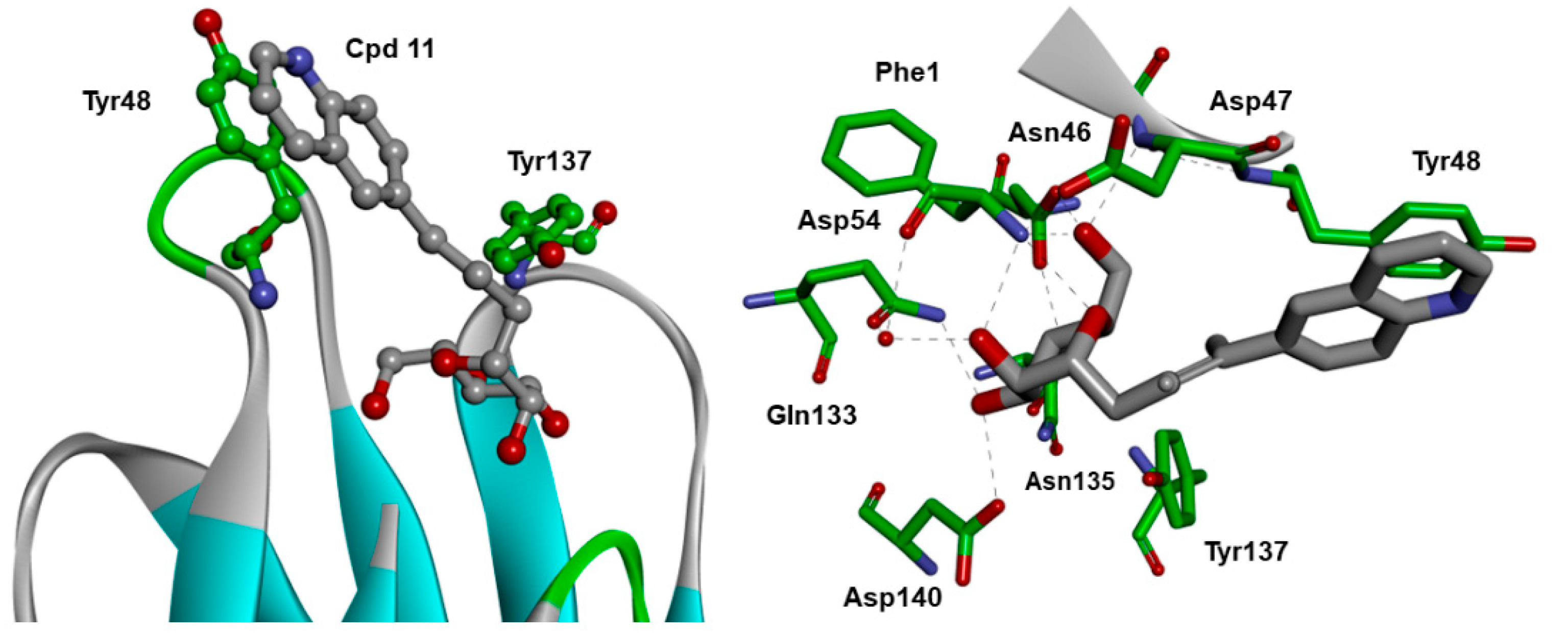
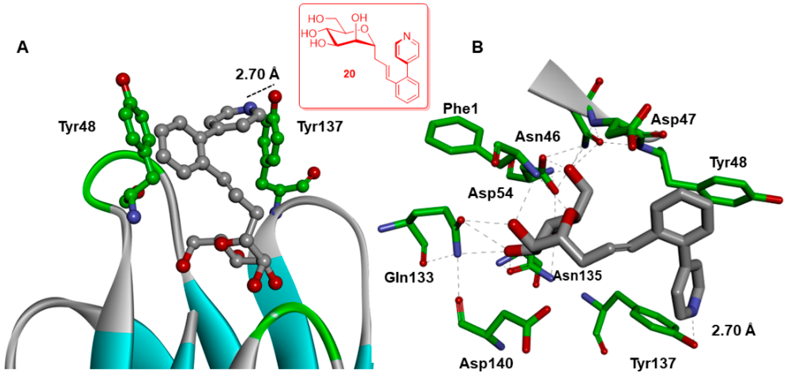
2.4. X-ray and Molecular Dynamic Simulations

Unfortunately, no crystalline structure could be obtained from our best inhibitor 20. However, co-crystallized FimH with the second-best inhibitor 11 was obtained at a resolution of 3 Å (Figure S38, Table S1, PDB entry code 8BVD). The structure was solved using the PHENIX [44] software to visualize the detailed binding interactions which took place between the FimH binding domain and the inhibitor.
Delightfully, the co-crystal structure of compound 11 and E. coli C43 (DE3) FimH [45,46] also revealed the expected 4C1 chair conformation of the ligand 11 bound into the active site of the tyrosine gate. In the mannose-binding site of FimH, we observed Tyr48 in parallel, while the Tyr137 residue was in T-aromatic stacking with the quinoline group of ligand 11. Four FimH lectin domains were observed in the asymmetric unit of the crystal (Figure S38) and the mannose-binding site of each FimH lectin domain was in proximity with a symmetry-related neighbor in the crystal packing (Figure 2) [34]. The two binding sites of FimH molecules were held together by the two quinoline substituents crossed over in a parallel stacking (Figure 2), while their mannosides projected into the binding pocket of FimH. The distance between the axial O2 hydroxyl group of the non-reducing end mannoside was 12 A° which exactly matched the distance previously observed in divalently bound trimannoside to FimH (crystal structures with PDB entry codes 6GTV and 6GTW). Therefore, the binding of FimH to compound 11 appeared to simulate the bivalent binding of the natural oligomannosidic N-glycan structures [47]. Inhibitor 11 interacted via different amino acids that included Ile 13, Phe 1, Asp 47, Gln 133, Asp 54, Asn 135, Asp 140, Phe 142 in the open conformation of FimH (Figure 3).

Further Insights into the Design and Binding of Compound 20

Given that the best compound 20 failed to crystallize with the FimH lectin domain, we tried to explain its improved potency using molecular dynamics (MD) simulations. The best low energy score was obtained from its docking within the FimH half-open tyrosine gate (−90.70 kcal/mol) (PDB 4AUY) (Figure 4). For comparison, the lowest FimH closed and open energy conformations were at −60.34 and −89.76 kcal/mol, respectively [48,49,50,51].
According to previous work [41], the ortho-biphenyl substituted C-linked mannopyranoside with KD = 6.9 ± 5.7 nM had a higher affinity over the para-substituted analog (compound 18, Table 1) with KD = 17 ± 3.5 [41]. Previously published MD simulation with an ortho-substituted biphenyl derivative could nicely explain the origin of the better affinity observed with 20. Indeed, the MD simulation allowed us to postulate that the higher affinity might originate from a π-stacking between the first phenyl ring of 20 with Tyr48, while the second ortho pyridyl moiety can interact with the hydroxyl group of Tyr137 through a hydrogen bond (2.70 A°). Thus, the MD simulation was instrumental in the replacement of the second phenyl group with a heterocyclic pyridyl moiety to improve the solubility as well as the affinity of ligand 20.

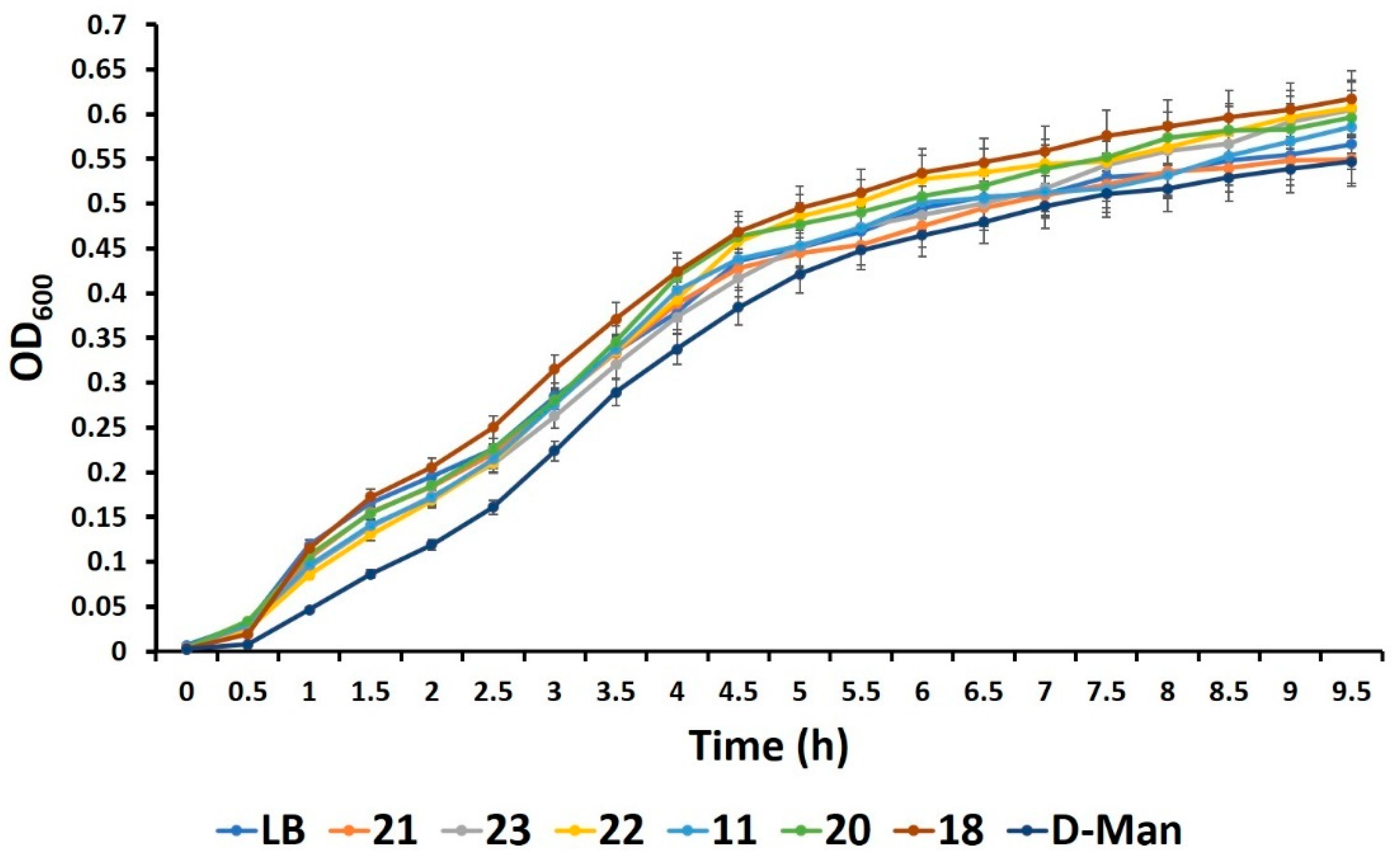
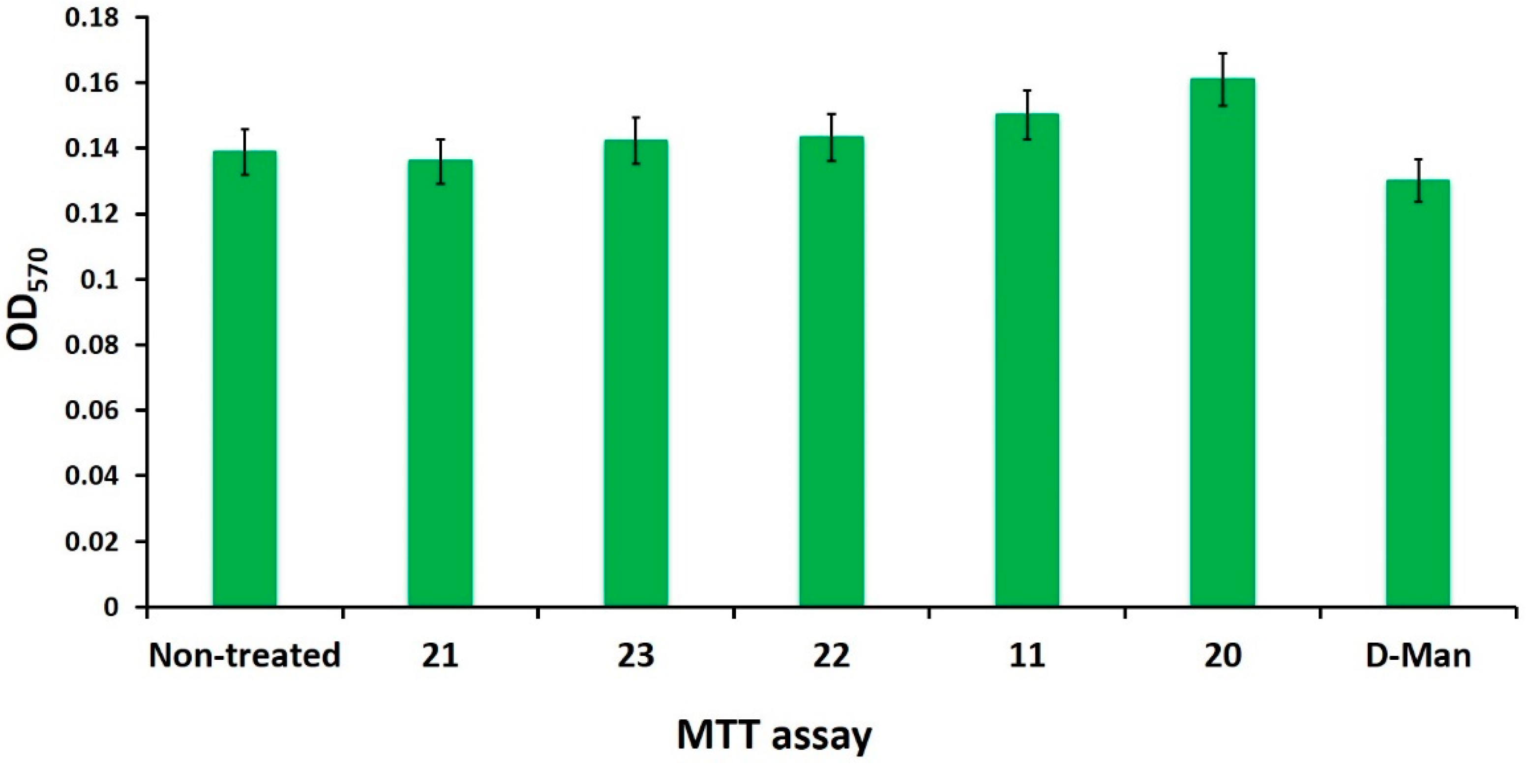
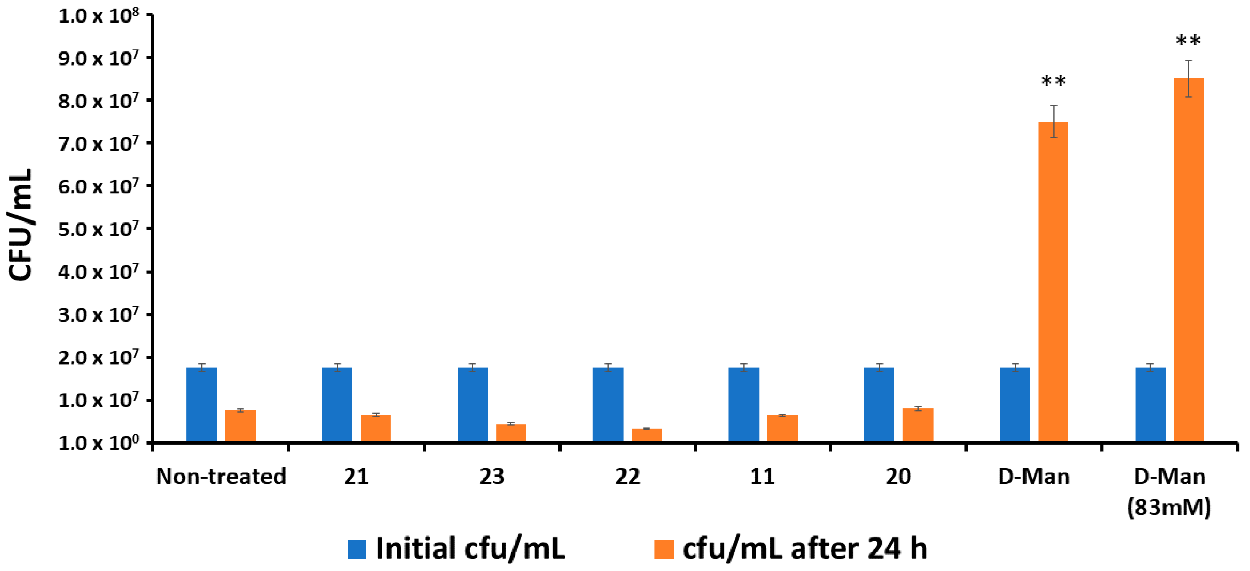
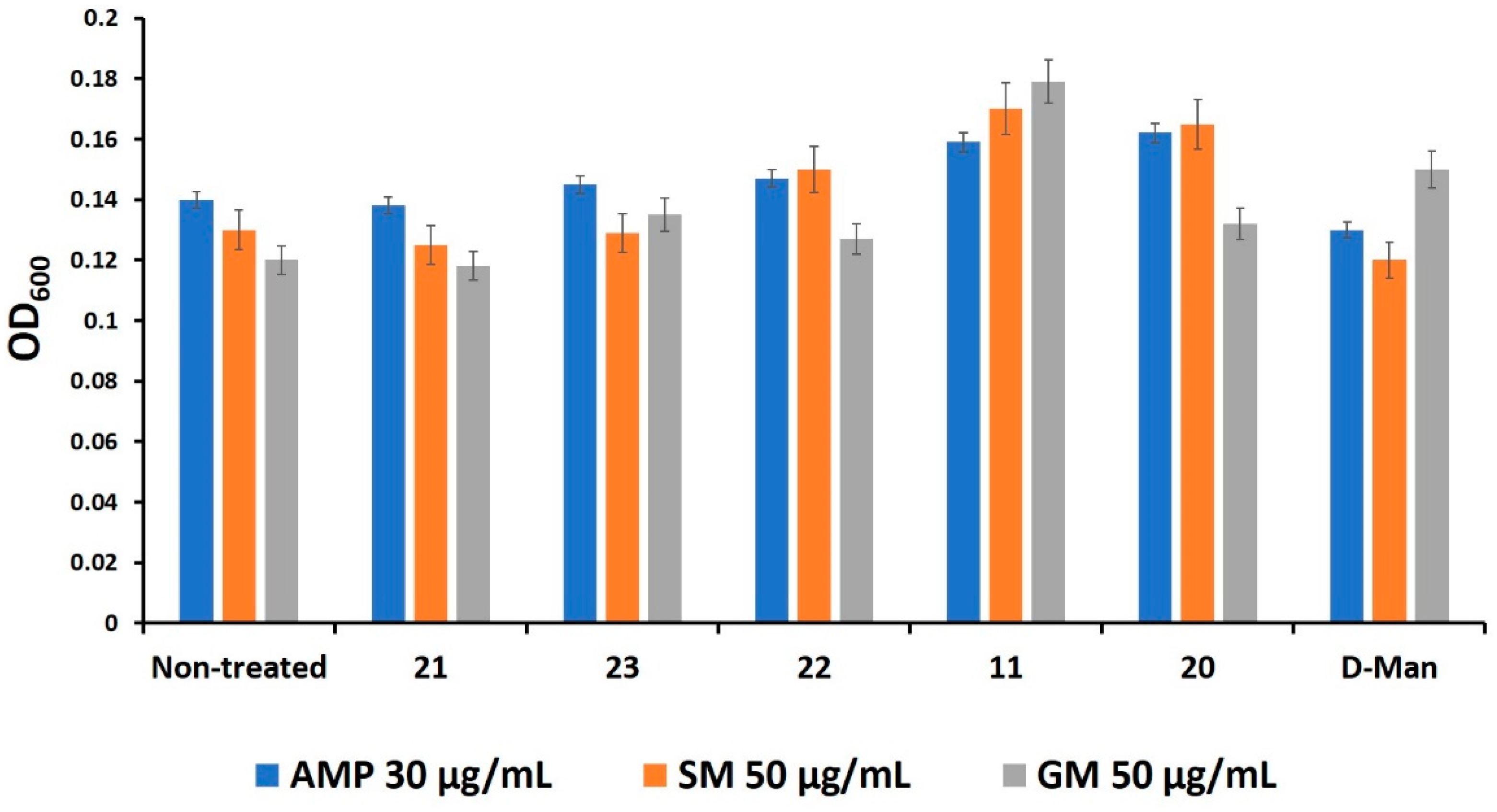
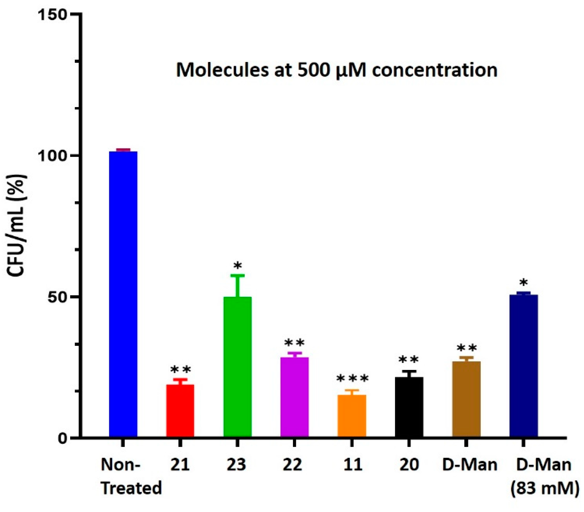
2.5. Mannosides Do Not Affect Bacterial Growth, Cell Viability, and Antibiotic Activities

To test whether the above mannoside derivatives could exert bactericidal and/or cytotoxic activities, bacterial growth and cell viability assays were undertaken in the presence of each mannoside at different concentrations. Natural D-mannose (D-Man) was included as control. As shown in Figure 5, bacteria grown in the presence of mannoside analogs showed similar growth rates compared to bacteria grown in LB (Luria–Bertani) medium that represents the positive control for bacterial replication. Moreover, no cytotoxic effect was recorded for urinary bladder cell line 5637 (HTB-9 cell) monolayers [48,52], as measured by the MTT test (Figure 6). Since D-mannose can serve as nutrient replacement for E. coli when there is a shortage of D-glucose, we tested whether these new derivatives could be used as carbon sources as well [5]. To further this aim, bacteria were cultured in LB medium for 12 h and then diluted to 1.5 × 10⁷ colony forming unit/mL (CFU/mL) in PBS buffer. Bacteria were incubated for an additional 24 h in the absence and in the presence of different mannosides and the number of viable bacteria was assessed by CFU/mL counting. Results show no statistically significant differences in the number of mannoside-treated bacteria compared to non-treated control (Figure 7). Conversely, the number of bacteria increased significantly in the presence of D-mannose, at both concentrations tested (500 μM and 83 mM) (Figure 7) [5]. The results indicated that the synthetic mannoside antagonists did not enter into the metabolic cycle of the bacterial cells to support their growth even in the absence of any other carbon sources. Differently, D-mannose as a natural sugar molecule is metabolized by bacteria, thereby maintaining bacterial replication [5]. Since carbohydrates can influence the activity of conventional antibiotics, the activity of different classes of antibiotics such as ampicillin (AMP 30 μg/mL), streptomycin (SM 50 µg/mL), and gentamycin (GM 50 µg/mL) in the presence of the synthetic mannoside analogs was evaluated. A broth-dilution test showed no differences in bacterial susceptibility, irrespectively in the presence of C-mannoside antagonists (Figure 8).
Altogether these results demonstrate the lack of toxicity of the synthetic mannoside derivatives toward both bacteria and eukaryotic cells. Moreover, these molecules are not used for bacterial metabolism and, unlike natural D-mannose, do not favor bacterial replication.

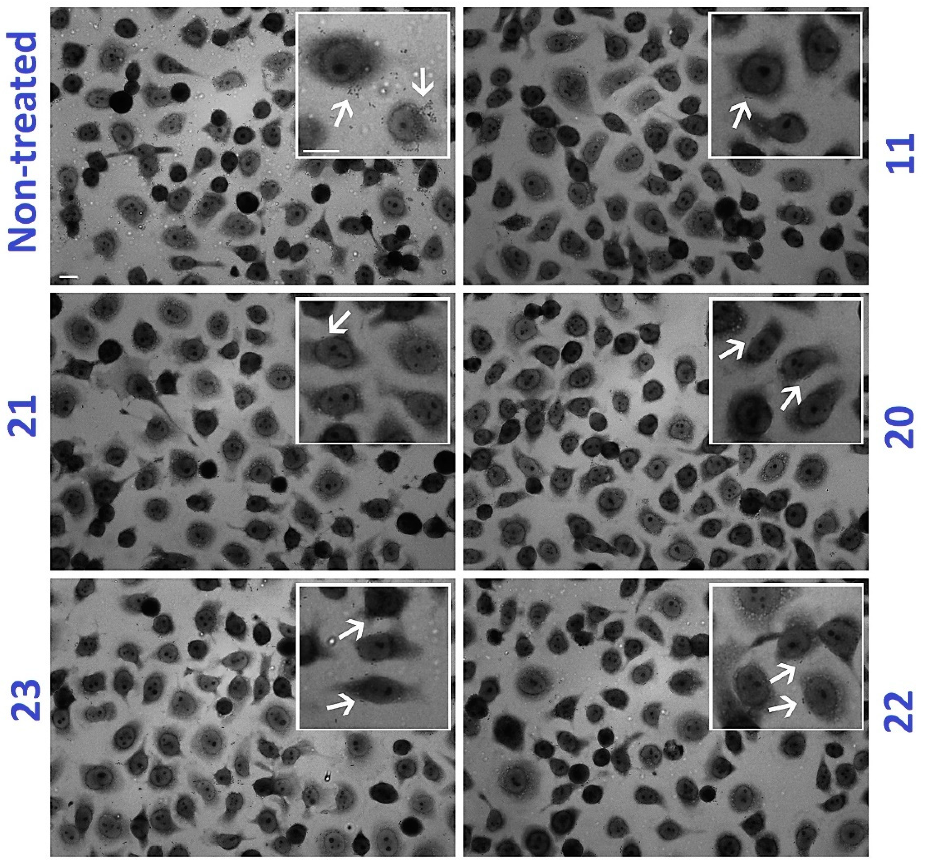
2.6. C-Mannoside Antagonists Are Effective in Decreasing Bacterial Adhesion to Human Bladder Epithelial Cells

To evaluate the efficacy of the synthetic mannoside antagonists to inhibit the ability of CFT073 strain to adhere to epithelial cells, an in vitro adhesion assay was performed. For this purpose, equal amounts of strain CFT073 strain were inoculated in PBS supplemented with different concentrations of each mannoside at final concentrations of 100, 500 μM, and 1 mM and incubated for 3h under static conditions. Bacterial inoculation was used to infect cell line 5637 (HTB-9 cell) monolayers at a multiplicity of infection (MOI) of 10. At 2.5 h post-infection, the number of cell-associated bacteria was calculated by CFU/mL counting (Figure 9). No significant inhibition of bacterial adhesion to bladder cells was obtained using mannoside inhibitors at 100 µM. Conversely, a significant reduction (more than 1 log) in the number of adherent bacteria was observed by increasing the concentration of the mannosides to 500 μM in comparison to non-treated bacteria (Figure 10). The same extent of reduction in bacterial adhesion to bladder cells was obtained by increasing mannosides concentration to 1 mM, thereby showing a dose-dependent effect. Interestingly, among the mannosides studied, the quinoline analog 11, having an IC50 of 3.17 nM, was the most efficient at reducing FimH-mediated bacterial adherence (Figure 9 and Figure 10). On the other hand, compound 23 (MeMan) was the less efficient; however, it achieved the same extent of bacterial adhesion inhibition of natural D-mannose but at a 164-fold lower concentration. These results nicely confirmed previous observations from the control reference compounds 21, 23, and D-mannose [46]. To appraise qualitatively the reduction of bacterial adhesion, parallel infected cells were fixed, and Giemsa stained. As shown in Figure 10, no macroscopic differences in the shape, integrity, adhesiveness, cytoplasmic vacuolization, proliferation, or cytotoxic effects were observed in HTB-9 cell monolayers incubated with the synthesized mannosides, in line with the results of the MTT assay [5]. Overall, these results revealed that these synthetic inhibitors finely mimic the interaction between FimH and its natural receptor, thereby significantly decreasing the adhesion of strain CFT073 to bladder cells.

3. Materials and Methods

3.1. General Information

Nuclear magnetic resonance (NMR) spectra were recorded on Varian Geminin 300 MHz or Innova 600 MHz spectrometer. Chemical shifts are reported in parts per million (ppm) (δ) relative to CDCl3 (δ 7.27 and 77.23 ppm for 1H- and 13C-NMR, respectively) or relative to the signal of CD3OD (δ. 3.31 and 4.8 ppm for 1H- and 49.7 ppm for 13C-NMR, respectively). Where necessary, DEPT, APT, and two-dimensional 1H-1H COSY and HSQC experiments were performed for complete signal assignments. Coupling constants (J) are reported in Hertz (Hz). Signal multiplicities are used as singlet (s), doublet (d), doublet of doublet (dd), triplet (t), multiplet (m). Accurate mass measurements were performed on a LC-MSD-Tof instrument from Agilent technologies in positive electrospray with protonated ions [M + H]+; sodium adducts [M + Na]+ and [M + NH4]+ were used for empirical formula confirmation. Flash chromatography was performed using Merck silica gel 60 (40–63 µm). TLC was performed on Kiesel gel 60 F254 plates from Merck. Detection was carried out under UV light or by spraying with 20% ethanolic sulfuric acid or molybdate solution followed by heating. Optical rotations were measured with a JASCO P-1010 polarimeter. Melting points were measured on a Fisher Jones apparatus and are uncorrected. Purification of some compounds were done by semi-preparative HPLC (Agilent, Santa Clara, CA, USA). All eluents contained 0.1% formic acid and flow rate was set to 5 mL/min. Solvents were dried by distillation from drying agents as follows: DMF (Barium oxide), CH2Cl2 (P2O5), Et3N and pyridine (CaH2), MeOH was stored over 4A molecular sieves. Some aryl iodides were synthesized according to literature procedures [53,54,55]: 2-iodophenyl acetate, 4-iodophenyl acetate, 4-iodobenzyl acetate and methyl 4-iodobenzoate. Compound 9 and 18 were prepared as previously published [30].

3.2. Synthetic Methods and Analytic Data of Compounds

3.2.1. General procedure for Heck Coupling of Protected C-Mannopyranosides 2–10

To a solution of mannopyranosides 1 [40] in degassed anhydrous DMF, were added the various iodoaryl derivatives (2 equiv.), 10% palladium(II) acetate, tetrabutylammonium bromide (1 equiv.), and sodium bicarbonate (3 equiv.). The reaction mixture was heated at 85 °C under N2. The course of the reactions was followed by TLC. The solution was evaporated under reduced pressure and the residue was purified by flash column chromatography on silica gel (from 0 to 425% AcOEt-Hexanes).
Compound 2: 40 mg, 0.053 mmol, 66%, [α]20D = −9.13 (c = 0.5, CHCl3). Rf = 0.29 (Hexane/EtOAc, 5.5: 4.5), m.p: 149–150 °C. 1H-NMR (600 MHz, CDCl3): δ ppm 8.8–7.19 (m, 26H, H-arom ), 6.75 (d, 1H, J2’,3’ = 15.8 Hz, CH=CH-), 6.46–6.36 (m, 1H, CH=CH-), 5.98–5.88 (m, 2H, H-4, H-3), 5.27 (dd, 1H, J1,2 = J2, 3 = 2.7 Hz, H-2), 4.75 (dd, 1H, J6a,6b = 12.0 Hz, J5, 6a = 6.9 Hz, H-6a), 4.50 (dd, 1H, J6a,6b = 12.0 Hz, J5,6b = 2.6 Hz, H-6b), 4.56–4.52 (m, 1H, H-1), 4.50–4.47 (m, 1H, H-5), 3.05–2.96 (m, 1H, H-1’a), 2.85–2.75 (m, 1H, H-1’b). 13CNMR (151 MHz, CDCl3): δ ppm, 166.2, 165.9, 165.6, 165.4 (4×CO), 150.1, 147.9, 135.89, 135.1, 133.5, 133.4, 132.9, 129.8, 129.6, 129.5, 128.59, 128.5, 128.2, 127.0, 121.3 (Carom, C-3’, C-2’), 74.2 (C-1), 71.2 (C-5), 71.1 (C-4), 69.6 (C-3), 68.0 (C-2), 62.9 (C-6), 33.1 (C-1’). ESI+-HRMS: [M + H]+ calcd for C46H37NO9 + H+: 748.2541; found, 748.2479.
Compound 3: 44.6 mg, 0.064 mmol, 80 %. [α]20D = −28.86 (c = 0.2, CHCl3). Rf = 0.23 (Hexane/EtOAc, 1.8:1.2). 1H-NMR (600 MHz, CDCl3): δ ppm 8.44 (d, 1H, 4JH-H = 2.2 Hz, H-arom), 8.36 (dd, 1H, 3JH-H = 4.7 Hz, 4JH-H = 1.5 Hz, H-arom), 7.91–7.21 (m, 21H, H-arom), 7.11–6.93 (m, 1H, H-arom), 6.51 (d, 1H, J2’,3’ = 16 Hz, CH=CH-Py), 6.24 (m, 1H, J2’,3’ = 16.3 Hz, J2,’H1’a = 13.4 Hz, J2’,H1’b = 5.8 Hz CH=CH-Py), 6.02 (dd, 1H, J3,4 = J4,5 = 8.3 Hz, H-4), 5.81 (dd, 1H, J2,3 = 3.1 Hz, J3,4 = 8.6 Hz, H-3), 5.63 (dd, 1H, J1,2 = J2, 3 = 3.4 Hz, H-2), 4.68 (dd, 1H, J6a,6b = 12.0 Hz, J5,6a = 6.7 Hz, H-6a), 4.52 (dd, 1H, J6a,6b = 12. Hz, J5,6b = 3.2 Hz, H-6b), 4.46–4.41 (m, 1H, H-1), 4.38–4.35 (m, 1H, H-5), 2.94–2.82 (m, 1H, H-1’a), 2.74–2.68 (m, 1H, H-1’b). 13CNMR (151 MHz, CDCl3): δ ppm, 166.2, 165.6, 165.6, 165.4 (4 × CO), 148.4, 148.2, 133.5, 133.4, 133.0, 132.4, 129.6, 128.9, 128.5, 126.7, 123.5 (Carom, C-3’, C-2’), 74.1 (C-1), 71.2 (C-5), 71.1 (C-2), 69.4 (C-4), 67.80 (C-3), 62.7 (C-6), 33.2 (C-1’). ESI+-HRMS: [M + H]+ calcd for C42H35NO9 + H+: 698.2385; found, 698.4013.
Compound 4:115 mg, 0.152 mmol, 80% yield. [α]20D = −9.92 (c = 0.5, CHCl3). Rf = 0.17 (Hexane/EtOAc, 2:1). 1H-NMR (300 MHz, CDCl3): δ, 8.21–7.00 (m, 24H, H-arom), 6.67 (d, 1H, J2’,3’ = 15.9 Hz, CH=CHPh), 6.39–6.2 (m, 1H, CH=CHPh), 6.00 (dd, 1H, J3,4 =J4,5 = 8.5 Hz, H-4), 5.91 (dd, 1H, J2,3 = 3.1 Hz, J3,4 = 8.9 Hz, H-3), 5.74 (dd, 1H J1,2 = J2,3 = 3.1 Hz, H-2), 4.66 (m, 2H, H-6a, H-6b), 4.74 (dd, 1H, J6a, 6b = 12 Hz, J5, 6a = 6.5 Hz, H-6a), 4.62 (dd, 1H, J5, 6b = 12 Hz, J6a, 6b = 2.8 Hz, H-6b), 4.59–4.36 (m, 2H, H-1, H-5), 3.09–2.92 (m, 1H, H-1’a), 2.85–2.78 (m, 1H, H-1’b), 2.37 (s,3H, Ac). 13C-NMR (75 MHz, CDCl3): δ 169.3, 166.2, 165.6, 165.4, 147.5 (5 × CO), 135.5, 133.4, 133.0, 129.8, 129.6, 129.5, 129.0, 128.4, 128.2, 126.7, 126.1, 122.5 (Carom, C-3’, C-2’), 74.4 (C-1), 71,4 (C-5), 71.0 (C-4), 69.7 (C-3), 67.8 (C-2), 62.8 (C-6), 33.23 (C-1’) and 21.1 (Ac). ESI+-HRMS: [M + H]+ calcd for C45 H39 O11, 755.2487; found, 755.2469.
Compound 5: 49 mg, 0.064 mmol 80% yield. [α]20D = −16.63 (c = 0.5, CHCl3)). Rf = 0.22 (Hexane/Toluene/EtOAc, 71: 24: 5). 1H-NMR (300 MHz, CDCl3): δ, 8.14–7.29 (m, 22H, H-arom), 6.98 (dd, 1H, 3JH-H = 9.0 Hz, 4JH-H = 2.4 Hz, H-arom), 6.61 (d, 1H, J2’,3’ = 15.9 Hz, CH=CHPh), 6.32–6.17 (m, 1H, CH=CHPh), 5.95 (dd, 1H, J3,4 =J4,5 = 8.8 Hz, H-4), 5.90 (dd, 1H, J2,3 = 3.0 Hz, J3,4 = 8.8 Hz, H-3), 5.74 (dd, 1H J1,2 = J2,3 = 3.1 Hz, H-2), 4.72 (dd, 1H, J6a,6b = 12 Hz, J5,6a = 6.5 Hz, H-6a), 4.62 (dd, 1H, J6a,6b = 12.1 Hz, J5,6b = 2.8 Hz, H-6b), 4.56–3.36 (m, 2H, H-1, H-5), 3.05–2.87 (m, 1H, H-1’a), 2.83–2.79 (m, 1H, H-1’b), 2.32 (s, 3H, Ac). 13C-NMR (75 MHz, CDCl3): δ 169.3, 166.3, 165.6, 165.4, 147.5 (5 × CO), 135.4, 133.3, 133.0, 129.8, 129.5, 129.0, 128.4, 126.7, 126.1, 122.5 (Carom, C-3’, C-2’), 74.4 (C-1), 71,4 (C-5), 71.0 (C-4), 69.7 (C-3), 67.8 (C-2), 62.8 (C-6), 33.23 (C-1’) and 21.0 (Ac). ESI+-HRMS: [M + H]+ calcd for C45H39O11, 755.2487; found, 755.2453.
Compound 6: 52 mg, 0.067 mmol, 90% yield. [α]20D = −8.3 (c = 0.5, CHCl3). Rf = 0.25 (Hexane/ EtOAc, 3:1). 1H-NMR (300 MHz, CDCl3): δ, 8.16.7.12 (m, 24H, H-arom), 6.63 (d, 1H, J2’,3’ = 15.8 Hz, CH=CHPh), 6.49–6.21 (m, 1H, CH=CHPh), 6.08–5.84 (m, 2H, H-4, H-3), 5.75 (dd, 1H J1,2 = J2,3 = 2.9 Hz, H-2), 5.11 (s, 2H, CH2Ph), 4.72 (dd,1H, J6a,6b = 11.9 Hz, J5,6a = 6.3 Hz, H-6a), 4.63 (dd, 1H, J6a,6b = 11.9 Hz, J5,6b = 2.6 Hz, H-6b), 4.57–4.39 (m, 2H, H-1, H-5), 3.01–2.93 (m, 1H, H-1’a), 2.84–2.77 (m, 1H, H-1’b), 2.84–2.77 (s, 3H, Ac). 13C-NMR (75 MHz, CDCl3): δ 166.2, 165.6, 165.6, 165.4, 137.6 (5 × CO), 134.9, 133.4, 133.0, 132.8, 129.8, 129.5, 128.9, 128.5, 126.3, 124.7 (Carom, C-3’, C-2’), 74.5 (C-1), 71,2 (C-5), 70.9 (C-4), 69.7 (C-3), 67.8 (C-2), 66.1 (CH2Ph), 63.0 (C-6), 29.6 (C-1’) and 21.0 (Ac). ESI+-HRMS: [M+NH4]+ calcd for C46H44NO11, 786.2909; found, 786.2919.
Compound 7: 194 mg, 0.237, 73% yield. [α]20D = −16.6 (c = 0.3, CHCl3). Rf = 0.18 (Hexane/EtOAc, 3:1). 1H-NMR (300 MHz, CDCl3): δ, 8.11–7.08 (m, 24H, H-arom), 6.65 (d, 1H, J2’,3’ = 15.9 Hz, CH=CHPh), 6.45–6.35 (m, 1H, CH=CHPh), 6.01–5.88 (m, 2H, H-4, H-3), 5.75 (dd, 1H, J1,2 = 3.9 Hz, J2,3 = 7.3 Hz, H-2), 4.78 (dd, 1H, J6a,6b = 12.0 Hz, J5,6a = 6.7 Hz, H-6a), 4.63 (dd, 1H, J6a,6b = 12 Hz, J5,6b = 2.8 Hz, H-6b), 4.58–4.42 (m, 2H, H-1, H-5), 3.93 (s, 3H, COOMe), 3.03–2.93 (m, 1H, H-1’a), 2.92.-2.71 (m, 1H, H-1’b). 13C-NMR (75 MHz, CDCl3): δ 166.8, 166.2, 165.7, 165.4, 141.4 (5 × CO), 133.4, 133.1, 132.4, 129.7, 129.6, 128.9, 128.5, 128.3, 127.1, 126.0 (Carom, C-3’, C-2’), 74.1 (C-1), 71.3 (C-5), 71.2 (C-4), 69.6 (C-3), 67.9 (C-2), 62.8 (C-6), 51.9 (COOMe), 33.1 (C-1’). ESI+-HRMS: [M + H]+ calcd for C45H39O11, 755.2487; found, 755.2493.
Compound 8: 158 mg, 88% yield. [α]20D = −6.12 (c = 0.5, CHCl3). Rf = 0.27 (Hexane/EtOAc, 3:1)1H-NMR (300 MHz, CDCl3): δ, 8.09–7.31 (m, 24H, H-arom), 6.65 (d, 1H, J2’,3’ = 15.9 Hz, CH=CHPh), 6.44 (m, 1H, CH=CHPh), 5.90 (m, 2H, H-4, H-3), 5.74 (dd, 1H J1,2 =3.7, Hz, J2,3 = 2.4 Hz, H-2), 4.86 (dd, 1H, J6a,6b = 12 Hz, J5,6a = 7.2 Hz, H-6a), 4.63–4.44 (m, 3H, H-6b, H-1, H-5), 3.12–2.98 (m, 1H, H-1’a), 2.76–2.85 (m, 1H, H-1’b). 13C-NMR (75 MHz, CDCl3): δ 166.2, 165.6, 165.4, 164.7 (4 × CO), 146.7, 143.3, (Carom-q),133.5, 133.1, 131.3, 129.8, 128.9, 128.3, 126.5, 123.6, (Carom, C-3’, C-2’), 73.4 (C-1), 71.4 (C-5), 71.0 (C-4), 69.4 (C-3), 68.2 (C-2), 62.6 (C-6), 33. 3 (C-1’). ESI+-HRMS: [M + Na]+ calcd for C45H35NO11, 764.2102; found, 764.2157.
Compound 10: This compound was synthesized according to general procedure A. 56 mg, 0.072 mmol, 90% yield, [α]20D = −14.16 (c = 0. 39, CHCl3). Rf = 0.27. (Hexane/EtOAc, 9:3). 1H-NMR (300 MHz, CDCl3): δ, 8.69 (d, 1H, 3JH-H = 5.6 Hz), 8.10–7.23 (m, 23H, H-arom), 6.56 (d, 1H, J2’,3’ = 15.7 Hz, CH=CHPh), 6.33–6.12 (m, 1H, CH=CHPh), 6.01 (dd, 1H, J3,4 = 12 Hz, J4,5 = 9.0 Hz, H-4), 5.92 (dd, 1H, J2,3 = 3.1 Hz, J3,4 = 9.0 Hz, H-3), 5.15 (dd, 1H J1,2 = J2,3 = 3.1 Hz, H-2), 3.93–3.90 (m, 1H, H-1), 4.72 (dd, 1H, J6a,6b = 12.1, Hz, J5,6a = 6 Hz, H-6a), 4.58–4.41 (m, 2H, H-5, H-1), 3.09–2.98 (m, 1H, H-1’a), 2.93–2.76 (m, 1H, H-1’b). 13C-NMR (75 MHz, CDCl3): δ 166.2, 166.6, 165.6, 165.4, (4 × CO), 136.8, 133.3, 133.0, 132.8, 132.2, 129.7, 128.5, 128.9, 128.4, 127.5, 127.3, 127.1, 123.3 (Carom- C-3’,C-2’), (Carom), 127.5 (Carom, C-2’), 127.3, 127.1 (Carom), 74.5 (C-1), 71,3 (C-5), 70.9 (C-4), 69.7 (C-3), 67.8 (C-2), 62.9 (C-6), 32.9 (C-1’). ESI+-HRMS: [M + NH4]+: calcd for C43H39BrNO9, 794.1792; found, 794.1770

3.2.2. General Procedure for De-O-Benzoylation

Compounds 11–18 and 20 were deprotected by treatment with 1M ammonia in methanol at room temperature for 36 h. Removal of solvent under vacuum afforded the crude residues which were purified by column chromatography (MeCN/MeOH, 9.5/0.5) and the compounds (11, 13, 15) were purified by semi-preparative HPLC (A: H2O + 0.1% trifluoroacetic acid, B: ACN + 0.1% trifluoroacetic acid, 5 mL/min).
Compound 11: 15 mg, 0.045 mmol, 85% yield. [α]20D = +41.3 (c = 0.1, CH3OH). 1H-NMR (600 MHz, CD3OD): δ ppm 8.87 (dd, 1H, 3JH-H = 4.4 Hz, 4JH-H = 1.6 Hz, H-arom), 8.33 (dd, 1H, 3JH-H = 8.4 Hz, 4JH-H = 1.6 Hz, H-arom), 7.97 (d, 2H, 3JH-H = 1.1 Hz, H-arom), 7.84 (s, 1H, H-arom), 7.52 (dd, 1H, 3JH-H = 8.3 Hz, 4JH-H = 4.4 Hz, H-arom), 6.75 (d, 1H, J2’,3’ = 16.0 Hz, CH=CH-), 6.65–6.48 (m, 1H, CH=CH-), 4.07 (ddd, 1H, J1,2 = 2.7 Hz, J1,H1’a = 8.7 Hz, J1, H1’b = 6.0 Hz, H-1), 3.84–3.65 (m, 5H, H-4, H-3, H-2, H-6a H-6b), 3.64–3.55 (m, 1H, H-5), 2.84–2.69 (m, 1H, H-1’a), 2.68–2.51 (m, 1H, H-1’b). 13C-NMR (151 MHz, CD3OD): δ ppm: 149.2, 146.8, 136.9, (Carom-q), 136.4, 131.1, 128.4, 128.2, 127.6, 127.4, 125.2, 121.4, (Carom, C-3’, C-2’), 76.6 (C-1), 75,0 (C-5), 71.3 (C-4), 70.7 (C-3), 68.1 (C-2), 61.6 (C-6), 32.6 (C-1’). ESI+-HRMS: [M + H]+ calcd for C18H21NO5, 332.1492; found, 332.1515.
Compound 12: 15 mg, 0.053 mmol, 84% yield. [α]20D = +27.8 (c = 0.1, CH3OH). 1H-NMR (300 MHz, CD3OD): δ ppm 8.53 (s, 1H, H-arom), 8.38 (d, 1H, 3JH-H = 3.8 Hz, H-arom), 7.94 (dd, 1H, 3JH-H = 3.5 Hz, 4JH-H = 1.8 Hz, H-arom), 7.42- (dd, 1H, 3JH-H = 8.0 Hz, 4JH-H = 4.9 Hz, H-arom), 6.60 (d, 1H, J2’,3’ = 16.0 Hz, CH=CH-Py), 6.50–6.41 (m, 1H, CH=CH-Py), 4.08 (ddd, 1H, J1,2 = 2.7 Hz, J1,H1’a = 8.7 Hz, J1,H1’b’ = 6 Hz, H-1) 3.91–3.65 (m, 5H, H-4, H-3, H-2, H-6a, H-6b), 3.62–3.57 (m, 1H, H-5), 2.81–2.69 (m, 1H, H-1’a), 2.62–2.51 (m, 1H, H-1’b). 13C-NMR (75 MHz, CD3OD): δ ppm: 146.9, 146.6, 133.6, 129.3, 128.2, 124.2, 121.1 (Carom, C-3’, C-2’), 76.8 (C-1), 74.1 (C-5), 70.7 (C-4), 70.7 (C-3), 67.5 (C-2), 61.4 (C-6), 32.5 (C-1’). ESI+-HRMS: [M + H]+: calcd for C14H19NO5, 282.1336; found, 282.1331.
Compound 13: 38 mg, 0.128 mmol, 85% yield. [α]20D = +8.54 (c = 0.2, CH3OH). 1H-NMR (300 MHz, CD3OD): δ, 7.38 (dd, 1H, 3JH-H = 8.3 Hz, 4JH-H = 1.3 Hz, H-arom), 7.03 (td, 1H, 3JH-H = 7.7 Hz, 4JH-H = 1.6 Hz, H-arom), 6.76 (m, 3H, H-arom, CH=CHPh), 6.31–6.21 (m, 1H, CH=CHPh), 4.06–3.96 (m, 1H, H-1), 3.89–3.61 (m, 5H, H-4, H-3, H-6a, H-6b, H-2), 3.56 (ddd, 1H, J4,5 = 8.5 Hz, J5,6a = 5.4 Hz, J5,6b = 2.9 Hz, H-5), 3.01 (s, 1H, PhOH), 2.66 (ddd, 1H, 1H, JH1’a, H1’b = 14.7 Hz, J1,H1’a = 1.4 Hz, J1’,2’ = 8.1 Hz, H-1’a), 2.54 (ddd, 1H, 1H, JH1’a,H1’b = 14.4 Hz, J1,H1’b = 3.9 Hz, JH1’b,2’ = 10.6 Hz, H-1’b). 13C-NMR (75 MHz, CD3OD): δ 154.1 (C-PhOH), 127.5 (C-3’), 127.3, 126.2 (Carom-q), 122.0 (C-2’), 124.4, 118.9, 115.0 (Carom), 77.5 (C-1), 74,8 (C-5), 71.4 (C-4), 70.6 (C-3), 67.9 (C-2), 61.6 (C-6), 33.09 (C-1’). ESI+-HRMS: [M+Na]+: calcd for C15H20NaO6, 319.1158; found, 319.1175.
Compound 14: 16 mg, 0.054 mmol, 85% yield. [α]20D = +31 (c = 0.1, CH3OH). 1H-NMR (300 MHz, CD3OD): δ, 7.23 (d, 2H, 3JH-H = 8.6 Hz, H-arom), 6.72 (d, 2H, 3JH-H = 8.6 Hz, H-arom), 6.42 (d, 1H, J2’,3’ = 15.7 Hz, CH=CHPh), 6.14–6.04 (m, 1H, CH=CHPh),4.10–3.91 (m, 1H, H-1), 3.87–3.61 (m, 5H, H-4, H-3, H-6a, H-6b, H-2), 3.62–3.44 (m, 1H, H-5), 2.66–2.57 (m, 1H, H-1’a), 2.53–2.43 (m, 1H, H-1’b). 13C-NMR (75 MHz, CD3OD): δ 153.6 (C-PhOH), 131.7 (C-3’), 129.1, 126.9 (Carom-q), 122.6 (C-2’), 115.1 (Carom), 77.5 (C-1), 74,8 (C-5), 71.4 (C-4), 70.6 (C-3), 67.9 (C-2), 61.6 (C-6), 33.09 (C-1’). ESI+-HRMS: [M+Na]+: calcd for C15H20NaO6, 319.1152; found, 319.1165.
Compound 15: 16.2 mg, 0.052 mmol, 80% yield. [α]20D = +20 (c = 0.1, CH3OH). 1H-NMR (300 MHz, CD3OD): δ, 7.38 (d, 2H, 3JH-H = 8.2 Hz, H-arom), 7.29 (d, 2H, 3JH-H = 8.2 Hz, H-arom), 6.52 (d, 1H, J2’,3’ = 15.9 Hz, CH=CHPh), 6.39–6.24 (m, 1H, CH=CHPh),4.58 (s, 2H, CH2Ph), 4.07–3.94 (m, 1H, H-1), 3.86–3.61 (m, 5H, H-4, H-3, H-6a, H-6b, H-2), 3.56 (ddd, 1H, J4,5 = 8.5 Hz, J5,6a = 5.5 Hz, J5,6b = 3.0 Hz, H-5), 2.72–2.61 (m, 1H, H-1’a), 2.57–2.61 (m, 1H, H-1’b). 13C-NMR (75 MHz, CD3OD): δ 140.2,136.6 (Carom-q), 131.8, (C-3’), 126.8, 125.7 (Carom), 125.6, (C-2’), 77.2 (C-1), 74,9 (C-5), 71.2 (C-4), 70.7 (C-3), 68.0 (C-2), 63.5 (CH2Ph), 61.6 (C-6), 32.4 (C-1’). ESI+-HRMS: [M+NH4]+: calcd for C16H26NO6, 328.1416; found, 328.1771.
Compound 16: 16 mg, 0.047 mmol, 85% yield. [α]20D = +42.62 (c = 0.1, CH3OH). 1H-NMR (300 MHz, CD3OD): δ, 7.95 (d, 2H, 3JH-H = 8.4 Hz, H-arom), 7.51 (d, 2H, 3JH-H = 8.4 Hz, H-arom), 6.64–6.44 (m, 2H, CH=CHPh, CH=CHPh), 4.04 (ddd, 1H, J1,2 = 6.0 Hz, J1,1’a = 8.7 Hz, J1,1’b = 2.7 Hz, H-1), 3.83 (dd, 1H, J3,4 =6.1 Hz, J4,5= 3.1 Hz, H-4,), 3.81–3.64 (m, 4H, H-3, H-6a, H-6b, H-2), 3.55 (ddd, 1H, J4,5 = 9.9 Hz, J5,6a = 6.5 Hz, J5,6b = 3.8 Hz, H-5), 2.76–2.66 (m, 1H, H-1’a), 2.64–2.47 (m, 1H, Hb). 13C-NMR (75 MHz, CD3OD): δ 167.0 (COOMe), 142.3 (Carom-q), 131.0 (C-3’), 129.4 (Carom-q), 128.2, 125.9 (C-2’), 77.1 (C-1), 75,2 (C-5), 70.9 (C-4), 70.7 (C-3), 67.8 (C-2), 61.5 (C-6), 51.4 (COOMe), 32.6 (C-1’). ESI+-HRMS: [M+Na]+: calcd for C17H22NaO7, 361.1258; found, 361.1253.
Compound 17: 16 mg, 0.049 mmol, 85% yield. [α]20D = +40.6 (c = 0.1, CH3OH). 1H-NMR (300 MHz, CD3OD): δ, 8.19 (d, 2H, 3JH-H = 5 Hz, H-arom), 7.64 (d, 2H, 3JH-H = 4.9 Hz, H-arom), 6.95–6.45 (m, 2H, CH=CHPh, CH=CHPh), 4.26–3.89 (m, 1H, H-1), 3.84–3.54 (m, 6H, H-4, H-3, H-6a, H-6b, H-2, H-5), 3.10–2.64 (m, 1H, H-1’a), 2.71–2.37 (m, 1H, H-1’b). 13C-NMR (75 MHz, CD3OD): δ 146.4, 144.3 (Carom-q), 132.1, 129.9, 126.2, 123.7 (Carom, C-3’, C-2’), 76.5 (C-1), 74.9 (C-5), 71.1 (C-4), 76.5 (C-3), 67.9 (C-2), 61.2 (C-6), 32.4 (C-1’). ESI+-HRMS: [M+NH4]+: calcd for C15H23N2O7, 343.15; found, 343.1512.

3.2.3. Synthesis of Compounds 19 and 20 by Suzuki Reaction

To a solution of compound 10 (40 mg, 0.051 mmol) in degassed (N2) dioxane-water (5:1) were added 4-piridinylboronicacid (10 mg, 2 equiv.), 15% Pd2Cl2 (PPh3)2 ferrocene, and cesium carbonate (48 mg, 3 equiv.). The reaction mixture was heated at 80 0C. The reaction was followed by TLC. The solution was evaporated under reduced pressure and the residue was purified by flash column chromatography on silica gel (6:4 Hexane/EtOAc) to afford compound 19 as colorless oil. 22 mg, 0.028 mmol, 55%, [α]20D = −10.7 (c = 0. 4, CHCl3). Rf = 0.17 (Hexane/EtOAc, 6:4). 1H-NMR (300 MHz, CDCl3): δ, 8.69 (d, 1H, H-arom), 8.06.7.23 (m, 27H, H-arom), 6.56 (d, 1H, J2’,3’ = 15.6 Hz, CH=CHPh), 6.28 (ddd, 1H, J1’a,2’ = J1’b, 2’=6.9 Hz, J2’,3’ = 15.6 Hz, CH=CHPh), 6.01 (dd, 1H, J3,4 = J4,5 = 8.6 Hz, H-4), 5.92 (dd, 1H, J2,3 = 3.3 Hz, J3,4 = 9.0 Hz, H-3), 5.72 (dd, 1H J1,2 = J2,3 = 3.2 Hz, H-2), 4.71 (dd, 1H, J6a,6b = 12.1 Hz, J5,6a = 5.9 Hz, H-6a), 4.62 (dd, 1H, J6a,6b = 12.2 Hz, J5,6b = 2.9 Hz, H-6a), 4.52–4.44 (m, 1H, H-1), 4.41–4.34 (m, 1H, H-5), 2.94–2.68 (m, 2H, H-1’a, H-1’b). 13C-NMR (75 MHz, CDCl3): δ 166.2, 165.6, 165.5, 165.4, (4 × CO), 148.9 (Carom-N), 137.3, 135.0, 133.5, 133.4, 133.3, 133.0, 131.3, 129.8, 129.7, 128.9, 128.5, 127.7, 126.5, 125.2 (Carom, C-3’, C-2’), 74.5 (C-1), 70,4 (C-5), 69.1 (C-4), 67.8 (C-3), 66.0 (C-2), 62.8 (C-6), 33.3 (C-1’). ESI+-HRMS: [M + H]+: calcd for C48H40NO9, 774.26; found, 774.2690.
Compound 19 was deprotected according to general procedure above to afford compound 20: 7 mg, 0.02 mmol, 80%, [α]20D = +17.38 (c = 0.13, CH3OH). 1H-NMR (300 MHz, CD3OD): δ 8.61 (s, 2H, Harom), 7.69 (d, 1H, 3JH-H = 7.9 Hz, H-arom), 7.49–7.29 (m, 5H, 3JH-H = 7.9 Hz, H-arom), 6.43 (d, 1H, J2’,3’ = 15.7 Hz, CH=CHPh), 6.33–6.14 (m, 1H, CH=CHPh), 4.04–3.91 (m, 1H, H-1), 3.83–3.62 (m, 5H, H-4, H-3, H-2, H-6a,H-6b) 3.5–3.47 (m, 1H, H-5), 2.69–2.51 (m, 1H, H-1’a), 2.50–2.38 (m, 1H, H-1’b). 13C-NMR (151 MHz, CD3OD): δ, 148.6, 136.9, 135.4, 129.9 (Carom), 129.1 (C-2’), 128.7, 128.6 (C-3’), 128.0, 127.2, 126.3, 125.1 (Carom), 76.5 (C-1), 74,8 (C-5), 71.1 (C-4), 70.6 (C-3), 68.9 (C-2), 61.7 (C-6), 32.5 (C-1’). ESI+-HRMS: [M + H]+: calcd for C20H24NO5, 357.16; found, 357.1666.

3.3. Bioactivity Assay

3.3.1. Expression and Purification of FimH

The FimH lectin (Phe1-Thr158) was produced and purified as described previously [46] by expression from the pET-24a vector in E. coli C43(DE3) [45] in MinA medium complemented with the 20 amino acids, the vitamins biotin and thiamin, glucose and MgCl2. Soluble FimH lectin secreted in the periplasm was extracted by applying 30% sucrose and 2.5 mM EDTA, in 20 mM HEPES at pH 7.4, onto the washed bacterial pellet followed by a 30-fold dilution in the same buffer to cause the desired osmotic shock. A 30’ centrifugation at 13,000× g was carried out to eliminate the cellular debris present in the pellet and isolate the soluble proteins in the supernatant. The supernatant was acidified using HCl to pH = 3.9 and centrifuged again before cation exchange chromatography onto an HiTrap Sulfopropyl Fast Flow column (SPFF, Cytiva). The SPFF column was washed in 20 mM formic acid (pH = 3.9) and eluted using a salt gradient. The fractions containing FimH lectin eluted between 150–250 mM NaCl and were immediately neutralized upon elution by adding a drop of 1 M HEPES at pH 7.4. Finally, pure protein fractions were pooled and dialyzed against 20 mM HEPES at pH 7.4 containing 150 mM NaCl.

3.3.2. Co-Crystallization of Antagonist 11 with FimH

Purified FimH lectin was concentrated to 17.19 mg·mL−1 and 1 mM of ligand 11 was added for co-crystallization at 20 °C using the sitting-drop vapor-diffusion method. A single crystal was obtained in a condition from the JCSG crystallization screen (Molecular Dimensions), containing 3.0 M NaCl and 0.1 M BIS-TRIS at pH = 5.5 (Figure S5). Cryoprotection prior to flash-freezing in liquid nitrogen was performed by dragging the crystal through a drop containing 3.5 M NaCl, 50 mM BIS-TRIS at pH 5.5 and 30% glycerol. The crystal diffracted to 3 Å resolution at the PX1 beamline of the French Soleil synchrotron. Molecular replacement using PDB entry 2VCO (same as above) [46] of its oligomannose-3 ligands, led to the placement of four FimH lectin protomers in the unit cell of the hexagonal crystal. Iterative rounds of refinements were performed using PHENIX and the model was adjusted manually using Coot [56]. The crystal structure was run through PDB_REDO [57], for further optimization, and validated using Molprobity [58] and Staraniso [59].

3.3.3. Affinity Evaluation of Mannosides through FimH LEctPROFILE Kit

FimH LEctPROFILE kit assays from GLYcoDiag (Orléans, France) were performed according to GlycoDiag’s protocol already described [60,61,62]. Briefly, the interaction profiles of each compound were determined through a competitive inhibition assay based on the inhibition by the compounds of the interaction between FimH lectin coated onto the microplate surface and a biotinylated neoglycoprotein NeoM (Man-BSA) as a tracer. A mix of biotinylated Man-BSA (fixed concentration) and the corresponding compounds (range of concentrations) prepared in PBS supplemented with 1 mM CaCl2 and 0.5 mM MgCl2 was deposited in each well (50 µL each) in triplicate and incubated for two hours at room temperature. After washing with PBS buffer, the conjugate streptavidin-DTAF (dichlorotriazinylamino fluorescein) was added (50 µL) and incubated 30 min more. The plate was washed again with PBS. Finally, 100 µL of PBS was added for the readout of fluorescent plate performed with a fluorescence reader (Pherastar microplate reader, BMG labtech, λex = 485 nm, λem = 530 nm). The signal intensity is inversely correlated with the capacity of the compound to be recognized by the lectin and expressed as inhibition percentage with comparison with the corresponding tracer alone. Data analysis was performed with GraphPadPrism software (version 5.03 for windows, San Diego, CA, USA). 50% inhibitory concentration (IC50) was determined according to a standard dose-response/inhibition fitting model with the following equation: y = 100 / (1 + [inhibitor]/IC50) and expressed in nanomolar units.

3.3.4. Bacterial Strains and Cell Line

The well characterized UPEC strain CFT073 (ATCC 700928) was used as the uropathotype in this study. Strain CFT073 was grown at 37 °C in LB or seeded onto MacConkey agar plates. The presence of the fimH gene was confirmed by PCR using the primers fimH-F 5′-TGCAGAACGGATAAGCCGTGG-3′ and fimH-R 5′-GCAGTCACCTGCCCTCCGGTA-3′ and E. coli fimH-proficient and -deficient strains served as positive and negative controls (E1P and I2P strains), respectively [5,63,64]. The human bladder epithelial cell line 5637, (ATCC HTB-9) (ATCC-LGC, Milan, Italy) was routinely cultured in T25 flasks at 37 °C in a humidified atmosphere with 5% CO2 using Roswell Park Memorial Institute (RPMI) 1640 medium supplemented with 10% FBS (both Gibco, Milan, Italy).

3.3.5. Effect of Mannosides on Bacterial Growth and Metabolism

Mannosides dissolved in dimethyl sulfoxide DMSO or water at a final concentration of 5 mM were prepared. To test the mannosides’ toxicity, strain CFT073 was grown on LB medium supplemented with each molecule reported in Table 1, at a final concentration of 50, 100 and 500 μM. Natural D-mannose (D-Man) at equal concentration to the mannosides was used as control. LB supplemented with DMSO was included as growth control. Bacterial cultures were then incubated in a 96-well plate at 37 °C over a period of 10 h with a 30 min temperature equilibration period before data acquisition started. Readings of culture turbidity (OD600) were determined using a plate reader (POLARstar Omega BMG Labtech plate reader, Germany). To evaluate whether mannosides could be used as carbon sources, an initial inoculum of strain CFT073 of ~1.5 × 10⁷ CFU/mL was incubated in PBS supplemented with each molecule at a final concentration of 500 μM for 24 h at 37 °C. Following incubation, the growth of strain CFT073 was determined by colony forming unit (CFU/mL) counting by spot-plating serial dilutions.

3.3.6. Cell Viability and Toxicity Assay

To evaluate whether synthesized mannosides can affect eukaryotic cells viability, HTB-9 cells were seeded onto 24-well plates at 5 × 10⁵ cells per mL and incubated in RPMI supplemented with 10% FBS in the presence of each mannoside molecule at a final concentration of 50, 100 and 500 μM at 37 °C in a 5% CO2 atmosphere for 24 h. At this point, cell viability was determined by the MTT assay. The medium was replaced by fresh RPMI supplemented with 10% FBS and 1 mg/mL MTT, and the cells were further incubated for 1 h. Viable cells, with active metabolism able to metabolize yellow tetrazole (MTT) into purple formazan crystals, were quantified by measuring the absorbance at 570 nm of formazan crystals formed and solubilized with isopropanol.

3.3.7. Antibiotic-Mannoside Interference Assay

To assess any interference with antibiotic activities, strain CFT073 (approximately 105–106 CFU/well) was inoculated into a 96-well microplate supplemented with LB containing ampicillin (AMP 30 μg/mL), streptomycin (SM 50 µg/mL) and gentamycin (GM 50 µg/mL) with or without the addition of each mannoside antagonist (500 μM concentration). LB supplemented with DMSO was included as growth control in this experiment. The microplate was incubated at 37 °C and bacterial growth kinetics were monitored by measuring the OD600 over a period of 16 h.

3.3.8. Bacterial Adhesion Assay

HTB-9 cells were routinely seeded in cell culture plates and maintained 2–4 days at 37 °C in a humidified atmosphere containing 5% CO2. For the adhesion assay, cells were seeded in 35 mm tissue culture plates at a density of 1 × 10⁵ cells/well and incubated at 37 °C for 48 h to reach confluency. CFT073 was grown in LB under mild shaking conditions overnight and resuspended in phosphate buffer (PBS) to an inoculum of ~106 CFU/mL (normalized according to OD600). Each mannoside molecule was added to CFT073 inoculant at the final concentrations of 100, 500 μM and 1 mM and incubated for 3 h in static conditions. One ml of these bacterial/mannosides mixtures was used to infect HTB-9 cell monolayers at a multiplicity of infection (MOI) of 10; monolayers were centrifuged (10’ at 2000× g) and incubated at 37 °C with 5% CO2 for 2.5 h. The CFT073 strain incubated without any mannoside molecules was used as control. Monolayers were extensively washed (seven times) with PBS and lysed with 0.1% Triton X-100 in PBS. Cell lysates were serially diluted and spot-plated onto LB agar plates for CFU/mL counting. Parallel infected cells were Giemsa stained for qualitative assessment of bacterial adhesion, as previously described [5,65]. Images were recorded with a Leica DM5000B microscope equipped with DFX340/DFX300 camera and processed using the Leica Application Suite 2.7.0.R1 software (Leica).

3.3.9. Molecular Dynamics Simulations

The complex of compound 20 and the FimH lectin domain (a.a. 1-158) with the best score using induced fit was used as the starting configuration for the molecular dynamics (MD) simulations. The complex was solvated and the structural waters were added using the same structural information as in the docking (PDB code: 4AUY) [66] and the ionic concentration was set to 0.15 M NaCl. In accordance with propKa [67] the standard protonation state at pH 7 was used for all protonatable groups of FimH. The generated molecular system comprised about 45,000 atoms including around 15,000 water molecules. The CHARMM36 force field with CMAP corrections was used to describe protein, water, and ion atoms [68,69,70]. Missing force field parameters for compound 20 were initially generated with CGenFF [68] with standard parameters and afterwards adapted. The integrity of the compound was verified in a 50-ns long MD simulation of the compound alone in water using the adapted force field.
Two independent simulations of the so generated system were performed. In each of them a three-step equilibration was applied: first, a 2.5 ns long equilibration of the water and ions molecules, second a 2.5 ns long equilibration in which only the protein backbone was fixed, and third unrestrained simulations was carried out for 2.5 ns. This was followed by a 30-ns long production run.
All MD calculations were performed in the isothermal-isobaric ensemble at 300 K with the program NAMD2.9 [48]. Long-range electrostatic interactions were calculated using the particle-mesh Ewald method [49]. A smoothing function was applied to truncate short-range electrostatic interactions. The Verlet-I/r-RESPA multiple time-step propagator [51] was used to integrate the equation of motions using a time step of 2 and 4 fs for short- and long-range forces, respectively. All bonds involving hydrogen atoms were constrained using the Rattle algorithm [50].

4. Conclusions

In this study, the design, synthesis, and function of a small library of C-mannose inhibitors containing heteroaryl moieties were reported. Their relative binding affinity was measured using a competitive inhibition assay against the binding of FimH with mannosylated BSA conjugate. Among them, the best results were obtained with compounds 20 and 11, respectively. Although compound 20 had a higher inhibitory potency (IC50 0.82 ± 0.4) against isolated FimH lectin binding domain, ligand 11 (IC50 3.17 ± 2.3) showed better potency in inhibiting bacterial adhesion to bladder HTB-9 cell monolayers without adverse side effects. The crystal structure of the FimH lectin-binding domain co-crystallized with inhibitor 11 was obtained at a resolution of 3 Å. It also confirmed the expected 4C1 chair conformation of antagonist 11 bound into the active site of the tyrosine gate. Interestingly, the inter-molecular π-π stacking of two quinolines residues of 11 triggered the interlacing of two FimH lectins, providing a “bidentate’’ complex. Furthermore, in the mannose-binding site between FimH and ligand 11, Tyr48 was shown to be p-stacked in parallel to the quinoline moiety, while its Tyr137 appeared to form T-aromatic stacking. On the other hand, molecular dynamics (MD) simulations were done for the best antagonist 20, which unfortunately failed to provide co-crystals with FimH. The results indicated that the lowest potential energy was obtained with the FimH in its half-open conformation. The docking of compound 20 to this conformer helped raising the hypothesis that its high affinity may originate from a p-stacking of the first phenyl ring with Tyr48, as well as from the interaction of the ortho-pyridyl moiety with Tyr137 through a potential hydrogen bond.
In addition, the synthetic C-linked mannopyranoside inhibitors discussed herein neither affected bacterial growth or cell viability, nor interfered with antibiotic activity. The latter aspect is particularly important because antibiotics still represent the standard treatment for UTIs. However, literature data evidenced the increase of the number of cleared infections when antibiotics were administrated in combination with D-mannose [71]. Moreover, the preventive use of D-mannose showed a reduced number of UTIs in patients suffering from rUTIs. Hence, the reported mannoside derivatives, and in particular molecules 11 and 20, represent good candidates to be analyzed in clinical trials to definitively accelerate the inclusion of mannoside-based FimH inhibitors in the clinical guidelines for the treatment of UTIs.

Supplementary Materials

The following supporting information can be downloaded at: https://www.mdpi.com/article/10.3390/pharmaceutics15020527/s1, Figures S1–S3 related to D1, D2 and Noesy NMR structural analyses of compound 2. Figure S4: ORTEP diagram of compound 2, and Figure S5: ES. HRMS of compound 2, Figures S6–S37: related to 1HNMR, 13CNMR, and ES. HRMS of compound 3 to 20, S38: Co-crystal structure of FimH with the ligand 11. Table S1: X-ray data collection and refinement statistics.; and Figure S39: a perform serial dilution of FimH with ligands.

Author Contributions

Conceptualization, L.M. and R.R.; methodology, L.M., M.S., C.B., B.R., D.S., C.A., A.T.P., G.V., L.L. and B.R.; validation, L.M., J.B. and R.R.; formal analysis, L.M., J.B. and R.R.; investigation, L.M., M.S., C.B., B.R., D.S., C.A., A.T.P.,G.V., L.L. and B.R.; resources, J.B. and R.R.; data curation, L.M., M.S., C.B., B.R., D.S., C.A., A.T.P., L.L., B.R., J.B. and R.R.; writing—original draft preparation, L.M. and R.R.; writing—review and editing, L.M., M.S., C.B., B.R., D.S., C.A., A.T.P., L.L, B.R., J.B. and R.R.; visualization, L.M., M.S., C.B., B.R., D.S., C.A., A.T.P., G.V., L.L., B.R., J.B. and R.R.; supervision.; R.R.; project administration, L.M. and R.R.; funding acquisition, R.R. All authors have read and agreed to the published version of the manuscript.

Funding

This work was supported from Natural Sciences and Engineering Research Council of Canada (NSERC) Discovery Grant (Project Grant RGPIN-2018-05570) and a Canadian Research Chair in Therapeutic Chemistry (Grant CRC-202061) to R.R. This research was also supported by the Ricerca Corrente IRCCS San Raffaele Roma to C.A. and from Dani Di Giò Foundation-Onlus, Rome, Italy to D.S. Salary of D.S. and M.S. were supported by POR Lazio FSE 2014-2020 and Sapienza Ateneo fundings and by the Italian Ministry of Health (starting grant SG-2018-12365432), respectively.

Institutional Review Board Statement

Not applicable.

Informed Consent Statement

Not applicable.

Data Availability Statement

Not applicable.

Conflicts of Interest

The authors declare no conflict of interest. The funding sponsors had no role in the design of the study; in the collection, analyses, or interpretation of data; in the writing of the manuscript, and in the decision to publish the results.

References

  1. Spaulding, C.N.; Klein, R.D.; Ruer, S.; Kau, A.L.; Schreiber, H.L.; Cusumano, Z.T.; Dodson, K.W.; Pinkner, J.S.; Fremont, D.H.; Janetka, J.W.; et al. Selective depletion of uropathogenic E. coli from the gut by a FimH antagonist. Nature 2017, 546, 528–532. [Google Scholar] [CrossRef]
  2. Terlizzi, M.E.; Gribaudo, G.; Maffei, M.E. Uropathogenic Escherichia coli (UPEC) infections: Virulence factors, bladder responses, antibiotic, and non-antibiotic antimicrobial strategies. Front. Microbiol. 2017, 8, 1566. [Google Scholar] [CrossRef]
  3. Abe, C.M.; Salvador, A.; Falsetti, I.N.; Blanco, E.; Blanco, M. Uropathogenic Escherichia coli ( UPEC ) strains may carry virulence properties of diarrhoeagenic E. coli. FEMS Immunol. Med. Microbiol. 2008, 52, 397–406. [Google Scholar] [CrossRef]
  4. Mousavifar, L.; Touaibia, M.; Roy, R. Development of Mannopyranoside Therapeutics against Adherent-Invasive Escherichia coli Infections. Acc. Chem. Res. 2018, 51, 2937–2948. [Google Scholar] [CrossRef]
  5. Scribano, D.; Sarshar, M.; Prezioso, C.; Lucarelli, M.; Angeloni, A.; Zagaglia, C.; Palamara, A.T.; Ambrosi, C. D-Mannose Treatment neither Affects Uropathogenic Escherichia coli Properties nor Induces Stable FimH Modifications. Molecules. 2020, 25, 316. [Google Scholar] [CrossRef]
  6. Sarshar, M.; Behzadi, P.; Ambrosi, C.; Zagaglia, C. FimH and Anti-Adhesive Therapeutics: A Disarming Strategy Against Uropathogens. Antibiotics. 2020, 9, 397. [Google Scholar] [CrossRef]
  7. Palmela, C.; Chevarin, C.; Xu, Z.; Torres, J.; Sevrin, G.; Hirten, R.; Barnich, N.; Ng, S.C.; Colombel, J.F. Adherent-invasive Escherichia coli in inflammatory bowel disease. Gut 2018, 67, 574–587. [Google Scholar] [CrossRef]
  8. Wagenlehner, F.; Lorenz, H.; Ewald, O.; Gerke, P. Why D -Mannose May Be as Efficient as Antibiotics in the Treatment of Acute Uncomplicated Lower Urinary Tract Infections—Preliminary Considerations and Conclusions from a Non-Interventional Study. Antibiotics 2022, 11, 314. [Google Scholar] [CrossRef]
  9. Hudson, R.E.; Job, K.M.; Sayre, C.L.; Krepkova, L.V.; Sherwin, C.M.; Enioutina, E.Y. Examination of Complementary Medicine for Treating Urinary Tract Infections Among Pregnant Women and Children. Front. Pharmacol. 2022, 13, 1541. [Google Scholar] [CrossRef]
  10. Ohman, L.; Magnusson, K.E.; Stendahl, O. Effect OF Monosaccharides and Ethyleneglycol on the Interaction Between Escherichia coli Bacteria and Octyl-Sepharose. Acta Path. Microbiol. Immunol. Scand. Sect. B 1985, 93, 133–138. [Google Scholar]
  11. Feenstra, T.; Thøgersen, M.S.; Wieser, E.; Peschel, A.; Ball, M.J.; Brandes, R.; Satchell, S.C.; Stockner, T.; Aarestrup, F.M.; Rees, A.J.; et al. Adhesion of Escherichia coli under flow conditions reveals potential novel effects of FimH mutations. Eur. J. Clin. Microbiol. Infect. Dis. 2017, 36, 467–478. [Google Scholar] [CrossRef] [Green Version]
  12. Scaglione, F.; Musazzi, U.M.; Minghetti, P. Considerations on D-mannose Mechanism of Action and Consequent Classification of Marketed Healthcare Products. Front. Pharmacol. 2021, 12, 636377. [Google Scholar] [CrossRef] [PubMed]
  13. Hatton, N.E.; Baumann, C.G.; Fascione, M.A. Developments in Mannose-Based Treatments for Uropathogenic Escherichia coli -Induced Urinary Tract Infections. Chembiochem 2021, 22, 613–629. [Google Scholar] [CrossRef] [PubMed]
  14. Mousavifar, L.; Roy, R. Recent development in the design of small ‘ drug-like ’ and nanoscale glycomimetics against Escherichia coli infections. Drug Discov. Today. 2021, 26, 2124–2137. [Google Scholar] [CrossRef]
  15. Mydock-McGrane, L.K.; Cusumano, Z.T.; Janetka, J.W. Mannose-derived FimH antagonists: A promising anti-virulence therapeutic strategy for urinary tract infections and Crohn’s disease. Expert Opin. Ther. Pat. 2016, 26, 175–197. [Google Scholar] [CrossRef]
  16. Pang, L.; Kleeb, S.; Lemme, K.; Rabbani, S.; Scharenberg, M.; Zalewski, A.; Schädler, F.; Schwardt, O.; Ernst, B. FimH Antagonists: Structure-Activity and Structure-Property Relationships for Biphenyl α-D-Mannopyranosides. ChemMedChem 2012, 7, 1404–1422. [Google Scholar] [CrossRef]
  17. Sivignon, A.; Yan, X.; Dorta, D.A.; Bonnet, R.; Bouckaert, J.; Fleury, E.; Bernard, J.; Gouin, S.G.; Darfeuille-Michaud, A.; Barnich, N. Development of heptylmannoside-based glycoconjugate antiadhesive compounds against adherent-invasive Escherichia coli bacteria associated with crohn’s disease. MBio 2015, 6, e01298-15. [Google Scholar] [CrossRef]
  18. Bouckaert, J.; Mackenzie, J.; de Paz, J.L.; Chipwaza, B.; Choudhury, D.; Zavialov, A.; Mannerstedt, K.; Anderson, J.; Piérard, D.; Wyns, L.; et al. The affinity of the FimH fimbrial adhesin is receptor-driven and quasi-independent of Escherichia coli pathotypes. Mol. Microbiol. 2006, 61, 1556–1568. [Google Scholar] [CrossRef]
  19. Touaibia, M.; Wellens, A.; Tze, C.S.; Wang, Q.; Sirois, S.; Bouckaert, J.; Roy, R. Mannosylated G(0) dendrimers with nanomolar affinities to Escherichia coli FimH. ChemMedChem 2007, 2, 1190–1201. [Google Scholar] [CrossRef]
  20. Bernardi, A.; Jiménez-Barbero, J.; Casnati, A.; De Castro, C.; Darbre, T.; Fieschi, F.; Finne, J.; Funken, H.; Jaeger, K.E.; Lahmann, M.; et al. Multivalent glycoconjugates as anti-pathogenic agents. Chem. Soc. Rev. 2013, 42, 4709–4727. [Google Scholar] [CrossRef]
  21. Lindhorst, T.K.; Dubber, M.; Krallmann-Wenzel, U.; Ehlers, S. Cluster Mannosides as Inhibitors of Type 1 Fimbriae-Mediated Adhesion of Escherichia coli: Pentaerythritol Derivatives as Scaffolds. Eur. J. Org. Chem. 2000, 2000, 2027–2034. [Google Scholar] [CrossRef]
  22. Twibanire, J.A.K.; Paul, N.K.; Grindley, T.B. Synthesis of novel types of polyester glycodendrimers as potential inhibitors of urinary tract infections. New J. Chem. 2015, 39, 4115–4127. [Google Scholar] [CrossRef]
  23. Almant, M.; Moreau, V.; Kovensky, J.; Bouckaert, J.; Gouin, S.G. Clustering of Escherichia coli type-1 fimbrial adhesins by using multimeric heptyl α- D -mannoside probes with a carbohydrate core. Chem.-A Eur. J. 2011, 17, 10029–10038. [Google Scholar] [CrossRef]
  24. Bouckaert, J.; Li, Z.; Xavier, C.; Almant, M.; Caveliers, V.; Lahoutte, T.; Weeks, S.D.; Kovensky, J.; Gouin, S.G. Heptyl a-D-Mannosides Grafted on a b-Cyclodextrin Core to Interfere with Escherichia coli Adhesion: An In Vivo Multivalent Effect. Chem. Eur. J. 2013, 19, 7847–7855. [Google Scholar] [CrossRef] [PubMed]
  25. Yan, X.; Sivignon, A.; Yamakawa, N.; Crepet, A.; Travelet, C.; Borsali, R.; Dumych, T.; Li, Z.; Bilyy, R.; Deniaud, D.; et al. Glycopolymers as Antiadhesives of E. coli Strains Inducing In fl ammatory Bowel Diseases. Bio Mac. 2015, 16, 1827–1836. [Google Scholar] [CrossRef]
  26. Schwardt, O.; Rabbani, S.; Hartmann, M.; Abgottspon, D.; Wittwer, M.; Kleeb, S.; Zalewski, A.; Smieško, M.; Cutting, B.; Ernst, B. Design, synthesis and biological evaluation of mannosyl triazoles as FimH antagonists. Bioorg. Med. Chem 2011, 19, 6454–6473. [Google Scholar] [CrossRef] [PubMed]
  27. Tomašić, T.; Rabbani, S.; Gobec, M.; Raščan, I.M.; Podlipnik, Č.; Ernst, B.; Anderluh, M. Branched a-D-mannopyranosides: A new class of potent FimH antagonists. Med. Chem. Commun. 2014, 5, 1247–1253. [Google Scholar] [CrossRef]
  28. Vetterli, S.U.; Moehle, K.; Robinson, J.A. Synthesis and antimicrobial activity against Pseudomonas aeruginosa of macrocyclic b -hairpin peptidomimetic antibiotics containing N -methylated amino acids. Bioorg. Med. Chem. 2016, 24, 6332–6339. [Google Scholar] [CrossRef]
  29. Han, Z.; Pinkner, J.S.; Ford, B.; Obermann, R.; Nolan, W.; Wildman, S.A.; Hobbs, D.; Ellenberger, T.; Cusumano, C.K.; Hultgren, S.J.; et al. Structure-based drug design and optimization of mannoside bacterial fimH antagonists. J. Med. Chem. 2010, 53, 4779–4792. [Google Scholar] [CrossRef]
  30. Mousavifar, L.; Vergoten, G.; Charron, G.; Roy, R. Comparative Study of Aryl O-, C-, and S-Mannopyranosides as potential Adhesion Inhibitors toward Uropathogenic E. coli FimH. Molecules. 2019, 24, 3566. [Google Scholar] [CrossRef]
  31. Krammer, E.M.; De Ruyck, J.; Roos, G.; Bouckaert, J.; Lensink, M.F. Targeting dynamical binding processes in the design of non-antibiotic anti-adhesives by molecular simulation—The example of FimH. Molecules 2018, 23, 1641. [Google Scholar] [CrossRef] [PubMed] [Green Version]
  32. Alvarez Dorta, D.; Sivignon, A.; Chalopin, T.; Dumych, T.I.; Roos, G.; Bilyy, R.O.; Deniaud, D.; Krammer, E.M.; De Ruyck, J.; Lensink, M.F.; et al. The Antiadhesive Strategy in Crohn’s Disease: Orally Active Mannosides to Decolonize Pathogenic Escherichia coli from the Gut. ChemBioChem 2016, 17, 936–952. [Google Scholar] [CrossRef] [PubMed]
  33. Chalopin, T.; Dorta, D.A.; Sivignon, A.; Caudan, M.; Dumych, T.I.; Bilyy, R.O.; Deniaud, D.; Barnich, N.; Bouckaert, J.; Gouin, S.G. Second generation of thiazolylmannosides, FimH antagonists for E. coli-induced Crohn’s disease. Org. Biomol. Chem. 2016, 14, 3913–3925. [Google Scholar] [CrossRef]
  34. Brument, S.; Sivignon, A.; Dumych, T.I.; Moreau, N.; Roos, G.; Guérardel, Y.; Gouin, S.G. Thiazolylaminomannosides As Potent Antiadhesives of Type 1 Piliated Escherichia coli Isolated from Crohn’ s Disease Patients. J. Med. Chem. 2013, 56, 5395–5406. [Google Scholar] [CrossRef] [PubMed]
  35. Han, Z.; Pinkner, J.S.; Ford, B.; Chorell, E.; Crowley, J.M.; Cusumano, C.K.; Campbell, S.; Henderson, J.P.; Hultgren, S.J.; Janetka, J.W. Lead optimization studies on FimH antagonists: Discovery of potent and orally bioavailable ortho-substituted biphenyl mannosides. J. Med. Chem. 2012, 55, 3945–3959. [Google Scholar] [CrossRef]
  36. Mydock-McGrane, L.K.; Hannan, T.J.; Janetka, J.W. Rational Design Strategies for FimH Antagonists: New Drugs on the Horizon for Urinary Tract Infection and Crohn’s Disease. Expert Opin. Drug Discov. 2017, 12, 711–731. [Google Scholar] [CrossRef]
  37. Huggins, D.J.; Sherman, W.; Tidor, B. Rational approaches to improving selectivity in drug design. J. Med.Chem. 2012, 55, 1424–1444. [Google Scholar] [CrossRef]
  38. Toma, T.; Maier, T.; Ernst, B.; Anderluh, M. Does targeting Arg98 of FimH lead to high af fi nity antagonists? Eur. J. Med. Chem. 2021, 211, 113093. [Google Scholar] [CrossRef]
  39. Mydock-McGrane, L.; Cusumano, Z.; Han, Z.; Binkley, J.; Kostakioti, M.; Hannan, T.; Pinkner, J.S.; Klein, R.; Kalas, V.; Crowley, J.; et al. Antivirulence C-Mannosides as Antibiotic-Sparing, Oral Therapeutics for Urinary Tract Infections. J. Med. Chem. 2016, 59, 9390–9408. [Google Scholar] [CrossRef]
  40. Mousavifar, L.; Vergoten, G.; Roy, R. Deciphering the conformation of C -linked α-D-mannopyranosides and their application toward the synthesis of low nanomolar E. coli FimH ligands. ARKIVOC Free. Online J. Org. Chem. 2018, 2018, 384–397. [Google Scholar] [CrossRef]
  41. Touaibia, M.; Krammer, E.-M.; Shiao, T.C.; Yamakawa, N.; Wang, Q.; Glinschert, A.; Papadopoulos, A.; Mousavifar, L.; Maes, E.; Oscarson, S.; et al. Sites for dynamic protein-carbohydrate interactions of O- and C-linked mannosides on the E. coli FimH adhesin. Molecules 2017, 22, 1101. [Google Scholar] [CrossRef] [Green Version]
  42. Thakur, A.; Zhang, K.; Louie, J. Suzuki-Miyaura coupling of heteroaryl boronic acids and vinyl chlorides. Chem. Commun. 2012, 48, 203–205. [Google Scholar] [CrossRef]
  43. Suzuki, A. Recent advances in the cross-coupling reactions of organoboron derivatives with organic electrophiles, 1995–1998. J. Organomet. Chem. 1999, 576, 147–168. [Google Scholar] [CrossRef]
  44. Liebschner, D.; Afonine, P.V.; Baker, M.L.; Bunkóczi, G.; Chen, V.B.; Croll, T.I.; Hintze, B.; Hung, L.W.; Jain, S.; McCoy, A.J.; et al. Macromolecular structure determination using X-rays, neutrons and electrons: Recent developments in Phenix. Acta Crystallogr. Sect. D Struct. Biol. 2019, D75, 861–877. [Google Scholar] [CrossRef]
  45. Miroux, B.; Walker, J.E. Over-production of Proteins in Escherichia coli: Mutant Hosts that Allow Synthesis of some Membrane Proteins and Globular Proteins at High Levels. J. Mol. Biol. 1996, 260, 289–298. [Google Scholar] [CrossRef] [PubMed]
  46. Wellens, A.; Garofalo, C.; Nguyen, H.; Van Gerven, N.; Slättegard, R.; Hernalsteens, J.P.; Wyns, L.; Oscarson, S.; De Greve, H.; Hultgren, S.; et al. Intervening with urinary tract infectious using anti-adhesives based on the crystal structure of the FimH-oligomannose-3 complex. PLoS ONE 2008, 3, e2040. [Google Scholar] [CrossRef]
  47. Sauer, M.M.; Jakob, R.P.; Luber, T.; Canonica, F.; Ernst, B.; Unverzagt, C.; Maier, T.; Glockshuber, R. Binding of the bacterial adhesin FimH to its natural, multivalent high-mannose type glycan targets Binding of the bacterial adhesin FimH to its natural, multivalent high- mannose type glycan targets. J. Am. Chem. Soc. 2018, 141, 936–944. [Google Scholar] [CrossRef]
  48. Phillips, J.C.; Braun, R.; Wang, W.; Gumbart, J.; Tajkhorshid, E.; Villa, E.; Chipot, C.; Skeel, R.D.; Kalé, L.; Schulten, K. Scalable molecular dynamics with NAMD. J. Comput. Chem. 2005, 26, 1781–1802. [Google Scholar] [CrossRef]
  49. Darden, T.; York, D.; Pedersen, L. Particle mesh Ewald:An N*log(N) method for Ewald sums in large systems. J. Chem. Phys. 1993, 98, 10089–10092. [Google Scholar] [CrossRef]
  50. Andersen, H.C. Rattle: A “velocity” version of the shake algorithm for molecular dynamics calculations. J. Comput. Phys. 1983, 52, 24–34. [Google Scholar] [CrossRef]
  51. Tuckerman, M.; Berne, B.J.; Martyna, G.J. Reversible multiple time scale molecular dynamics. J. Chem. Phys. 1992, 97, 1990. [Google Scholar] [CrossRef] [Green Version]
  52. Hung, C.S.; Bouckaert, J.; Hung, D.; Pinkner, J.; Widberg, C.; DeFusco, A.; Auguste, C.G.; Strouse, R.; Langermann, S.; Waksman, G.; et al. Structure basis of tropism of Escherichia coli to the bladder during urinary tract infection. Mol. Microbiol. 2002, 44, 903–915. [Google Scholar] [CrossRef]
  53. Bianco, A.; Cavarischia, C.; Guiso, M. Total synthesis of anthocyanidins via Heck reaction. Nat. Prod. Res. 2006, 20, 93–97. [Google Scholar] [CrossRef]
  54. Lindgren, A.E.G.; Öberg, C.T.; Hillgren, J.M.; Elofsson, M. Total synthesis of the resveratrol oligomers (±)-Ampelopsin B and (±)-σ-Viniferin. Eur. J. Org. Chem. 2016, 2016, 426–429. [Google Scholar] [CrossRef]
  55. Cabrera-Afonso, M.J.; Lu, Z.P.; Kelly, C.B.; Lang, S.B.; Dykstra, R.; Gutierrez, O.; Molander, G.A. Engaging Sulfinate Salts via Ni/Photoredox Dual Catalysis Enables Facile C–SO2R Coupling María. Chem. Sci. 2018, 9, 3186–3191. [Google Scholar] [CrossRef] [PubMed]
  56. Emsley, P.; Cowtan, K. Coot: Model-building tools for molecular graphics research papers. Acta Crystallogr. Sect. D Biol. Crystallogr. 2004, 60, 2126–2132. [Google Scholar] [CrossRef] [PubMed]
  57. Joosten, R.P.; Long, F.; Murshudov, G.N.; Perrakis, A. The PDB_REDO server for macromolecular structure model optimization. IUCrJ 2014, 1, 213–220. [Google Scholar] [CrossRef]
  58. Williams, C.J.; Headd, J.J.; Moriarty, N.W.; Prisant, M.G.; Videau, L.L.; Deis, L.N.; Verma, V.; Keedy, D.A.; Hintze, B.J.; Chen, V.B.; et al. MolProbity: More and better reference data for improved all-atom structure validation. Protein Sci. 2018, 27, 293–315. [Google Scholar] [CrossRef]
  59. Vonrhein, C.; Tickle, I.J.; Flensburg, C.; Keller, P.; Paciorek, W.; Sharff, A.; Bricogne, G. Advances in automated data and processing within autoPROC, combined with improved characterization, mitigation, and visualization of the anisotropy of diffraction limits using STARANISO. Acta Crystallogr. Sect. A 2018, 74, a360. [Google Scholar] [CrossRef]
  60. Cauwel, M.; Sivignon, A.; Bridot, C.; Nongbe, M.C.; Deniaud, D.; Roubinet, B.; Landemarre, L. Heptylmannose-functionalized cellulose for the binding and specific detection of pathogenic E. coli. Chem. Commun. 2019, 55, 10158–10161. [Google Scholar] [CrossRef]
  61. Landemarre, L.; Duverger, E. Lectin glycoprofiling of recombinant therapeutic interleukin-7. In Glycosylation Engineering of Biopharmaceuticals. Methods in Molecular Biology; Alain beck, Ed.; Humana Press of Springer Science: Totowa, NJ, USA, 2013; pp. 221–226. ISBN 9781627033268. [Google Scholar]
  62. Brissonnet, Y.; Compain, G.; Renoux, B.; Krammer, E.; Daligault, F.; Deniaud, D. Monitoring glycosidase activity for clustered sugar substrates, a study on b -glucuronidase. RSC Adv. 2019, 9, 40263–40267. [Google Scholar] [CrossRef] [Green Version]
  63. Sarshar, M.; Scribano, D.; Marazzato, M.; Ambrosi, C.; Rita, M.; Aleandri, M.; Pronio, A.; Longhi, C.; Nicoletti, M.; Zagaglia, C.; et al. Genetic diversity, phylogroup distribution and virulence gene pro fi le of pks positive Escherichia coli colonizing human intestinal polyps. Microb. Pathog. 2017, 112, 274–278. [Google Scholar] [CrossRef]
  64. Ambrosi, C.; Sarshar, M.; Rita, M.; Pompilio, A.; Di, G.; Strati, F.; Pronio, A.; Nicoletti, M.; Zagaglia, C.; Teresa, A.; et al. Colonic adenoma-associated Escherichia coli express speci fi c phenotypes. Microbes Infect. 2019, 21, 305–312. [Google Scholar] [CrossRef]
  65. Ambrosi, C.; Pompili, M.; Scribano, D.; Zagaglia, C.; Ripa, S.; Nicoletti, M. Outer Membrane Protein A ( OmpA ): A New Player in Shigella flexneri Protrusion Formation and Inter-Cellular Spreading. PLoS ONE 2012, 7, e49625. [Google Scholar] [CrossRef]
  66. Wellens, A.; Lahmann, M.; Touaibia, M.; Vaucher, J.; Oscarson, S.; Roy, R.; Remaut, H.; Bouckaert, J. The tyrosine gate as a potential entropic lever in the receptor-binding site of the bacterial adhesin FimH. Biochemistry 2012, 51, 4790–4799. [Google Scholar] [CrossRef]
  67. Bas, D.C.; Rogers, D.M.; Jensen, J.H. Very fast prediction and rationalization of pKa values for protein-ligand complexes. Proteins 2008, 73, 765–783. [Google Scholar] [CrossRef]
  68. Vanommeslaeghe, K.; Hatcher, E.; Acharya, C.; Kundu, S.; Zhong, S.; Shim, J.; Darian, E.; Guvench, O.; Lopes, P.; Vorobyov, I.; et al. CHARMM general force field: A force field for drug-like molecules compatible with the CHARMM all-atom additive biological force fields. J. Comput. Chem. 2010, 31, 671–690. [Google Scholar] [CrossRef]
  69. MacKerell, A.D.; Bashford, D.; Bellott, M.; Dunbrack, R.L.; Evanseck, J.D.; Field, M.J.; Fischer, S.; Gao, J.; Guo, H.; Ha, S.; et al. All-atom empirical potential for molecular modeling and dynamics studies of proteins. J. Phys. Chem. B 1998, 102, 3586–3616. [Google Scholar] [CrossRef]
  70. MacKerell, A.D.; Feig, M.; Brooks, C.L. Extending the treatment of backbone energetics in protein force fields: Limitations of gas-phase quantum mechanics in reproducing protein conformational distributions in molecular dynamics simulations. J. Comput. Chem. 2004, 25, 1400–1415. [Google Scholar] [CrossRef]
  71. De Nunzio, C.; Bartoletti, R.; Tubaro, A.; Simonato, A. Role of D-Mannose in the Prevention of Recurrent Uncomplicated Cystitis: State of the Art and Future Perspectives. Antibiotics 2021, 10, 373. [Google Scholar] [CrossRef]
Scheme 1. Synthesis of a family of C-linked mannopyranosides using palladium-catalyzed Heck reaction.
Pharmaceutics 15 00527 sch001
Scheme 2. Synthesis of compound 20 by Suzuki–Miyaura coupling after initial palladium-catalyzed Heck reaction from 10. Reference standards used in affinity measurements are identified as: HepMan: heptyl α-D-mannopyranoside (21); PNPMan (para-nitrophenyl α-D-mannopyranoside (22); MeMan (methyl α-D-mannopyranoside (23).
Pharmaceutics 15 00527 sch002
Figure 1. Determination of relative affinity of the lectin binding domain of E. coli FimH with mannoside derivatives using solid phase competitive inhibition assay (LEctProfile from GLYcoDiag).
Pharmaceutics 15 00527 g001
Figure 2. (A) Binding mode of quinoline inhibitor 11 (resolution 3Å). (B) The different colors show two neighboring FimH molecules (gold and blue) in the crystal packing. The distances between O2 of each mannose and between the two Ile13 side chains, were 12 A°. (Protein PDB entry 8BVD).
Pharmaceutics 15 00527 g002
Figure 3. (Left) FimH lectin domain (amino acids 1-158) complexed with compound 11. (Right) detailed molecular interactions of 11 with amino acids in the protein binding pocket of FimH: Phe1, Asp47, Gln133, Asp54, Asn135, Asp140 (PDB entry 8BVD). The carbon atom of the sugar ligand is grey and green for the FimH carbons.
Pharmaceutics 15 00527 g003
Figure 4. (A) Results of the Molecular Dynamics simulations in the docking of the ortho-biphenyl derivative 20 in the half-open FimH conformer (PDB entry 4AUY) [41]). (B) Detailed binding interactions between compound 20 within the E. coli-FimH lectin domain. Notice a potential hydrogen binding between the pyridine moiety and Tyr137. Colors of atoms are as in Figure 3.
Pharmaceutics 15 00527 g004
Figure 5. Bacterial growth is not affected by mannoside antagonists. Strain CFT073 was grown in LB medium supplemented with each mannoside derivative at a final concentration of 500 μM. Natural D-mannose (D-Man) at equal concentration to the mannosides was used as control. LB supplemented with DMSO (LB) was included as growth control. Bacterial cultures were incubated in a 96-well plate at 37 °C over a period of 10 h and the OD600 reads were determined every 30 min.
Pharmaceutics 15 00527 g005
Figure 6. Mannoside antagonists do not alter the viability of urinary bladder cell line 5637 (HTB-9 cell). Cell monolayers were incubated with RPMI medium supplemented or not (non-treated) with mannoside molecules and natural D-Man at a final concentration of 500 μM. Cell viability was assessed by the MTT assay by measuring the OD570. Bars represent the means ± SDs of three experiments carried out in duplicate. p values were evaluated by one-way ANOVA; p > 0.05.
Pharmaceutics 15 00527 g006
Figure 7. Only natural D-mannose (D-Man) supports the growth of strain CFT073. An initial inoculum of ~1.5 × 10⁷ CFU/mL of strain CFT073 (blue bars) was incubated in PBS in the presence or absence (non-treated) of mannoside molecules at a final concentration of 500 μM for 24 h at 37 °C (orange bars). Natural D-mannose was added also at 83 mM. Bacterial CFU/mL were determined at endpoints. Bars represent the means ± SDs of two experiments carried out in duplicate. p values were evaluated by one-way ANOVA; ** p ≤ 0.01.
Pharmaceutics 15 00527 g007
Figure 8. Mannoside antagonists do not affect antibiotic activity. The antibiotic susceptibility of strain CFT073 was assessed in the presence or absence (non-treated) of 500 μM mannosides and natural D-Man by broth-dilution assay. Bars represent the means ± SDs of two experiments carried out in duplicate. p values were evaluated by one-way ANOVA; p > 0.05.
Pharmaceutics 15 00527 g008
Figure 9. Mannoside antagonists efficiently inhibit bacterial adhesion to bladder cells. Bacteria were pre-incubated or not (non-treated) with mannose molecules at a final concentration of 500 μM. One mL of inoculation was used to infect HTB-9 cells at a MOI of 10. The total number of adherent bacteria was determined after 2.5 h of incubation and expressed as a percentage of CFU/mL (%) relative to the non-treated bacteria considered as 100%. Data represent the means ± SDs of three independent experiments performed in triplicate. p values were evaluated by one-way ANOVA; * p < 0.05, ** p < 0.01, *** p < 0.001.
Pharmaceutics 15 00527 g009
Figure 10. Mannoside antagonists 11, 2023 efficiently inhibit bacterial adhesion to bladder cells without adverse side effects. HTB-9 cell monolayers incubated with 500 μM mannosides were Giemsa-stained after 2.5 h cell incubation. Representative images of three independent experiments are shown. Scale bar, 10 μm. Images were recorded with the 40× objective using a Leica DM5000B microscope and processed using the Leica Application Suite 2.7.0.R1 software (Leica). Arrows show the adherent bacteria.
Pharmaceutics 15 00527 g010
Table 1. Relative binding affinities as measured by FimH LEctPROFILE kit for mannoside derivatives.
CpdStructureIC50
(nM)
RIP acLogP
11Pharmaceutics 15 00527 i0013.17 ± 2.38871.16
18Pharmaceutics 15 00527 i00230.28 ± 9.0933.16
20Pharmaceutics 15 00527 i0030.82 ± 0.434281.66
21Pharmaceutics 15 00527 i00419.4 ± 5.21451.44
22Pharmaceutics 15 00527 i00574.13 ± 48.1380.02
23Pharmaceutics 15 00527 i0062810.74 ± 25461−1.58
a The relative inhibitory potency (RIP) factor obtained by dividing the IC50 value of the compounds by the IC50 of the reference (MeMan, 23).
Disclaimer/Publisher’s Note: The statements, opinions and data contained in all publications are solely those of the individual author(s) and contributor(s) and not of MDPI and/or the editor(s). MDPI and/or the editor(s) disclaim responsibility for any injury to people or property resulting from any ideas, methods, instructions or products referred to in the content.

