

Arginine-Coated Nanoglobules for the Nasal Delivery of Insulin

Abstract
:1. Introduction
2. Materials and Methods
2.1. Materials
2.2. HPLC Method
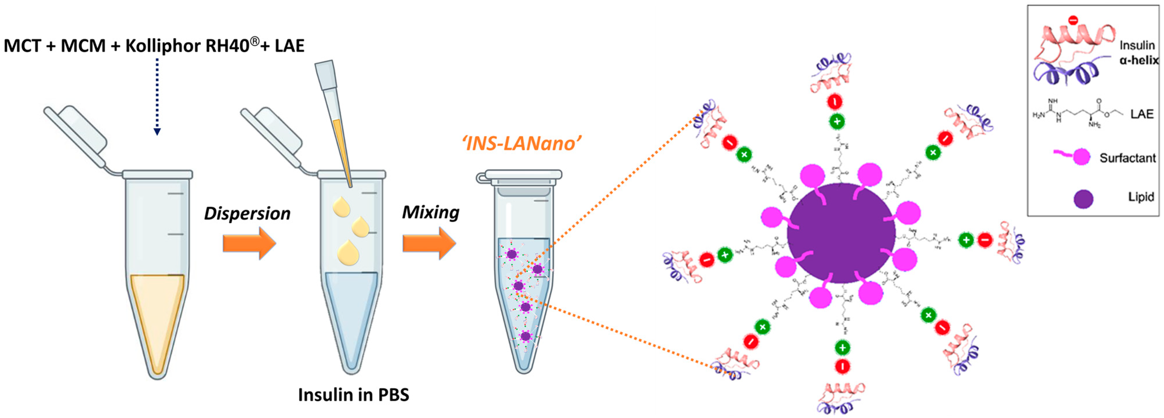
2.3. Preparation of Arginine-Coated Self-Emulsifying Nanoglobule System (Lanano)
2.4. Size and Zeta Potential
2.5. Circular Dichroism (Cd) Spectroscopy
2.6. Binding Efficiency
2.7. Cytocompatibiliy Study
2.8. Permeability Study
2.9. In Vivo Study
2.10. Statistical Analysis
3. Results
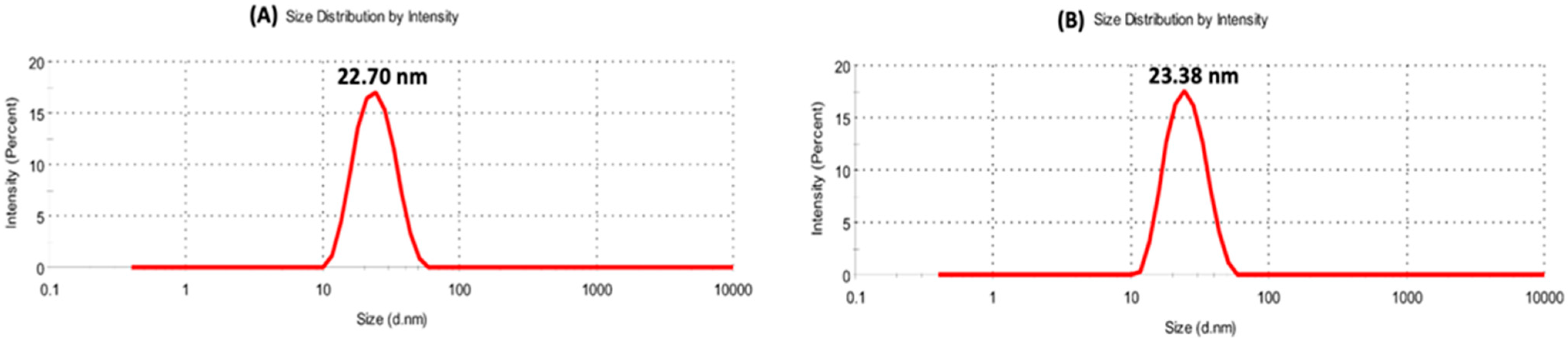
3.1. Particle Size and Zeta Potential
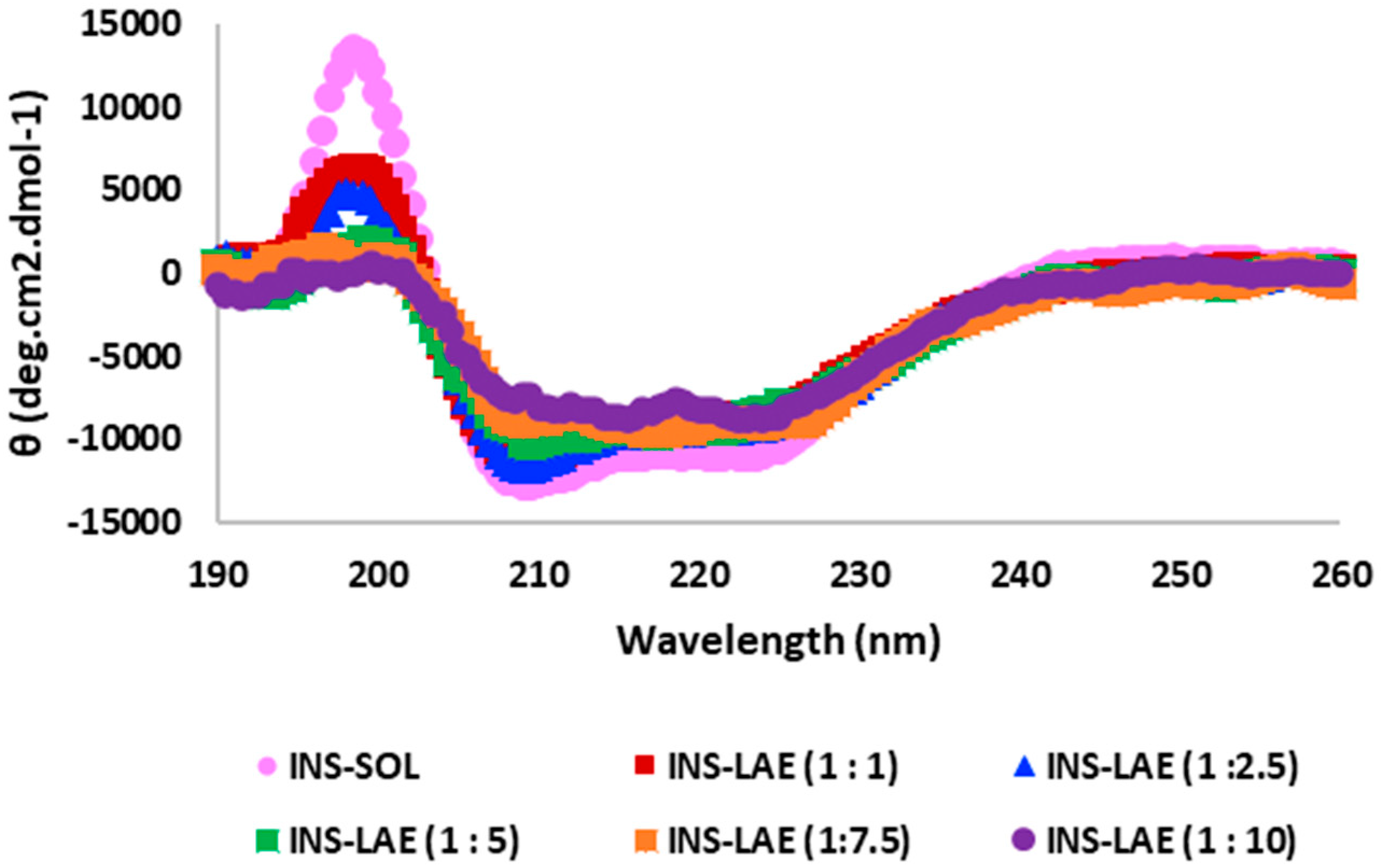
3.2. Circular Dichroism (Cd) Spectroscopy Study
3.3. Binding Efficiency Study
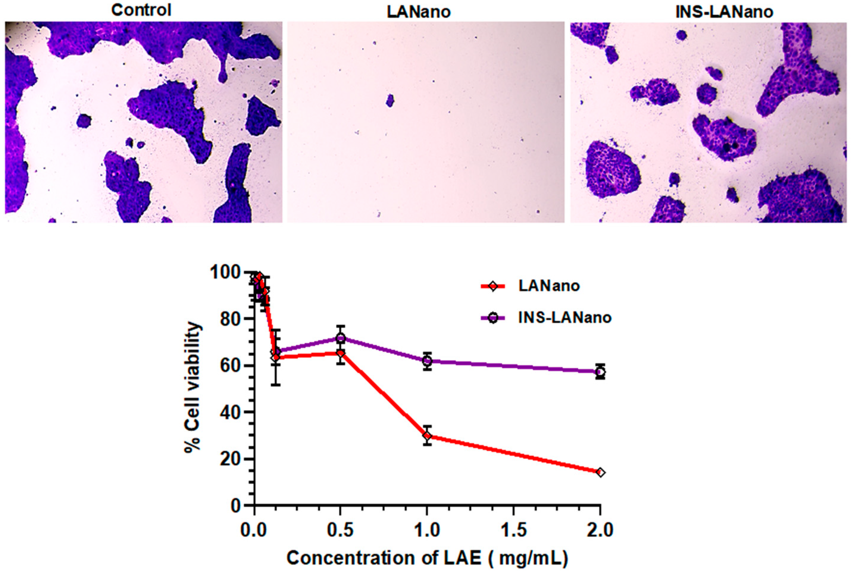
3.4. Cytocompatibility Study
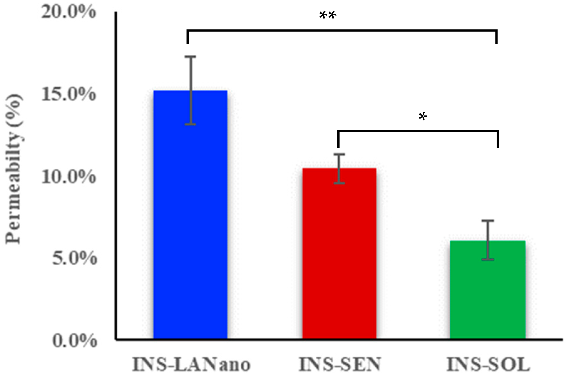
3.5. Permeability Study
3.6. In Vivo Study
4. Discussion
5. Conclusions
Author Contributions
Funding
Institutional Review Board Statement
Data Availability Statement
Acknowledgments
Conflicts of Interest
Abbreviations
References
- Easa, N.; Alany, R.; Carew, M.; Vangala, A. A review of non-invasive insulin delivery systems for diabetes therapy in clinical trials over the past decade. Drug Discov. Today 2019, 24, 440–451. [Google Scholar] [CrossRef] [Green Version]
- Radermecker, R.; Pierard, G.; Scheen, A. Lipodystrophy reactions to insulin: Effects of continuous insulin infusion and new insulin analogs. Am. J. Clin. Dermatol. 2007, 8, 21–28. [Google Scholar] [CrossRef]
- Brod, M.; Kongso, J.; Lessard, S.; Christensen, T. Psychological insulin resistance: Patient beliefs and implications for diabetes management. Qual. Life Res. 2009, 18, 23–32. [Google Scholar] [CrossRef] [Green Version]
- Goldberg, T.; Wong, E. Afrezza (Insulin Human) Inhalation Powder: A New Inhaled Insulin for the Management Of Type-1 or Type-2 Diabetes Mellitus. P T 2015, 40, 735–741. [Google Scholar]
- Beals, J.; DeFelippis, M.; Paavola, C.; Allen, D.; Garg, A.; Baldwin, D.B. Insulin, Pharmaceutical Biotechnology; Springer: Berlin/Heidelberg, Germany, 2019; pp. 403–427. [Google Scholar]
- Duan, X.; Mao, S. New strategies to improve the intranasal absorption of insulin. Drug Discov. Today 2010, 15, 416–427. [Google Scholar] [CrossRef]
- Javaid, M.; Selim, M.; Ortega-Gutierrez, S.; Lattanzi, S.; Zargar, S.; Alaouieh, D.; Hong, E.; Divani, A. Potential Application of Intranasal Insulin Delivery for Treatment of Intracerebral Hemorrhage: A Review of The Literature. J. Stroke Cerebrovasc. Dis. 2022, 31, 106489. [Google Scholar] [CrossRef]
- Bunchongprasert, K.; Shao, J. Effect of fatty acid ester structure on cytotoxicity of self-emulsified nanoemulsion and transport of nanoemulsion droplets. Colloids Surf. B Biointerfaces 2020, 194, 111220. [Google Scholar] [CrossRef]
- Vartak, R.; Menon, S.; Patki, M.; Billack, B.; Patel, K. Ebselen nanoemulgel for the treatment of topical fungal infection. Eur. J. Pharm. Sci. 2020, 148, 105323. [Google Scholar] [CrossRef]
- Gandhi, T.; Patki, M.; Kong, J.; Koya, J.; Yoganathan, S.; Reznik, S.; Patel, K. Development of an Arginine Anchored Nanoglobule with Retrograde Trafficking Inhibitor (Retro-2) for the Treatment of an Enterohemorrhagic Escherichia coli Outbreak. Mol. Pharm. 2019, 16, 4405–4415. [Google Scholar] [CrossRef]
- Menon, S.; Vartak, R.; Patel, K.; Billack, B. Evaluation of the antifungal activity of an ebselen-loaded nanoemulsion in a mouse model of vulvovaginal candidiasis. Nanomed. Nanotechnol. Biol. Med. 2021, 37, 102428. [Google Scholar] [CrossRef]
- Ostrozka-Cieslik, A.; Maciazek-Jurczyk, M.; Pozycka, J.; Dolinska, B. Pre-Formulation Studies: Physicochemical Characteristics and In Vitro Release Kinetics of Insulin from Selected Hydrogels. Pharmaceutics 2021, 13, 1215. [Google Scholar] [CrossRef] [PubMed]
- Micsonai, A.; Wien, F.; Bulyaki, E.; Kun, J.; Moussong, E.; Lee, Y.; Goto, Y.; Refregiers, M.; Kardos, J. BeStSel: A web server for accurate protein secondary structure prediction and fold recognition from the circular dichroism spectra. Nucleic Acids Res. 2018, 46, W315–W322. [Google Scholar] [CrossRef] [PubMed]
- Vartak, R.; Patil, S.; Saraswat, A.; Patki, M.; Kunda, N.; Patel, K. Aerosolized nanoliposomal carrier of remdesivir: An effective alternative for COVID-19 treatment in vitro. Nanomedicine 2021, 16, 1187–1202. [Google Scholar] [CrossRef] [PubMed]
- Goebel-Stengel, M.; Stengel, A.; Tache, Y.; Reeve, J., Jr. The importance of using the optimal plasticware and glassware in studies involving peptides. Anal. Biochem. 2011, 414, 38–46. [Google Scholar] [CrossRef] [PubMed] [Green Version]
- Illum, L. Nasal drug delivery—Recent developments and future prospects. J. Control. Release 2012, 161, 254–263. [Google Scholar] [CrossRef]
- Sharma, G.; Wilson, K.; van der Walle, C.; Sattar, N.; Petrie, J.; Kumar, M.R. Microemulsions for oral delivery of insulin: Design, development and evaluation in streptozotocin induced diabetic rats. Eur. J. Pharm. Biopharm. 2010, 76, 159–169. [Google Scholar] [CrossRef] [PubMed]
- Pocker, Y.; Biswas, S. Conformational dynamics of insulin in solution. Circular dichroic studies. Biochemistry 1980, 19, 5043–5049. [Google Scholar] [CrossRef]
- Ettinger, M.; Timasheff, S. Optical activity of insulin. I. On the nature of the circular dichroism bands. Biochemistry 1971, 10, 824–831. [Google Scholar]
- Rave, K.; Potocka, E.; Boss, A.; Marino, M.; Costello, D.; Chen, R. Pharmacokinetics and linear exposure of AFRESA™ compared with the subcutaneous injection of regular human insulin. Diabetes Obes. Metab. 2009, 11, 715–720. [Google Scholar] [CrossRef]
- Keller, L.-A.; Merkel, O.; Popp, A. Intranasal drug delivery: Opportunities and toxicologic challenges during drug development. Drug Deliv. Transl. Res. 2022, 12, 735–757. [Google Scholar] [CrossRef]
- Rodger, A.; Marrington, R.; Roper, D.; Windsor, S. Circular dichroism spectroscopy for the study of protein-ligand interactions. Methods Mol. Biol. 2005, 305, 343–364. [Google Scholar]
- Miguel, M.S.; Marrington, R.; Rodger, P.; Rodger, A.; Robinson, C. An Escherichia coli twin-arginine signal peptide switches between helical and unstructured conformations depending on the hydrophobicity of the environment. Eur. J. Biochem. 2003, 270, 3345–3352. [Google Scholar] [CrossRef] [Green Version]
- Shah, D.; Guo, Y.; Ban, I.; Shao, J. Intranasal delivery of insulin by self-emulsified nanoemulsion system: In vitro and in vivo studies. Int. J. Pharm. 2022, 616, 121565. [Google Scholar] [CrossRef]
- Bortolotti, F.; Balducci, A.; Sonvico, F.; Russo, P.; Colombo, G. In vitro permeation of desmopressin across rabbit nasal mucosa from liquid nasal sprays: The enhancing effect of potassium sorbate. Eur. J. Pharm. Sci. 2009, 37, 36–42. [Google Scholar] [PubMed]
- GIZURARSON, S.; BECHGAARD, E. Study of nasal enzyme activity towards insulin. In vitro. Chem. Pharm. Bull. 1991, 39, 2155–2157. [Google Scholar] [CrossRef] [PubMed] [Green Version]
- Zhang, L.; Du, S.; Lu, Y.; Liu, C.; Tian, Z.; Yang, C.; Wu, H.; Wang, Z. Puerarin transport across a Calu-3 cell monolayer—An in vitro model of nasal mucosa permeability and the influence of paeoniflorin and menthol. Drug Des. Devel. Ther. 2016, 10, 2227–2237. [Google Scholar] [CrossRef] [Green Version]
- Amidi, M.; Romeijn, S.; Borchard, G.; Junginger, H.; Hennink, W.; Jiskoot, W. Preparation and characterization of protein-loaded N-trimethyl chitosan nanoparticles as nasal delivery system. J. Control. Release 2006, 111, 107–116. [Google Scholar] [CrossRef] [PubMed]
- Zheng, C.; Guo, Q.; Wu, Z.; Sun, L.; Zhang, Z.; Li, C.; Zhang, X. Amphiphilic glycopolymer nanoparticles as vehicles for nasal delivery of peptides and proteins. Eur. J. Pharm. Sci. 2013, 49, 474–482. [Google Scholar] [CrossRef] [PubMed]
- Natsume, H.; Iwata, S.; Ohtake, K.; Miyamoto, M.; Yamaguchi, M.; Hosoya, K.; Kobayashi, D.; Sugibayashi, K.; Morimoto, Y. Screening of cationic compounds as an absorption enhancer for nasal drug delivery. Int. J. Pharm. 1999, 185, 1–12. [Google Scholar] [CrossRef]
- Ohtake, K.; Maeno, T.; Ueda, H.; Ogihara, M.; Natsume, H.; Morimoto, Y. Poly-L-arginine enhances paracellular permeability via serine/threonine phosphorylation of ZO-1 and tyrosine dephosphorylation of occludin in rabbit nasal epithelium. Pharm. Res. 2003, 20, 1838–1845. [Google Scholar] [CrossRef]
- Yamaki, T.; Kamiya, Y.; Ohtake, K.; Uchida, M.; Seki, T.; Ueda, H.; Kobayashi, J.; Morimoto, Y.; Natsume, H. A mechanism enhancing macromolecule transport through paracellular spaces induced by Poly-L-Arginine: Poly-L-Arginine induces the internalization of tight junction proteins via clathrin-mediated endocytosis. Pharm. Res. 2014, 31, 2287–2296. [Google Scholar] [CrossRef]
- Bunchongprasert, K.; Shao, J. Cytotoxicity and permeability enhancement of Capmul(R)MCM in nanoemulsion formulation. Int. J. Pharm. 2019, 561, 289–295. [Google Scholar] [CrossRef]
- Awsiuk, K.; Stetsyshyn, Y.; Raczkowska, J.; Lishchynskyi, O.; Dąbczyński, P.; Kostruba, A.; Ohar, H.; Shymborska, Y.; Nastyshyn, S.; Budkowski, A. Temperature-controlled orientation of proteins on temperature-responsive grafted polymer brushes: Poly (butyl methacrylate) vs poly (butyl acrylate): Morphology, wetting, and protein adsorption. Biomacromolecules 2019, 20, 2185–2197. [Google Scholar] [CrossRef]
- Nastyshyn, S.; Stetsyshyn, Y.; Raczkowska, J.; Nastishin, Y.; Melnyk, Y.; Panchenko, Y.; Budkowski, A. Temperature-responsive polymer brush coatings for advanced biomedical applications. Polymers 2022, 14, 4245. [Google Scholar] [CrossRef]
- Khafagy El, S.; Morishita, M.; Isowa, K.; Imai, J.; Takayama, K. Effect of cell-penetrating peptides on the nasal absorption of insulin. J. Control. Release 2009, 133, 103–108. [Google Scholar] [CrossRef]
- Dyer, A.; Hinchcliffe, M.; Watts, P.; Castile, J.; Jabbal-Gill, I.; Nankervis, R.; Smith, A.; Illum, L. Nasal delivery of insulin using novel chitosan based formulations: A comparative study in two animal models between simple chitosan formulations and chitosan nanoparticles. Pharm. Res. 2002, 19, 998–1008. [Google Scholar] [CrossRef]
- Schipper, N.; Romeijn, S.; Verhoef, J.; Merkus, F. Nasal insulin delivery with dimethyl-beta-cyclodextrin as an absorption enhancer in rabbits: Powder more effective than liquid formulations. Pharm. Res. 1993, 10, 682–686. [Google Scholar] [CrossRef]
- Mayor, S.; Illum, L. Investigation of the effect of anaesthesia on nasal absorption of insulin in rats. Int. J. Pharm. 1997, 149, 123–129. [Google Scholar] [CrossRef]


| Formulation | Zeta (mV) | Size (nm) | PDI |
|---|---|---|---|
| SEN (Water) | −0.241 ± 0.942 | 26.84 ± 0.19 | 0.051 ± 0.018 |
| LANano (Water) | +13.10 ± 0.794 | 14.04 ± 0.12 | 0.383 ± 0.001 |
| LANano (PBS) | +3.040 ± 0.335 | 22.70 ± 0.60 | 0.134 ± 0.015 |
| Conc. of Insulin (µg/mL) | LAE to Insulin Ratio | Zeta (mV) | Size (nm) | PDI |
|---|---|---|---|---|
| 100 | 1:0.1 | +1.341 ± 0.941 | 21.74 ± 0.07 | 0.022 ± 0.016 |
| 300 | 1:0.3 | +0.559 ± 0.497 | 21.69 ± 0.19 | 0.042 ± 0.014 |
| 500 | 1:0.5 | +0.118 ± 0.095 | 23.38 ± 0.62 | 0.089 ± 0.013 |
| 700 | 1:0.7 | +0.281 ± 0.089 | 21.78 ± 0.42 | 0.077 ± 0.023 |
| 900 | 1:0.9 | −0.081 ± 0.153 | 22.87 ± 0.47 | 0.068 ± 0.021 |
| 1000 | 1:1.0 | −0.375 ± 0.540 | 23.38 ± 0.40 | 0.080 ± 0.005 |
| Insulin to LAE Ratio | Peak/Valley Wavelength (nm) | α-Helix Content (%) |
|---|---|---|
| 1 to 0.0 | 198.5/208.5, 223.0 | 42.0 |
| 1 to 1.0 | 198.5/209.0, 225.0 | 35.7 |
| 1 to 2.5 | 198.0/209.5, 224.0 | 35.5 |
| 1 to 5.0 | 199.0/209.5, 221.5 | 24.9 |
| 1 to 7.5 | 196.5/209.0, 224.0 | 22.9 |
| 1 to 10.0 | 199.5/215.5, 224.0 | 13.4 |
| Formulations | Recovery (%) |
|---|---|
| INS-SOL | 91.5 ± 1.5 |
| INS-SEN | 86.4 ± 3.6 |
| INS-LANano | 53.6 ± 0.1 * |
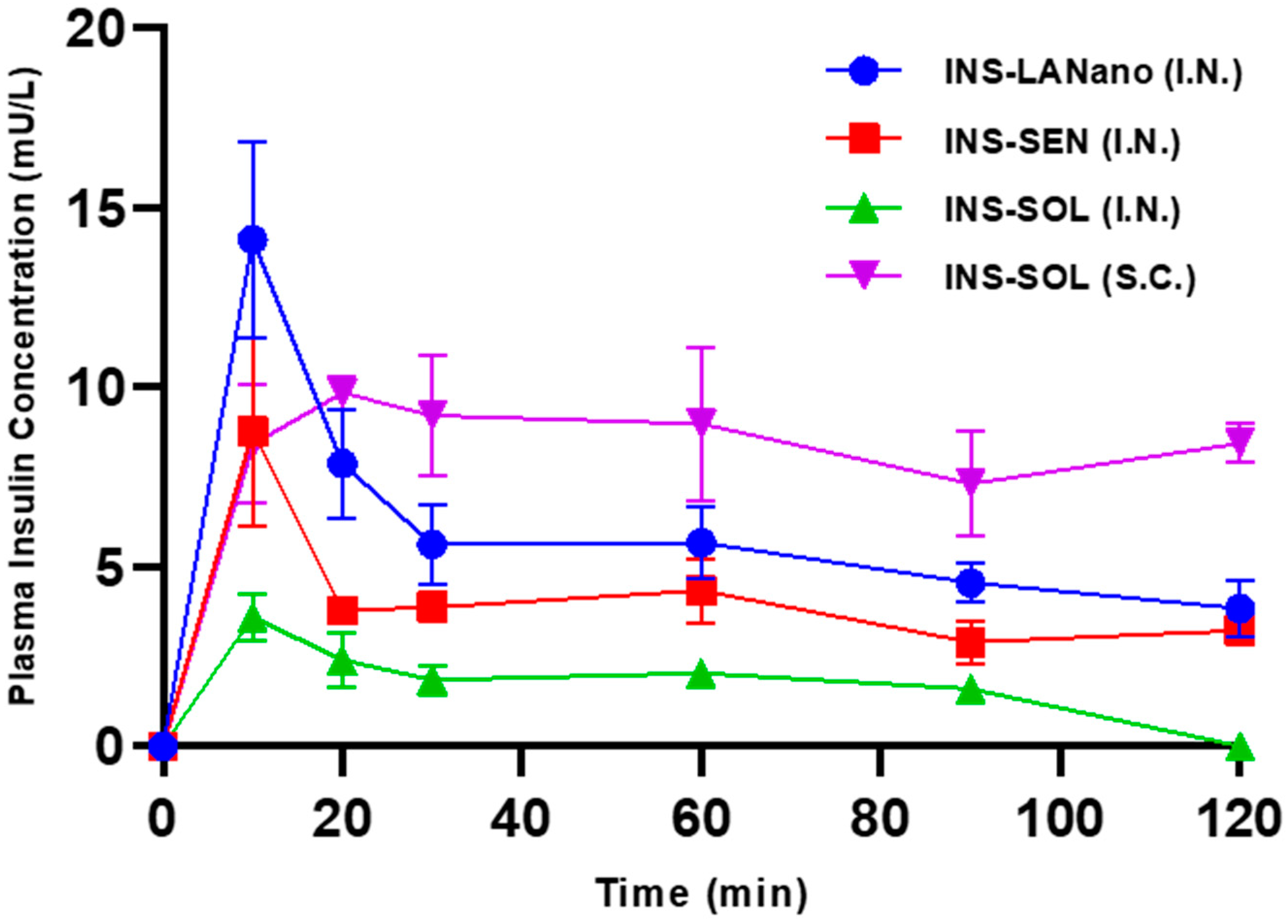
| Groups | Dose (IU/kg) | Cmax (mU/L) | Tmax (min) | AUC 0–120 min (mU/L × h) | BA (%) |
|---|---|---|---|---|---|
| INS-SOL (S.C.) | 1 | 11.1 ± 1.1 | 36.7 ± 12.0 | 16.2 ± 2.6 | 100 |
| INS-LANano (I.N.) | 2 | 14.3 ± 2.7 * | 12.5 ± 2.2 | 11.3 ± 0.9 | 23.3 ± 1.8 *,** |
| INS-SEN (I.N.) | 2 | 8.8 ± 2.6 | 10.0 ± 0.0 | 7.7 ± 0.3 | 15.8 ± 0.6 * |
| INS-SOL (I.N.) | 2 | 4.1 ± 0.2 | 13.3 ± 3.3 | 3.4 ± 0.4 | 6.9 ± 0.8 |
| Criteria | Afrezza® | INS-LANano | |
|---|---|---|---|
| 1 | Formulation technology | It is a dry powder formulation where recombinant human insulin is adsorbed onto fumaryl diketopiperazine particles as carrier to deliver insulin via the pulmonary route [4] | It is an aqueous formulation where recombinant human insulin is bound to the surface of Arginine-coated nanoglobules as carrier to deliver insulin via nasal route |
| 2 | Comparison of Pharmacokinetic Profile | In non-smoking healthy volunteers, the BA (compared to subcutaneous) is about 24.6%, 22.9% and 20.6% and the Tmax is about 12 min, 15 min and 17 min at 25 U, 50 U and 100 U doses, respectively [20] | In diabetic rats the BA and Tmax is about 23.3% and 12.5 min |
| 3 | Adverse Effects |
| Further studies needed to understand;
|
| 4 | Effect of Smoking | Not recommended for smoker patients [4] | Further studies needed to understand it. For Miacalcin Nasal Spray, smoking did not show a contributory effect on the occurrence of nasal adverse reactions) |
| 5 | Complexity of delivery device | Complex | Simple |
Disclaimer/Publisher’s Note: The statements, opinions and data contained in all publications are solely those of the individual author(s) and contributor(s) and not of MDPI and/or the editor(s). MDPI and/or the editor(s) disclaim responsibility for any injury to people or property resulting from any ideas, methods, instructions or products referred to in the content. |
© 2023 by the authors. Licensee MDPI, Basel, Switzerland. This article is an open access article distributed under the terms and conditions of the Creative Commons Attribution (CC BY) license (https://creativecommons.org/licenses/by/4.0/).
Share and Cite
Das, A.; Vartak, R.; Islam, M.A.; Kumar, S.; Shao, J.; Patel, K. Arginine-Coated Nanoglobules for the Nasal Delivery of Insulin. Pharmaceutics 2023, 15, 353. https://doi.org/10.3390/pharmaceutics15020353
Das A, Vartak R, Islam MA, Kumar S, Shao J, Patel K. Arginine-Coated Nanoglobules for the Nasal Delivery of Insulin. Pharmaceutics. 2023; 15(2):353. https://doi.org/10.3390/pharmaceutics15020353
Chicago/Turabian StyleDas, Atanu, Richa Vartak, Md Asrarul Islam, Sunil Kumar, Jun Shao, and Ketan Patel. 2023. "Arginine-Coated Nanoglobules for the Nasal Delivery of Insulin" Pharmaceutics 15, no. 2: 353. https://doi.org/10.3390/pharmaceutics15020353