

Silver Nanoparticles and Their Therapeutic Applications in Endodontics: A Narrative Review


Abstract
:1. Introduction
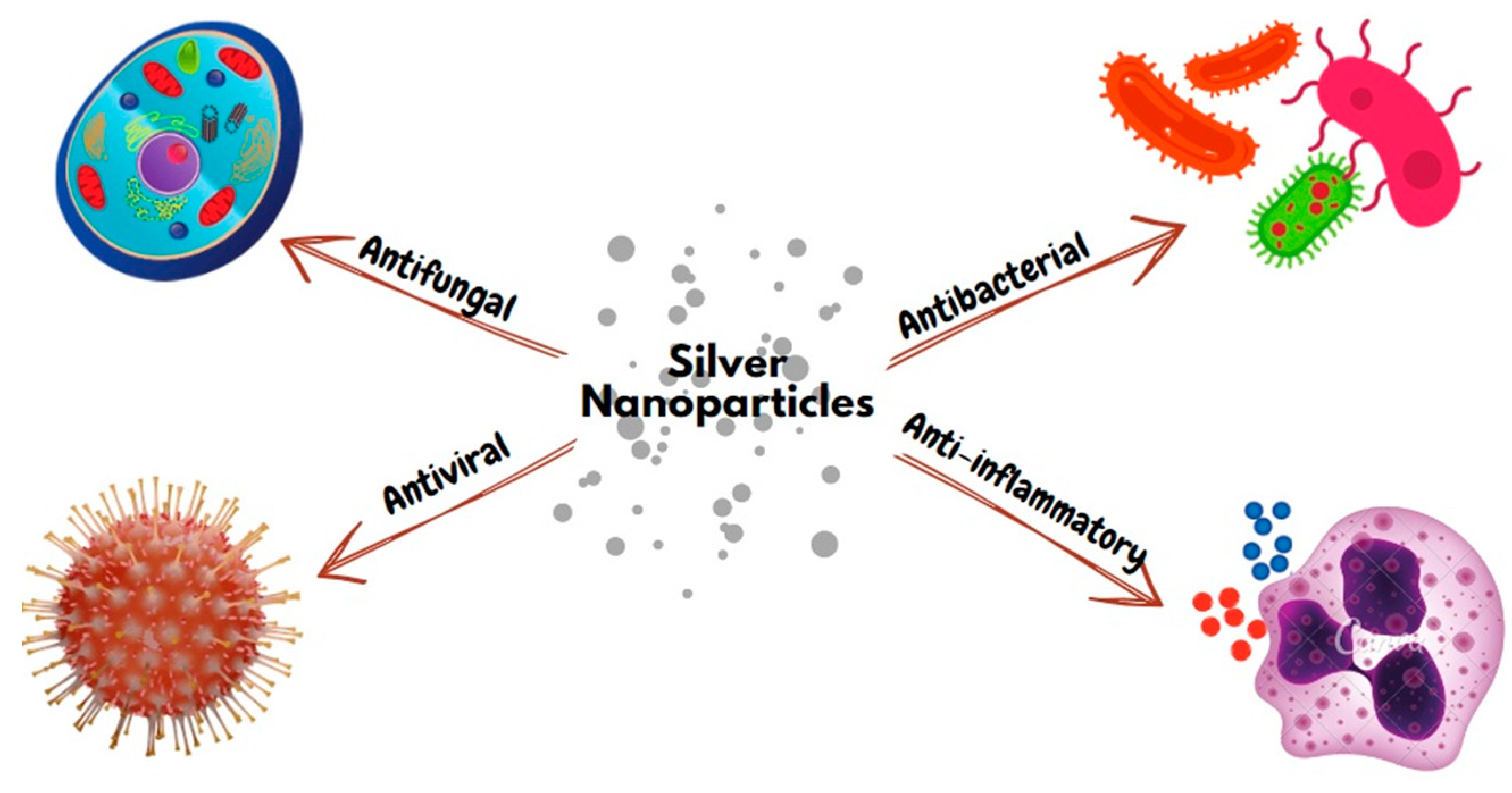
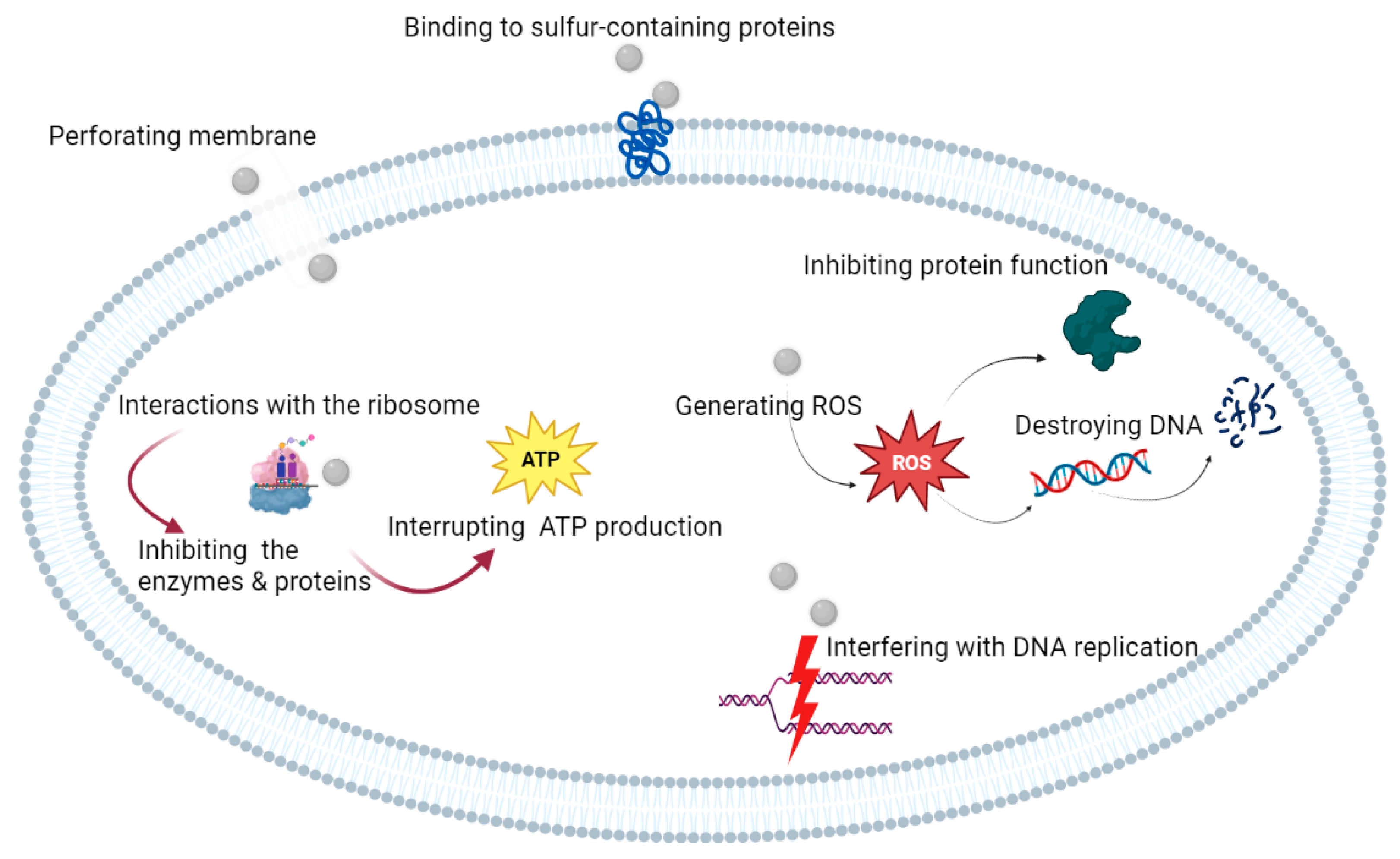
1.1. Antibacterial Properties
1.2. Antiviral Properties
1.3. Antifungal Properties
1.4. Anti-Inflammatory Properties
1.5. Toxicity of AgNPs
2. Application of AgNPs in Endodontics
2.1. Novel AgNP-Based Irrigants and Medicaments
2.2. Effects of AgNP-Based Irrigants Compared with Conventional Irrigants
2.3. Effect of AgNP-Based Medicaments Compared with Conventional Medicaments
2.4. Effect of AgNP-Based Sealers and Root-Filling Materials Compared with Traditional Root-Canal-Filling Materials
2.5. Effect of Addition of AgNPs to MTA
2.6. Effect of Addition of AgNPs to Fiber Posts
2.7. Application of AgNPs in Endodontic Surgery
2.8. Effect of AgNPs on Postoperative Pain
3. Conclusions and Prospects
Author Contributions
Funding
Institutional Review Board Statement
Informed Consent Statement
Data Availability Statement
Conflicts of Interest
References
- Bapat, R.A.; Chaubal, T.V.; Joshi, C.P.; Bapat, P.R.; Choudhury, H.; Pandey, M.; Gorain, B.; Kesharwani, P. An overview of application of silver nanoparticles for biomaterials in dentistry. Mater. Sci. Eng. C 2018, 91, 881–898. [Google Scholar] [CrossRef] [PubMed]
- Tsuzuki, T.; McCormick, P.G. Mechanochemical synthesis of nanoparticles. J. Mater. Sci. 2004, 39, 5143–5146. [Google Scholar] [CrossRef]
- Kalbassi, M.R.; Salari-joo, H.; Johari, A. Toxicity of silver nanoparticles in aquatic ecosystems: Salinity as the main cause in reducing toxicity. Iran. J. Toxicol. 2011, 5, 436–443. [Google Scholar]
- Song, W.; Ge, S. Application of antimicrobial nanoparticles in dentistry. Molecules 2019, 24, 1033. [Google Scholar] [CrossRef] [PubMed] [Green Version]
- Fernandez, C.C.; Sokolonski, A.R.; Fonseca, M.S.; Stanisic, D.; Araújo, D.B.; Azevedo, V.; Portela, R.D.; Tasic, L. Applications of Silver Nanoparticles in Dentistry: Advances and Technological Innovation. Int. J. Mol. Sci. 2021, 22, 2485. [Google Scholar] [CrossRef]
- Peng, J.J.Y.; Botelho, M.G.; Matinlinna, J.P. Silver compounds used in dentistry for caries management: A review. J. Dent. 2012, 40, 531–541. [Google Scholar] [CrossRef] [PubMed]
- Morones, J.R.; Elechiguerra, J.L.; Camacho, A.; Holt, K.; Kouri, J.B.; Ramírez, J.T.; Yacaman, M.J. The bactericidal effect of silver nanoparticles. Nanotechnology 2005, 16, 2346–2353. [Google Scholar] [CrossRef] [Green Version]
- Sotiriou, G.A.; Pratsinis, S.E. Antibacterial activity of nanosilver ions and particles. Environ. Sci. Technol. 2010, 44, 5649–5654. [Google Scholar] [CrossRef]
- Kaur, P.; Luthra, R. Silver nanoparticles in dentistry: An emerging trend. SRM J. Res. Dent. Sci. 2016, 7, 162–165. [Google Scholar] [CrossRef]
- Yamanaka, M.; Hara, K.; Kudo, J. Bactericidal actions of a silver ion solution on Escherichia coli, studied by energy-filtering transmission electron microscopy and proteomic analysis. Appl. Environ. Microbiol. 2005, 71, 7589–7593. [Google Scholar] [CrossRef] [Green Version]
- Abbaszadegan, A.; Ghahramani, Y.; Gholami, A.; Hemmateenejad, B.; Dorostkar, S.; Nabavizadeh, M.; Sharghi, H. The effect of charge at the surface of silver nanoparticles on antimicrobial activity against gram-positive and gram-negative bacteria: A preliminary study. J. Nanomater. 2015, 2015, 720654. [Google Scholar] [CrossRef] [Green Version]
- Mishra, P.; Tyagi, S.; Tripathi, D. Comparative evaluation of silver nanoparticles and 5.25% sodium hypochlorite for rapid chairside decontamination of artificially infected gutta-percha with Escherichia coli: An in vitro study. J. Int. Dent. Med. Res. 2019, 7, 23–27. [Google Scholar] [CrossRef]
- Ibrahim, A.I.O.; Petrik, L.; Moodley, D.S.; Patel, N. Use of antibacterial nanoparticles in endodontics. S. Afr. Dent. J. 2017, 72, 105–112. [Google Scholar]
- Zheng, T.; Huang, X.; Chen, J.; Feng, D.; Mei, L.; Huang, Y.; Quan, G.; Zhu, C.; Singh, V.; Ran, H.; et al. A liquid crystalline precursor incorporating chlorhexidine acetate and silver nanoparticles for root canal disinfection. Biomater. Sci. 2018, 6, 596–603. [Google Scholar] [CrossRef]
- Hou, X.; Fu, H.; Han, Y.; Xue, Y.; Li, C. Analysis of transcriptome in Enterococcus faecalis treated with silver nanoparticles. J. Nanosci. Nanotechnol. 2020, 20, 1046–1055. [Google Scholar] [CrossRef]
- Salas-Orozco, M.; Niño-Martínez, N.; Martínez-Castañón, G.A.; Méndez, F.T.; Jasso, M.E.; Ruiz, F. Mechanisms of resistance to silver nanoparticles in endodontic bacteria: A literature review. J. Nanomater. 2019, 2019, 7630316. [Google Scholar] [CrossRef] [Green Version]
- Kishen, A. Nanotechnology in Endodontics; Springer: Cham, Switzerland, 2016. [Google Scholar]
- Aydın, H.; Er, K.; Kuştarcı, A.; Akarsu, M.; Gencer, G.; Er, H.; Felek, R. Antibacterial activity of silver nanoparticles activated by photodynamic therapy in infected root canals. Dent. Med. Probl. 2020, 57, 393–400. [Google Scholar] [CrossRef]
- Galdiero, S.; Falanga, A.; Vitiello, M.; Cantisani, M.; Marra, V.; Galdiero, M. Silver nanoparticles as potential antiviral agents. Molecules 2011, 16, 8894–8918. [Google Scholar] [CrossRef] [Green Version]
- Şuhani, M.F.; Băciuţ, G.; Băciuţ, M.; Şuhani, R.; Bran, S. Current perspectives regarding the application and incorporation of silver nanoparticles into dental biomaterials. Clujul Med. 2018, 91, 274–279. [Google Scholar] [CrossRef]
- Kim, K.J.; Sung, W.S.; Suh, B.K.; Moon, S.K.; Choi, J.S.; Kim, J.G.; Lee, D.G. Antifungal activity and mode of action of silver nano-particles on Candida albicans. Biometals 2009, 22, 235–242. [Google Scholar] [CrossRef]
- Meneses, M.L.; Recalde, M.; Martin, P.L.; Pardo, A.G. Antifungal activity of silver nanoparticles and clotrimazole against Candida spp. Braz. J. Pharm. Sci. 2022, 58, e18719. [Google Scholar] [CrossRef]
- Mozayeni, M.A.; Hadian, A.; Bakhshaei, P.; Dianat, O. Comparison of antifungal activity of 2% chlorhexidine, calcium hydroxide, and nanosilver gels against Candida albicans. J. Dent. 2015, 12, 109–117. [Google Scholar]
- Chinnasamy, G.; Chandrasekharan, S.; Koh, T.W.; Bhatnagar, S. Synthesis, characterization, antibacterial and wound healing efficacy of silver nanoparticles from Azadirachta indica. Front. Microbiol. 2021, 12, 611560. [Google Scholar] [CrossRef]
- Gunasekaran, T.; Nigusse, T.; Dhanaraju, M.D. Silver nanoparticles as real topical bullets for wound healing. J. Am. Coll. Clin. Wound. Spec. 2012, 3, 82–96. [Google Scholar] [CrossRef] [Green Version]
- Yin, I.X.; Zhang, J.; Zhao, I.S.; Mei, M.L.; Li, Q.; Chu, C.H. The antibacterial mechanism of silver nanoparticles and its application in dentistry. Int. J. Nanomed. 2020, 15, 2555–2562. [Google Scholar] [CrossRef] [PubMed] [Green Version]
- Tyavambiza, C.; Elbagory, A.M.; Madiehe, A.M.; Meyer, M.; Meyer, S. The antimicrobial and anti-inflammatory effects of silver nanoparticles synthesised from Cotyledon orbiculata aqueous extract. Nanomaterials 2021, 11, 1343. [Google Scholar] [CrossRef]
- Noronha, V.T.; Paula, A.J.; Durán, G.; Galembeck, A.; Cogo-Müller, K.; Franz-Montan, M.; Durán, N. Silver nanoparticles in dentistry. Dent. Mater. 2017, 33, 1110–1126. [Google Scholar] [CrossRef]
- Zhang, X.F.; Shen, W.; Gurunathan, S. Silver nanoparticle-mediated cellular responses in various cell lines: An in vitro model. Int. J. Mol. Sci. 2016, 17, 1603. [Google Scholar] [CrossRef] [Green Version]
- Lebda, M.A.; Sadek, K.M.; Tohamy, H.G.; Abouzed, T.K.; Shukry, M.; Umezawa, M.; El-Sayed, Y.S. Potential role of α-lipoic acid and Ginkgo biloba against silver nanoparticles-induced neuronal apoptosis and blood-brain barrier impairments in rats. Life Sci. 2018, 212, 251–260. [Google Scholar] [CrossRef]
- Rai, M.; Ingle, A.P.; Gade, A.K.; Duarte, M.C.; Duran, N. Three Phoma spp. synthesised novel silver nanoparticles that possess excellent antimicrobial efficacy. IET Nanobiotechnol. 2015, 9, 280–287. [Google Scholar] [CrossRef]
- Takamiya, A.S.; Monteiro, D.R.; Bernabe, D.G.; Gorup, L.F.; Camargo, E.R.; Gomes-Filho, J.E.; Oliveira, S.H.; Barbosa, D.B. In vitro and in vivo toxicity evaluation of colloidal silver nanoparticles used in endodontic treatments. J. Endod. 2016, 42, 953–960. [Google Scholar] [CrossRef] [PubMed] [Green Version]
- Munger, M.A.; Radwanski, P.; Hadlock, G.C.; Stoddard, G.; Shaaban, A.; Falconer, J.; Grainger, D.W.; Deering-Rice, C.E. In vivo human time-exposure study of orally dosed commercial silver nanoparticles. Nanomedicine 2014, 10, 1–9. [Google Scholar] [CrossRef] [PubMed] [Green Version]
- Van der Zande, M.; Vandebriel, R.J.; Van Doren, E.; Kramer, E.; Herrera Rivera, Z.; Serrano-Rojero, C.S.; Gremmer, E.R.; Mast, J.; Peters, R.J.; Hollman, P.C.; et al. Distribution, elimination, and toxicity of silver nanoparticles and silver ions in rats after 28-day oral exposure. ACS Nano. 2012, 6, 7427–7442. [Google Scholar] [CrossRef] [PubMed]
- Kishen, A. Advanced therapeutic options for endodontic biofilms. Endod. Top. 2010, 22, 99–123. [Google Scholar] [CrossRef]
- Shaik, I.; Goyal, S.; Bhowmick, S.; Shetty, S.V.; Shetty, V.; Sharma, S.; Singh, S. Knowledge and assessment of endodontists in the field of nanotechnology in Endodontics: A Qualitative Research. J. Adv. Med. Dent. Sci. Res. 2020, 8, 84–87. [Google Scholar]
- Betancourt, J.A.; Romero, C.C.; Delgadillo, R.H.; Villarreal, M.M.; Rodriguez, L.E.; Quintanilla, N.C.; Kim, H.; Soto, J.M. Analysis of the antimicrobial and antibiotic activity of nanoparticles for endodontic use. Int. J. Appl. Dent. Sci. 2020, 6, 85–89. [Google Scholar] [CrossRef]
- Oncu, A.; Huang, Y.; Amasya, G.; Sevimay, F.S.; Orhan, K.; Celikten, B. Silver nanoparticles in endodontics: Recent developments and applications. Restor. Dent. Endod. 2021, 46, e38. [Google Scholar] [CrossRef]
- Bhushan, J.; Maini, C. Nanoparticles: A promising novel adjunct for dentistry. Indian J. Dent. Sci. 2019, 11, 167–173. [Google Scholar] [CrossRef]
- Shrestha, A.; Kishen, A. Antibacterial nanoparticles in endodontics: A review. J. Endod. 2016, 42, 1417–1426. [Google Scholar] [CrossRef]
- Halkai, K.R.; Mudda, J.A.; Shivanna, V.; Rathod, V.; Halkai, R. Biosynthesised silver nanoparticles from fungi as antimicrobial agents for endo-perio lesions–a review. Annu. Res. Rev. Biol. 2016, 10, 1–7. [Google Scholar] [CrossRef]
- Halkai, K.R.; Halkai, R.; Mudda, J.A.; Shivanna, V.; Rathod, V. Antibiofilm efficacy of biosynthesized silver nanoparticles against endodontic-periodontal pathogens: An in vitro study. J. Conserv. Dent. 2018, 21, 662–666. [Google Scholar] [CrossRef] [PubMed]
- Parvekar, P.; Palaskar, J.; Metgud, S.; Maria, R.; Dutta, S. The minimum inhibitory concentration (MIC) and minimum bactericidal concentration (MBC) of silver nanoparticles against Staphylococcus aureus. Biomater. Investig. Dent. 2020, 7, 105–109. [Google Scholar] [CrossRef] [PubMed]
- Samiei, M.; Farjami, A.; Dizaj, S.M.; Lotfipour, F. Nanoparticles for antimicrobial purposes in Endodontics: A systematic review of in vitro studies. Mater. Sci. Eng. C 2016, 58, 1269–1278. [Google Scholar] [CrossRef] [PubMed]
- Ertem, E.; Gutt, B.; Zuber, F.; Allegri, S.; Le Ouay, B.; Mefti, S.; Formentin, K.; Stellacci, F.; Ren, Q. Core–shell silver nanoparticles in endodontic disinfection solutions enable long-term antimicrobial effect on oral biofilms. ACS Appl. Mater. Interfaces 2017, 9, 34762–34772. [Google Scholar] [CrossRef]
- Fan, W.; Wu, D.; Tay, F.R.; Ma, T.; Wu, Y.; Fan, B. Effects of adsorbed and templated nanosilver in mesoporous calcium-silicate nanoparticles on inhibition of bacteria colonization of dentin. Int. J. Nanomed. 2014, 9, 5217–5230. [Google Scholar] [CrossRef] [PubMed] [Green Version]
- Sadek, R.W.; Moussa, S.M.; El Backly, R.M.; Hammouda, A.F. Evaluation of the efficacy of three antimicrobial agents used for regenerative endodontics: An in vitro study. Microb. Drug Resist. 2019, 25, 761–771. [Google Scholar] [CrossRef] [PubMed]
- Ioannidis, K.; Niazi, S.; Mylonas, P.; Mannocci, F.; Deb, S. The synthesis of nano silver-graphene oxide system and its efficacy against endodontic biofilms using a novel tooth model. Dent. Mater. 2019, 35, 1614–1629. [Google Scholar] [CrossRef] [PubMed]
- Bhandi, S.; Mehta, D.; Mashyakhy, M.; Chohan, H.; Testarelli, L.; Thomas, J.; Dhillon, H.; Raj, A.T.; Madapusi Balaji, T.; Varadarajan, S.; et al. Antimicrobial efficacy of silver nanoparticles as root canal irrigant’s: A systematic review. J. Clin. Med. 2021, 10, 1152. [Google Scholar] [CrossRef]
- Jowkar, Z.; Hamidi, S.A.; Shafiei, F.; Ghahramani, Y. The effect of silver, zinc oxide, and titanium dioxide nanoparticles used as final irrigation solutions on the fracture resistance of root-filled teeth. Clin. Cosmet. Investig. Dent. 2020, 12, 141–148. [Google Scholar] [CrossRef] [Green Version]
- Al-Fahham, B.; Al-Haidar, A. Evaluation of the antibacterial efficacy of silver nanoparticles as an irrigant against Enterococcus faecalis in vitro study. J. Res. Med. Dent. Sci. 2019, 7, 21–27. [Google Scholar]
- Cecilia, S.; Divyarani, S.; Lakshya, K. Preparation of silver nano particles using aqueous solution of Ocimum sanctum and Piper betle and evaluation of its antimicrobial activity against Enterococcus faecalis. Int. J. Pharm. Clin. Res. 2016, 8, 1118–1120. [Google Scholar]
- Halkai, K.R.; Mudda, J.A.; Shivanna, V.; Rathod, V.; Halkai, R. Evaluation of antibacterial efficacy of fungal-derived silver nanoparticles against Enterococcus faecalis. Contemp. Clin. Dent. 2018, 9, 45–48. [Google Scholar] [CrossRef] [PubMed]
- Krishnan, R.; Arumugam, V.; Vasaviah, S.K. The MIC and MBC of silver nanoparticles against Enterococcus faecalis-a facultative anaerobe. J. Nanomed. Nanotechnol. 2015, 6, 285. [Google Scholar]
- Alsamhari, M.M.; AlKhawlani, M.M.; Al-Kholani, A.I.; Al-Najhi, M.M.; Al-Shamahy, H.A.; Al-Sharani, A.A.; Ismael, O.A.; Al-dossary, K.A.; Al-labani, M.A. Antimicrobial activity of Sodium hypochlorite, nano Silver and Chlorhexidine against mono-species biofilms of selected microorganisms of oral sources. Univers. J. Pharm. Res. 2022, 7, 11–16. [Google Scholar] [CrossRef]
- Moradi, F.; Haghgoo, R. Evaluation of antimicrobial efficacy of nanosilver solution, sodium hypochlorite and normal saline in root canal irrigation of primary teeth. Contemp. Clin. Dent. 2018, 9, S227–S232. [Google Scholar] [PubMed]
- Othman, A.M.; Elsayed, M.A.; Al-Balakocy, N.G.; Hassan, M.M.; Elshafei, A.M. Biosynthesis and characterization of silver nanoparticles induced by fungal proteins and its application in different biological activities. J. Genet. Eng. Biotechnol. 2019, 17, 8. [Google Scholar] [CrossRef] [PubMed] [Green Version]
- Makkar, S.; Aggarwal, A.; Pasricha, S.; Kapur, I. To evaluate the antibacterial properties of silver nano particle based irrigant as endodontic root canal irrigant. Int. J. Dent. Health Sci. 2014, 1, 485–492. [Google Scholar]
- Gomes-FilHo, J.E.; Silva, F.O.; Watanabe, S.; Tendoro, K.V.; Dalto, L.G.; Pacanaro, S.V.; Lodi, C.S.; De Melo, F.F.; Dezan Júnior, E.; Cintra, L.T. Evaluation of silver nanoparticles as irrigating solution. Dent. Press Endod. 2013, 3, 16–23. [Google Scholar] [CrossRef]
- Ambalavanan, N.; Kavitha, M.; Jayakumar, S.; Raj, A.; Nataraj, S. Comparative evaluation of bactericidal effect of silver nanoparticle in combination with Nd-YAG laser against Enterococcus faecalis: An in vitro study. J. Contemp. Dent. Pract. 2020, 21, 1141–1145. [Google Scholar] [PubMed]
- Sadony, D.M.; Montasser, K. Evaluation and comparison between the bactericidal effect of diode laser irradiation (970 nm) and silver nanoparticles on Enterococcus faecalis bacterial strain (an in vitro study). Bull. Natl. Res. Cent. 2019, 43, 155. [Google Scholar] [CrossRef] [Green Version]
- Sadony, D.M.; Abozaid, H.E.S. Antibacterial effect of metallic nanoparticles on Streptococcus mutans bacterial strain with or without diode laser (970 nm). Bull. Natl. Res. Cent. 2020, 44, 2. [Google Scholar] [CrossRef] [Green Version]
- Afkhami, F.; Akbari, S.; Chiniforush, N. Entrococcus faecalis elimination in root canals using silver nanoparticles, photodynamic therapy, diode laser, or laser-activated nanoparticles: An in vitro study. J. Endod. 2017, 43, 279–282. [Google Scholar] [CrossRef] [PubMed]
- Afkhami, F.; Ahmadi, P.; Chiniforush, N.; Sooratgar, A. Effect of different activations of silver nanoparticle irrigants on the elimination of Enterococcus faecalis. Clin. Oral Investig. 2021, 25, 6893–6899. [Google Scholar] [CrossRef] [PubMed]
- Kushwaha, V.; Yadav, R.K.; Tikku, A.P.; Chandra, A.; Verma, P.; Gupta, P.; Shakya, V.K. Comparative evaluation of antibacterial effect of nanoparticles and lasers against endodontic microbiota: An in vitro study. J. Clin. Exp. Dent. 2018, 10, e1155–e1160. [Google Scholar] [CrossRef]
- Charannya, S.; Duraivel, D.; Padminee, K.; Poorni, S.; Nishanthine, C.; Srinivasan, M.R. Comparative evaluation of antimicrobial efficacy of silver nanoparticles and 2% chlorhexidine gluconate when used alone and in combination assessed using agar diffusion method: An in vitro study. Contemp. Clin. Dent. 2018, 9, S204–S209. [Google Scholar] [CrossRef] [PubMed]
- González-Luna, I.V.P.; Martínez-Castañón, G.A.; Zavala-Alonso, N.V.; Patiño-Marin, N.; Niño-Martínez, N.; Morán-Martínez, J. Bactericide effect of silver nanoparticles as a final irrigation agent in endodontics on Enterococcus faecalis: An ex vivo study. J. Nanomater. 2016, 2016, 7597295. [Google Scholar] [CrossRef] [Green Version]
- Rajasekhar, R.; James, B.; Devadathan, A.; Soman, S.; Sebastian, V.M.; Sathyan, M. An In Vitro Evaluation of Antibacterial and Smear Layer Removal Efficacy of Silver Nanoparticles as Final Irrigant against Enterococcus Faecalis. World 2022, 13, 148–154. [Google Scholar]
- Martinez-Andrade, J.M.; Avalos-Borja, M.; Vilchis-Nestor, A.R.; Sanchez-Vargas, L.O.; Castro-Longoria, E. Dual function of EDTA with silver nanoparticles for root canal treatment–A novel modification. PLoS ONE 2018, 13, e0190866. [Google Scholar] [CrossRef] [Green Version]
- Chávez-Andrade, G.M.; Tanomaru-Filho, M.; Bernardi, M.I.; de Toledo Leonardo, R.; Faria, G.; Guerreiro-Tanomaru, J.M. Antimicrobial and biofilm anti-adhesion activities of silver nanoparticles and farnesol against endodontic microorganisms for possible application in root canal treatment. Arch. Oral Biol. 2019, 107, 104481. [Google Scholar] [CrossRef]
- Rodríguez-Chang, S.; Ramírez-Mora, T.; Valle-Bourrouet, G.; Rojas-Campos, N.; Chavarría-Bolaño, D.; Montero-Aguilar, M. Antibacterial efficacy of a dispersion of silver nanoparticles in citrate medium for the treatment of E. faecalis: An In Vitro Study. Odovtos-Int. J. Dent. Sc. 2016, 18, 99–107. [Google Scholar] [CrossRef]
- Sabry, H.A.; Nashaat, Y.M.; Omar, N.; Negm, A.; Shaheen, N.A. Comparative study of the antibacterial effect of nano-silver irrigant, sodium hypochlorite and chlorhexidine against Enterococcus faecalis biofilm. Egypt. Dent. J. 2019, 65, 1503–1509. [Google Scholar] [CrossRef]
- Rodrigues, C.T.; De Andrade, F.B.; De Vasconcelos, L.R.; Midena, R.Z.; Pereira, T.C.; Kuga, M.C.; Duarte, M.A.; Bernardineli, N. Antibacterial properties of silver nanoparticles as a root canal irrigant against Enterococcus faecalis biofilm and infected dentinal tubules. Int. Endod. J. 2018, 51, 901–911. [Google Scholar] [CrossRef]
- Nabavizadeh, M.; Abbaszadegan, A.; Gholami, A.; Kadkhoda, Z.; Mirhadi, H.; Ghasemi, Y.; Safari, A. Antibiofilm efficacy of positively charged imidazolium-based silver nanoparticles in Enterococcus faecalis using quantitative real-time PCR. Jundishapur J. Microbiol. 2017, 10, e55616. [Google Scholar] [CrossRef] [Green Version]
- Kangarlou Haghighi, A.; Tashfam, B.; Nasseri, M.; Dianat, O.; Taheri, S. In vitro comparison of antibacterial efficacy of a new irrigation solution containing nanosilver with sodium hypochlorite and chlorhexidine. Shahid Beheshti Uni. Dent. J. 2013, 31, 1–7. [Google Scholar]
- Nia, A.F.; Ataei, M.; Zeighami, H. A comparative study on the antimicrobial activity of irreversible hydrocolloid mixed with silver nanoparticles and chlorhexidine. Dent. Res. J. 2020, 17, 120–125. [Google Scholar]
- Wu, D.; Fan, W.; Kishen, A.; Gutmann, J.L.; Fan, B. Evaluation of the antibacterial efficacy of silver nanoparticles against Enterococcus faecalis biofilm. J. Endod. 2014, 40, 285–290. [Google Scholar] [CrossRef] [PubMed]
- Moazami, F.; Sahebi, S.; Ahzan, S. Tooth discoloration induced by imidazolium based silver nanoparticles as an intracanal irrigant. J. Dent. 2018, 19, 280–286. [Google Scholar]
- Saygi, K.O.; Bayram, H.M.; Bayram, E. Green synthesis of silver nanoparticles using artichoke flower petals and application in endodontic dentistry. Biomass. Convers. Biorefin. 2022, 13, 1–9. [Google Scholar] [CrossRef]
- Yuan, Z.; Chen, Y.; Li, T.; Yu, C.-P. Reaction of silver nanoparticles in the disinfection process. Chemosphere 2013, 93, 619–625. [Google Scholar] [CrossRef]
- Elkillany, R.; Sabet, N.; Fakhr, M. The Antimicrobial Efficacy of Nanoparticles Intracanal Medicaments Against Enterococcus Faecalis Biofilm. Egypt. Dent. J. 2022, 68, 1789–1796. [Google Scholar] [CrossRef]
- Lorena, C.; Georgescu, R.V.; Diaconu, O.A.; Scrieciu, M.; Petcu, C.; Popescu, S.M.; Rîcă, A.M.; Turcu, A.; Andreea, N.; Țuculină, M.J. Review of endodontic drugs and their antibacterial effectiveness. Rom. J. Med. Dent. Educ. 2021, 10, 33–41. [Google Scholar]
- Balto, H.; Bukhary, S.; Al-Omran, O.; BaHammam, A.; Al-Mutairi, B. Combined effect of a mixture of silver nanoparticles and calcium hydroxide against Enterococcus faecalis biofilm. J. Endod. 2020, 46, 1689–1694. [Google Scholar] [CrossRef] [PubMed]
- Siddiqi, K.S.; Husen, A.; Rao, R.A.K. A review on biosynthesis of silver nanoparticles and their biocidal properties. J. Nanobiotechnology 2018, 16, 14. [Google Scholar] [CrossRef] [PubMed]
- Marín-Correa, B.M.; Guzmán-Martínez, N.; Gómez-Ramírez, M.; Pless, R.C.; Mundo, J.R.; García-Ramos, J.C.; Rojas-Avelizapa, N.G.; Pestryakov, A.; Bogdanchikova, N.; Fierros-Romero, G. Nanosilver gel as an endodontic alternative against Enterococcus faecalis in an in vitro root canal system in Mexican dental specimens. New Microbiol. 2020, 43, 166–170. [Google Scholar]
- Hassan, R.; Khallaf, M. Effect of a silver nanoparticle intracanal-based medicament on the microhardness of human radicular dentine. Endod. Pract. Today 2018, 12, 125–131. [Google Scholar]
- Javidi, M.; Afkhami, F.; Zarei, M.; Ghazvini, K.; Rajabi, O. Efficacy of a combined nanoparticulate/calcium hydroxide root canal medication on elimination of Enterococcus faecalis. Aust. Endod. J. 2013, 40, 61–65. [Google Scholar] [CrossRef]
- Afkhami, F.; Pourhashemi, S.J.; Sadegh, M.; Salehi, Y.; Fard, M.J. Antibiofilm efficacy of silver nanoparticles as a vehicle for calcium hydroxide medicament against Enterococcus faecalis. J. Dent. 2015, 43, 1573–1579. [Google Scholar] [CrossRef]
- Riaz, Z.; Raza, M.; Hanif, A.; Haider, B.; Akram, S.; Safdar, S. Antibacterial Efficacy of Silver Nanoparticles Impregnated Calcium Hydroxide: An in Vitro Study. J. Pak. Dent. Assoc. 2022, 31, 1–4. [Google Scholar] [CrossRef]
- Nasim, I.; Shamly, M.; Jaju, K.; Vishnupriya, V.; Jabin, Z. Antioxidant and anti-inflammatory activity of a nanoparticle based intracanal drugs. Bioinformation 2022, 18, 450–454. [Google Scholar] [CrossRef]
- Arora, S.S.; Shetty, R.; Hemagiriyappa, M.S.; Thakur, S.S.; Mishra, N.; Lokhande, N.M. Comparative Evaluation of Antibacterial Efficacy of Silver and Cadmium Nanoparticles and Calcium Hydroxide against Enterococcus faecalis Biofilm. J. Contemp. Dent. Pract. 2022, 22, 1438–1443. [Google Scholar]
- Liu, T.; Aman, A.; Ainiwaer, M.; Ding, L.; Zhang, F.; Hu, Q.; Song, Y.; Ni, Y.; Tang, X. Evaluation of the anti-biofilm effect of poloxamer-based thermoreversible gel of silver nanoparticles as a potential medication for root canal therapy. Sci. Rep. 2021, 11, 12577. [Google Scholar] [CrossRef] [PubMed]
- Bruniera, J.F.; Silva-Sousa, Y.T.; Lara, M.G.; Pitondo-Silva, A.; Marcaccini, A.M.; Miranda, C.E. Development of intracanal formulation containing silver nanoparticles. Braz. Dent. J. 2014, 25, 302–306. [Google Scholar] [CrossRef] [PubMed]
- Raura, N.; Garg, A.; Arora, A.; Roma, M. Nanoparticle technology and its implications in endodontics: A review. Biomater. Res. 2020, 24, 21. [Google Scholar] [CrossRef] [PubMed]
- Afkhami, F.; Elahy, S.; Mahmoudi-Nahavandi, A. Spectrophotometric analysis of crown discoloration following the use of silver nanoparticles combined with calcium hydroxide as intracanal medicament. J. Clin. Exp. Dent. 2017, 9, e842–e847. [Google Scholar] [CrossRef] [PubMed] [Green Version]
- Chandra, A.; Yadav, R.K.; Shakya, V.K.; Luqman, S.; Yadav, S. Antimicrobial efficacy of silver nanoparticles with and without different antimicrobial agents against Enterococcus faecalis: Ex vivo study. J. Dent. Oral. Biol. 2017, 2, 1047. [Google Scholar]
- Chandra, A.; Yadav, R.K.; Shakya, V.K.; Luqman, S.; Yadav, S. Antimicrobial efficacy of silver nanoparticles with and without different antimicrobial agents against Enterococcus faecalis and Candida albicans. Dent. Hypotheses 2017, 8, 94–99. [Google Scholar]
- Alabdulmohsen, Z.A.; Saad, A.Y. Antibacterial effect of silver nanoparticles against Enterococcus faecalis. Saudi Endod. J. 2017, 7, 29–35. [Google Scholar]
- Chandana, C.S.; Gayathri, R.; Priya, V.V.; Geetha, R.V. Synthesis and characterization of silver nano particles from Plectranthus ambionicus extract and its antimicrobial activity against Enterococcus faecalis and Candida albicans. J. Pharm. Sci. Res. 2017, 9, 2423–2425. [Google Scholar]
- Salas-Orozco, M.F.; Martínez, N.N.; Martínez-Castañón, G.A.; Méndez, F.T.; Patiño-Marín, N.; Ruiz, F. Detection of genes related to resistance to silver nanoparticles in bacteria from secondary endodontic infections. J. Nanomater. 2019, 2019, 8742975. [Google Scholar] [CrossRef] [Green Version]
- ElKateb, W.M.; Massoud, A.G.; Mokhless, N.A.; Shalaby, T.I. Measurement of tubular penetration depth of three types of nanoparticles mixed with endodontic sealer using scanning electron microscope (an in vitro study). Am. J. Sci. 2015, 11, 111–122. [Google Scholar]
- Alzaidy, F.A.; Khalifa, A.K.; Emera, R.M. The antimicrobial efficacy of nanosilver modified root canal sealer. Eur. J. Med. Res. 2018, 6, 1–6. [Google Scholar]
- Farahat, M.; Elfaramawy, M.; Yehia, T. The effect of addition of silver nanoparticles on the antibacterial effect of three different root canal sealers (an in vitro study). Egypt. Dent. J. 2022, 68, 1775–1779. [Google Scholar] [CrossRef]
- Duque-Aristizábal, J.C.; Isaza-Areiza, L.M.; Tobón-Calle, D.; Londoño, M.E. Antibacterial activity of silver nanoparticles immobilized in zinc oxide-eugenol cement against Enterococcus faecalis: An in vitro study. Rev. Fac. Odontol. Univ. Antioq. 2019, 30, 154–165. [Google Scholar] [CrossRef]
- Emad, A.; Al-Abodi, E.E. Anti-Inflammation Effects of Silver Nanoparticles-Zinc Polycarboxylate Cement (AGNPS-ZPCCEM). Pakistan J. Med. Health Sci. 2022, 16, 943–946. [Google Scholar] [CrossRef]
- Baras, B.H.; Melo, M.A.; Thumbigere-Math, V.; Tay, F.R.; Fouad, A.F.; Oates, T.W.; Weir, M.D.; Cheng, L.; Xu, H.H. Novel bioactive and therapeutic root canal sealers with antibacterial and remineralization properties. Materials 2020, 13, 1096. [Google Scholar] [CrossRef] [Green Version]
- Baras, B.H.; Sun, J.; Melo, M.A.; Tay, F.R.; Oates, T.W.; Zhang, K.; Weir, M.D.; Xu, H.H. Novel root canal sealer with dimethylaminohexadecyl methacrylate, nano-silver and nano-calcium phosphate to kill bacteria inside root dentin and increase dentin hardness. Dent. Mater. 2019, 35, 1479–1489. [Google Scholar] [CrossRef]
- Baras, B.H.; Melo, M.A.S.; Sun, J.; Oates, T.W.; Weir, M.D.; Xie, X.; Bai, Y.; Xu, H.H. Novel endodontic sealer with dual strategies of dimethylaminohexadecyl methacrylate and nanoparticles of silver to inhibit root canal biofilms. Dent. Mater. 2019, 35, 1117–1129. [Google Scholar] [CrossRef]
- Afkhami, F.; Nasri, S.; Valizadeh, S. Bacterial leakage assessment in root canals sealed with AH Plus sealer modified with silver nanoparticles. BMC Oral Health 2021, 21, 577. [Google Scholar] [CrossRef]
- Haghgoo, R.; Ahmadvand, M.; Nyakan, M.; Jafari, M. Antimicrobial efficacy of mixtures of nanosilver and zinc oxide eugenol against Enterococcus faecalis. J. Contemp. Dent. Pract. 2017, 18, 177–181. [Google Scholar] [CrossRef] [Green Version]
- Shantiaee, Y.; Dianat, O.; Mohammadkhani, H.; Akbarzadeh, B.A. Cytotoxicity comparison of nanosilver coated gutta-percha with Guttaflow and normal gutta-percha on L929 fibroblast with MTT assay. J. Dent. Sch. 2011, 29, 63–69. [Google Scholar]
- Shantiaee, Y.; Maziar, F.; Dianat, O.; Mahjour, F. Comparing microleakage in root canals obturated with nanosilver coated gutta-percha to standard gutta-percha by two different methods. Iran. Endod. J. 2011, 6, 140–145. [Google Scholar] [PubMed]
- Mohan, A.; Dipallini, S.; Lata, S.; Mohanty, S.; Pradhan, P.K.; Patel, P.; Makkar, H.; Verma, S.K. Oxidative stress induced antimicrobial efficacy of chitosan and silver nanoparticles coated Gutta-percha for endodontic applications. Mater Today Chem. 2020, 17, 100299. [Google Scholar] [CrossRef]
- Mozayeni, M.A.; Dianat, O.; Tahvildari, S.; Mozayani, M.; Paymanpour, P. Subcutaneous reaction of rat tissues to nanosilver coated gutta-percha. Iran. Endod. J. 2017, 12, 157–161. [Google Scholar] [PubMed]
- Samiei, M.; Aghazadeh, M.; Lotfi, M.; Shakoei, S.; Aghazadeh, Z.; Pakdel, S.M. Antimicrobial efficacy of mineral trioxide aggregate with and without silver nanoparticles. Iran. Endod. J. 2013, 8, 166–170. [Google Scholar] [PubMed]
- Nasri, S.; Afkhami, F. Efficacy of MTA modified by nanosilver for the prevention of coronal leakage. Open Dent. J. 2021, 15, 204–209. [Google Scholar] [CrossRef]
- Mendes, M.; Resende, L.D.; Pinto, C.A.; Raldi, D.P.; Cardoso, F.G.; Habitante, S.M. Radiopacity of mineral trioxide aggregate with and without inclusion of silver nanoparticles. J. Contemp. Dent. Pract. 2017, 18, 448–451. [Google Scholar] [PubMed]
- Vazquez-Garcia, F.; Tanomaru-Filho, M.; Chávez-Andrade, G.M.; Bosso-Martelo, R.; Basso-Bernardi, M.I.; Guerreiro-Tanomaru, J.M. Effect of silver nanoparticles on physicochemical and antibacterial properties of calcium silicate cements. Braz. Dent. J. 2016, 27, 508–514. [Google Scholar] [CrossRef] [Green Version]
- Samiei, M.; Ghasemi, N.; Asl-Aminabadi, N.; Divband, B.; Golparvar-Dashti, Y.; Shirazi, S. Zeolite-silver-zinc nanoparticles: Biocompatibility and their effect on the compressive strength of mineral trioxide aggregate. J. Clin. Exp. Dent. 2017, 9, e356–e360. [Google Scholar] [CrossRef] [Green Version]
- Bahador, A.; Pourakbari, B.; Bolhari, B.; Hashemi, F.B. In vitro evaluation of the antimicrobial activity of nanosilver-mineral trioxide aggregate against frequent anaerobic oral pathogens by a membrane-enclosed immersion test. Biomed. J. 2015, 38, 77–83. [Google Scholar]
- Ghasemi, N.; Salarinasab, S.; Rahbarghazi, R.; Sedghi, S.; Davoudi, P. Effect of incorporation of zeolite containing silver-zinc nanoparticles into mineral trioxide aggregate on odontogenic activity of human dental pulp stem cells. J. Dent. 2021, 22, 187–192. [Google Scholar]
- Afkhami, F.; Razavi, S.; Ghabraei, S. The effect of different intracanal medicaments on the dislodgement resistance of mineral trioxide aggregate. BMC Oral Health 2022, 22, 207. [Google Scholar] [CrossRef] [PubMed]
- Bahador, A.; Khaledi, A.; Ghorbanzadeh, R. Evaluation of antibacterial properties of nano silver Iranian MTA against Fusobacterium nucleatum. Eur. J. Exp. Biol. 2013, 3, 88–94. [Google Scholar]
- Bahador, A.; Esmaeili, D.; Ghorbanzadeh, R. An in vitro assessment of the antibacterial properties of nanosilver Iranian MTA against Porphyromonas gingivalis. J. Chem. Pharm. Res. 2013, 5, 65–71. [Google Scholar]
- Jonaidi-Jafari, N.; Izadi, M.; Javidi, P. The effects of silver nanoparticles on antimicrobial activity of ProRoot mineral trioxide aggregate (MTA) and calcium enriched mixture (CEM). J. Clin. Exp. Dent. 2016, 8, e22–e26. [Google Scholar] [CrossRef] [Green Version]
- Nevarez-Rascon, A.; Orrantia-Borunda, E.; González-Hernández, J.; Flores-Gallardo, S.; Hurtado-Macías, A. Mechanical characterization of optical glass fiber coated with a thin film of silver nanoparticles by nanoindentation. Mater. Lett. 2014, 136, 63–66. [Google Scholar] [CrossRef]
- Poggio, C.; Trovati, F.; Ceci, M.; Chiesa, M.; Colombo, M.; Pietrocola, G. Biological and antibacterial properties of a new silver fiber post: In vitro evaluation. J. Clin. Exp. Dent. 2017, 9, e387–e393. [Google Scholar] [CrossRef] [Green Version]
- Sheethal Dsouza, T.; Shetty, A.; Dsouza, N. Evaluation of pH, calcium ion release, and dimensional stability of an experimental silver nanoparticle-incorporated calcium silicate-based cement. Bioinorg. Chem. Appl. 2021, 2021, 3919543. [Google Scholar] [CrossRef]
- Gomes-Filho, J.E.; Silva, F.O.; Watanabe, S.; Cintra, L.T.; Tendoro, K.V.; Dalto, L.G.; Pacanaro, S.V.; Lodi, C.S.; de Melo, F.F. Tissue reaction to silver nanoparticles dispersion as an alternative irrigating solution. J. Endod. 2010, 36, 1698–1702. [Google Scholar] [CrossRef]
- Zand, V.; Lotfi, M.; Aghbali, A.; Mesgariabbasi, M.; Janani, M.; Mokhtari, H.; Tehranchi, P.; Pakdel, S.M. Tissue reaction and biocompatibility of implanted mineral trioxide aggregate with silver nanoparticles in a rat model. Iran. Endod. J. 2016, 11, 13–16. [Google Scholar]
- Dsouza, T.S.; Hegde, M.N.; Radhakrishna, V.; Dsouza, N.; Kumari, S. In vitro cytotoxic evaluation of mineral trioxide aggregate with silver and titanium dioxide nanoparticles. World J. Dent. 2019, 10, 432–434. [Google Scholar] [CrossRef]
- Dsouza, T.S.; Shetty, A.; Hegde, M.N.; Packayam, J.E.; Monteiro, A.D. Biocompatibility of calcium silicate-based cement incorporated with silver or gold nanoparticles—An in vitro study. Ann. Dent. 2020, 8, 62–66. [Google Scholar]
- Leng, D.; Li, Y.; Zhu, J.; Liang, R.; Zhang, C.; Zhou, Y.; Li, M.; Wang, Y.; Rong, D.; Wu, D.; et al. The antibiofilm activity and mechanism of nanosilver- and nanozinc-incorporated mesoporous calcium-silicate nanoparticles. Int. J. Nanomed. 2020, 15, 3921–3936. [Google Scholar] [CrossRef] [PubMed]
- Nam, K.Y. Characterization and antimicrobial efficacy of Portland cement impregnated with silver nanoparticles. J. Adv. Prosthodont 2017, 9, 217–223. [Google Scholar] [CrossRef] [PubMed] [Green Version]
- El Abbasy, F.E.Z.; Ibrahim, S.; Shaker, O.; Ahmed, G. Intra-canal medication containing silver nanoparticle versus calcium hydroxide in reducing postoperative pain: A randomized clinical trial. F1000Research 2018, 7, 1949. [Google Scholar] [CrossRef] [Green Version]
- Hassan, N.; Diab, A.; Ahmed, G. Post-operative pain and antibacterial efficacy of silver nanoparticles formulations intracanal medication: A randomized controlled clinical study. Open Access Maced. J. Med. Sci. 2021, 9, 248–256. [Google Scholar] [CrossRef]
- Fahim, M.M.; Saber, S.E.M.; Elkhatib, W.F.; Nagy, M.M.; Schafer, E. The antibacterial effect and the incidence of post-operative pain after the application of nano-based intracanal medications during endodontic retreatment: A randomized controlled clinical trial. Clin. Oral Investig. 2022, 26, 2155–2163. [Google Scholar] [CrossRef]


| Author | Study Design | Usage | Experimental Groups | Control Group | Microorganism | Main Findings |
|---|---|---|---|---|---|---|
| Ertem et al., 2017 [45] | Human root model | Irrigant | 0.18 mM AgNPs@SiO2 + 0.75 mM Tris + 3% (w/w) NaOCl + 35% (w/w) SP, 0.18 mM AgNPs@SiO2 + 0.75 mM Tris + 3% (w/w) NaOCl + 35% (w/w) EGTA in UPW | Untreated biofilms | Fusobacterium nucleatum Actinomyces naeslundii E. faecalis Streptococcus sanguinis Streptococcus sobrinus | In comparison with classically used solutions, AgNPs/SiO2-containing solutions have shown to be less cytotoxic. Biomedical devices may benefit from this proactive long-term disinfection approach based on nanomaterials. |
| Fan et al., 2014 [46] | Human root model | Irrigant | Mesoporous calcium-silicate (MCSNs) Ag-MCSNs-A, AgNPs -incorporated MCSNs prepared by the adsorption method Ag-MCSNs-T, AgNPs -incorporated MCSNs prepared by the template method | Bacteria inoculum without nanoparticles | E. faecalis | In planktonic or colonized forms, Ag-MCSNs-T showed similar antibacterial effects to Ag-MCSNs-A but were significantly less toxic. |
| Zheng et al., 2018 [14] | Human root model | Medicament | Glycerol monooleate (GMO) LLC precursor incorporation with chlorhexidine (CHX) and AgNPs GMO–ethanol–water (48%: 12%: 40%, w/w) | Ca(OH)2 | E. faecalis | In comparison with Ca(OH)2, cubic precursors incorporated with 0.5% CHX and 0.02% AgNPs showed a significant increase in antibacterial activity against E. faecalis |
| Author | Study Design | Usage | Experimental Groups | Control Group | Microorganism | Main Findings |
|---|---|---|---|---|---|---|
| Ioannidis et al., 2019 [48] | Human root model | Irrigant | Aqueous suspension of 0.25% Ag-GO, 1% and 2.5% NaOCl, 2% CHX, 17% EDTA | Sterile saline | Propionibacterium acnes Actinomyces radicidentis Staphylococcus epidermidis Streptococcus mitis E. faecalis | All sampling sites showed superior antimicrobial efficacy with NaOCl 2.5% and the least affected area was found to be the middle root third lateral canal. |
| AL-Fahham et al., 2019 [51] | Human root model | Irrigant | AgNPs, NaOCl CHX | Normal saline | E. faecalis | Using AgNPs as irrigation solutions can effectively remove E. faecalis biofilms similar to sodium hypochlorite. |
| Moradi et al., 2018 [56] | Human root model | Irrigant | AgNPs solution, NaOCl | Normal Saline | E. faecalis | Other root canal irrigants can be replaced with AgNPs solution. |
| Halkai et al., 2018 [42] | Human dentin block model | Irrigant | AgNPs, 2% and 0.2% CHX | Distilled water | Porphyromonas gingivalis Bacillus pumilus E. faecalis | Endoperio pathogens are susceptible to fungal-derived AgNPs. |
| Makkar et al., 2018 [58] | Brain Heart infusion agar plate | Irrigant | Combination of AgNPs ethanol and NaOCl | NaOCl 3% | E. faecalis S.aureus C. albicans | It is effective to use AgNPs based irrigant for endodontic treatment. |
| Gomes-filho et al., 2013 [59] | Wistar albino rats received infected or uninfected tubes | Irrigant | AgNPs dispersion (23 and 47 ppm) 2.5% NaOCl | Saline solution | - | Especially at 23 ppm, AgNPs dispersion may be able to act as disinfectants in contaminated tubes. |
| Ambalavanan et al., 2020 [60] | Trypticase soy agar plates. | Irrigant | AgNPs in combination with or without Nd-YAG laser | No treatment | E. faecalis | The use of AgNPs alone or in conjunction with Nd: YAG laser irradiation would be effective against E. faecalis. |
| Sadony et al., 2019 [61] | Human root model | Irrigant | AgNPs diode laser | No treatment | E. faecalis | The antibacterial properties of diode lasers allow them to be used as adjunctive endodontic disinfection modalities. |
| Alsamhari et al., 2022 [55] | Tissue culture method/microtiter plate method | Irrigant | 2.5%, 5.25% NaOCl, 2.0% CHX liquid and 60 mg/L AgNPs | Sterile saline | E. faecalis, S. aureus Pseudomonas aeruginosa C. albicans | 5.25% NaOCl and 60mg/L AgNPs liquid are preferred for removing biofilm microorganisms from liquid supplies. |
| Rajasekhar et al., 2022 [68] | Human root model | Irrigant | AgNp, AgNPs + 17% EDTA 5.25% NaOCl, NaOCl 5.25% +17% EDTA | Distilled water | E. faecalis | The most effective antibacterial irrigant is 5.25% NaOCl, followed by AgNps and AgNPs + 17% EDTA. When AgNPs + 17% EDTA irrigant and 5.25% NaOCl + 17% EDTA irrigant were used, the greatest smear layer removal efficacy was seen. |
| Author | Study Design | Usage | Experimental Groups | Control Groups | Microorganism | Main Findings |
|---|---|---|---|---|---|---|
| Afkhami et al., 2015 [88] | Human root model | Medicament | Ca(OH)2/normal saline Ca(OH)2/CHX, Ca(OH)2/AgNPs suspension | Saline | E. faecalis | For short term treatment, Ca(OH)2/AgNPs were more effective than other tested vehicles against E. faecalis biofilms. |
| Javidi et al., 2013 [87] | Human root model | Medicament | Ca(OH)2 with or without a AgNPs suspension | Sterile water | E. faecalis | A combination of Ca(OH)2 and AgNPs significantly reduced the number of intracanal E. faecalis. |
| Chandra et al., 2017 [97] | Human root model | Medicament | Ca(OH)2 2% CHX AgNPs AgNPs with Ca(OH)2 AgNPs with 2% CHX | Saline | E. faecalis C. albicans | In both short- and long-term studies, 2% CHX was more effective as other intracanal medicaments against E. faecalis and C. albicans biofilms. |
| Mozayeni et al., 2015 [23] | Human root model | Medicament | Ca(OH)2 CHX | Saline | C. albicans | The antifungal activity of Ca(OH)2 and 2% CHX gels are significantly higher than AgNPs gel. |
| Elkillany et al., 2022 [81] | Human root model | Medicament | CaOH2, CaOH2 nanoparticles, CHX, CHX loaded by AgNPs, CHX loaded by chitosan nanoparticles | No medicament | E. faecalis | There was a reduction in bacterial counts with all tested medicaments. Medicaments that were nanosized were more effective than normal sized. |
| Raza et al., 2022 [89] | Human root model | Medicament | Ca(OH)2 impregnated with 0.1% by weight AgNPs | unmodified Ca(OH)2 | E. faecalis | Ca(OH)2 impregnated with AgNPs showed improved ability to eliminate biofilms of E. faecalis. |
| Arora et al., 2021 [91] | Standard size dentin sections | Medicament | Ca(OH)2 AgNPs gels CdNPs gels | No treatment | E. faecalis | Both AgNPs gel and CdNPs gel eliminated E. faecalis biofilms during root canal disinfection and can be used as a medicament. |
| Author | Study Design | Usage | Experimental Groups | Control Groups | Microorganism | Main Findings |
|---|---|---|---|---|---|---|
| Alzaidy et al., 2018 [102] | Agar diffusion brain-heart infusion | Sealer | 0.5%, 1%, 2% and 4% additive of AgNPs particles to the weighted powder | AgNPs free | E. faecalis | Antimicrobial activity of the root-canal sealer increased significantly by adding AgNPs to the powder of the root canal sealer. |
| Baras et al., 2019 [107] | Human dentin block | Sealer | Dimethylaminohexadecyl methacrylate (DMAHDM) + AgNPs DMAHDM + AgNPs + 10NACP DMAHDM + AgNPs + 20NACP DMAHDM + AgNPs + 30NACP | AH Plus | E. faecalis | Endodontic therapy and tooth root strengthening can both be improved by the use of this new sealer with highly desirable antibacterial and remineralization properties. |
| Baras et al., 2019 [108] | linear dye penetration method Colony-forming units (CFU), live/dead assay, polysaccharide production of biofilms grown on sealers | Sealer | DMAHDM and AgNPs each alone and in combination using DMAHDM mass fractions of 0%, 2.5% and 5%, and AgNPs mass fractions of 0.05%, 0.1% and 0.15% | AH Plus | E. faecalis | As compared to AH Plus and experimental controls, the sealer containing 5% DMAHDM and 0.15% AgNPs significantly reduced biofilm polysaccharide production and decreased CFU. |
| Haghgoo et al., 2017 [110] | Disk Diffusion Test | Sealer | 0, 0.5, 2, and 5 wt% AgNPs in conjunction with zinc oxide eugenol (ZOE) | N/A | E. faecalis | Adding AgNPs to ZOE sealer up to 5 wt% would not improve its antibacterial properties against E. faecalis. |
| Farahat et al., 2022 [103] | Brain Heart Infusion broth | Sealer | MTA Fillapex MF-AgNPs GuttaFlow 2 GF-AgNPs AD Seal AD-AgNPs | Sealer and culture media and saline solution Culture media and bacterial suspension without any sealer | E. faecalis | Antibacterial activity of sealers was enhanced by adding AgNPs. |
| Emad et al., 2022 [105] | Agar diffusion and broth dilution | Sealer | AgNPs-zinc polycarboxylate cement (ZPCCEM) | N/A | E. coli S. aureus C. albicans | In order to enhance ZPCCEM’s antimicrobial activity, AgNPs can be added in small amounts. |
| Author | Study Design | Experimental Groups | Control Groups | Microorganism | Main Findings |
|---|---|---|---|---|---|
| Samiei et al., 2013 [115] | Agar diffusion | MTA MTA/AgNPs 1% weight | Control plates without adding any materials | E. faecalis Pseudomonas aeruginosa S. aureus C. albicans | MTA’s antimicrobial efficacy was improved by adding AgNPs. |
| Nasri et al., 2021 [116] | Human root model | MTA Ag-MTA | The entire root surfaces were covered with two layers of nail varnish Root canals were filled with a single gutta-percha cone without a sealer and no orifice plug | C. albicans, S. aureus Streptococcus mutans, E. faecalis E. coli Streptococcus sanguinis | The Gray ProRoot MTA modified by AgNPs has the potential to be used in endodontic treatment as an orifice plug. |
| Bahador et al., 2015 [120] | Agar diffusion membrane-enclosed immersion | MTA AgNPs-MTA | 1 mL of the bacterial suspension in wells not containing MTA or AgNPs-MTA Control wells were treated identically, except for Bacterial inoculation | Aggregatibacter actinomycetemcomitans Fusobacterium nucleatum Porphyromonas gingivalis Prevotella intermedia | AgNPs can be used as an excellent additive for MTA against anaerobic endodontic–periodontal bacteria with a clinical application for infection control in endodontics |
| Bahador et al., 2013 [123] | Agar diffusion broth dilution | MTA AgNPs-MTA | 1 mL of Bacterial suspension in a well free of MTA and AgNPs-MTA A well without F. Nucleatum | Fusobacterium nucleatum | In dose dependent manner, AgNPs-MTA were found to completely inhibit the proliferation of F. nucleatum that may affect root perforation prognosis. |
| Bahador et al., 2013 [124] | Agar diffusion broth dilution | IMTA AgNPs-MTA | 1 mL of bacterial suspension in a well free of MTA and AgNPs-MTA A well without P. gingivalis | Porphyromonas gingivalis | A dose-dependent effect of AgNPs -MTA on gingival proliferation may have a significant impact on root perforation prognosis. |
Disclaimer/Publisher’s Note: The statements, opinions and data contained in all publications are solely those of the individual author(s) and contributor(s) and not of MDPI and/or the editor(s). MDPI and/or the editor(s) disclaim responsibility for any injury to people or property resulting from any ideas, methods, instructions or products referred to in the content. |
© 2023 by the authors. Licensee MDPI, Basel, Switzerland. This article is an open access article distributed under the terms and conditions of the Creative Commons Attribution (CC BY) license (https://creativecommons.org/licenses/by/4.0/).
Share and Cite
Afkhami, F.; Forghan, P.; Gutmann, J.L.; Kishen, A. Silver Nanoparticles and Their Therapeutic Applications in Endodontics: A Narrative Review. Pharmaceutics 2023, 15, 715. https://doi.org/10.3390/pharmaceutics15030715
Afkhami F, Forghan P, Gutmann JL, Kishen A. Silver Nanoparticles and Their Therapeutic Applications in Endodontics: A Narrative Review. Pharmaceutics. 2023; 15(3):715. https://doi.org/10.3390/pharmaceutics15030715
Chicago/Turabian StyleAfkhami, Farzaneh, Parisa Forghan, James L. Gutmann, and Anil Kishen. 2023. "Silver Nanoparticles and Their Therapeutic Applications in Endodontics: A Narrative Review" Pharmaceutics 15, no. 3: 715. https://doi.org/10.3390/pharmaceutics15030715