
Ciprofloxacin-Loaded Titanium Nanotubes Coated with Chitosan: A Promising Formulation with Sustained Release and Enhanced Antibacterial Properties

 ,
,  ,
,
Abstract
:1. Introduction
2. Materials and Methods
2.1. Materials and Methods
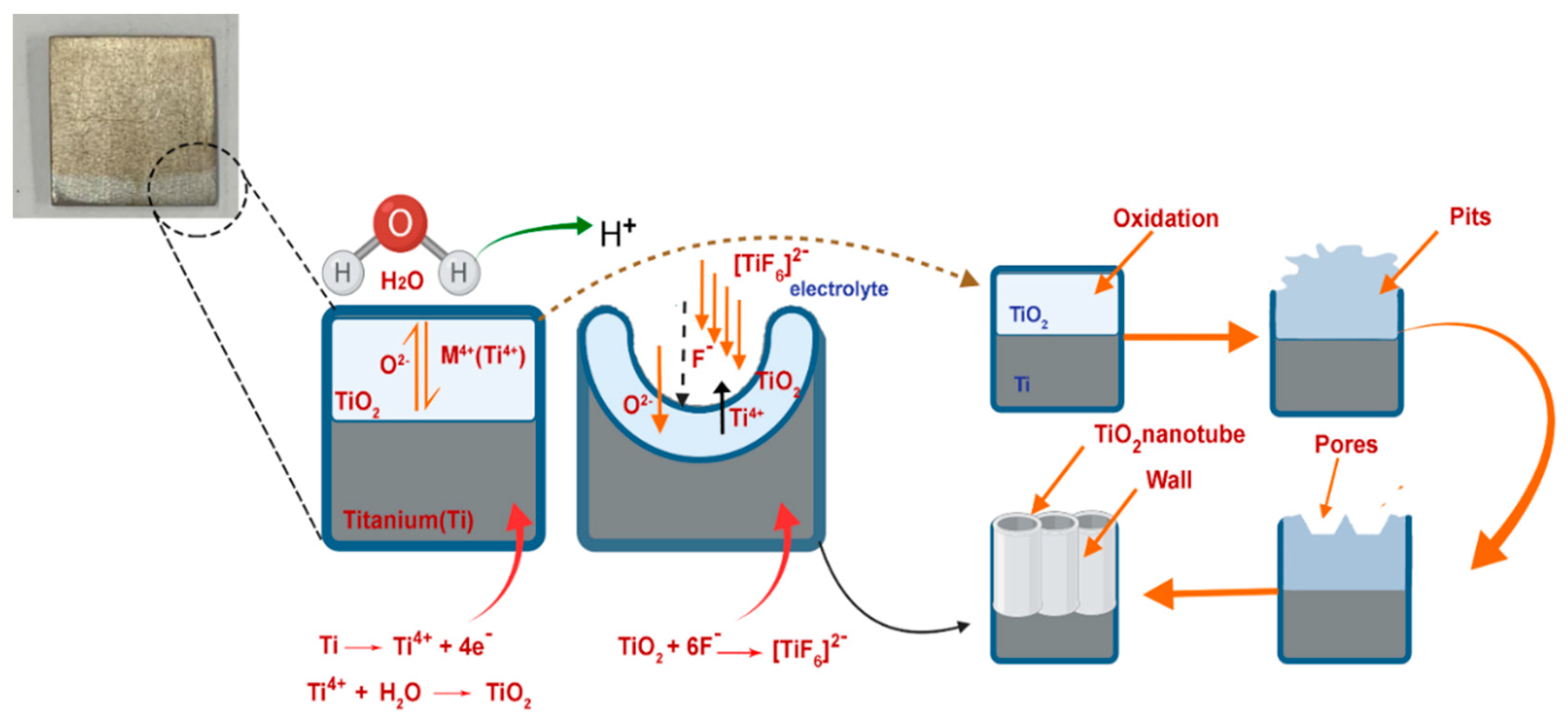
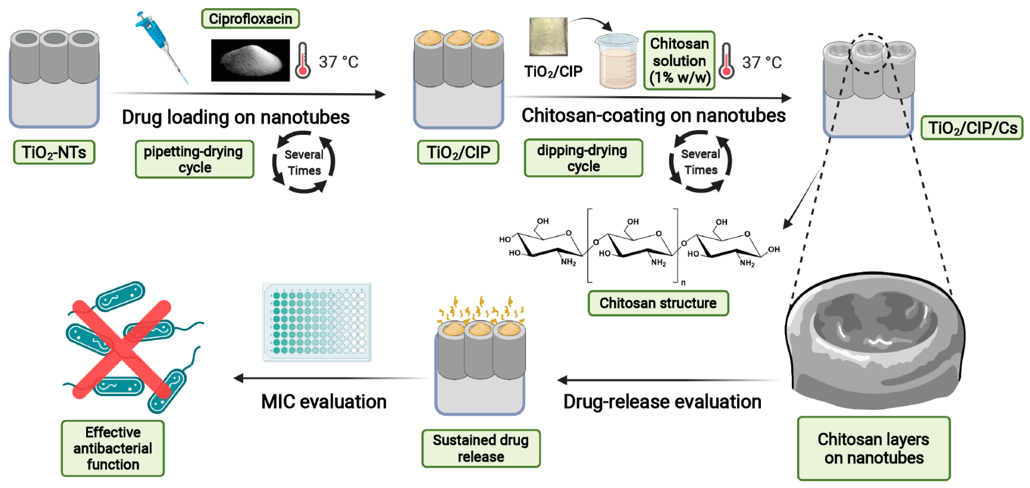
2.2. Synthesis of TiO2-NTs
2.3. Characterization of TiO2/CIP/Cs
2.4. The Effect of Electrolyte Water Percentage on the Morphology of TiO2-NTs
2.5. Drug Encapsulation and Polymer Coating on the TiO2-NTs
2.6. Investigation of the Effect of Chitosan Layer Thickness on Release Behavior
2.7. Investigation of Drug Release from Polymer-Coated TiO2-NTs
2.8. Kinetic of Release
2.8.1. Zero-Order Model
2.8.2. First-Order Model
2.8.3. Higuchi Model
2.8.4. Korsmeyer–Peppas Kinetics
2.9. Antibacterial Experiment
2.9.1. Bacterial Strains
2.9.2. Determination of Minimum Inhibitory Concentrations (MICs)
2.9.3. Determination of Minimum Bactericidal Concentration (MBCs)
2.9.4. Assessment of Biofilm Formation
2.10. Hemolysis Assay
2.11. Statistical Analysis
3. Results
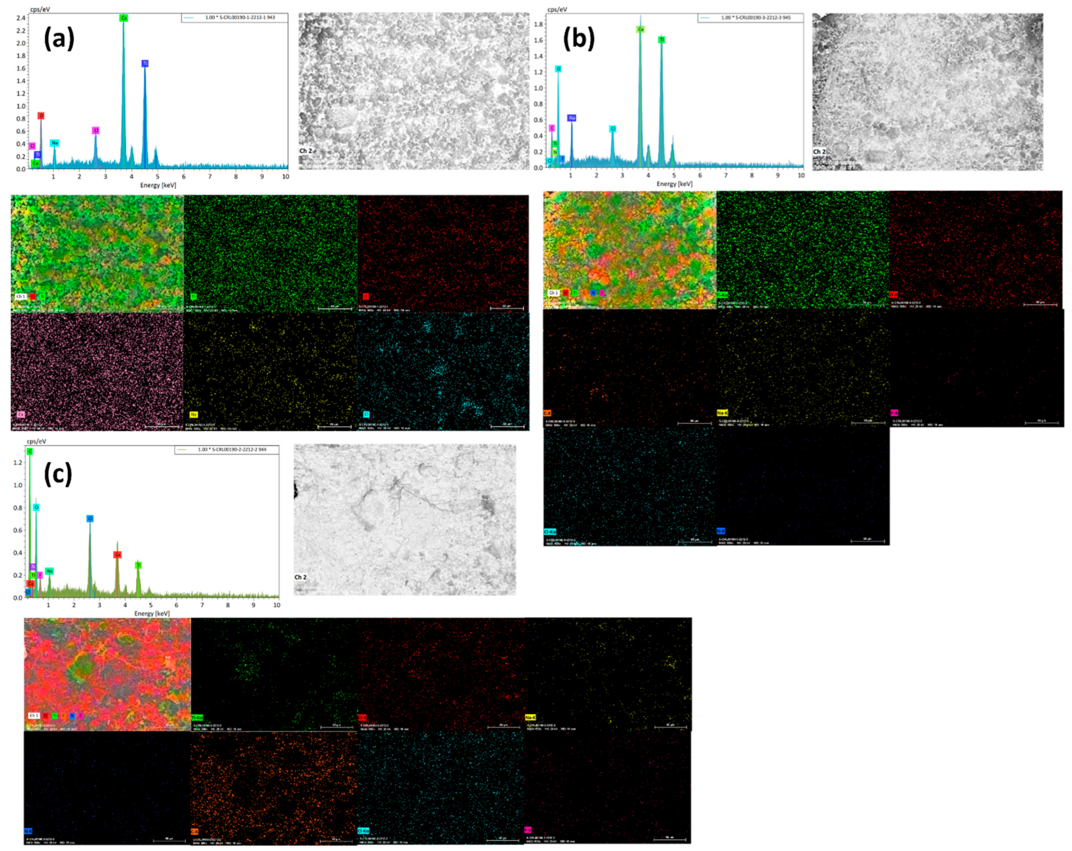
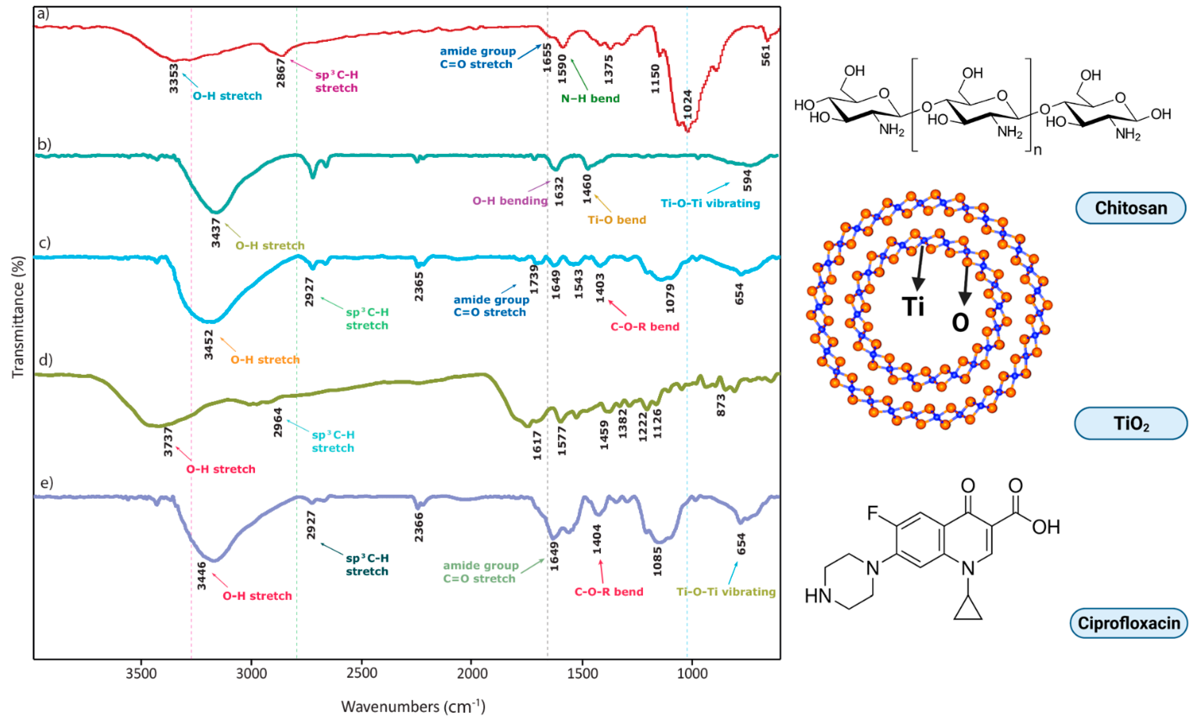
3.1. Characterization of TiO2/CIP/Cs
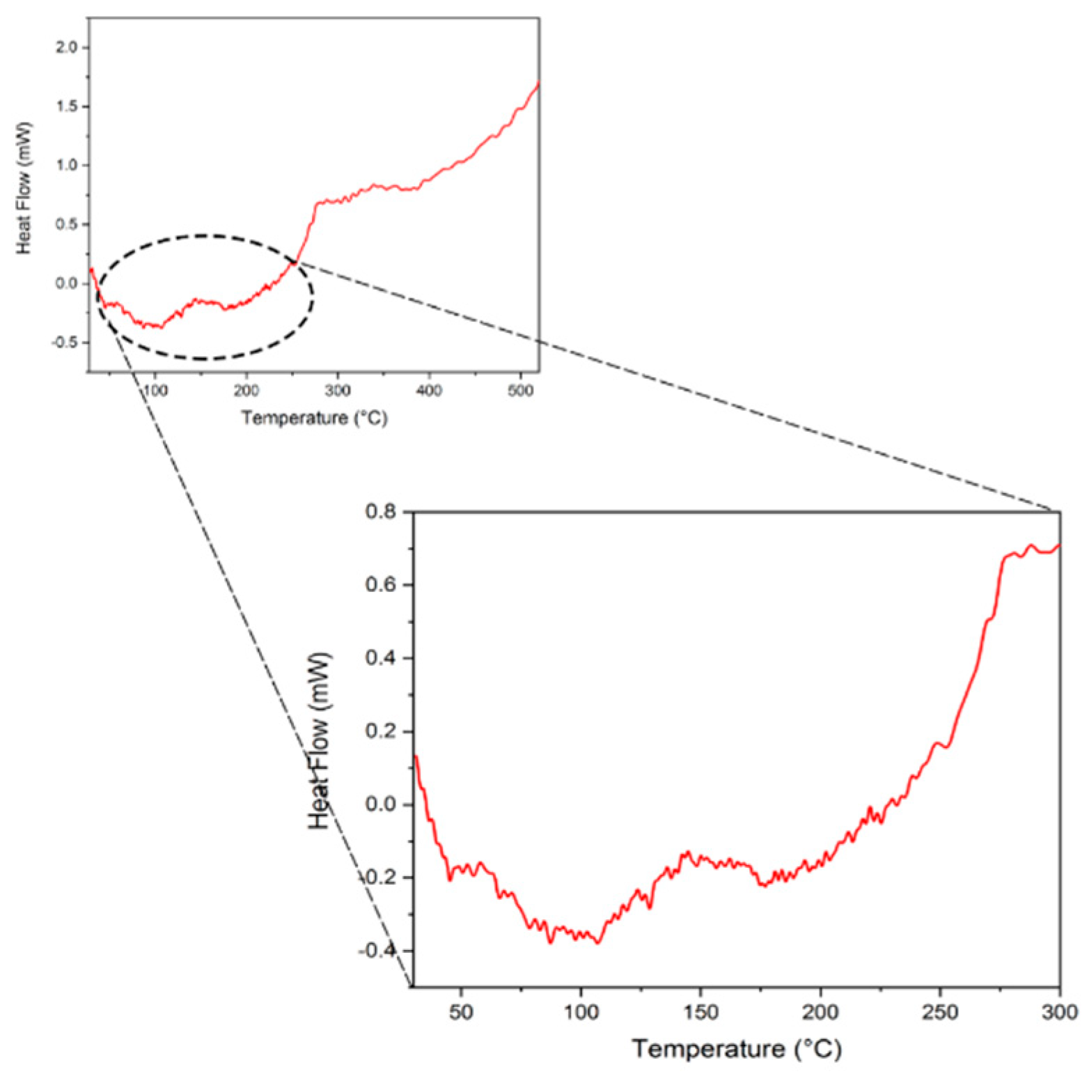
3.2. DSC Analysis
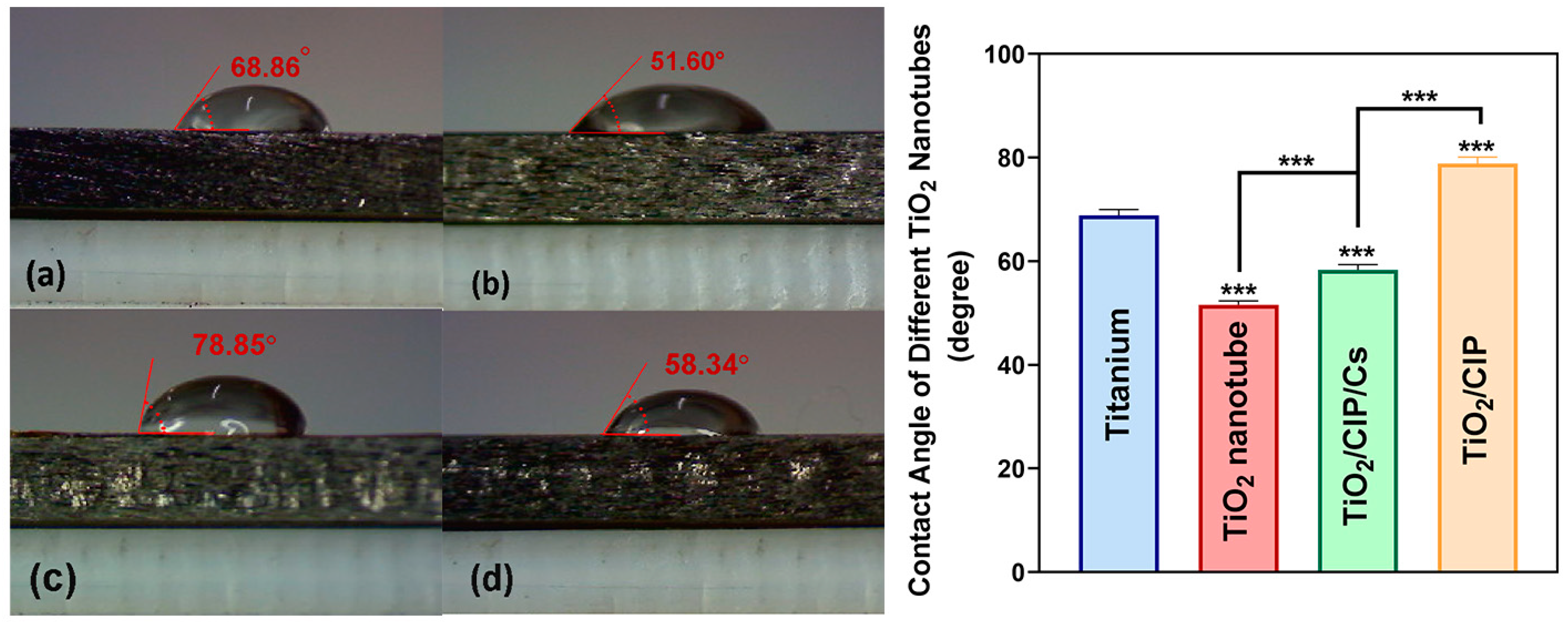
3.3. Contact Angle
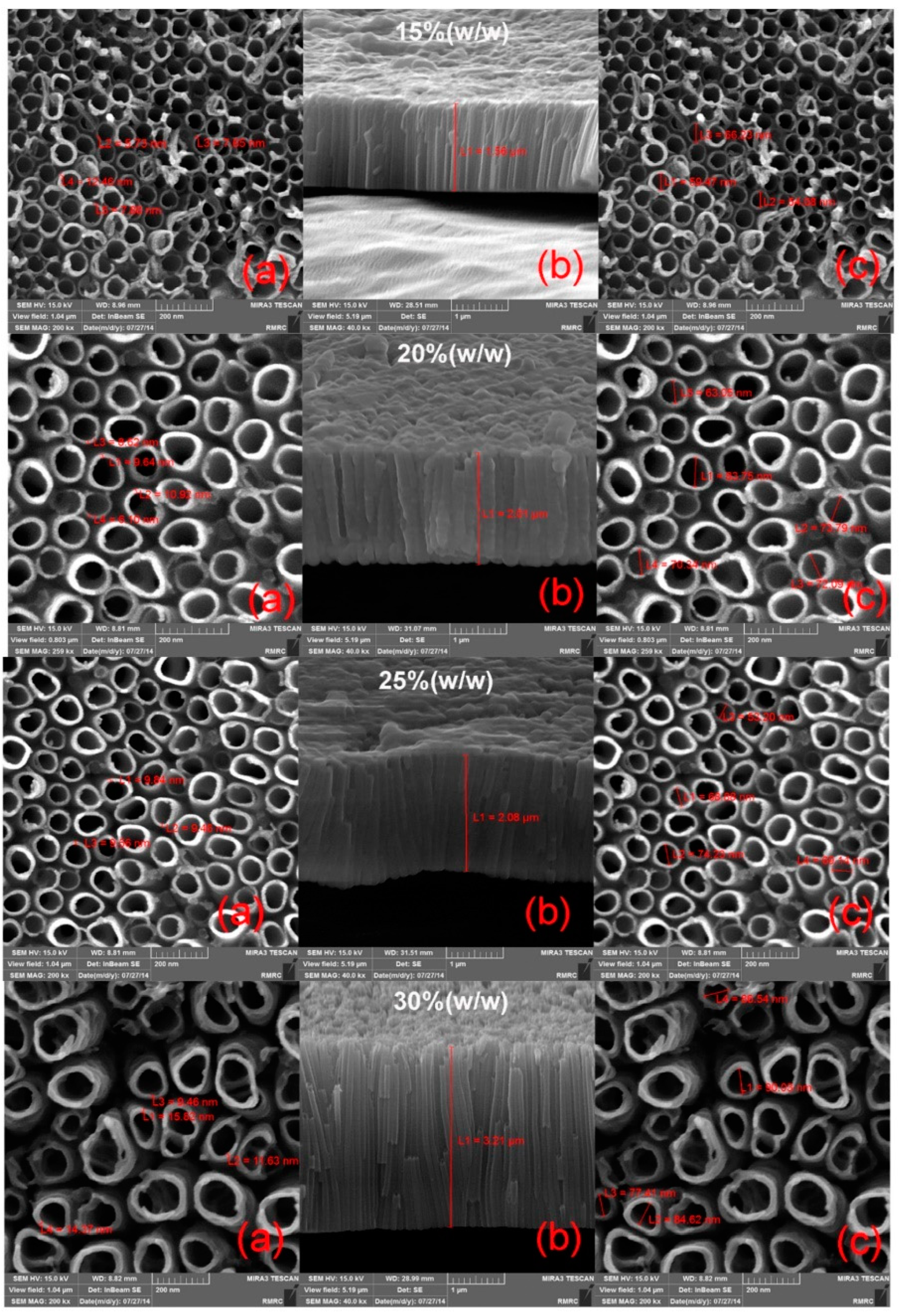
3.4. Effect of Water Percentage of Electrolyte on Morphology
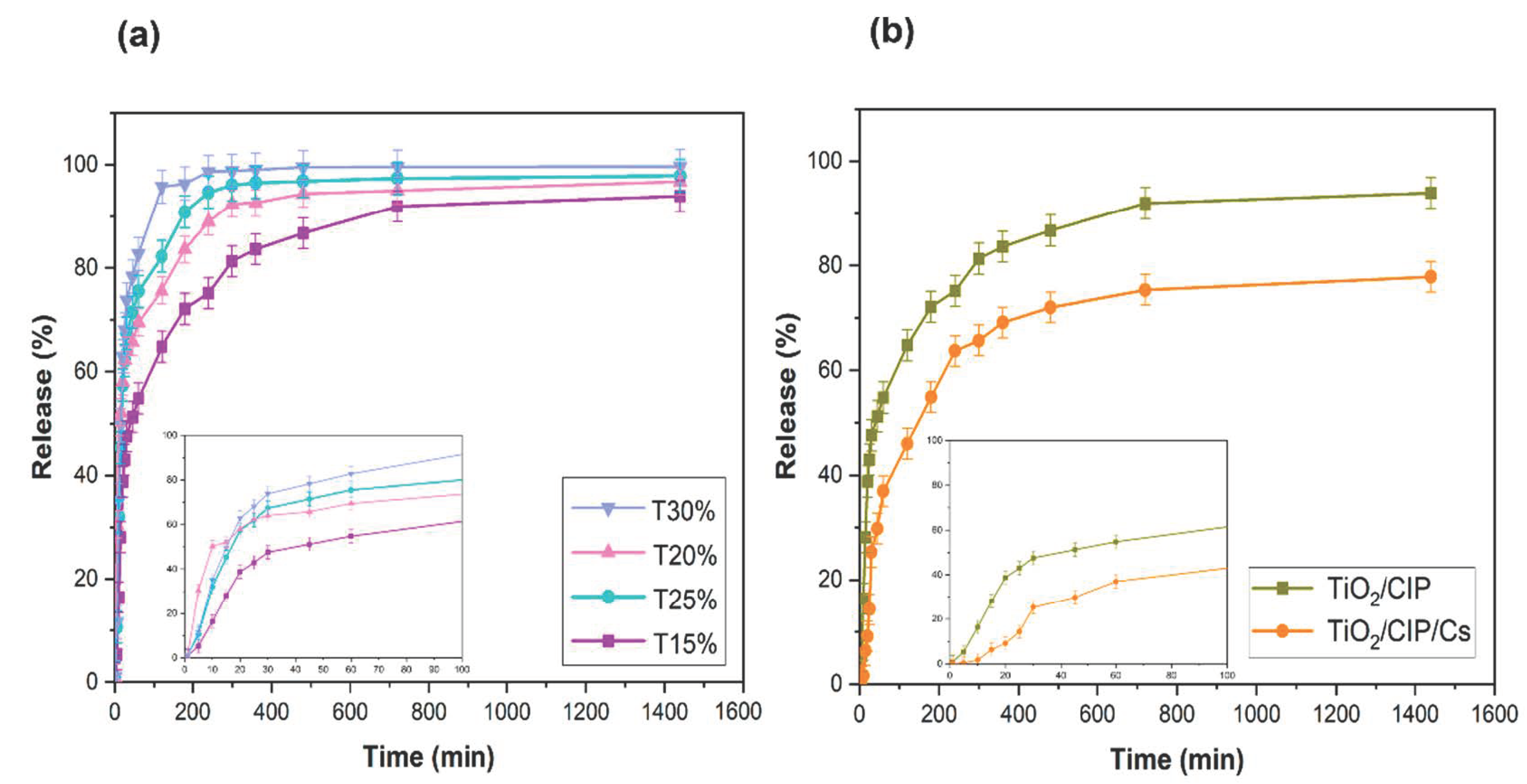
3.5. Drug Release from TiO2-NTs
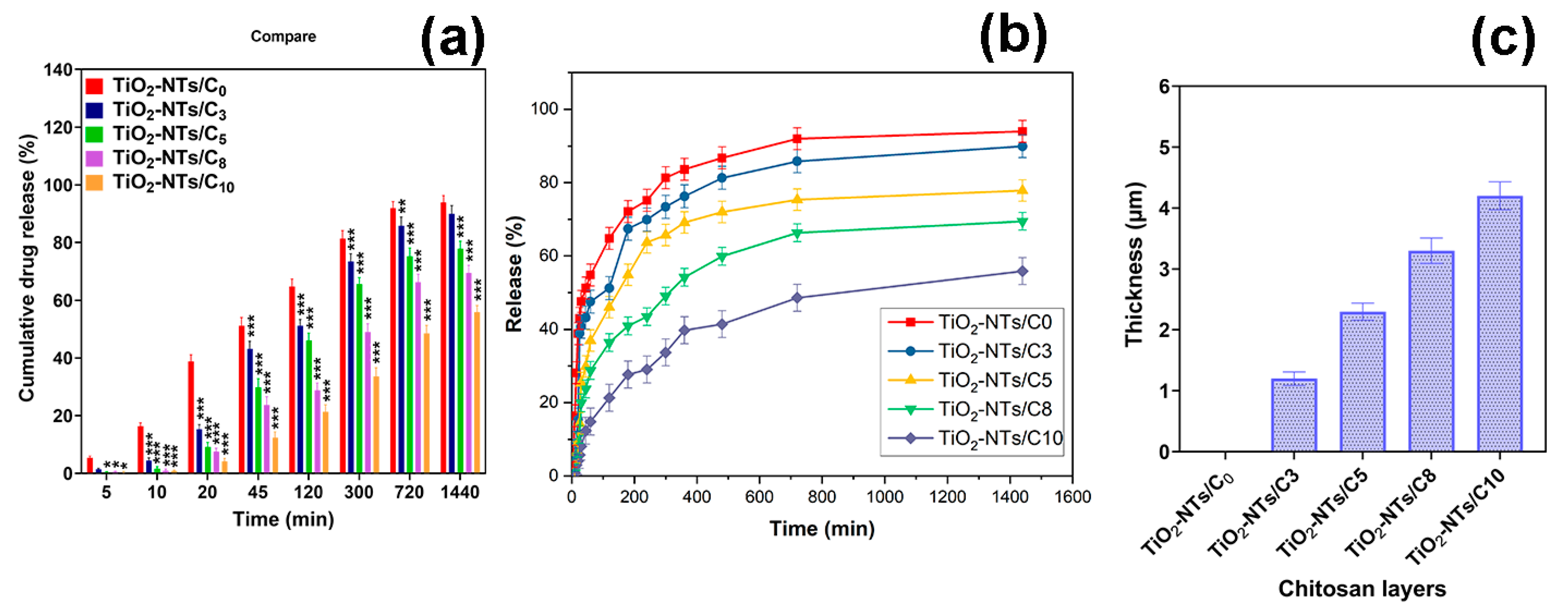
3.6. Investigation of Chitosan-Coating Thickness on Release Behavior
3.7. Kinetic of Release
3.8. Antibacterial Properties
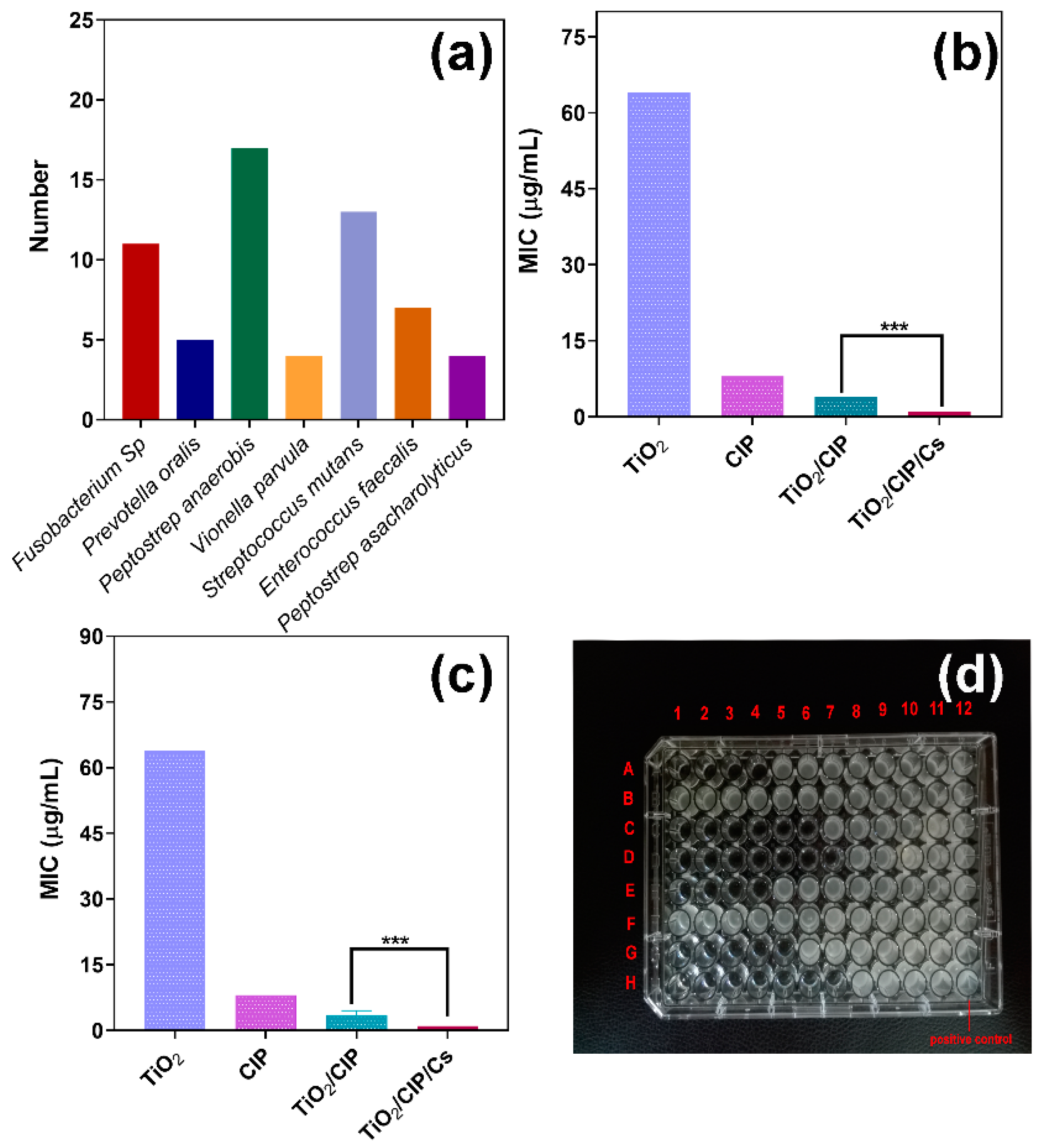
3.8.1. Determination of Minimum Inhibitory Concentrations (MICs)
3.8.2. Investigation of Minimum Bactericidal Concentrations (MBCs)
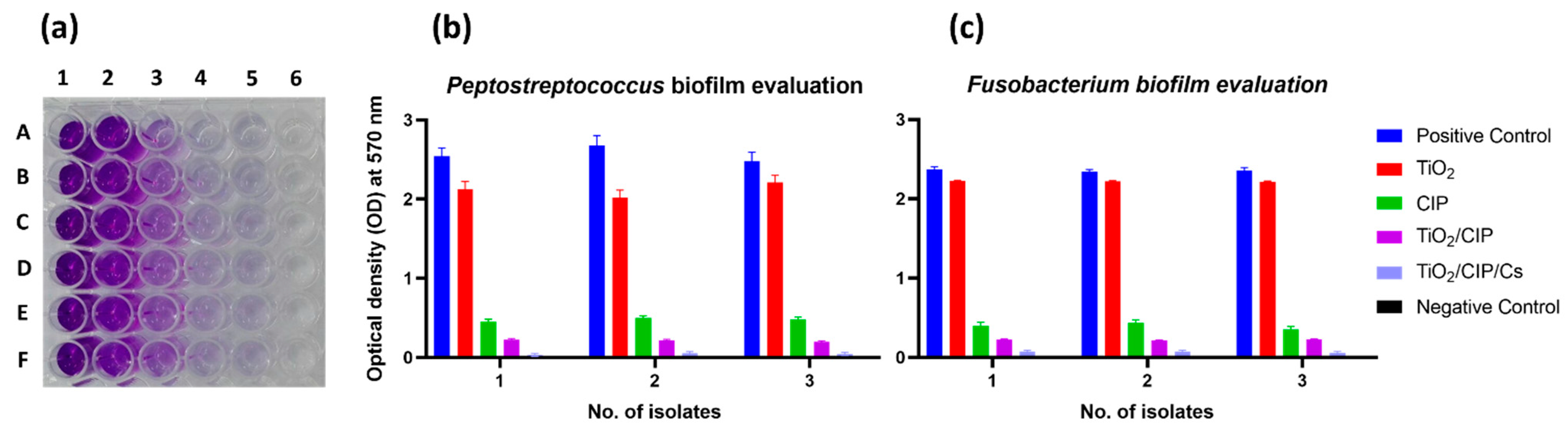
3.8.3. Effect of TiO2-NTs on Biofilm Formation
3.8.4. Mechanism of Bactericidal Effect
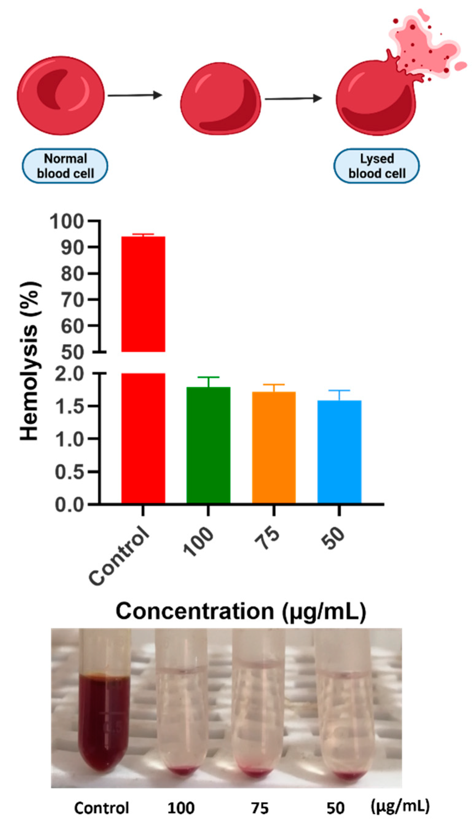
3.9. Hemocompatability Properties
4. Discussion
5. Conclusions
Supplementary Materials
Author Contributions
Funding
Institutional Review Board Statement
Informed Consent Statement
Data Availability Statement
Acknowledgments
Conflicts of Interest
References
- Zhang, L.; Niu, X.; Sun, L.; She, Z.; Tan, R.; Wang, W. Immune response of bovine sourced cross-linked collagen sponge for hemostasis. J. Biomater. Appl. 2018, 32, 920–931. [Google Scholar] [CrossRef] [PubMed]
- Yang, F.; Niu, X.; Gu, X.; Xu, C.; Wang, W.; Fan, Y. Biodegradable Magnesium-Incorporated Poly(l-lactic acid) Microspheres for Manipulation of Drug Release and Alleviation of Inflammatory Response. ACS Appl. Mater. Interfaces 2019, 11, 23546–23557. [Google Scholar] [CrossRef] [PubMed]
- Gao, A.; Hang, R.; Chu, P.K. Recent advances in anti-infection surfaces fabricated on biomedical implants by plasma-based technology. Surf. Coatings Technol. 2017, 312, 2–6. [Google Scholar] [CrossRef]
- Niu, X.; Liu, Z.; Hu, J.; Rambhia, K.J.; Fan, Y.; Ma, P.X. Microspheres Assembled from Chitosan-Graft-Poly(lactic acid) Micelle-Like Core-Shell Nanospheres for Distinctly Controlled Release of Hydrophobic and Hydrophilic Biomolecules. Macromol. Biosci. 2016, 16, 1039–1047. [Google Scholar] [CrossRef] [Green Version]
- Besinis, A.; Hadi, S.D.; Le, H.R.; Tredwin, C.; Handy, R.D. Antibacterial activity and biofilm inhibition by surface modified titanium alloy medical implants following application of silver, titanium dioxide and hydroxyapatite nanocoatings. Nanotoxicology 2017, 11, 327–338. [Google Scholar] [CrossRef] [Green Version]
- Alavi, S.E.; Esfahani, M.K.M.; Raza, A.; Adelnia, H.; Shahmabadi, H.E. PEG-grafted liposomes for enhanced antibacterial and antibiotic activities: An in vivo study. NanoImpact 2022, 25, 100384. [Google Scholar] [CrossRef]
- Joseph, R.R.; Venkatraman, S.S. Drug delivery to the eye: What benefits do nanocarriers offer? Nanomedicine 2017, 12, 683–702. [Google Scholar] [CrossRef] [PubMed] [Green Version]
- Patra, J.K.; Das, G.; Fraceto, L.F.; Campos, E.V.R.; del Pilar Rodriguez-Torres, M.; Acosta-Torres, L.S.; Diaz-Torres, L.A.; Grillo, R.; Swamy, M.K.; Sharma, S.; et al. Nano based drug delivery systems: Recent developments and future prospects. J. Nanobiotechnol. 2018, 16, 71. [Google Scholar] [CrossRef] [Green Version]
- Peng, Z.; Ni, J. Surface properties and bioactivity of TiO2 nanotube array prepared by two-step anodic oxidation for biomedical applications. R. Soc. Open Sci. 2019, 6, 181948. [Google Scholar] [CrossRef] [Green Version]
- Fatima, S.; Ali, K.; Ahmed, B.; Al Kheraif, A.A.; Syed, A.; Elgorban, A.M.; Musarrat, J.; Lee, J. Titanium Dioxide Nanoparticles Induce Inhibitory Effects against Planktonic Cells and Biofilms of Human Oral Cavity Isolates of Rothia mucilaginosa, Georgenia sp. and Staphylococcus saprophyticus. Pharmaceutics 2021, 13, 1564. [Google Scholar] [CrossRef]
- Soonnarong, R.; Tungsukruthai, S.; Nutho, B.; Rungrotmongkol, T.; Vinayanuwattikun, C.; Maluangnont, T.; Chanvorachote, P. Titania Nanosheet Generates Peroxynitrite-Dependent S-Nitrosylation and Enhances p53 Function in Lung Cancer Cells. Pharmaceutics 2021, 13, 1233. [Google Scholar] [CrossRef] [PubMed]
- Vighetto, V.; Racca, L.; Canta, M.; Matos, J.C.; Dumontel, B.; Gonçalves, M.C.; Cauda, V. Smart Shockwave Responsive Titania-Based Nanoparticles for Cancer Treatment. Pharmaceutics 2021, 13, 1423. [Google Scholar] [CrossRef] [PubMed]
- Xu, J.; Zhou, X.; Gao, Z.; Song, Y.-Y.; Schmuki, P. Visible-Light-Triggered Drug Release from TiO2 Nanotube Arrays: A Controllable Antibacterial Platform. Angew. Chem. 2016, 128, 603–607. [Google Scholar] [CrossRef]
- Kafshgari, M.H.; Kah, D.; Mazare, A.; Nguyen, N.T.; Distaso, M.; Peukert, W.; Goldmann, W.H.; Schmuki, P.; Fabry, B. Anodic Titanium Dioxide Nanotubes for Magnetically Guided Therapeutic Delivery. Sci. Rep. 2019, 9, 13439. [Google Scholar] [CrossRef] [Green Version]
- Song, Y.-Y.; Schmidt-Stein, F.; Bauer, S.; Schmuki, P. Amphiphilic TiO2 Nanotube Arrays: An Actively Controllable Drug Delivery System. J. Am. Chem. Soc. 2009, 131, 4230–4232. [Google Scholar] [CrossRef]
- Lai, Y.-K.; Wang, Q.; Huang, J.-Y.; Li, H.-Q.; Chen, Z.; Zhao, A.Z.-J.; Wang, Y.; Zhang, K.-Q.; Sun, H.-T.; Al-Deyab, S.S. TiO2 nanotube platforms for smart drug delivery: A review. Int. J. Nanomed. 2016, 11, 4819–4834. [Google Scholar] [CrossRef] [Green Version]
- Jin, M.; Yao, S.; Wang, L. Surface Modification of Metallic Implants with Nanotubular Arrays via Electrochemical Anodization. In Nanobiomaterials; John Wiley & Sons: Hoboken, NJ, USA, 2017; pp. 211–238. [Google Scholar] [CrossRef]
- A, L.; Xu, W.; Zhao, J.; Li, C.; Qi, M.; Li, X.; Wang, L.; Zhou, Y. Surface functionalization of TiO2 nanotubes with minocycline and its in vitro biological effects on Schwann cells. Biomed. Eng. Online 2018, 17, 88. [Google Scholar] [CrossRef] [Green Version]
- Li, H.; Cui, Q.; Feng, B.; Wang, J.; Lu, X.; Weng, J. Antibacterial activity of TiO2 nanotubes: Influence of crystal phase, morphology and Ag deposition. Appl. Surf. Sci. 2013, 284, 179–183. [Google Scholar] [CrossRef]
- Yao, C.; Webster, T.J. Prolonged antibiotic delivery from anodized nanotubular titanium using a co-precipitation drug loading method. J. Biomed. Mater. Res. Part B Appl. Biomater. 2009, 91, 587–595. [Google Scholar] [CrossRef]
- Goodman, S.B.; Yao, Z.; Keeney, M.; Yang, F. The future of biologic coatings for orthopaedic implants. Biomaterials 2013, 34, 3174–3183. [Google Scholar] [CrossRef] [Green Version]
- Feng, W.; Geng, Z.; Li, Z.; Cui, Z.; Zhu, S.; Liang, Y.; Liu, Y.; Wang, R.; Yang, X. Controlled release behaviour and antibacterial effects of antibiotic-loaded titania nanotubes. Mater. Sci. Eng. C 2016, 62, 105–112. [Google Scholar] [CrossRef] [PubMed]
- Niu, X.; Sun, L.; Zhang, X.; Sun, Y.; Wang, J. Fabrication and antibacterial properties of cefuroxime-loaded TiO2 nanotubes. Appl. Microbiol. Biotechnol. 2020, 104, 2947–2955. [Google Scholar] [CrossRef] [PubMed]
- Tong, S.; Sun, X.; Wu, A.; Guo, S.; Zhang, H. Improved Biocompatibility of TiO2 Nanotubes via Co-Precipitation Loading with Hydroxyapatite and Gentamicin. Coatings 2021, 11, 1191. [Google Scholar] [CrossRef]
- Ramu, C.; Padmanabhan, T.V. Indications of antibiotic prophylaxis in dental practice–Review. Asian Pac. J. Trop. Biomed. 2012, 2, 749–754. [Google Scholar] [CrossRef] [Green Version]
- Thabit, A.K.; Fatani, D.F.; Bamakhrama, M.S.; Barnawi, O.A.; Basudan, L.O.; Alhejaili, S.F. Antibiotic penetration into bone and joints: An updated review. Int. J. Infect. Dis. 2019, 81, 128–136. [Google Scholar] [CrossRef] [Green Version]
- Jikia, D.; Chkhaidze, N.; Imedashvili, E.; Mgaloblishvili, I.; Tsitlanadze, G.; Katsarava, R.; Glenn Morris, J., Jr.; Sulakvelidze, A. The use of a novel biodegradable preparation capable of the sustained release of bacteriophages and ciprofloxacin, in the complex treatment of multidrug-resistant Staphylococcus aureus-infected local radiation injuries caused by exposure to Sr90. Clin. Exp. Dermatol. 2005, 30, 23–26. [Google Scholar] [CrossRef]
- Suter, C.; Leemann, H.; Twerenbold, R. “Ciprofloxacin-induced” bilateral quadriceps tendon rupture: A case report and conclusions of the recent literature. Trauma Case Rep. 2021, 32, 100423. [Google Scholar] [CrossRef]
- Schlüter, G. Ciprofloxacin: Toxicologic evaluation of additional safety data. Am. J. Med. 1989, 87, S37–S39. [Google Scholar] [CrossRef]
- Ibrahim, N.A.; Elmorshedy, K.E.; Radwan, D.A.; Buabeid, M.A. The impact of oral ciprofloxacin on the structure and functions of rat gastric mucosa. Saudi J. Biol. Sci. 2021, 29, 2187–2198. [Google Scholar] [CrossRef]
- Berhe, A.; Russom, M.; Bahran, F.; Hagos, G. Ciprofloxacin and risk of hypolycemia in non-diabetic patients. J. Med. Case Rep. 2019, 13, 142. [Google Scholar] [CrossRef] [Green Version]
- Mohammed, M.A.; Syeda, J.T.M.; Wasan, K.M.; Wasan, E.K. An Overview of Chitosan Nanoparticles and Its Application in Non-Parenteral Drug Delivery. Pharmaceutics 2017, 9, 53. [Google Scholar] [CrossRef] [PubMed] [Green Version]
- Mohan, L.; Anandan, C.; Rajendran, N. Drug release characteristics of quercetin-loaded TiO2 nanotubes coated with chitosan. Int. J. Biol. Macromol. 2016, 93, 1633–1638. [Google Scholar] [CrossRef] [PubMed]
- Hashemi, A.; Ezati, M.; Mohammadnejad, J.; Houshmand, B.; Faghihi, S. Chitosan Coating of TiO2 Nanotube Arrays for Improved Metformin Release and Osteoblast Differentiation. Int. J. Nanomed. 2020, 15, 4471–4481. [Google Scholar] [CrossRef] [PubMed]
- Ahmadi, S.; Mohammadi, I.; Sadrnezhaad, S. Hydroxyapatite based and anodic Titania nanotube biocomposite coatings: Fabrication, characterization and electrochemical behavior. Surf. Coatings Technol. 2016, 287, 67–75. [Google Scholar] [CrossRef]
- Fray, D.J. Novel methods for the production of titanium. Int. Mater. Rev. 2013, 53, 317–325. [Google Scholar] [CrossRef]
- Zhang, G.; Zhang, L.; Yang, D.; Zhang, N.; He, L.; Du, G.; Lu, Y. Salt screening and characterization of ciprofloxacin. Acta Crystallogr. Sect. B Struct. Sci. Cryst. Eng. Mater. 2016, 72, 20–28. [Google Scholar] [CrossRef]
- Kaushik, S.N.; Scoffield, J.; Andukuri, A.; Alexander, G.C.; Walker, T.; Kim, S.; Choi, S.C.; Brott, B.C.; Eleazer, P.D.; Lee, J.-Y.; et al. Evaluation of ciprofloxacin and metronidazole encapsulated biomimetic nanomatrix gel on Enterococcus faecalis and Treponema denticola. Biomater. Res. 2015, 19, 9. [Google Scholar] [CrossRef] [Green Version]
- Newman, M.G. Anaerobic Oral and Dental Infection. Clin. Infect. Dis. 1984, 6, S107–S114. [Google Scholar] [CrossRef]
- Giti, R.; Zomorodian, K.; Firouzmandi, M.; Zareshahrabadi, Z.; Rahmannasab, S. Antimicrobial Activity of Thermocycled Polymethyl Methacrylate Resin Reinforced with Titanium Dioxide and Copper Oxide Nanoparticles. Int. J. Dent. 2021, 2021, 6690806. [Google Scholar] [CrossRef]
- Iga, C.; Agata, T.; Marcin; Natalia, F.; Justyna, K.-L. Ciprofloxacin-Modified Degradable Hybrid Polyurethane-Polylactide Porous Scaffolds Developed for Potential Use as an Antibacterial Scaffold for Regeneration of Skin. Polymers 2020, 12, 171. [Google Scholar] [CrossRef] [Green Version]
- Dostert, K.-H.; O’Brien, C.P.; Mirabella, F.; Ivars-Barceló, F.; Schauermann, S. Adsorption of acrolein, propanal, and allyl alcohol on Pd(111): A combined infrared reflection–absorption spectroscopy and temperature programmed desorption study. Phys. Chem. Chem. Phys. 2016, 18, 13960–13973. [Google Scholar] [CrossRef] [PubMed] [Green Version]
- Roy, S.; Priyadarshi, R.; Rhim, J.-W. Development of Multifunctional Pullulan/Chitosan-Based Composite Films Reinforced with ZnO Nanoparticles and Propolis for Meat Packaging Applications. Foods 2021, 10, 2789. [Google Scholar] [CrossRef] [PubMed]
- Fernandes Queiroz, M.; Melo, K.R.T.; Sabry, D.A.; Sassaki, G.L.; Rocha, H.A.O. Does the Use of Chitosan Contribute to Oxalate Kidney Stone Formation? Mar. Drugs 2015, 13, 141–158. [Google Scholar] [CrossRef]
- Lagat, M.K.; Were, S.; Ndwigah, F.; Kemboi, V.J.; Kipkoech, C.; Tanga, C.M. Antimicrobial Activity of Chemically and Biologically Treated Chitosan Prepared from Black Soldier Fly (Hermetia illucens) Pupal Shell Waste. Microorganisms 2021, 9, 2417. [Google Scholar] [CrossRef]
- Shamsipur, M.; Gholivand, M.B.; Dehdashtian, S.; Feyzi, M.; Jafari, F. Synthesis of Co/TiO2 Nanocomposite and its Use in Construction of a Sensitive and Selective Sensor for Determination of Ciprofloxacin. Adv. Mater. Res. 2014, 829, 563–567. [Google Scholar] [CrossRef]
- Manalu, S.P.; Natarajan, T.S.; De Guzman, M.; Wang, Y.-F.; Chang, T.-C.; Yen, F.-C.; You, S.-J. Synthesis of ternary g-C3N4/Bi2MoO6/TiO2 nanotube composite photocatalysts for the decolorization of dyes under visible light and direct sunlight irradiation. Green Process. Synth. 2018, 7, 493–505. [Google Scholar] [CrossRef]
- Dey, S.C.; Al-Amin, M.; Rashid, T.U.; Sultan, M.Z.; Ashaduzzaman, M.; Sarker, M.; Shamsuddin, S.M. Preparation, Characterization and Performance Evaluation of Chitosan as an Adsorbent for Remazol Red. Int. J. Latest Res. Eng. Technol. 2016, 2, 52–62. [Google Scholar]
- Sullo, A.; Norton, I. Food Colloids and Emulsions. In Encyclopedia of Food and Health; Elsevier: Amsterdam, The Netherlands, 2015; pp. 7–15. [Google Scholar] [CrossRef]
- Mohsenzadeh, A.; Fazel, A.; Bavari, S.; Borji, S.; Pourasghar, S.; Azimi, T.; Sabati, H. Detecting of biofilm formation in the clinical isolates of Pseudomonas aeruginosa and Escherichia coli: An evaluation of different screening methods. J. Curr. Biomed. Rep. 2021, 2, 56–61. [Google Scholar] [CrossRef]
- Tang, T.; Peng, Z.; Ni, J.; Zheng, K.; Shen, Y.; Wang, X.; He, G.; Jin, S. Dual effects and mechanism of TiO2 nanotube arrays in reducing bacterial colonization and enhancing C3H10T1/2 cell adhesion. Int. J. Nanomed. 2013, 8, 3093–3105. [Google Scholar] [CrossRef] [Green Version]
- Pietsch, F.; Bergman, J.M.; Brandis, G.; Marcusson, L.L.; Zorzet, A.; Huseby, D.; Hughes, D. Ciprofloxacin selects for RNA polymerase mutations with pleiotropic antibiotic resistance effects. J. Antimicrob. Chemother. 2017, 72, 75–84. [Google Scholar] [CrossRef] [Green Version]
- De Smet, J.; Wagemans, J.; Boon, M.; Ceyssens, P.-J.; Voet, M.; Noben, J.-P.; Andreeva, J.; Ghilarov, D.; Severinov, K.; Lavigne, R. The bacteriophage LUZ24 “Igy” peptide inhibits the Pseudomonas DNA gyrase. Cell Rep. 2021, 36, 109567. [Google Scholar] [CrossRef] [PubMed]
- Duan, C.; Meng, X.; Meng, J.; Khan, I.H.; Dai, L.; Khan, A.; An, X.; Zhang, J.; Huq, T.; Ni, Y. Chitosan as A Preservative for Fruits and Vegetables: A Review on Chemistry and Antimicrobial Properties. J. Bioresour. Bioprod. 2019, 4, 11–21. [Google Scholar] [CrossRef]
- Zhang, H.; Yu, S.; Tian, A.; Lin, W.; Ali, A.; Xue, X.X.; Bai, X.Z. Improved antibacterial activity and biocompatibility on vancomycin-loaded TiO2 nanotubes: In vivo and in vitro studies. Int. J. Nanomed. 2013, 8, 4379–4389. [Google Scholar] [CrossRef] [Green Version]
- Jafari, S.; Mahyad, B.; Hashemzadeh, H.; Janfaza, S.; Gholikhani, T.; Tayebi, L. Biomedical Applications of TiO2 Nanostructures: Recent Advances. Int. J. Nanomed. 2020, 15, 3447–3470. [Google Scholar] [CrossRef]
- Anitha, V.C.; Lee, J.-H.; Lee, J.; Banerjee, A.N.; Joo, S.W.; Min, B.K. Biofilm formation on a TiO2nanotube with controlled pore diameter and surface wettability. Nanotechnology 2015, 26, 065102. [Google Scholar] [CrossRef]
- Gold, K.; Slay, B.; Knackstedt, M.; Gaharwar, A.K. Antimicrobial Activity of Metal and Metal-Oxide Based Nanoparticles. Adv. Ther. 2018, 1, 1700033. [Google Scholar] [CrossRef]
- Zhao, J.; Xu, J.; Jian, X.; Xu, J.; Gao, Z.; Song, Y.-Y. NIR Light-Driven Photocatalysis on Amphiphilic TiO2 Nanotubes for Controllable Drug Release. ACS Appl. Mater. Interfaces 2020, 12, 23606–23616. [Google Scholar] [CrossRef]
- Wang, Z.; Jin, S.; Zhang, F.; Wang, D. Combined Toxicity of TiO2 Nanospherical Particles and TiO2 Nanotubes to Two Microalgae with Different Morphology. Nanomaterials 2020, 10, 2559. [Google Scholar] [CrossRef]
- Yan, H.; Liu, L.; Wang, R.; Zhu, W.; Ren, X.; Luo, L.; Zhang, X.; Luo, S.; Ai, X.; Wang, J. Binary composite MoS2/TiO2 nanotube arrays as a recyclable and efficient photocatalyst for solar water disinfection. Chem. Eng. J. 2020, 401, 126052. [Google Scholar] [CrossRef]
- Yeganeh, F.E.; Yeganeh, A.E.; Yousefi, M.; Far, B.F.; Akbarzadeh, I.; Bokov, D.O.; Raahemifar, K.; Soltani, M. Formulation and Characterization of Poly (Ethylene Glycol)-Coated Core-Shell Methionine Magnetic Nanoparticles as a Carrier for Naproxen Delivery: Growth Inhibition of Cancer Cells. Cancers 2022, 14, 1797. [Google Scholar] [CrossRef]
- Khatoon, Z.; McTiernan, C.D.; Suuronen, E.J.; Mah, T.-F.; Alarcon, E.I. Bacterial biofilm formation on implantable devices and approaches to its treatment and prevention. Heliyon 2018, 4, e01067. [Google Scholar] [CrossRef] [Green Version]
- Harawaza, K.; Cousins, B.; Roach, P.; Fernandez, A. Modification of the surface nanotopography of implant devices: A translational perspective. Mater. Today Bio 2021, 12, 100152. [Google Scholar] [CrossRef]
- Pokrowiecki, R. The paradigm shift for drug delivery systems for oral and maxillofacial implants. Drug Deliv. 2018, 25, 1504–1515. [Google Scholar] [CrossRef] [Green Version]
- Zheng, S.; Bawazir, M.; Dhall, A.; Kim, H.-E.; He, L.; Heo, J.; Hwang, G. Implication of Surface Properties, Bacterial Motility, and Hydrodynamic Conditions on Bacterial Surface Sensing and Their Initial Adhesion. Front. Bioeng. Biotechnol. 2021, 9, 82. [Google Scholar] [CrossRef]
- Luo, X.; Matranga, C.; Tan, S.; Alba, N.; Cui, X.T. Carbon nanotube nanoreservior for controlled release of anti-inflammatory dexamethasone. Biomaterials 2011, 32, 6316–6323. [Google Scholar] [CrossRef] [Green Version]
- Chennell, P.; Feschet-Chassot, E.; Devers, T.; Awitor, K.; Descamps, S.; Sautou, V. In vitro evaluation of TiO2 nanotubes as cefuroxime carriers on orthopaedic implants for the prevention of periprosthetic joint infections. Int. J. Pharm. 2013, 455, 298–305. [Google Scholar] [CrossRef]
- Bai, K.; Hong, B.; Huang, W.; He, J. Selenium-Nanoparticles-Loaded Chitosan/Chitooligosaccharide Microparticles and Their Antioxidant Potential: A Chemical and In Vivo Investigation. Pharmaceutics 2020, 12, 43. [Google Scholar] [CrossRef] [Green Version]
- Mandal, A.; Bisht, R.; Rupenthal, I.D.; Mitra, A.K. Polymeric micelles for ocular drug delivery: From structural frameworks to recent preclinical studies. J. Control. Release 2017, 248, 96–116. [Google Scholar] [CrossRef] [Green Version]
- Dang, M.; Saunders, L.; Niu, X.; Fan, Y.; Ma, P.X. Biomimetic delivery of signals for bone tissue engineering. Bone Res. 2018, 6, 25. [Google Scholar] [CrossRef]
- Rigo, S.; Cai, C.; Gunkel-Grabole, G.; Maurizi, L.; Zhang, X.; Xu, J.; Palivan, C.G. Nanoscience-Based Strategies to Engineer Antimicrobial Surfaces. Adv. Sci. 2018, 5, 1700892. [Google Scholar] [CrossRef]
- Lee, M.-J.; Kwon, J.-S.; Jiang, H.B.; Choi, E.H.; Park, G.; Kim, K.-M. The antibacterial effect of non-thermal atmospheric pressure plasma treatment of titanium surfaces according to the bacterial wall structure. Sci. Rep. 2019, 9, 1938. [Google Scholar] [CrossRef] [Green Version]
- Mirzaie, A.; Peirovi, N.; Akbarzadeh, I.; Moghtaderi, M.; Heidari, F.; Yeganeh, F.E.; Noorbazargan, H.; Mirzazadeh, S.; Bakhtiari, R. Preparation and optimization of ciprofloxacin encapsulated niosomes: A new approach for enhanced antibacterial activity, biofilm inhibition and reduced antibiotic resistance in ciprofloxacin-resistant methicillin-resistance Staphylococcus aureus. Bioorg. Chem. 2020, 103, 104231. [Google Scholar] [CrossRef] [PubMed]


| Sample | Element | Mass Percent (%) | Atom Percent (%) | abs. Error [%] (1 Sigma) |
|---|---|---|---|---|
| TiO2-NTs | O | 41.34 | 64.29 | 8.82 |
| Ti | 27.94 | 14.52 | 0.85 | |
| Ca | 23.20 | 14.40 | 0.72 | |
| Na | 3.96 | 4.28 | 0.38 | |
| Cl | 3.57 | 2.50 | 0.19 | |
| TiO2/CIP | O | 44.39 | 58.30 | 9.23 |
| C | 10.36 | 18.12 | 2.93 | |
| Ti | 22.88 | 10.04 | 0.82 | |
| Ca | 16.20 | 8.49 | 0.62 | |
| Na | 3.99 | 3.65 | 0.39 | |
| Cl | 2.01 | 1.19 | 0.13 | |
| F | 0.19 | 0.21 | 0.31 | |
| TiO2/CIP/Cs | C | 40.71 | 51.99 | 10.03 |
| O | 33.82 | 32.43 | 9.21 | |
| N | 5.05 | 5.53 | 3.59 | |
| F | 4.91 | 3.97 | 2.39 | |
| Ca | 5.37 | 2.06 | 0.31 | |
| Cl | 3.92 | 1.70 | 0.25 | |
| Ti | 5.22 | 1.67 | 0.32 | |
| Na | 1.00 | 0.67 | 0.16 |
| Water Content (w/w%) | 15 | 20 | 25 | 30 |
|---|---|---|---|---|
| Code | T15% | T20% | T25% | T30% |
| Nanotube wall thickness (nm) | 5.92 | 8.77 | 9.53 | 15.8 |
| Inner diameter of nanotubes (nm) | 59.92 | 72.64 | 66.3 | 84.61 |
| Nanotube length (µm) | 1.56 | 2.01 | 2.08 | 3.21 |
| Sample | Entrapment Efficiency (EE)% |
|---|---|
| T15% (w/w) | 83.19 ± 1.68 |
| T20% (w/w) | 83.70 ± 1.77 |
| T25% (w/w) | 86.48 ± 1.4 |
| T30% (w/w) | 92.95 ± 2.25 |
| Samples | Chitosan Layers | Thickness (µm) | Release Profile (%) | EE (%) | ||||||||
|---|---|---|---|---|---|---|---|---|---|---|---|---|
| 5 min | 10 min | 20 min | 45 min | 120 min | 300 min | 720 min | 1440 min | |||||
| TiO2 NTs/C0 | 0 | mean | 0 | 5.35 | 16.38 | 38.8 | 51.23 | 64.76 | 81.32 | 91.94 | 93.96 | 83.19 |
| SD | 0 | 0.65 | 1.21 | 2.34 | 2.79 | 2.66 | 2.85 | 2.31 | 2.35 | 1.12 | ||
| TiO2-NTs/C3 | 3 | mean | 1.2 | 1.42 | 4.41 | 15.32 | 43.17 | 51.23 | 73.4 | 85.83 | 89.9 | 87.68 |
| SD | 0.12 | 0.35 | 0.92 | 1.58 | 2.65 | 2.15 | 2.75 | 3.05 | 2.93 | 1.85 | ||
| TiO2-NTs/C5 | 5 | mean | 2.3 | 0.4 | 1.63 | 9.15 | 29.82 | 46.02 | 65.68 | 75.32 | 77.8 | 90.22 |
| SD | 0.23 | 0.22 | 0.68 | 1.59 | 2.99 | 2.64 | 2.21 | 2.8 | 2.56 | 2.59 | ||
| TiO2-NTs/C8 | 8 | mean | 3.3 | 0.47 | 0.95 | 7.52 | 23.74 | 28.77 | 49.05 | 66.31 | 69.45 | 92.04 |
| SD | 0.16 | 0.35 | 0.58 | 1.25 | 2.89 | 2.6 | 2.85 | 2.75 | 2.64 | 1.48 | ||
| TiO2-NTs /C10 | 10 | mean | 4.2 | 0.18 | 0.87 | 4.1 | 12.36 | 21.29 | 33.67 | 48.6 | 55.83 | 93.97 |
| SD | 0.24 | 0.54 | 0.3 | 1.05 | 2.1 | 2.54 | 2.94 | 2.73 | 2.37 | 1.21 | ||
| Release Model | Equation | R2 | |
|---|---|---|---|
| without Chitosan (A) | with Chitosan (B) | ||
| Zero-order | Ct = C0 + K0t | R2 = 0.4159 | R2 = 0.5444 |
| Korsmeyer-Peppas | Mt/M∞ = Ktn | R2 = 0.5542 N = 0.3953 | R2 = 0.6811 N = 0.5234 |
| First-order | LogC = LogC0 + Kt/2.303 | R2 = 0.8667 | R2 = 0.6565 |
| Higuchi | R2 = 0.6821 | R2 = 0.8018 | |
| Sources. | Strain | CIP (µg/mL) | TiO2 (µg/mL) | TiO2/CIP (µg/mL) | TiO2/CIP/Cs (µg/mL) |
|---|---|---|---|---|---|
| 1 | Peptostreptococcus | 8 ± 0 | 64 ± 0 | 2 ± 0 | 1 ± 0 |
| 2 | Peptostreptococcus | 8 ± 0 | 64 ± 0 | 2 ± 0 | 1 ± 0 |
| 3 | Peptostreptococcus | 8 ± 0 | 64 ± 0 | 2 ± 0 | 1 ± 0 |
| 4 | Peptostreptococcus | 8 ± 0 | 64 ± 0 | 2 ± 0 | 1 ± 0 |
| 5 | Peptostreptococcus | 8 ± 0 | 64 ± 0 | 2 ± 0 | 1 ± 0 |
| 6 | Peptostreptococcus | 8 ± 0 | 64 ± 0 | 2 ± 0 | 1 ± 0 |
| 1 | Fusobacterium | 8 ± 0 | 64 ± 0 | 4 ± 0 | 1 ± 0 |
| 2 | Fusobacterium | 8 ± 0 | 64 ± 0 | 4 ± 0 | 1 ± 0 |
| 3 | Fusobacterium | 8 ± 0 | 64 ± 0 | 4 ± 0 | 1 ± 0 |
| Bacterial Strain | Sample | MIC (μg/mL) | MBC (Pour Plate) (μg/mL) |
|---|---|---|---|
| Fusobacterium | TiO2-NTs | 64 | 64 |
| CIP | 8 | 16 | |
| TiO2/CIP | 2 | 2 | |
| TiO2/CIP/Cs | 1 | 2 | |
| Pepto streptococcus | TiO2-NTs | 64 | 64 |
| CIP | 8 | 8 | |
| TiO2/CIP | 4 | 4 | |
| TiO2/CIP/Cs | 1 | 1 |
Publisher’s Note: MDPI stays neutral with regard to jurisdictional claims in published maps and institutional affiliations. |
© 2022 by the authors. Licensee MDPI, Basel, Switzerland. This article is an open access article distributed under the terms and conditions of the Creative Commons Attribution (CC BY) license (https://creativecommons.org/licenses/by/4.0/).
Share and Cite
Asadi, S.; Mortezagholi, B.; Hadizadeh, A.; Borisov, V.; Ansari, M.J.; Shaker Majdi, H.; Nishonova, A.; Adelnia, H.; Farasati Far, B.; Chaiyasut, C. Ciprofloxacin-Loaded Titanium Nanotubes Coated with Chitosan: A Promising Formulation with Sustained Release and Enhanced Antibacterial Properties. Pharmaceutics 2022, 14, 1359. https://doi.org/10.3390/pharmaceutics14071359
Asadi S, Mortezagholi B, Hadizadeh A, Borisov V, Ansari MJ, Shaker Majdi H, Nishonova A, Adelnia H, Farasati Far B, Chaiyasut C. Ciprofloxacin-Loaded Titanium Nanotubes Coated with Chitosan: A Promising Formulation with Sustained Release and Enhanced Antibacterial Properties. Pharmaceutics. 2022; 14(7):1359. https://doi.org/10.3390/pharmaceutics14071359
Chicago/Turabian StyleAsadi, Soada, Bardia Mortezagholi, Alireza Hadizadeh, Vitaliy Borisov, Mohammad Javed Ansari, Hasan Shaker Majdi, Azizakhon Nishonova, Hossein Adelnia, Bahareh Farasati Far, and Chaiyavat Chaiyasut. 2022. "Ciprofloxacin-Loaded Titanium Nanotubes Coated with Chitosan: A Promising Formulation with Sustained Release and Enhanced Antibacterial Properties" Pharmaceutics 14, no. 7: 1359. https://doi.org/10.3390/pharmaceutics14071359