

Biosynthesized Gold, Silver, Palladium, Platinum, Copper, and Other Transition Metal Nanoparticles

 , , ,
, , ,
Abstract
:1. Introduction
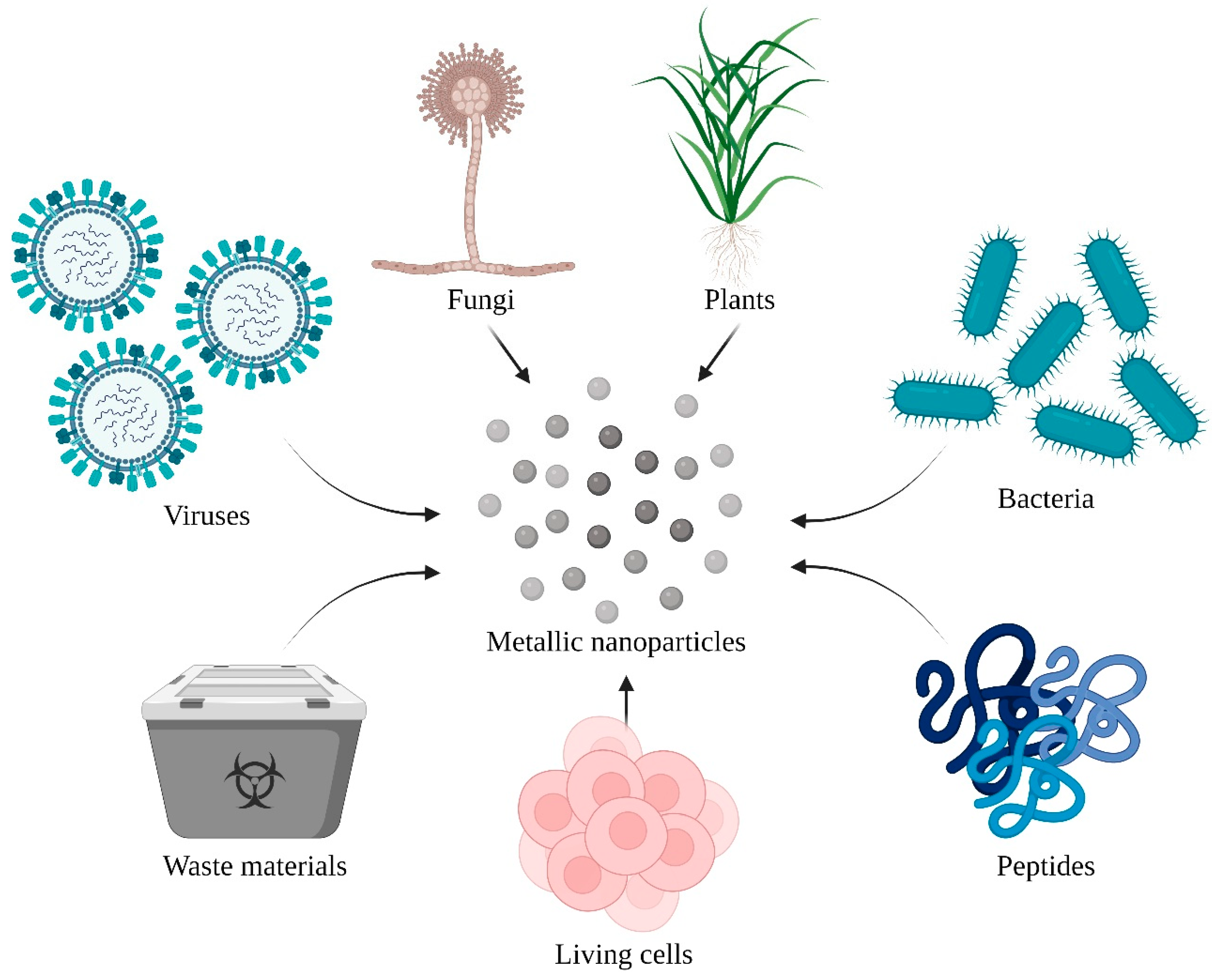
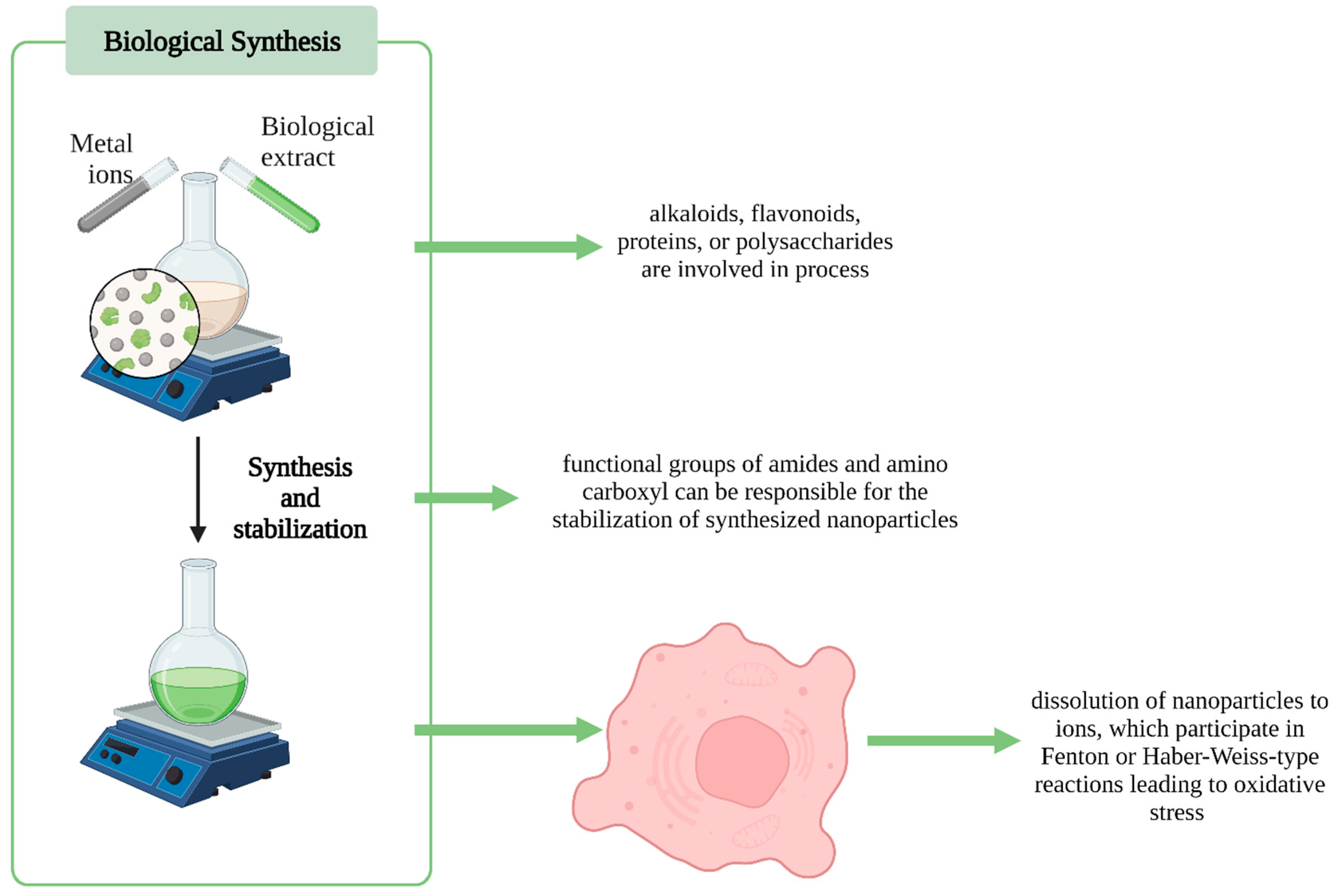
2. Nanoparticle Synthesis and Analysis
3. Gold Nanoparticles
3.1. Anticancer Activity
3.1.1. Plants
3.1.2. Fungi
3.1.3. Pure Substances
3.2. Antibacterial Activity
Bacteria
3.3. Multidirectional Activity
Waste Materials
3.4. Toxicity
Bacteria
4. Silver Nanoparticles
4.1. Anticancer Activity
Plants
4.2. Antibacterial Activity
Waste Materials
4.3. Alternative Biomedical Applications
Plants
4.4. Multidirectional Activity
Plants
4.5. Toxicity
Plants
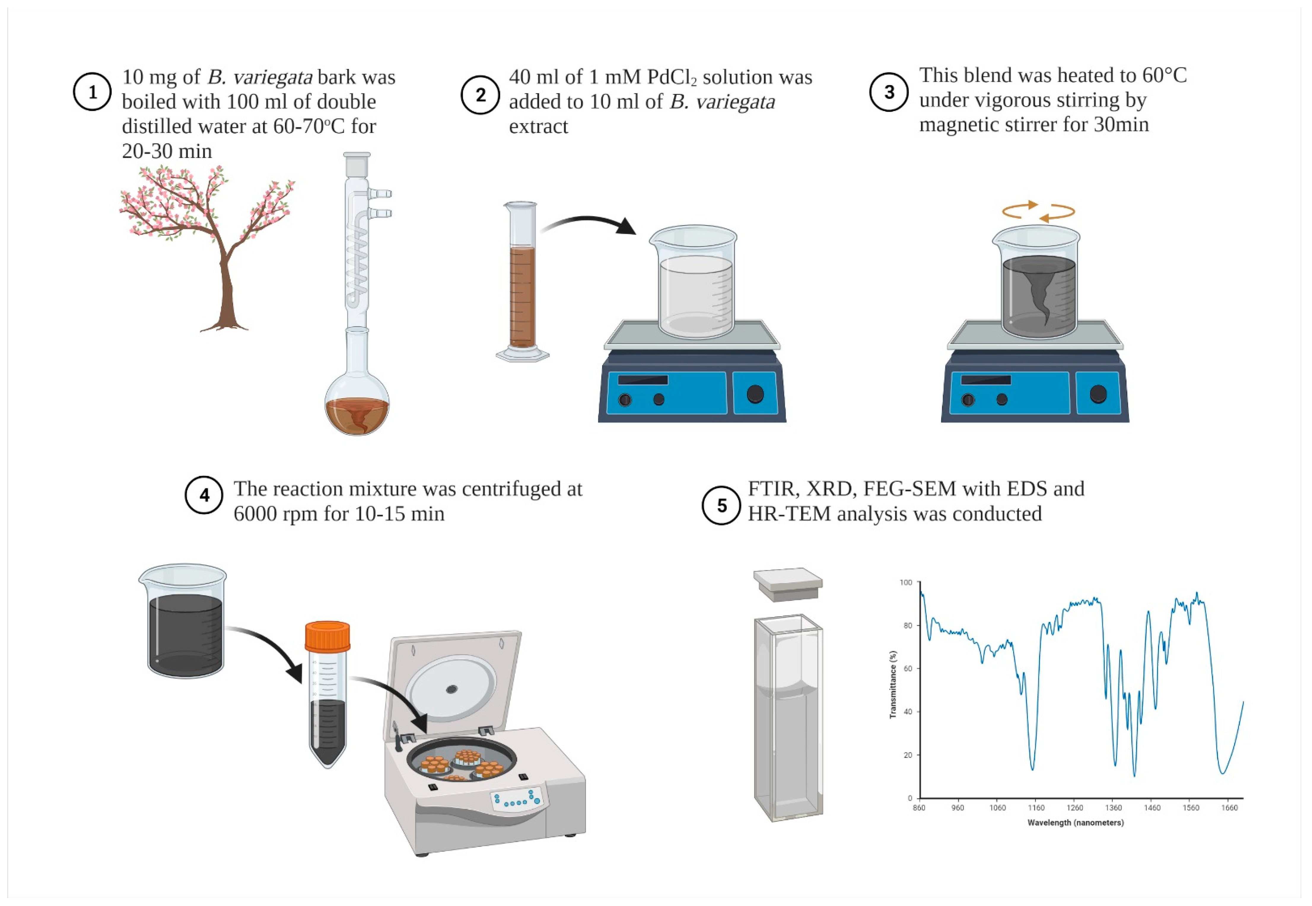
5. Palladium Nanoparticles
5.1. Anticancer Activity
Plants
5.2. Antibacterial Activity
Plants
5.3. Alternative Biomedical Applications
Plants
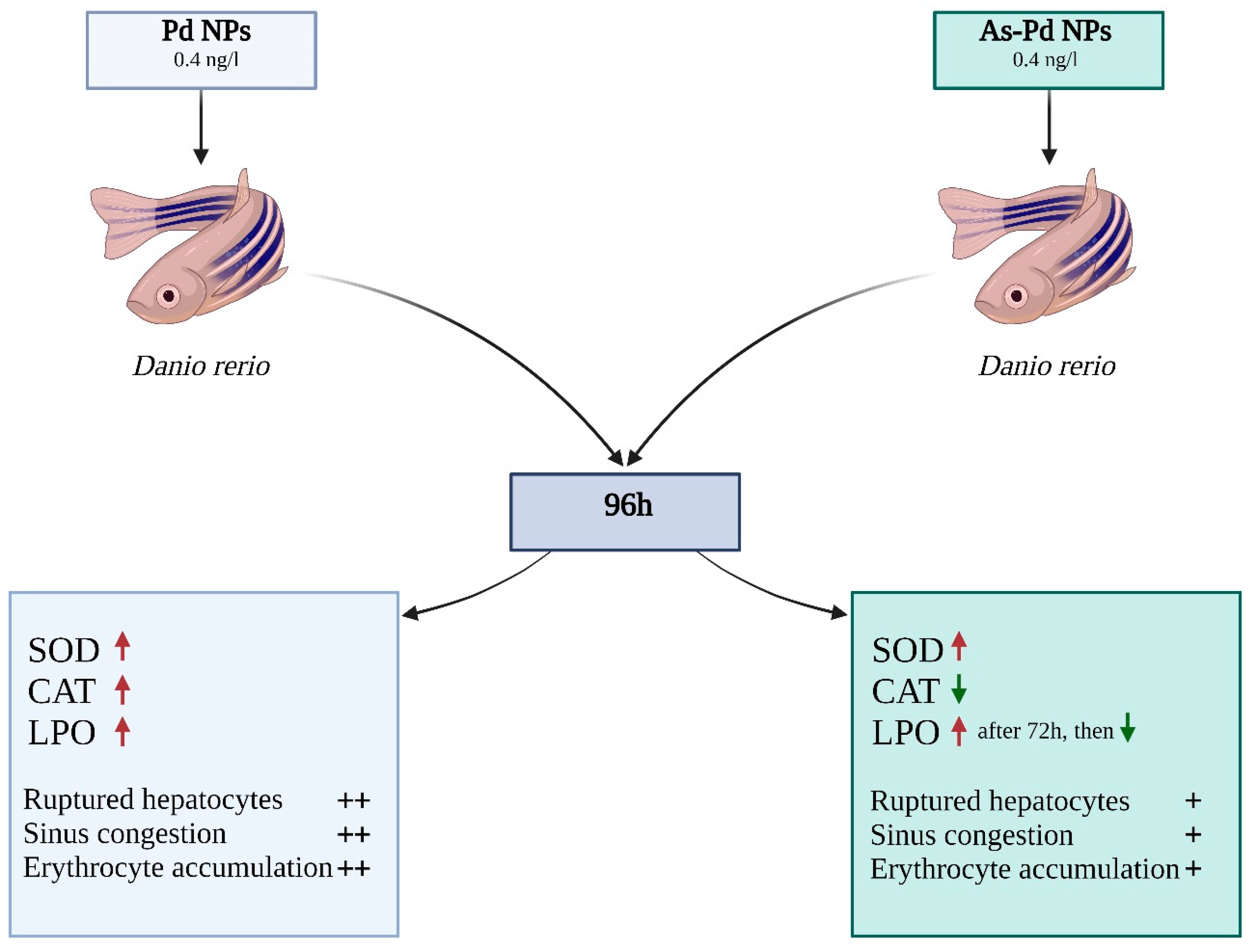
5.4. Toxicity
Plants
6. Platinum Nanoparticles
6.1. Anticancer Activity
Waste Materials
6.2. Antibacterial Activity
Waste Materials
6.3. Alternative Biomedical Applications
Plants
6.4. Toxicity
Plants
7. Copper Nanoparticles
7.1. Anticancer Activity
Plants
7.2. Antibacterial Activity
Plants
7.3. Alternative Biomedical Applications
Plants
8. Other Transition Metal Nanoparticles
8.1. Anticancer Activity
8.1.1. Plants
8.1.2. Bacteria
8.2. Antibacterial Activity
8.2.1. Plants
8.2.2. Bacteria
8.3. Alternative Biomedical Applications
Plants
9. Conclusions
Author Contributions
Funding
Institutional Review Board Statement
Informed Consent Statement
Data Availability Statement
Conflicts of Interest
References
- Gozzi, M.; Schwarze, B.; Hey-Hawkins, E. Preparing (Metalla)carboranes for Nanomedicine. ChemMedChem 2021, 16, 1533–1565. [Google Scholar] [CrossRef]
- Chugh, H.; Sood, D.; Chandra, I.; Tomar, V.; Dhawan, G.; Chandra, R. Role of gold and silver nanoparticles in cancer. Artif. Cells Nanomed. Biotechnol. 2018, 46, S1210–S1220. [Google Scholar] [CrossRef]
- Khan, A.U.; Malik, N.; Khan, M.; Cho, M.H.; Khan, M.M. Fungi-assisted silver nanoparticle synthesis and their applications. Bioprocess Biosyst. Eng. 2018, 41, 1–20. [Google Scholar] [CrossRef] [PubMed]
- Czarnomysy, R.; Radomska, D.; Szewczyk, O.K.; Roszczenko, P.; Bielawski, K. Platinum and palladium complexes as promising sources for antitumor treatments. Int. J. Mol. Sci. 2021, 22, 8271. [Google Scholar] [CrossRef] [PubMed]
- Tweedle, M.F. Alternatives to Gadolinium-Based Contrast Agents. Investig. Radiol. 2021, 56, 35–41. [Google Scholar] [CrossRef]
- Zeng, X.; Sun, J.; Li, S.; Shi, J.; Gao, H.; Sun Leong, W.; Wu, Y.; Li, M.; Liu, C.; Li, P.; et al. Blood-triggered generation of platinum nanoparticle functions as an anti-cancer agent. Nat. Commun. 2020, 11, 567. [Google Scholar] [CrossRef] [Green Version]
- Clegg, J.R.; Irani, A.S.; Ander, E.W.; Ludolph, C.M.; Venkataraman, A.K.; Zhong, J.X.; Peppas, N.A. Synthetic networks with tunable responsiveness, biodegradation, and molecular recognition for precision medicine applications. Sci. Adv. 2019, 5, eaax7946. [Google Scholar] [CrossRef] [PubMed] [Green Version]
- Suma, P.R.; Padmanabhan, R.A.; Telukutla, S.R.; Ravindran, R.; Velikkakath, A.K.G.; Dekiwadia, C.D.; Paul, W.; Laloraya, M.; Srinivasula, S.M.; Bhosale, S.V.; et al. Vanadium pentoxide nanoparticle mediated perturbations in cellular redox balance and the paradigm of autophagy to apoptosis. Free Radic. Biol. Med. 2020, 161, 198–211. [Google Scholar] [CrossRef] [PubMed]
- Szewczyk, O.K.; Roszczenko, P.; Czarnomysy, R.; Bielawska, A.; Bielawski, K. An Overview of the Importance of Transition-Metal Nanoparticles in Cancer Research. Int. J. Mol. Sci. 2022, 23, 6688. [Google Scholar] [CrossRef] [PubMed]
- Zaimy, M.A.; Saffarzadeh, N.; Mohammadi, A.; Pourghadamyari, H.; Izadi, P.; Sarli, A.; Moghaddam, L.K.; Paschepari, S.R.; Azizi, H.; Torkamandi, S.; et al. New methods in the diagnosis of cancer and gene therapy of cancer based on nanoparticles. Cancer Gene Ther. 2017, 24, 233–243. [Google Scholar] [CrossRef] [PubMed]
- Ferlay, J.; Ervik, M.; Lam, F.; Colombet, M.; Mery, L. Global Cancer Observatory: Cancer Today. Lyon: International Agency for Research on Cancer. 2020. Available online: https://gco.iarc.fr/today (accessed on 10 February 2022).
- Mba, I.E.; Nweze, E.I. Nanoparticles as therapeutic options for treating multidrug-resistant bacteria: Research progress, challenges, and prospects. World J. Microbiol. Biotechnol. 2021, 37, 108. [Google Scholar] [CrossRef] [PubMed]
- Fu, J.; Wang, Y.; Zhang, J.; Yuan, K.; Yan, J.; Yuan, B.; Guan, Y.; Wang, M. The safety and efficacy of transarterial chemoembolisation with bleomycin for hepatocellular carcinoma unresponsive to doxorubicin: A prospective single-centre study. Clin. Radiol. 2021, 76, 864.e7–864.e12. [Google Scholar] [CrossRef] [PubMed]
- Baindara, P.; Mandal, S.M. Bacteria and bacterial anticancer agents as a promising alternative for cancer therapeutics. Biochimie 2020, 177, 164–189. [Google Scholar] [CrossRef] [PubMed]
- Mitchell, M.J.; Billingsley, M.M.; Haley, R.M.; Wechsler, M.E.; Peppas, N.A.; Langer, R. Engineering precision nanoparticles for drug delivery. Nat. Rev. Drug Discov. 2021, 20, 101–124. [Google Scholar] [CrossRef]
- Liu, L.; Wu, J.; Gao, J.; Lu, X. Bacteria-Derived Nanoparticles: Multifunctional Containers for Diagnostic and Therapeutic Applications. Adv. Healthc. Mater. 2020, 9, 2000893. [Google Scholar] [CrossRef]
- Lahtinen, E.; Kukkonen, E.; Kinnunen, V.; Lahtinen, M.; Kinnunen, K.; Suvanto, S.; Vaïsänen, A.; Haukka, M. Gold Nanoparticles on 3D-Printed Filters: From Waste to Catalysts. ACS Omega 2019, 4, 16891–16898. [Google Scholar] [CrossRef] [Green Version]
- Islam, M.A.; Jacob, M.V.; Antunes, E. A critical review on silver nanoparticles: From synthesis and applications to its mitigation through low-cost adsorption by biochar. J. Environ. Manag. 2021, 281, 111918. [Google Scholar] [CrossRef]
- Akintelu, S.A.; Bo, Y.; Folorunso, A.S. A review on synthesis, optimization, mechanism, characterization, and antibacterial application of silver nanoparticles synthesized from plants. J. Chem. 2020, 2020, 3189043. [Google Scholar] [CrossRef]
- Truong, N.P.; Whittaker, M.R.; Mak, C.W.; Davis, T.P. The importance of nanoparticle shape in cancer drug delivery. Expert Opin. Drug Deliv. 2015, 12, 129–142. [Google Scholar] [CrossRef] [PubMed]
- Çapan, Y.; Sahin, A.; Tonbul, H. Drug Delivery with Targeted Nanoparticles: In Vitro and In Vivo Evaluation Methods; CRC Press: Boca Raton, FL, USA, 2022; ISBN 9789814877756. [Google Scholar]
- Jafarirad, S.; Mirzayinahr, S.; Pooresmaeil, M.; Salehi, R. Green and facile synthesis of gold/perlite nanocomposite using Allium Fistulosum L. for photothermal application. Photodiagnosis Photodyn. Ther. 2021, 34, 102243. [Google Scholar] [CrossRef]
- Ji, Y.; Cao, Y.; Song, Y. Green synthesis of gold nanoparticles using a Cordyceps militaris extract and their antiproliferative effect in liver cancer cells (HepG2). Artif. Cells Nanomed. Biotechnol. 2019, 47, 2737–2745. [Google Scholar] [CrossRef] [Green Version]
- Zhao, W.; Li, J.; Zhong, C.; Zhang, X.; Bao, Y. Green synthesis of gold nanoparticles from Dendrobium officinale and its anticancer effect on liver cancer. Drug Deliv. 2021, 28, 985–994. [Google Scholar] [CrossRef] [PubMed]
- Bharadwaj, K.K.; Rabha, B.; Pati, S.; Sarkar, T.; Choudhury, B.K.; Barman, A.; Bhattacharjya, D.; Srivastava, A.; Baishya, D.; Edinur, H.A.; et al. Green synthesis of gold nanoparticles using plant extracts as beneficial prospect for cancer theranostics. Molecules 2021, 26, 6389. [Google Scholar] [CrossRef]
- Dadashpour, M.; Firouzi-Amandi, A.; Pourhassan-Moghaddam, M.; Maleki, M.J.; Soozangar, N.; Jeddi, F.; Nouri, M.; Zarghami, N.; Pilehvar-Soltanahmadi, Y. Biomimetic synthesis of silver nanoparticles using Matricaria chamomilla extract and their potential anticancer activity against human lung cancer cells. Mater. Sci. Eng. C 2018, 92, 902–912. [Google Scholar] [CrossRef]
- Şahin, B.; Aygün, A.; Gündüz, H.; Şahin, K.; Demir, E.; Akocak, S.; Şen, F. Cytotoxic effects of platinum nanoparticles obtained from pomegranate extract by the green synthesis method on the MCF-7 cell line. Colloids Surfaces B Biointerfaces 2018, 163, 119–124. [Google Scholar] [CrossRef] [PubMed]
- Chinnathambi, A.; Awad Alahmadi, T.; Ali Alharbi, S. Biogenesis of copper nanoparticles (Cu-NPs) using leaf extract of Allium noeanum, antioxidant and in-vitro cytotoxicity. Artif. Cell Nanomed. Biotechnol. 2021, 49, 500–510. [Google Scholar] [CrossRef] [PubMed]
- Fahmy, S.A.; Preis, E.; Azzazy, H.M.E.-S. Palladium Nanoparticles Fabricated by Green Chemistry: Promising Chemotherapeutic, Antioxidant and Antimicrobial Agents. Materials 2020, 13, 3661. [Google Scholar] [CrossRef] [PubMed]
- Nishanthi, R.; Malathi, S.; Palani, P. Green synthesis and characterization of bioinspired silver, gold and platinum nanoparticles and evaluation of their synergistic antibacterial activity after combining with different classes of antibiotics. Mater. Sci. Eng. C 2019, 96, 693–707. [Google Scholar] [CrossRef]
- Khani, R.; Roostaei, B.; Bagherzade, G.; Moudi, M. Green synthesis of copper nanoparticles by fruit extract of Ziziphus spina-christi (L.) Willd.: Application for adsorption of triphenylmethane dye and antibacterial assay. J. Mol. Liq. 2018, 255, 541–549. [Google Scholar] [CrossRef]
- Das, K.R.; Tiwari, A.K.; Kerkar, S. Psychrotolerant Antarctic bacteria biosynthesize gold nanoparticles active against sulphate reducing bacteria. Prep. Biochem. Biotechnol. 2020, 50, 438–444. [Google Scholar] [CrossRef]
- Sarli, S.; Kalani, M.R.; Moradi, A. A potent and safer anticancer and antibacterial taxus-based green synthesized silver nanoparticle. Int. J. Nanomed. 2020, 15, 3791–3801. [Google Scholar] [CrossRef] [PubMed]
- Kalaivani, R.; Maruthupandy, M.; Muneeswaran, T.; Singh, M.; Sureshkumar, S.; Anand, M.; Ramakritinan, C.M.; Quero, F.; Kumaraguru, A.K. Chitosan mediated gold nanoparticles against pathogenic bacteria, fungal strains and MCF-7 cancer cells. Int. J. Biol. Macromol. 2020, 146, 560–568. [Google Scholar] [CrossRef]
- Moslah, M.; Fredj, Z.; Dridi, C. Development of a new highly sensitive serotonin sensor based on green synthesized silver nanoparticle decorated reduced graphene oxide. Anal. Methods 2021, 13, 5187–5194. [Google Scholar] [CrossRef] [PubMed]
- Lee, S.J.; Yu, Y.; Jung, H.J.; Naik, S.S.; Yeon, S.; Choi, M.Y. Efficient recovery of palladium nanoparticles from industrial wastewater and their catalytic activity toward reduction of 4-nitrophenol. Chemosphere 2021, 262, 128358. [Google Scholar] [CrossRef] [PubMed]
- Lebaschi, S.; Hekmati, M.; Veisi, H. Green synthesis of palladium nanoparticles mediated by black tea leaves (Camellia sinensis) extract: Catalytic activity in the reduction of 4-nitrophenol and Suzuki-Miyaura coupling reaction under ligand-free conditions. J. Colloid Interface Sci. 2017, 485, 223–231. [Google Scholar] [CrossRef] [PubMed]
- Nasrollahzadeh, M.; Bidgoli, N.S.S.; Issaabadi, Z.; Ghavamifar, Z.; Baran, T.; Luque, R. Hibiscus Rosasinensis L. aqueous extract-assisted valorization of lignin: Preparation of magnetically reusable Pd NPs@Fe3O4-lignin for Cr(VI) reduction and Suzuki-Miyaura reaction in eco-friendly media. Int. J. Biol. Macromol. 2020, 148, 265–275. [Google Scholar] [CrossRef]
- Rafi Shaik, M.; Ali, Z.J.Q.; Khan, M.; Kuniyil, M.; Assal, M.E.; Alkhathlan, H.Z.; Al-Warthan, A.; Siddiqui, M.R.H.; Khan, M.; Adil, S.F. Green synthesis and characterization of palladium nanoparticles using Origanum vulgare L. extract and their catalytic activity. Molecules 2017, 22, 165. [Google Scholar] [CrossRef]
- Pakzad, K.; Alinezhad, H.; Nasrollahzadeh, M. Green synthesis of Ni@Fe3O4 and CuO nanoparticles using Euphorbia maculata extract as photocatalysts for the degradation of organic pollutants under UV-irradiation. Ceram. Int. 2019, 45, 17173–17182. [Google Scholar] [CrossRef]
- Kamala Priya, M.R.; Iyer, P.R. Anticancer studies of the synthesized gold nanoparticles against MCF 7 breast cancer cell lines. Appl. Nanosci. 2015, 5, 443–448. [Google Scholar] [CrossRef] [Green Version]
- Rizwan, K.; Khan, S.A.; Ahmad, I.; Rasool, N.; Ibrahim, M.; Zubair, M.; Jaafar, H.Z.E.; Manea, R. A Comprehensive Review on Chemical and Pharmacological Potential of Viola betonicifolia: A plant with multiple benefits. Molecules 2019, 24, 3138. [Google Scholar] [CrossRef]
- Wang, M.; Meng, Y.; Zhu, H.; Hu, Y.; Xu, C.P.; Chao, X.; Li, W.; Li, C.; Pan, C. Green synthesized gold nanoparticles using viola betonicifolia leaves extract: Characterization, antimicrobial, antioxidant, and cytobiocompatible activities. Int. J. Nanomed. 2021, 16, 7319–7337. [Google Scholar] [CrossRef]
- Mahendran, G.; Rahman, L.-U. Ethnomedicinal, phytochemical and pharmacological updates on Peppermint (Mentha × piperita L.)-A review. Phytother. Res. 2020, 34, 2088–2139. [Google Scholar] [CrossRef]
- Ahmad, N.; Bhatnagar, S.; Saxena, R.; Iqbal, D.; Ghosh, A.K.; Dutta, R. Biosynthesis and characterization of gold nanoparticles: Kinetics, in vitro and in vivo study. Mater. Sci. Eng. C 2017, 78, 553–564. [Google Scholar] [CrossRef]
- Salehi, B.; Venditti, A.; Sharifi-Rad, M.; Kręgiel, D.; Sharifi-Rad, J.; Durazzo, A.; Lucarini, M.; Santini, A.; Souto, E.B.; Novellino, E.; et al. The Therapeutic Potential of Apigenin. Int. J. Mol. Sci. 2019, 20, 1305. [Google Scholar] [CrossRef] [Green Version]
- Amini, S.M.; Mohammadi, E.; Askarian-amiri, S.; Azizi, Y.; Shakeri-zadeh, A.; Neshastehriz, A. Investigating the in vitro photothermal effect of green synthesized apigenin-coated gold nanoparticle on colorectal carcinoma. IET Nanobiotechnol. 2021, 15, 329–337. [Google Scholar] [CrossRef] [PubMed]
- Rajasekar, T.; Karthika, K.; Muralitharan, G.; Maryshamya, A.; Sabarika, S.; Anbarasu, S.; Revathy, K.; Prasannabalaji, N.; Kumaran, S. Green synthesis of gold nanoparticles using extracellular metabolites of fish gut microbes and their antimicrobial properties. Braz. J. Microbiol. 2020, 51, 957–967. [Google Scholar] [CrossRef] [PubMed]
- Bordoni, V.; Sanna, L.; Lyu, W.; Avitabile, E.; Zoroddu, S.; Medici, S.; Kelvin, D.J.; Bagella, L. Silver nanoparticles derived by artemisia arborescens reveal anticancer and apoptosis-inducing effects. Int. J. Mol. Sci. 2021, 22, 8621. [Google Scholar] [CrossRef] [PubMed]
- Khan, F.; Shariq, M.; Asif, M.; Siddiqui, M.A.; Malan, P.; Ahmad, F. Green Nanotechnology: Plant-Mediated Nanoparticle Synthesis and Application. Nanomaterials 2022, 12, 673. [Google Scholar] [CrossRef] [PubMed]
- Baxter-Holland, M.; Dass, C.R. Doxorubicin, mesenchymal stem cell toxicity and antitumour activity: Implications for clinical use. J. Pharm. Pharmacol. 2018, 70, 320–327. [Google Scholar] [CrossRef] [PubMed] [Green Version]
- Saeidi, J.; Dolatabadi, S.; Esfahani, M.B.; Saeidi, M.; Mohtashami, M.; Mokhtari, K.; Ghasemi, A. Anticancer Potential of Doxorubicin in Combination with Green-Synthesized Silver Nanoparticle and its Cytotoxicity Effects on Cardio-Myoblast Normal Cells. Anti Cancer Agents Med. Chem. 2021, 21, 1842–1849. [Google Scholar] [CrossRef] [PubMed]
- Rodríguez-Félix, F.; López-Cota, A.G.; Moreno-Vásquez, M.J.; Graciano-Verdugo, A.Z.; Quintero-Reyes, I.E.; Del-Toro-Sánchez, C.L.; Tapia-Hernández, J.A. Sustainable-green synthesis of silver nanoparticles using safflower (Carthamus tinctorius L.) waste extract and its antibacterial activity. Heliyon 2021, 7, e06923. [Google Scholar] [CrossRef] [PubMed]
- Pradhan, B.; Patra, S.; Nayak, R.; Behera, C.; Dash, S.R.; Nayak, S.; Sahu, B.B.; Bhutia, S.K.; Jena, M. Multifunctional role of fucoidan, sulfated polysaccharides in human health and disease: A journey under the sea in pursuit of potent therapeutic agents. Int. J. Biol. Macromol. 2020, 164, 4263–4278. [Google Scholar] [CrossRef] [PubMed]
- Chen, X.; Li, H.; Qiao, X.; Jiang, T.; Fu, X.; He, Y.; Zhao, X. Agarose oligosaccharide- silver nanoparticle- antimicrobial peptide- composite for wound dressing. Carbohydr. Polym. 2021, 269, 118258. [Google Scholar] [CrossRef]
- Xiong, X.; Gou, J.; Liao, Q.; Li, Y.; Zhou, Q.; Bi, G.; Li, C.; Du, R.; Wang, X.; Sun, T.; et al. The Taxus genome provides insights into paclitaxel biosynthesis. Nat. Plants 2021, 7, 1026–1036. [Google Scholar] [CrossRef] [PubMed]
- Marefati, N.; Ghorani, V.; Shakeri, F.; Boskabady, M.; Kianian, F.; Rezaee, R.; Boskabady, M.H. A review of anti-inflammatory, antioxidant, and immunomodulatory effects of Allium cepa and its main constituents. Pharm. Biol. 2021, 59, 287–302. [Google Scholar] [CrossRef] [PubMed]
- Tan Sian Hui Abdullah, H.S.; Aqlili Riana Mohd Asseri, S.N.; Khursyiah Wan Mohamad, W.N.; Kan, S.Y.; Azmi, A.A.; Yong Julius, F.S.; Chia, P.W. Green synthesis, characterization and applications of silver nanoparticle mediated by the aqueous extract of red onion peel. Environ. Pollut. 2021, 271, 116295. [Google Scholar] [CrossRef] [PubMed]
- Ibrahim, A.T.A. Antagonistic effect of different selenium type on green synthesized silver nanoparticle toxicity on Oreochromis niloticus: Oxidative stress biomarkers. Environ. Sci. Pollut. Res. 2021, 28, 21900–21909. [Google Scholar] [CrossRef]
- Gurunathan, S.; Kim, E.S.; Han, J.W.; Park, J.H.; Kim, J.H.; Grumezescu, A.M. Green chemistry approach for synthesis of effective anticancer palladium nanoparticles. Molecules 2015, 20, 22476–22498. [Google Scholar] [CrossRef]
- Vaghela, H.; Shah, R.; Pathan, A. Palladium Nanoparticles Mediated Through Bauhinia variegata: Potent In vitro Anticancer Activity Against MCF-7 Cell Lines and Antimicrobial Assay. Curr. Nanomater. 2019, 3, 168–177. [Google Scholar] [CrossRef]
- Jayawardena, T.U.; Wang, L.; Sanjeewa, K.K.A.; Kang, S.I.; Lee, J.-S.; Jeon, Y.-J. Antioxidant Potential of Sulfated Polysaccharides from Padina boryana; Protective Effect against Oxidative Stress in In Vitro and In Vivo Zebrafish Model. Mar. Drugs 2020, 18, 212. [Google Scholar] [CrossRef]
- Sonbol, H.; Ameen, F.; AlYahya, S.; Almansob, A.; Alwakeel, S. Padina boryana mediated green synthesis of crystalline palladium nanoparticles as potential nanodrug against multidrug resistant bacteria and cancer cells. Sci. Rep. 2021, 11, 5444. [Google Scholar] [CrossRef]
- Anasdass, J.R.; Kannaiyan, P.; Raghavachary, R.; Gopinath, S.C.B.; Chen, Y. Palladium nanoparticle-decorated reduced graphene oxide sheets synthesized using Ficus carica fruit extract: A catalyst for Suzuki cross-coupling reactions. PLoS ONE 2018, 13, e0193281. [Google Scholar] [CrossRef] [PubMed] [Green Version]
- Anila, P.A.; Keerthiga, B.; Ramesh, M.; Muralisankar, T. Synthesis and characterization of palladium nanoparticles by chemical and green methods: A comparative study on hepatic toxicity using zebrafish as an animal model. Comp. Biochem. Physiol. Part C Toxicol. Pharmacol. 2021, 244, 108979. [Google Scholar] [CrossRef] [PubMed]
- Liu, K.; Zhao, Y.; Zhang, L.; He, M.; Lin, W.; Sun, H.; Liu, Z.; Hu, J.; Wang, L. Biocompatible Platinum Nanoclusters Prepared Using Bitter Gourd Polysaccharide for Colorimetric Detection of Ascorbic Acid. Biomolecules 2021, 11, 647. [Google Scholar] [CrossRef] [PubMed]
- Attia, A.I.; Reda, F.M.; Patra, A.K.; Elnesr, S.S.; Attia, Y.A.; Alagawany, M. Date (Phoenix dactylifera L.) by-Products: Chemical Composition, Nutritive Value and Applications in Poultry Nutrition, an Updating Review. Animals 2021, 11, 1133. [Google Scholar] [CrossRef]
- Ali, N.H.; Mohammed, A.M. Biosynthesis and characterization of platinum nanoparticles using Iraqi Zahidi dates and evaluation of their biological applications. Biotechnol. Rep. 2021, 30, e00635. [Google Scholar] [CrossRef]
- Almeer, R.S.; Ali, D.; Alarifi, S.; Alkahtani, S.; Almansour, M. Green Platinum Nanoparticles Interaction With HEK293 Cells: Cellular Toxicity, Apoptosis, and Genetic Damage. Dose Response 2018, 16, 1559325818807382. [Google Scholar] [CrossRef] [Green Version]
- Nasrollahzadeh, M.; Momeni, S.S.; Sajadi, S.M. Green synthesis of copper nanoparticles using Plantago asiatica leaf extract and their application for the cyanation of aldehydes using K4Fe(CN)6. J. Colloid Interface Sci. 2017, 506, 471–477. [Google Scholar] [CrossRef]
- Nasrollahzadeh, M.; Sajjadi, M.; Sajadi, S.M. Biosynthesis of copper nanoparticles supported on manganese dioxide nanoparticles using Centella asiatica L. leaf extract for the efficient catalytic reduction of organic dyes and nitroarenes. Chinese J. Catal. 2018, 39, 109–117. [Google Scholar] [CrossRef]
- Namvar, F.; Rahman, H.S.; Mohamad, R.; Baharara, J.; Mahdavi, M.; Amini, E.; Chartrand, M.S.; Yeap, S.K. Cytotoxic effect of magnetic iron oxide nanoparticles synthesized via seaweed aqueous extract. Int. J. Nanomed. 2014, 9, 2479–2488. [Google Scholar] [CrossRef]
- Fatemi, M.; Mollania, N.; Momeni-Moghaddam, M.; Sadeghifar, F. Extracellular biosynthesis of magnetic iron oxide nanoparticles by Bacillus cereus strain HMH1: Characterization and in vitro cytotoxicity analysis on MCF-7 and 3T3 cell lines. J. Biotechnol. 2018, 270, 1–11. [Google Scholar] [CrossRef] [PubMed]
- Mahmoudi, B.; Soleimani, F.; Keshtkar, H.; Ali Nasseri, M.; Kazemnejadi, M. Green synthesis of trimetallic oxide nanoparticles and their use as an efficient catalyst for the green synthesis of quinoline and spirooxindoles: Antibacterial, cytotoxicity and anti-colon cancer effects. Inorg. Chem. Commun. 2021, 133, 108923. [Google Scholar] [CrossRef]
- Altaf, M.; Zeyad, M.T.; Hashmi, M.A.; Manoharadas, S.; Hussain, S.A.; Ali Abuhasil, M.S.; Almuzaini, M.A.M. Effective inhibition and eradication of pathogenic biofilms by titanium dioxide nanoparticles synthesized usingCarum copticumextract. RSC Adv. 2021, 11, 19248–19257. [Google Scholar] [CrossRef] [PubMed]
- Narayanan, M.; Devi, P.G.; Natarajan, D.; Kandasamy, S.; Devarayan, K.; Alsehli, M.; Elfasakhany, A.; Pugazhendhi, A. Green synthesis and characterization of titanium dioxide nanoparticles using leaf extract of Pouteria campechiana and larvicidal and pupicidal activity on Aedes aegypti. Environ. Res. 2021, 200, 111333. [Google Scholar] [CrossRef] [PubMed]


| Biomedical Applications | Metal | Origin of the Extract | Analysis | Reference |
|---|---|---|---|---|
| Anticancer activity | Au | Allium fistulosum | FT-IR, zeta potential, DLS, XRD, UV-vis, SEM, and EDX | [22] |
| Cordyceps militaris | FT-IR, HR-TEM, and XRD | [23] | ||
| Dendrobium officinale | TEM, DLS, FT-IR, and EDX | [24] | ||
| Taxus baccata | UV-Vis, TEM, and FT-IR | [25] | ||
| Ag | Matricaria recutita | UV-Vis, TEM, XRD, EDX and FT-IR | [26] | |
| Pt | Punica granatum | UV-Vis, TEM, XRD, SEM and FT-IR | [27] | |
| Cu | Allium noeanum | EDX, FT-IR, FE-SEM, UV-Vis, TEM, and XRD | [28] | |
| Antibacterial | Pd | Santalum album | UV-vis, XRD, TEM, and FTIR | [29] |
| Pt | Garcinia mangostana | FT-IR, UV-Vis, XRD, zeta potential measurements, HR-TEM, and HR-SEM | [30] | |
| Cu | Ziziphus spina-christi | UV-Vis, FT-IR, FESEM, TEM and XRD | [31] | |
| Ag | Bacillus sp. | UV-Vis, TEM, XRD, EDS | [32] | |
| Anticancer and antimicrobial activity | Ag | Taxus brevifolia | UV-Vis, XRD, SEM, DLS, TEM and FT-IR | [33] |
| Au | Chitosan | UV-Vis, XRD, DLS, TEM and FT-IR | [34] | |
| Alternative biomedical applications | Ag | Salvia rosmarinus | UV-Vis, FTIR, TEM, and EIS | [35] |
| Indirect biomedical applications | Pd | Industrial wastewater | XRD, FE-SEM, TEM, ICP-OES | [36] |
| Camellia sinensis | UV-vis, XRD, FT-IR, FE-SEM, TEM, and EDS | [37] | ||
| Hibiscus Rosasinensis | TEM, FE-SEM, VSM, FT-IR, XRD, EDS, and UV-Vis | [38] | ||
| Origanum vulgare | UV-Vis, FT-IR, XRD, TEM, and TGA | [39] | ||
| Ni, Cu | Euphorbia maculata | FT-IR, FE-SEM, XRD, UV-Vis, EDS, BET, TGA | [40] |
Publisher’s Note: MDPI stays neutral with regard to jurisdictional claims in published maps and institutional affiliations. |
© 2022 by the authors. Licensee MDPI, Basel, Switzerland. This article is an open access article distributed under the terms and conditions of the Creative Commons Attribution (CC BY) license (https://creativecommons.org/licenses/by/4.0/).
Share and Cite
Roszczenko, P.; Szewczyk, O.K.; Czarnomysy, R.; Bielawski, K.; Bielawska, A. Biosynthesized Gold, Silver, Palladium, Platinum, Copper, and Other Transition Metal Nanoparticles. Pharmaceutics 2022, 14, 2286. https://doi.org/10.3390/pharmaceutics14112286
Roszczenko P, Szewczyk OK, Czarnomysy R, Bielawski K, Bielawska A. Biosynthesized Gold, Silver, Palladium, Platinum, Copper, and Other Transition Metal Nanoparticles. Pharmaceutics. 2022; 14(11):2286. https://doi.org/10.3390/pharmaceutics14112286
Chicago/Turabian StyleRoszczenko, Piotr, Olga Klaudia Szewczyk, Robert Czarnomysy, Krzysztof Bielawski, and Anna Bielawska. 2022. "Biosynthesized Gold, Silver, Palladium, Platinum, Copper, and Other Transition Metal Nanoparticles" Pharmaceutics 14, no. 11: 2286. https://doi.org/10.3390/pharmaceutics14112286