
Design, Preparation and Evaluation of Supramolecular Complexes with Curcumin for Enhanced Cytotoxicity in Breast Cancer Cell Lines
 ,
,  , and
, and
Abstract
:1. Introduction
2. Materials and Methods
2.1. Materials
2.2. Preparation of Curcumin:Cyclodextrin Physical and Coprecipitated Mixtures
2.3. Characterization of Curcumin: CD Mixtures
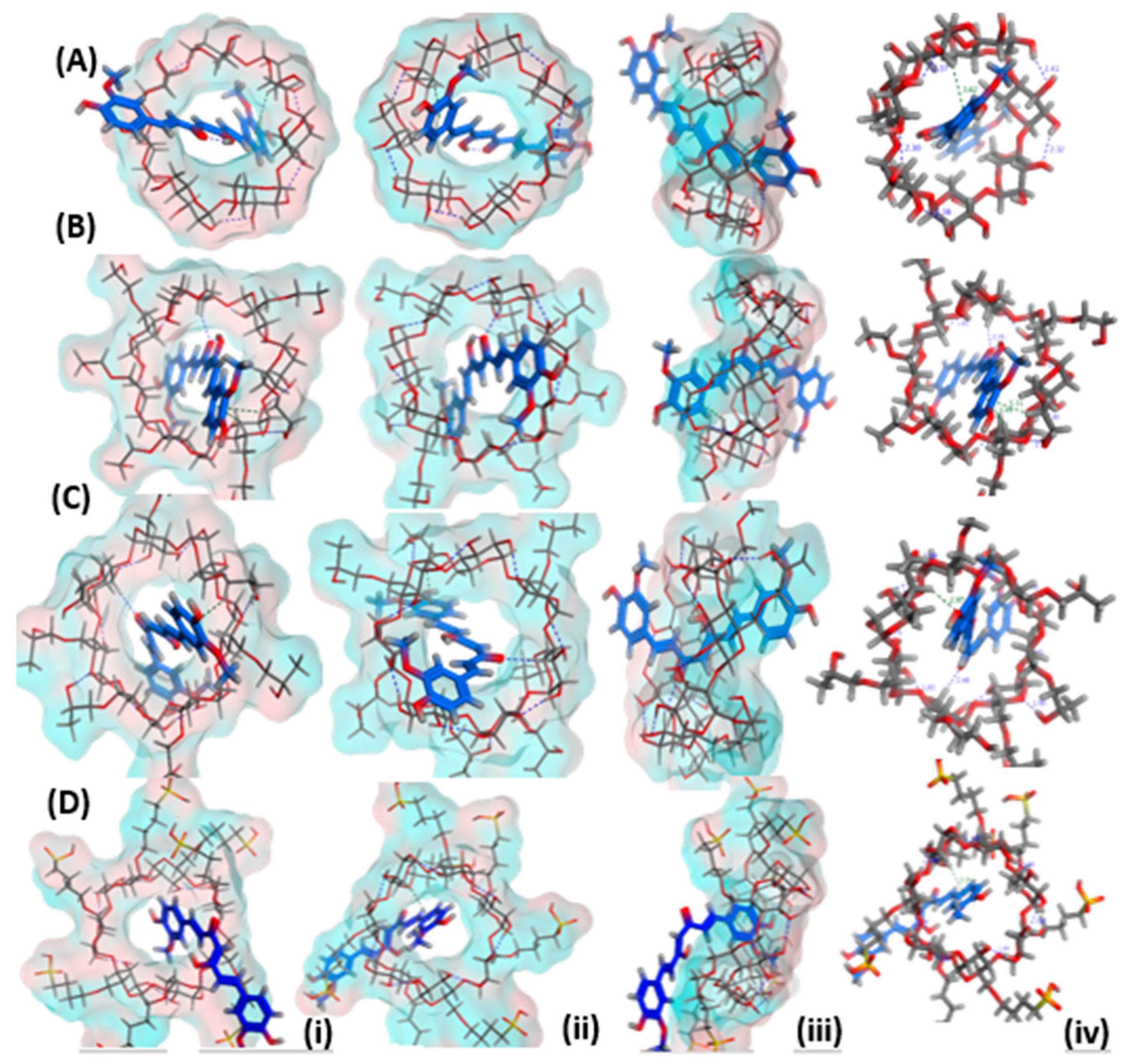
2.3.1. Molecular Docking
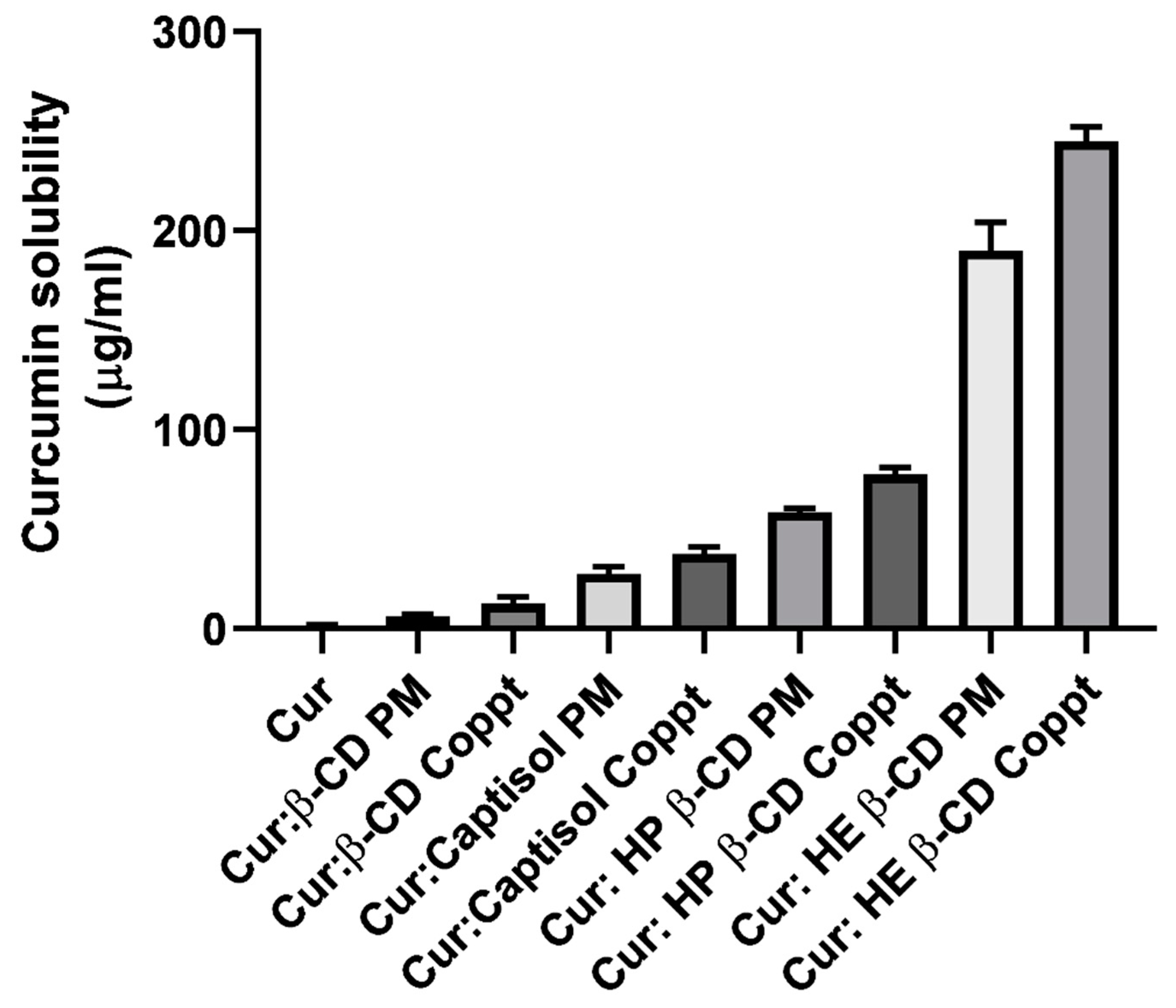
2.3.2. Solubility Studies
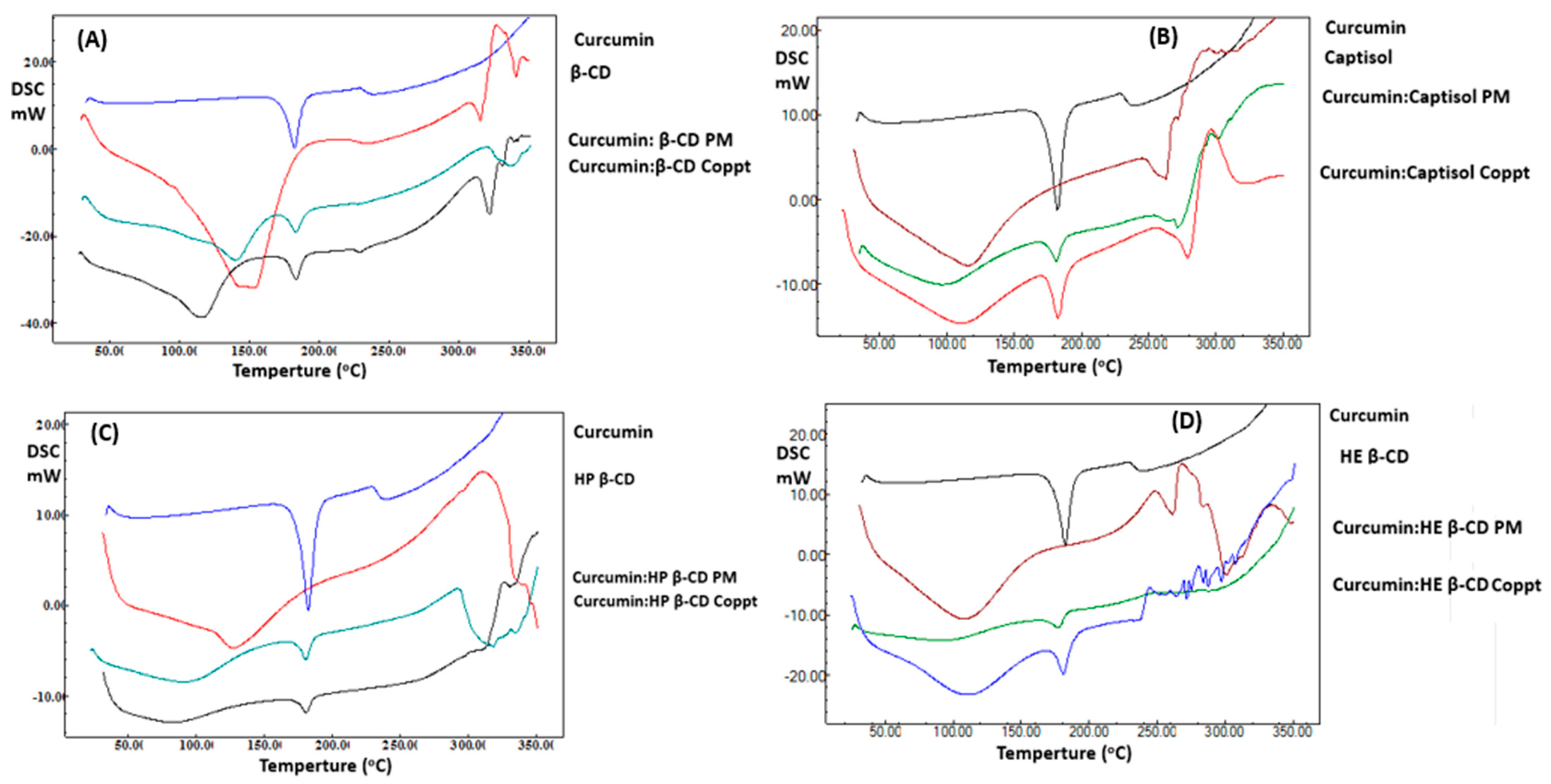
2.3.3. Differential Scanning Calorimetry (DSC)
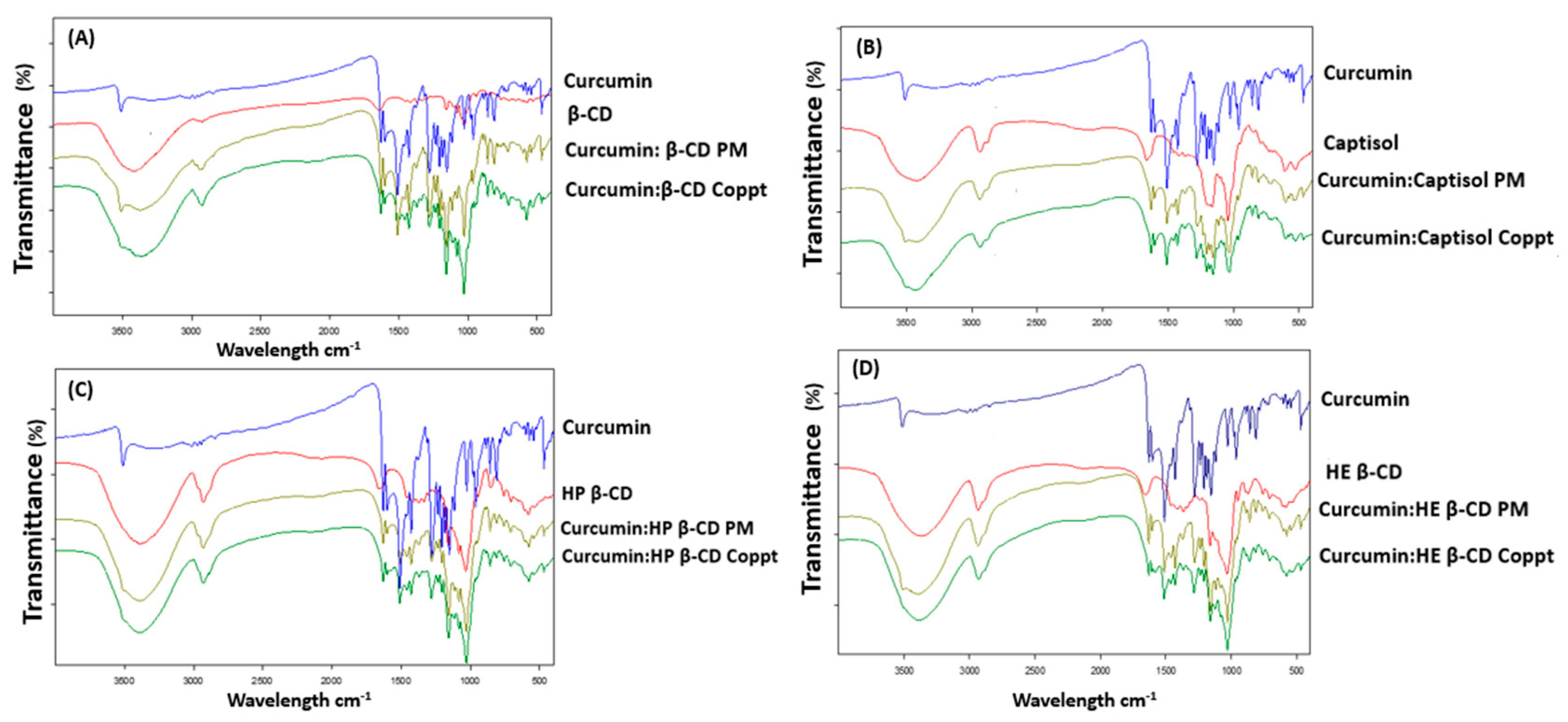
2.3.4. Fourier Transform Infrared Spectroscopic (FTIR) Study
2.3.5. X-ray Diffraction (XRD)
2.3.6. In Vitro Dissolution Studies
2.3.7. In Vitro Cytotoxicity Assay Using Human Breast MCF-7 Cells
2.4. Statistical Analysis
3. Results and Discussion
3.1. Docking Studies
3.2. Solubility Study
3.3. DSC
3.4. FTIR
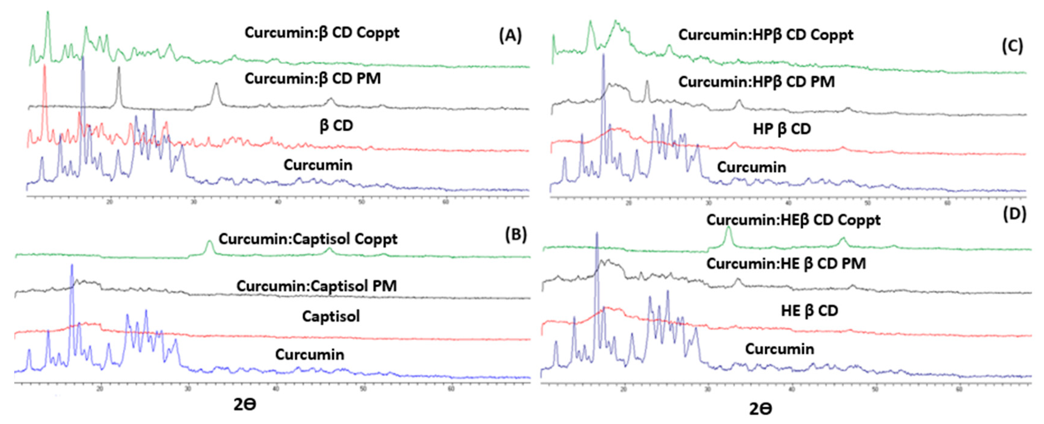
3.5. XRD
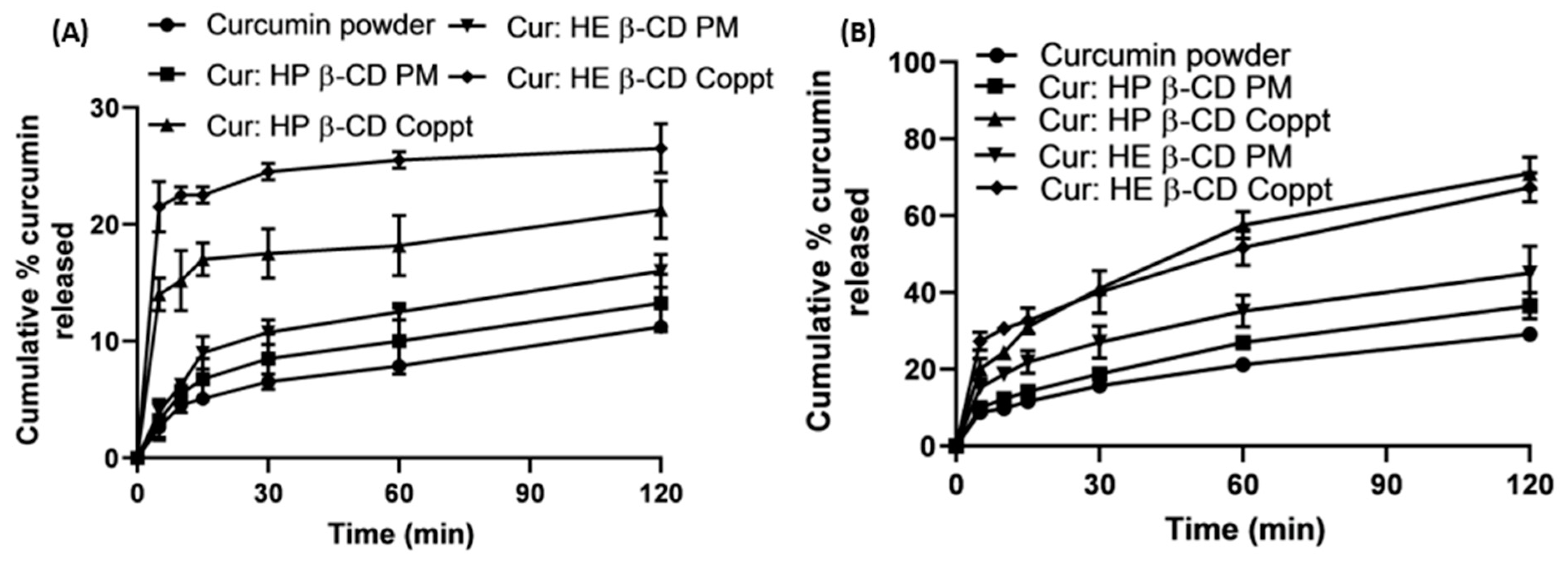
3.6. In Vitro Dissolution in Simulated Gastric and Intestinal Fluids
3.7. Cytotoxicity Assay Using Human Breast MCF-7 Cell Lines
4. Conclusions
Author Contributions
Funding
Institutional Review Board Statement
Informed Consent Statement
Data Availability Statement
Acknowledgments
Conflicts of Interest
References
- Tomeh, M.A.; Hadianamrei, R.; Zhao, X. A Review of Curcumin and Its Derivatives as Anticancer Agents. Int. J. Mol. Sci. 2019, 20, 1033. [Google Scholar] [CrossRef] [PubMed] [Green Version]
- International, W. Global Cancer Data by Country. 2022. Available online: https://www.wcrf.org/cancer-trends/global-cancer-data-by-country/#:~:text=Globally%2C%2018%2C094%2C716%20million%20cases%20of,women%20(178.1%20per%20100%2C000) (accessed on 23 August 2022).
- Siegel, R.L.; Miller, K.D.; Jemal, A. Cancer statistics. CA Cancer J. Clin. 2018, 68, 7–30. [Google Scholar] [CrossRef] [PubMed]
- Umar, A.; Dunn, B.K.; Greenwald, P. Future directions in cancer prevention. Nat. Cancer 2012, 12, 835–848. [Google Scholar] [CrossRef] [PubMed]
- Wang, J.; Jiang, W. Natural compounds as anticancer agents: Experimental evidence. World J. Exp. Med. 2012, 2, 45–57. [Google Scholar] [CrossRef] [PubMed]
- Kinghorn, A.D.; Chin, Y.-W.; Swanson, S.M. Discovery of natural product anticancer agents from biodiverse organisms. Curr. Opin. Drug Discov. Dev. 2009, 12, 189–196. [Google Scholar]
- Gupta, A.P.; Khan, S.; Manzoor, M.M.; Yadav, A.K.; Sharma, G.; Anand, R.; Gupta, S. Anticancer Curcumin: Natural Analogues and Structure-Activity Relationship. In Natural Products Chemistry; Attaur, R., Ed.; Elsevier: Amsterdam, The Netherlands, 2017; pp. 355–401. [Google Scholar]
- Anand, P.; Sundaram, C.; Jhurani, S.; Kunnumakkara, A.B.; Aggarwal, B.B. Curcumin and cancer: An “old-age” disease with an “age-old” solution. Cancer Lett. 2008, 267, 133–164. [Google Scholar] [CrossRef] [PubMed]
- Kunnumakkara, A.B.; Bordoloi, D.; Padmavathi, G.; Monisha, J.; Roy, N.K.; Prasad, S.; Aggarwal, B.B. Curcumin, the golden nutraceutical: Multitargeting for multiple chronic diseases. Br. J. Pharmacol. 2017, 174, 1325–1348. [Google Scholar] [CrossRef] [PubMed] [Green Version]
- Kunnumakkara, A.B.; Harsha, C.; Banik, K.; Vikkurthi, R.; Sailo, B.L.; Bordoloi, D.; Gupta, S.C.; Aggarwal, B.B. Is curcumin bioavailability a problem in humans: Lessons from clinical trials. Expert Opin. Drug Metab. Toxicol. 2019, 15, 705–733. [Google Scholar] [CrossRef]
- Liu, Q.; Loo, W.T.; Sze, S.; Tong, Y. Curcumin inhibits cell proliferation of mda-mb-231 and bt-483 breast cancer cells mediated by down-regulation of nfkb, cyclind and mmp-1 transcription. Phytomedicine 2009, 16, 916–922. [Google Scholar] [CrossRef] [Green Version]
- Abdelkader, H.; Mustafa, W.; Alqahtani, A.; Alsharani, S.; Al Fatease, A.; Alany, R. Glycation-induced age-related illnesses, antiglycation and drug delivery strategies. J. Pharm. Pharmacol. 2022, 1–22, in press. [Google Scholar] [CrossRef]
- Zheng, B.; McClements, D. Formulation of more effecacious curcumin delivery systems using colloid science: Enhanced solubility, stability, and bioavailability. Molecules 2020, 25, 2791. [Google Scholar] [CrossRef] [PubMed]
- McClements, D.J.; Decker, E.; Weiss, J. Emulsion-Based Delivery Systems for Lipophilic Bioactive Components. J. Food Sci. 2007, 72, R109–R124. [Google Scholar] [CrossRef] [PubMed]
- Loftsson, T.; Brewster, M.E.; Másson, M. Role of Cyclodextrins in Improving Oral Drug Delivery. Am. J. Drug Deliv. 2004, 2, 261–275. [Google Scholar] [CrossRef]
- Loftsson, T.; Duchêne, D. Cyclodextrins and their pharmaceutical applications. Int. J. Pharm. 2007, 329, 1–11. [Google Scholar] [CrossRef]
- Żandarek, J.; Gumułka, P.; Starek, M.; Dąbrowska, M. Characteristics and applications of cyclodextrin complexes. Farm. Polska 2022, 78, 308–316. [Google Scholar] [CrossRef]
- Zhang, L.; Man, S.; Qiu, H.; Liu, Z.; Zhang, M.; Ma, L.; Gao, W. Curcumin-cyclodextrin complexes enhanced the anti-cancer effects of curcumin. Environ. Toxicol. Pharmacol. 2016, 48, 31–38. [Google Scholar] [CrossRef]
- Yallapu, M.; Jaggi, M.; Chauhan, S. beta-Cyclodextrin-curcumin self-assembly enhances curcumin delivery in prostate cancer cells. Colloids Surf B Biointerfaces 2010, 79, 113–125. [Google Scholar] [CrossRef]
- Feng, L.; Fawaz, R.; Hovde, S.; Sheng, F.; Nosrati, M.; Geiger, J.H. Crystal structures of Escherichia coli branching enzyme in complex with cyclodextrins. Acta Crystallogr. Sect. D Struct. Biol. 2016, 72, 641–647. [Google Scholar] [CrossRef]
- Shityakov, S.; Salmas, R.E.; Durdagi, S.; Salvador, E.; Pápai, K.; Yáñez-Gascón, M.; Pérez-Sánchez, H.; Puskás, I.; Roewer, N.; Förster, C.; et al. Characterization, in vivo evaluation, and molecular modelling of different propofol–cyclodextrin complexes to assess their drug delivery potential at the blood-brain barrier level. J. Chem. Inf. Model. 2016, 56, 1914–1922. [Google Scholar] [CrossRef]
- van de Loosdrecht, A.; Beelen, R.; Ossenkoppele, G.; Broekhoven, M.; Langenhuijsen, M. A tetrazolium-based colorimetric MTT assay to quantitate human monocyte mediated cytotoxicity against leukemic cells from cell lines and patients with acute myeloid leukemia. J. Immunol. Methods 1994, 174, 311–320. [Google Scholar] [CrossRef]
- Ghosh, A.; Biswas, S.; Ghosh, T. Preparation and Evaluation of Silymarin β-cyclodextrin Molecular Inclusion Complexes. J. Young- Pharm. 2011, 3, 205–210. [Google Scholar] [CrossRef] [PubMed]
- Mai, N.N.S.; Nakai, R.; Kawano, Y.; Hanawa, T. Enhancing the Solubility of Curcumin Using a Solid Dispersion System with Hydroxypropyl-β-Cyclodextrin Prepared by Grinding, Freeze-Drying, and Common Solvent Evaporation Methods. Pharmacy 2020, 8, 203. [Google Scholar] [CrossRef] [PubMed]
- Maharjan, P.; Jin, M.; Kim, D.; Yang, J.; Maharjan, A.; Shin, M.C.; Cho, K.H.; Kim, M.S.; Min, K.A. Evaluation of epithelial transport and oxidative stress protection of nanoengineered curcumin derivative-cyclodextrin formulation for ocular delivery. Arch. Pharmacal Res. 2019, 42, 909–925. [Google Scholar] [CrossRef]
- Athira, G.; Jyothi, A. Preparation and characterization of curcumin loaded cassava starch nanoparticles with improved cellular absorption. In. J. Pharm. Pharm. Sci. 2014, 6, 171–176. [Google Scholar]
- Rachmawati, H.; Edityaningrum, C.; Mauludin, R. Molecular inclusion complex of curcumin-beta-cyclodextrin nanoparticles to enhance curcumin skin permeability from hysrophilic matrix gel. AAPS PharmSciTech 2013, 14, 1303. [Google Scholar] [CrossRef] [Green Version]
- Abdelkader, H.; Fathalla, Z.; Moharram, H.; Ali, T.; Pierscionek, B. Cyclodextrin Enhances Corneal Tolerability and Reduces Ocular Toxicity Caused by Diclofenac. Oxidative Med. Cell. Longev. 2018, 2018, 5260976. [Google Scholar] [CrossRef] [PubMed] [Green Version]
- Rassu, G.; Fancello, S.; Roldo, M.; Malanga, M.; Szente, L.; Migheli, R.; Gavini, E.; Giunchedi, P. Investigation of Cytotoxicity and Cell Uptake of Cationic Beta-Cyclodextrins as Valid Tools in Nasal Delivery. Pharmaceutics 2020, 12, 658. [Google Scholar] [CrossRef]


| Drug | Carrier | E Score (kcal/mol) | Number of Interactions | |
|---|---|---|---|---|
| H-Bond | Hydrophobic Interactions | |||
| Curcumin | β-cyclodextrin | −5.2617 | - | 1 |
| Hydroxyethyl-β-cyclodextrin | −6.0093 | 1 | 2 | |
| Hydroxypropyl-β-cyclodextrin | −5.8415 | 1 | 1 | |
| Sulfobutylether-β-cyclodextrin | −5.7392 | - | 1 | |
| Formulation/Preparation Method | Simulated Gastric Fluid (pH 1.2) | Simulated Intestinal Fluid (pH 6.8) | ||
|---|---|---|---|---|
| Q10min (%) | Q120min (%) | Q10min (%) | Q120min (%) | |
| Curcumin | 4 ± 0.5 | 10 ± 1.0 | 10 ± 0.5 | 28 ± 3.5 |
| Curcumin: HP β-CD PM | 5.5± 1.0 | 13 ± 1.0 | 12 ± 0.5 | 36 ± 3.0 |
| Curcumin: HP β-CD Coppt | 15 ± 2.5 | 21 ± 2.0 | 25 ± 3.0 | 72 ± 4.0 |
| Curcumin: HE β-CD PM | 6.5 ± 1.0 | 15 ± 1.0 | 18 ± 2.0 | 45 ± 3.0 |
| Curcumin: HE β-CD Coppt | 23 ± 4.0 | 26 ± 3.5 | 30 ± 3.5 | 70 ± 4.0 |
Publisher’s Note: MDPI stays neutral with regard to jurisdictional claims in published maps and institutional affiliations. |
© 2022 by the authors. Licensee MDPI, Basel, Switzerland. This article is an open access article distributed under the terms and conditions of the Creative Commons Attribution (CC BY) license (https://creativecommons.org/licenses/by/4.0/).
Share and Cite
Abdelkader, H.; Fatease, A.A.; Fathalla, Z.; Shoman, M.E.; Abou-Taleb, H.A.; Abourehab, M.A.S. Design, Preparation and Evaluation of Supramolecular Complexes with Curcumin for Enhanced Cytotoxicity in Breast Cancer Cell Lines. Pharmaceutics 2022, 14, 2283. https://doi.org/10.3390/pharmaceutics14112283
Abdelkader H, Fatease AA, Fathalla Z, Shoman ME, Abou-Taleb HA, Abourehab MAS. Design, Preparation and Evaluation of Supramolecular Complexes with Curcumin for Enhanced Cytotoxicity in Breast Cancer Cell Lines. Pharmaceutics. 2022; 14(11):2283. https://doi.org/10.3390/pharmaceutics14112283
Chicago/Turabian StyleAbdelkader, Hamdy, Adel Al Fatease, Zeinab Fathalla, Mai E. Shoman, Heba A. Abou-Taleb, and Mohammed A. S. Abourehab. 2022. "Design, Preparation and Evaluation of Supramolecular Complexes with Curcumin for Enhanced Cytotoxicity in Breast Cancer Cell Lines" Pharmaceutics 14, no. 11: 2283. https://doi.org/10.3390/pharmaceutics14112283