
Silver Nanoparticles as Potential Antiviral Agents

 , , , , and
Abstract
:1. Introduction
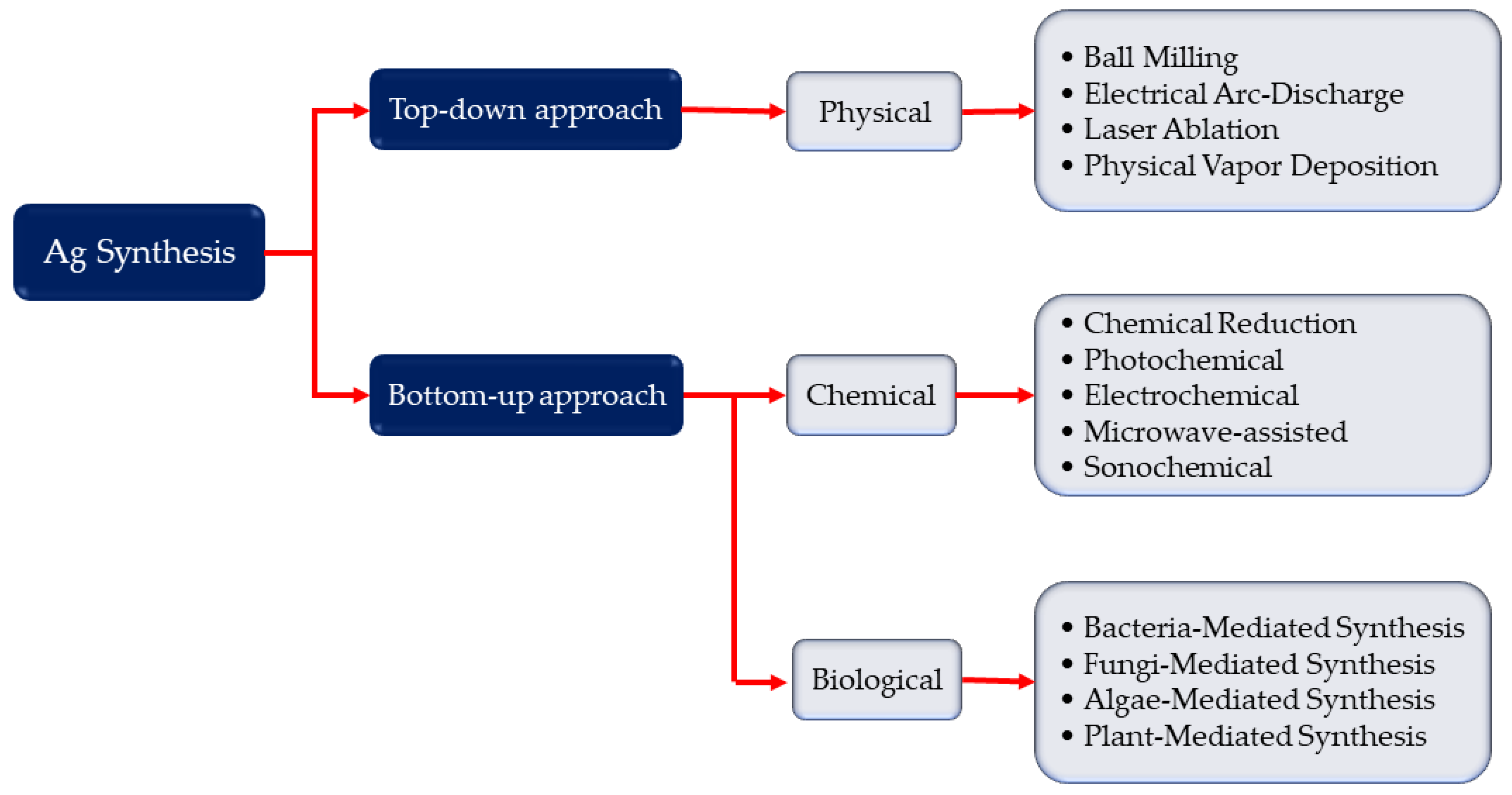
2. Synthesis of Silver Nanoparticles
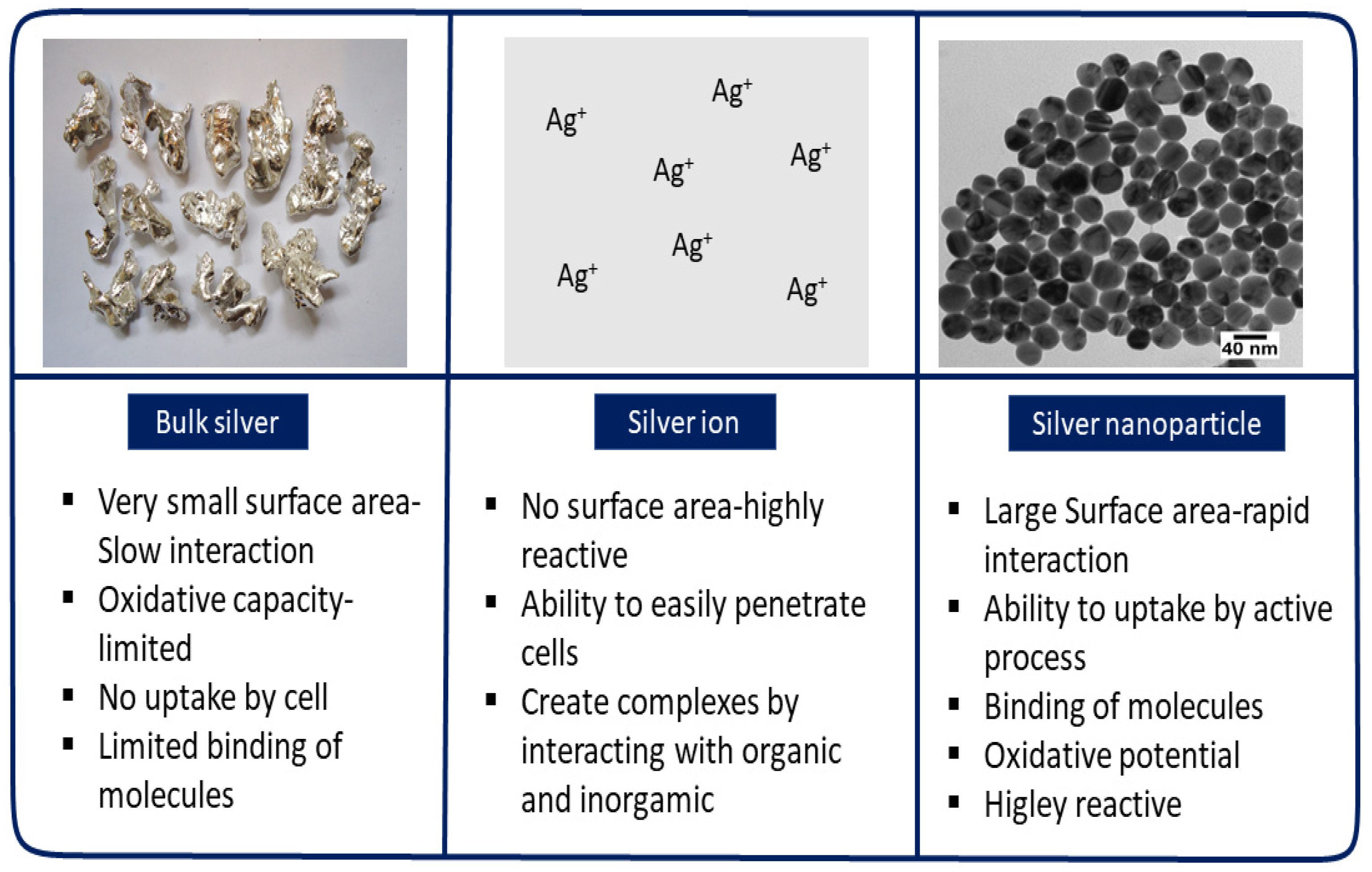
3. Properties of Silver Nanoparticles
3.1. Shape and Crystallinity
3.2. Melting Temperature
3.3. Optical Properties
3.4. Electrical Properties
4. Characterization of AgNPs
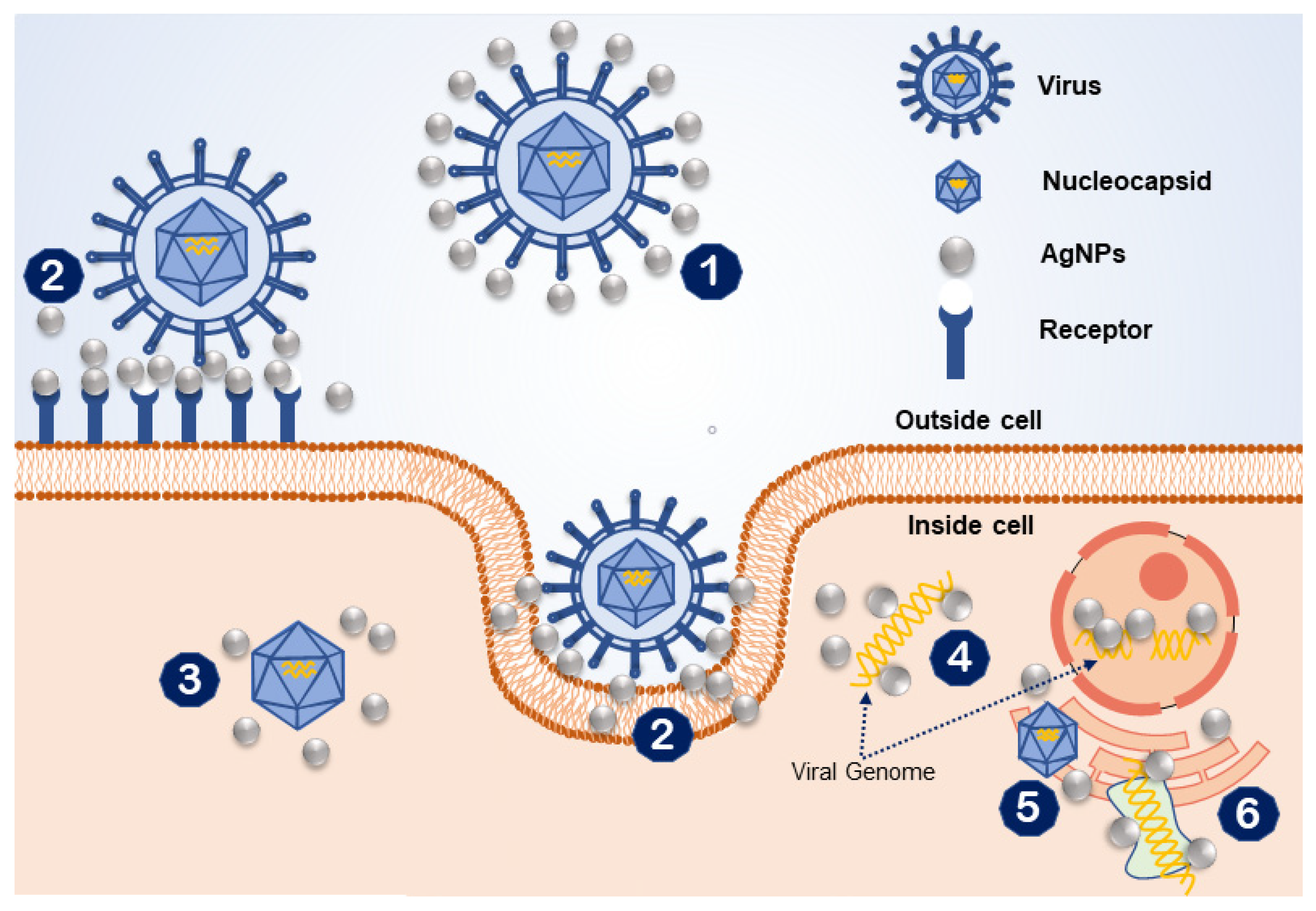
5. Antiviral Mechanisms of AgNPs
6. Activity of AgNPs against Viruses
6.1. Adenovirus
6.2. Hepatitis B
6.3. Herpes Simplex Virus
6.4. Human Immunodeficiency Virus-1 (HIV-1)
6.5. Influenza A
6.6. Noroviruses
6.7. Poliovirus
6.8. Respiratory Syncytial Virus
6.9. Rift Valley Fever Virus
6.10. SARS-CoV-2
6.11. Chikungunya Virus
6.12. Bunyamwera Orthobunyavirus
6.13. White Spot Syndrome Virus
6.14. Zika Virus
7. Toxicity and Safety Issue of AgNPs
8. Antiviral Application of AgNPs
9. Conclusions and Future Prospects
Author Contributions
Funding
Institutional Review Board Statement
Informed Consent Statement
Data Availability Statement
Acknowledgments
Conflicts of Interest
References
- Nasir, A.; Kim, K.M.; Caetano-Anollés, G. Viral evolution. Mob. Genet. Elem. 2012, 2, 247–252. [Google Scholar] [CrossRef] [PubMed] [Green Version]
- Passi, D.; Sharma, S.; Dutta, S.R.; Dudeja, P.; Sharma, V. Ebola virus disease (the killer virus): Another threat to humans and bioterrorism: Brief review and recent updates. J. Clin. Diagn. Res. 2015, 9, LE01. [Google Scholar] [CrossRef] [PubMed]
- Corstjens, M.P.L.A.; Abrams, W.R.; Malamud, D. Saliva and viral infections. Periodontology 2000 2016, 70, 93–110. [Google Scholar] [CrossRef] [PubMed]
- Freestone, D. The need for new antiviral agents. Antivir. Res. 1985, 5, 307–324. [Google Scholar] [CrossRef]
- Colpitts, C.C.; Verrier, E.R.; Baumert, T.F. Targeting viral entry for treatment of hepatitis B and C virus infections. ACS Infect. Dis. 2015, 1, 420–427. [Google Scholar] [CrossRef] [PubMed]
- Morens, D.M.; Folkers, G.K.; Fauci, A.S. The challenge of emerging and re-emerging infectious diseases. Nature 2004, 43, 242–249. [Google Scholar] [CrossRef]
- Bhattacharya, S. The World Health Organization and global smallpox eradication. J. Epidemiol. Community Health 2008, 62, 909–912. [Google Scholar] [CrossRef] [PubMed] [Green Version]
- Rauch, S.; Jasny, E.; Schmidt, K.E.; Petsch, B. New Vaccine Technologies to Combat Outbreak Situations. Front. Immunol. 2018, 9, 1963. [Google Scholar] [CrossRef] [PubMed] [Green Version]
- Excler, J.L.; Saville, M.; Berkley, S.; Kim, J.H. Vaccine development for emerging infectious diseases. Nat. Med. 2021, 27, 591–600. [Google Scholar] [CrossRef]
- Soares, S.; Sousa, J.; Pais, A.; Vitorino, C. Nanomedicine: Principles, properties, and regulatory issues. Front. Chem. 2018, 6, 360. [Google Scholar] [CrossRef] [PubMed]
- Lembo, D.; Donalisio, M.; Civra, A.; Argenziano, M.; Cavalli, R. Nanomedicine formulations for the delivery of antiviral drugs: A promising solution for the treatment of viral infections. Expert Opin. Drug Deliv. 2018, 15, 93–114. [Google Scholar] [CrossRef] [PubMed]
- Waris, A.; Ali, M.; Khan, A.U.; Ali, A.; Baset, A. Role of nanotechnology in diagnosing and treating COVID-19 during the Pandemic. Int. J. Clin. Virol. 2020, 4, 65–70. [Google Scholar]
- Aranda, P.; Wicklein, B.; Ruiz-Garcia, C.; Martín-Sampedro, R.; Darder, M.; Del Real, G.; Ruiz-Hitzky, E. Research and patents on coronavirus and COVID-19: A review. Recent Pat. Nanotechnol. 2020, 14, 328–350. [Google Scholar] [CrossRef] [PubMed]
- Jones, G.W.; Monopoli, M.P.; Campagnolo, L.; Pietroiusti, A.; Tran, L.; Fadeel, B. No small matter: A perspective on nanotechnology-enabled solutions to fight COVID-19. Nanomedicine 2020, 15, 2411–2427. [Google Scholar] [CrossRef] [PubMed]
- Talebian, S.; Wallace, G.G.; Schroeder, A.; Stellacci, F.; Conde, J. Nanotechnology-based disinfectants and sensors for SARS-CoV-2. Nat. Nanotechnol. 2020, 15, 618–621. [Google Scholar] [CrossRef]
- Galdiero, S.; Falanga, A.; Vitiello, M.; Cantisani, M.; Marra, V.; Galdiero, M. Silver nanoparticles as potential antiviral agents. Molecules 2011, 16, 8894–8918. [Google Scholar] [CrossRef] [Green Version]
- Ghiuță, I.; Cristea, D. Silver nanoparticles for delivery purposes. Nanoeng. Biomater. Adv. Drug Deliv. 2020, 347–371. [Google Scholar]
- Khaiboullina, S.; Uppal, T.; Dhabarde, N.; Subramanian, V.R.; Verma, S.C. Inactivation of Human Coronavirus by Titania Nanoparticle Coatings and UVC Radiation: Throwing Light on SARS-CoV-2. Viruses 2021, 13, 19. [Google Scholar] [CrossRef]
- Jagaran, K.; Singh, M. Nanomedicine for covid-19: Potential of copper nanoparticles. Biointerface Res. Appl. Chem. 2020, 11, 10716–10728. [Google Scholar]
- Sarkar, K.P.; Mukhopadhyay, C.D. Ayurvedic metal nanoparticles could be novel antiviral agents against SARS-CoV-2. Int. Nano Lett. 2021, 11, 197–203. [Google Scholar] [CrossRef]
- Campos, E.V.R.; Pereira, A.E.S.; De Oliveira, J.L.; Carvalho, L.B.; Guilger-Casagrande, M.; De Lima, R.; Fraceto, L.F. How can nanotechnology help to combat COVID-19? Opportunities and urgent need. J. Nanobiotechnology 2020, 18, 125. [Google Scholar] [CrossRef] [PubMed]
- Raj, S.; Jose, S.; Sumod, U.S.; Sabitha, M. Nanotechnology in cosmetics: Opportunities and challenges. J. Pharm. Bioallied Sci. 2012, 4, 186. [Google Scholar] [CrossRef] [PubMed]
- Chen, X.; Schluesener, H.J. Nanosilver: A nanoproduct in medical application. Toxicol. Lett. 2008, 176, 1–12. [Google Scholar] [CrossRef] [PubMed]
- Dorjnamjin, D.; Ariunaa, M.; Shim, Y.K. Synthesis of silver nanoparticles using hydroxyl functionalized ionic liquids and their antimicrobial activity. Int. J. Mol. Sci. 2008, 9, 807–820. [Google Scholar] [CrossRef] [PubMed]
- Sondi, I.; Goia, D.V.; Matijević, E. Preparation of highly concentrated stable dispersions of uniform silver nanoparticles. J. Colloid Interface Sci. 2003, 260, 75–81. [Google Scholar] [CrossRef]
- Aamodt, K.; Abelev, B.; Quintana, A.A.; Adamova, D.; Adare, A.M.; Aggarwal, M.M.; Rinella, G.A.; Agocs, A.G.; Salazar, S.A.; Ahammed, Z.; et al. Elliptic flow of charged particles in Pb-Pb collisions at s N N = 2.76 TeV. Phys. Rev. Lett. 2010, 105, 252302. [Google Scholar] [CrossRef] [PubMed] [Green Version]
- Ju-Nam, Y.; Lead, J.R. Manufactured nanoparticles: An overview of their chemistry, interactions and potential environmental implications. Sci. Total. Environ. 2008, 400, 396–414. [Google Scholar] [CrossRef]
- Yadav, P.T.; Yadav, R.M.; Singh, D.P. Mechanical milling: A top down approach for the synthesis of nanomaterials and nanocomposites. Nanosci. Nanotechnol. 2012, 2, 22–48. [Google Scholar] [CrossRef] [Green Version]
- Jeremiah, S.S.; Miyakawa, K.; Morita, T.; Yamaoka, Y.; Ryo, A. Potent antiviral effect of silver nanoparticles on SARS-CoV-2. Biochem. Biophys. Res. Commun. 2020, 533, 195–200. [Google Scholar] [CrossRef]
- Baram-Pinto, D.; Shukla, S.; Perkas, N.; Gedanken, A.; Sarid, R. Inhibition of herpes simplex virus type 1 infection by silver nanoparticles capped with mercaptoethane sulfonate. Bioconjugate Chem. 2009, 20, 1497–1502. [Google Scholar] [CrossRef]
- Zheng, X.; Peng, Y.; Lombardi, J.R.; Cui, X.; Zheng, W. Photochemical growth of silver nanoparticles with mixed-light irradiation. Colloid Polym. Sci. 2016, 294, 911–916. [Google Scholar] [CrossRef]
- Kuntyi, O.I.; Kytsya, A.R.; Mertsalo, I.P.; Mazur, A.S.; Zozula, G.I.; Bazylyak, L.I.; Topchak, R.V. Electrochemical synthesis of silver nanoparticles by reversible current in solutions of sodium polyacrylate. Colloid Polym. Sci. 2019, 297, 689–695. [Google Scholar] [CrossRef]
- Chung, D.S.; Kim, H.; Ko, J.; Lee, J.; Hwang, B.; Chang, S.; Kim, B.; Chung, S.J. Microwave Synthesis of Silver Nanoparticles Using Different Pentose Carbohydrates as Reducing Agents. J. Chem. 2018, 12, 1–10. [Google Scholar]
- Elsupikhe, F.R.; Shameli, K.; Ahmad, M.B. Sonochemical method for the synthesis of silver nanoparticles in κ-carrageenan from silver salt at different concentrations. Res. Chem. Intermed. 2015, 41, 8515–8525. [Google Scholar] [CrossRef] [Green Version]
- Ga’al, H.; Fouad, H.; Mao, G.; Tian, J.; Jianchu, M. Larvicidal and pupicidal evaluation of silver nanoparticles synthesized using Aquilaria sinensis and Pogostemon cablin essential oils against dengue and zika viruses vector Aedes albopictus mosquito and its histopathological analysis. Artif. Cells Nanomed. Biotechnol. 2018, 46, 1171–1179. [Google Scholar] [CrossRef] [Green Version]
- Powers, A.M. Vaccine and therapeutic options to control chikungunya virus. Clin. Microbiol. Rev. 2018, 31, e00104-16. [Google Scholar] [CrossRef] [PubMed] [Green Version]
- Pantidos, N.; Horsfall, L.E. Biological synthesis of metallic nanoparticles by bacteria, fungi and plants. J. Nanomed. Nanotechnol. 2014, 5, 1. [Google Scholar] [CrossRef]
- Khan, A.M.; Khan, T.; Nadhman, A. Applications of plant terpenoids in the synthesis of colloidal silver nanoparticles. Adv. Colloid Interface Sci. 2016, 234, 132–141. [Google Scholar]
- Kuppusamy, P.; Yusoff, M.M.; Maniam, G.P.; Govindan, N. Biosynthesis of metallic nanoparticles using plant derivatives and their new avenues in pharmacological applications—An updated report. Saudi Pharm. J. 2016, 24, 473–484. [Google Scholar] [CrossRef] [PubMed]
- Hamedi, S.; Ghaseminezhad, M.; Shokrollahzadeh, S.; Shojaosadati, S.A. Controlled biosynthesis of silver nanoparticles using nitrate reductase enzyme induction of filamentous fungus and their antibacterial evaluation. Artif. Cells Nanomed. Biotechnol. 2017, 45, 1588–1596. [Google Scholar] [CrossRef]
- Skonieczna, M.; Hudy, D. Biological Activity of Silver Nanoparticles and Their Applications in Anticancer Therapy; IntechOpen: London, UK, 2018. [Google Scholar]
- Haes, A.J.; Haynes, C.L.; McFarland, A.D.; Schatz, G.C.; Van Duyne, R.P.; Zou, S. Plasmonic materials for surface-enhanced sensing and spectroscopy. MRS Bull. 2005, 30, 368–375. [Google Scholar] [CrossRef] [Green Version]
- Kelly, K.L.; Coronado, E.; Zhao, L.L.; Schatz, G.C. Optical properties of metal nanoparticles: The influence of size, shape, and dielectric environment. J. Phys. Chem. B 2003, 107, 668–677. [Google Scholar] [CrossRef]
- Bastys, V.; Pastoriza-Santos, I.; Rodríguez-González, B.; Vaisnoras, R.; Liz-Marzán, L.M. Formation of silver nanoprisms with surface plasmons at communication wavelengths. Adv. Funct. Mater. 2006, 16, 766–773. [Google Scholar] [CrossRef]
- Sau, T.K.; Murphy, C.J. Room temperature, high-yield synthesis of multiple shapes of gold nanoparticles in aqueous solution. J. Am. Chem. Soc. 2004, 126, 8648–8649. [Google Scholar] [CrossRef] [PubMed]
- Millstone, E.J.; Métraux, G.S.; Mirkin, C.A. Controlling the edge length of gold nanoprisms via a seed-mediated approach. Adv. Funct. Mater. 2006, 16, 1209–1214. [Google Scholar] [CrossRef]
- Métraux, S.G.; Mirkin, C.A. Rapid thermal synthesis of silver nanoprisms with chemically tailorable thickness. Adv. Mater. 2005, 17, 412–415. [Google Scholar] [CrossRef]
- Wiley, B.J.; Im, S.H.; Li, Z.Y.; McLellan, J.; Siekkinen, A.; Xia, Y. Maneuvering the surface plasmon resonance of silver nanostructures through shape-controlled synthesis. J. Phys. Chem. B 2006, 110, 15666–15675. [Google Scholar] [CrossRef]
- Xiao, J.P.; Xie, Y.; Tang, R.; Chen, M.E.; Tian, X.B. Novel ultrasonically assisted templated synthesis of palladium and silver dendritic nanostructures. Adv. Mater. 2001, 13, 1887–1891. [Google Scholar] [CrossRef]
- Tsuji, M.; Hashimoto, M.; Nishizawa, Y.; Kubokawa, M.; Tsuji, T. Microwave-assisted synthesis of metallic nanostructures in solution. Chem. A Eur. J. 2005, 11, 440–452. [Google Scholar] [CrossRef]
- Tsuji, M.; Miyamae, N.; Lim, S.; Kimura, K.; Zhang, X.; Hikino, S.; Nishio, M. Crystal structures and growth mechanisms of Au@ Ag core−shell nanoparticles prepared by the microwave−polyol method. Cryst. Growth Des. 2006, 6, 1801–1807. [Google Scholar] [CrossRef]
- He, R.; Qian, X.; Yin, J.; Zhu, Z. Preparation of polychrome silver nanoparticles in different solvents. J. Mater. Chem. 2002, 12, 3783–3786. [Google Scholar] [CrossRef]
- Roduner, E. Nanoscopic Materials: Size-Dependent Phenomena; Royal Society of Chemistry: London, UK, 2006. [Google Scholar]
- Shandiz, M.A. Effective coordination number model for the size dependency of physical properties of nanocrystals. J. Phys. Condens. Matter. 2008, 20, 325237. [Google Scholar] [CrossRef]
- Magdassi, S.; Grouchko, M.; Berezin, O.; Kamyshny, A. Triggering the sintering of silver nanoparticles at room temperature. ACS Nano 2010, 4, 1943–1948. [Google Scholar] [CrossRef] [PubMed]
- Allen, G.L.; Bayles, R.A.; Gile, W.W.; Jesser, W.A. Small particle melting of pure metals. Thin Solid Film 1986, 144, 297–308. [Google Scholar] [CrossRef]
- Ide, E.; Angata, S.; Hirose, A.; Kobayashi, K.F. Metal–metal bonding process using Ag metallo-organic nanoparticles. Acta Mater. 2005, 53, 2385–2393. [Google Scholar] [CrossRef]
- Evanoff, D.D.; Chumanov, G. Size-controlled synthesis of nanoparticles. 2. Measurement of extinction, scattering, and absorption cross sections. J. Phys. Chem. B 2004, 108, 13957–13962. [Google Scholar] [CrossRef]
- Gonzalez, A.; Noguez, C. Optical properties of silver nanoparticles. Phys. Status Solidi C 2007, 4, 4118–4126. [Google Scholar] [CrossRef]
- Alshehri, A.H.; Jakubowska, M.; Młożniak, A.; Horaczek, M.; Rudka, D.; Free, C.; Carey, J.D. Enhanced electrical conductivity of silver nanoparticles for high frequency electronic applications. ACS Appl. Mater. Interfaces 2012, 4, 7007–7010. [Google Scholar] [CrossRef] [PubMed] [Green Version]
- Pleus, R. Nanotechnologies-Guidance on Physicochemical Characterization of Engineered Nanoscale Materials for Toxicologic Assessment; ISO: Geneva, Switzerland, 2012. [Google Scholar]
- Zhang, X.F.; Liu, Z.G.; Shen, W.; Gurunathan, S. Silver Nanoparticles: Synthesis, Characterization, Properties, Applications, and Therapeutic Approaches. Int. J. Mol. Sci. 2016, 17, 1534. [Google Scholar] [CrossRef] [PubMed]
- Sre, P.R.; Reka, M.; Poovazhagi, R.; Kumar, M.A.; Murugesan, K. Antibacterial and cytotoxic effect of biologically synthesized silver nanoparticles using aqueous root extract of Erythrina indica lam. Spectrochim. Acta Part A Mol. Biomol. Spectrosc. 2015, 135, 1137–1144. [Google Scholar]
- Firdhouse, M.J.; Lalitha, P. Biosynthesis of silver nanoparticles using the extract of Alternanthera sessilis—Antiproliferative effect against prostate cancer cells. Cancer Nanotechnol. 2013, 4, 137. [Google Scholar] [CrossRef] [PubMed] [Green Version]
- Shankar, S.S.; Rai, A.; Ahmad, A.; Sastry, M. Rapid synthesis of Au, Ag, and bimetallic Au core–Ag shell nanoparticles using Neem (Azadirachta indica) leaf broth. J. Colloid Interface Sci. 2004, 275, 496–502. [Google Scholar] [CrossRef] [PubMed]
- Vidhu, V.; Aromal, S.A.; Philip, D. Green synthesis of silver nanoparticles using Macrotyloma uniflorum. Spectrochim. Acta Part A Mol. Biomol. Spectrosc. 2011, 83, 392–397. [Google Scholar] [CrossRef] [PubMed]
- Fedlheim, D.L.; Foss, C.A. Metal Nanoparticles: Synthesis, Characterization, and Applications; CRC Press: Boca Raton, FL, USA, 2001. [Google Scholar]
- Titus, D.; Jebaseelan Samuel, E.J.; Roopan, S.M. Nanoparticle characterization techniques. In Green Synthesis, Characterization and Applications of Nanoparticles; Elsevier: Amsterdam, The Netherlands, 2019; pp. 303–319. [Google Scholar]
- Rao, C.; Biswas, K. Characterization of nanomaterials by physical methods. Annu. Rev. Anal. Chem. 2009, 2, 435–462. [Google Scholar] [CrossRef] [PubMed] [Green Version]
- Rajeshkumar, S.; Bharath, L.V.; Geetha, R. Broad spectrum antibacterial silver nanoparticle green synthesis: Characterization, and mechanism of action. In Green Synthesis, Characterization and Applications of Nanoparticles; Elsevier: Amsterdam, The Netherlands, 2019; pp. 429–444. [Google Scholar]
- Eppler, A.S.; Rupprechter, G.; Anderson, E.A.; Somorjai, G.A. Thermal and chemical stability and adhesion strength of Pt nanoparticle arrays supported on silica studied by transmission electron microscopy and atomic force microscopy. J. Phys. Chem. B 2000, 104, 7286–7292. [Google Scholar] [CrossRef]
- Engel, A.; Lyubchenko, Y.; Müller, D. Atomic force microscopy: A powerful tool to observe biomolecules at work. Trends Cell Biol. 1999, 9, 77–80. [Google Scholar] [CrossRef]
- Ratan, Z.A.; Haidere, M.F.; Nurunnabi, M.; Shahriar, S.M.; Ahammad, A.J.; Shim, Y.Y.; Reaney, M.J.; Cho, J.Y. Green Chemistry Synthesis of Silver Nanoparticles and Their Potential Anticancer Effects. Cancers 2020, 12, 855. [Google Scholar] [CrossRef] [Green Version]
- Jiang, J.; Oberdörster, G.; Biswas, P. Characterization of size, surface charge, and agglomeration state of nanoparticle dispersions for toxicological studies. J. Nanoparticle Res. 2009, 11, 77–89. [Google Scholar] [CrossRef]
- Chithrani, D.B.; Ghazani, A.A.; Chan, W.C. Determining the size and shape dependence of gold nanoparticle uptake into mammalian cells. Nano Lett. 2006, 6, 662–668. [Google Scholar] [CrossRef]
- Sun, S.; Murray, C.B.; Weller, D.; Folks, L.; Moser, A. Monodisperse FePt Nanoparticles and Ferromagnetic FePt Nanocrystal Superlattices. Science 2000, 287, 1989–1992. [Google Scholar] [CrossRef]
- Song, J.Y.; Kim, B.S. Rapid biological synthesis of silver nanoparticles using plant leaf extracts. Bioprocess Biosyst. Eng. 2009, 32, 79. [Google Scholar] [CrossRef] [PubMed]
- Khan, I.; Saeed, K.; Khan, I. Nanoparticles: Properties, applications and toxicities. Arab. J. Chem. 2019, 12, 908–931. [Google Scholar] [CrossRef]
- Dykman, L.A.; Khlebtsov, N.G. Gold nanoparticles in biology and medicine: Recent advances and prospects. Acta Naturae. 2011, 3, 34–55. [Google Scholar] [CrossRef] [PubMed] [Green Version]
- Lutz, M.; Engelbrecht, L.; Laurie, A.; Dyayiya, N. Using CLEM to investigate the distribution of nano-sized antimicrobial agents within an EVOH matrix. Int. J. Polym. Anal. Charact. 2018, 23, 300–312. [Google Scholar] [CrossRef]
- Luckner, M.; Wanner, G. From light microscopy to analytical scanning electron microscopy (SEM) and focused ion beam (FIB)/SEM in biology: Fixed coordinates, flat embedding, absolute references. Microsc. Microanal. 2018, 24, 526–544. [Google Scholar] [CrossRef] [Green Version]
- Sun, L.; Singh, A.K.; Vig, K.; Pillai, S.R.; Singh, S.R. Silver nanoparticles inhibit replication of respiratory syncytial virus. J. Biomed. Nanotechnol. 2008, 4, 149–158. [Google Scholar]
- Morris, D.; Ansar, M.; Speshock, J.; Ivanciuc, T.; Qu, Y.; Casola, A.; Garofalo, R.P. Antiviral and Immunomodulatory Activity of Silver Nanoparticles in Experimental RSV Infection. Viruses 2019, 11, 732. [Google Scholar] [CrossRef] [Green Version]
- Khandelwal, N.; Kaur, G.; Kumar, N.; Tiwari, A. Application of silver nanoparticles in viral inhibition: A new hope for antivirals. Dig. J. Nanomater. Biostructures DJNB 2014, 9, 175–186. [Google Scholar]
- Echavarria, M. Adenoviruses in immunocompromised hosts. Clin. Microbiol. Rev. 2008, 21, 704–715. [Google Scholar] [CrossRef] [PubMed] [Green Version]
- Lenaerts, L.; Naesens, L. Antiviral therapy for adenovirus infections. Antivir. Res. 2006, 71, 172–180. [Google Scholar] [CrossRef] [PubMed]
- Chen, N.; Zheng, Y.; Yin, J.; Li, X.; Zheng, C. Inhibitory effects of silver nanoparticles against adenovirus type 3 in vitro. J. Virol. Methods 2013, 193, 470–477. [Google Scholar] [CrossRef] [PubMed]
- Ganem, D.; Prince, A.M. Hepatitis B virus infection—Natural history and clinical consequences. N. Engl. J. Med. 2004, 350, 1118–1129. [Google Scholar] [CrossRef] [PubMed] [Green Version]
- World Health Organization. Hepatitis B-Fact Sheet. 2017 (Cited 3 2017 June). Available online: https://www.who.int/news-room/fact-sheets/detail/hepatitis-b (accessed on 5 October 2021).
- Tuttleman, S.J.; Pourcel, C.; Summers, J. Formation of the pool of covalently closed circular viral DNA in hepadnavirus-infected cells. Cell 1986, 47, 451–460. [Google Scholar] [CrossRef]
- Will, H.; Reiser, W.; Weimer, T.; Pfaff, E.; Büscher, M.; Sprengel, R. Replication strategy of human hepatitis B virus. J. Virol. 1987, 61, 904–911. [Google Scholar] [CrossRef] [PubMed] [Green Version]
- Seeger, C.; Ganem, D.; Varmus, H.E. Biochemical and genetic evidence for the hepatitis B virus replication strategy. Science 1986, 232, 477–485. [Google Scholar] [CrossRef] [PubMed]
- Lu, L.; Sun, R.W.; Chen, R.; Hui, C.K.; Ho, C.M.; Luk, J.M.; Lau, G.K.; Che, C.M. Silver nanoparticles inhibit hepatitis B virus replication. Antivir. Ther. 2008, 13, 253. [Google Scholar]
- Reske, A.; Pollara, G.; Krummenacher, C.; Chain, B.M.; Katz, D.R. Understanding HSV-1 entry glycoproteins. Rev. Med Virol. 2007, 17, 205–215. [Google Scholar] [CrossRef]
- Looker, K.J.; Magaret, A.S.; May, M.T.; Turner, K.M.; Vickerman, P.; Gottlieb, S.L.; Newman, L.M. Global and regional estimates of prevalent and incident herpes simplex virus type 1 infections in 2012. PLoS ONE 2015, 10, e0140765. [Google Scholar]
- Grünewald, K.; Desai, P.; Winkler, D.C.; Heymann, J.B.; Belnap, D.M.; Baumeister, W.; Steven, A.C. Three-dimensional structure of herpes simplex virus from cryo-electron tomography. Science 2003, 302, 1396–1398. [Google Scholar] [CrossRef] [Green Version]
- Arvin, A.; Campadelli-Fiume, G.; Mocarski, E.; Moore, P.S.; Roizman, B.; Whitley, R.; Yamanishi, K. (Eds.) Human Herpesviruses: Biology, Therapy, and Immunoprophylaxis; Cambridge University Press: Cambridge, UK, 2007. [Google Scholar]
- Mårdberg, K.; Trybala, E.; Glorioso, J.C.; Bergström, T. Mutational analysis of the major heparan sulfate-binding domain of herpes simplex virus type 1 glycoprotein C. J. Gen. Virol. 2001, 82, 1941–1950. [Google Scholar] [CrossRef]
- UNAIDS. More than 80 Countries Increase Their Domestic Investments for AIDS by over 50% between 2006 and 2011. 2012 Cited 3 June 2017). Available online: http://www.unaids.org/en/resources/presscentre/pressreleaseandstatementarchive/2012/july/20120718prunaidsreport (accessed on 20 September 2021).
- Bolduc, J.F.; Hany, L.; Barat, C.; Ouellet, M.; Tremblay, M.J. Epigenetic metabolite acetate inhibits class I/II histone deacetylases, promotes histone acetylation and increases HIV-1 integration in CD4+ T cells. J. Virol. 2017, 91, e01943-16. [Google Scholar] [CrossRef] [PubMed] [Green Version]
- Fayaz, A.M.; Ao, Z.; Girilal, M.; Chen, L.; Xiao, X.; Kalaichelvan, P.T.; Yao, X. Inactivation of microbial infectiousness by silver nanoparticles-coated condom: A new approach to inhibit HIV-and HSV-transmitted infection. Int. J. Nanomed. 2012, 7, 5007–5018. [Google Scholar]
- Lara, H.H.; Ixtepan-Turrent, L.; Garza-Treviño, E.N.; Rodriguez-Padilla, C. PVP-coated silver nanoparticles block the transmission of cell-free and cell-associated HIV-1 in human cervical culture. J. Nanobiotechnol. 2010, 8, 15. [Google Scholar] [CrossRef] [PubMed] [Green Version]
- Lara, H.H.; Ayala-Nuñez, N.V.; Ixtepan-Turrent, L.; Rodriguez-Padilla, C. Mode of antiviral action of silver nanoparticles against HIV-1. J. Nanobiotechnol. 2010, 8, 1. [Google Scholar] [CrossRef] [PubMed]
- Elechiguerra, J.L.; Burt, J.L.; Morones, J.R.; Camacho-Bragado, A.; Gao, X.; Lara, H.H.; Yacaman, M.J. Interaction of silver nanoparticles with HIV-1. J. Nanobiotechnol. 2005, 3, 6. [Google Scholar] [CrossRef] [PubMed] [Green Version]
- Sun, R.W.; Chen, R.; Chung, N.P.; Ho, C.M.; Lin, C.L.; Che, C.M. Silver nanoparticles fabricated in Hepes buffer exhibit cytoprotective activities toward HIV-1 infected cells. Chem. Commun. 2005, 40, 5059–5061. [Google Scholar] [CrossRef] [PubMed]
- Centers for Disease Control and Prevention. Influenza Type A Viruses. (Cited 4 June 2017). Available online: https://www.cdc.gov/flu/avianflu/influenza-a-virus-subtypes.htm (accessed on 20 September 2021).
- Xiang, D.X.; Chen, Q.; Pang, L.; Zheng, C.L. Inhibitory effects of silver nanoparticles on H1N1 influenza A virus in vitro. J. Virol. Methods 2011, 178, 137–142. [Google Scholar] [CrossRef]
- Mehrbod, P.; Motamed, N.; Tabatabaeian, M.; Soleymanier, R.; Amini, E.; Shahidi, M.; Kheyri, M.T. In vitro antiviral effect of “Nanosilver” on influenza virus. DARU J. Pharm. Sci. 2015, 17, 88–93. [Google Scholar]
- Park, S.; Ko, Y.S.; Lee, S.J.; Lee, C.; Woo, K.; Ko, G. Inactivation of influenza A virus via exposure to silver nanoparticle-decorated silica hybrid composites. Environ. Sci. Pollut. Res. Int. 2018, 25, 27021–27030. [Google Scholar] [CrossRef] [PubMed]
- Saadh, M.J.; Aldalaen, S.M. Inhibitory effects of epigallocatechin gallate (EGCG) combined with zinc sulfate and silver nanoparticles on avian influenza A virus subtype H5N1. Eur. Rev. Med. Pharmacol. Sci. 2021, 25, 2630–2636. [Google Scholar]
- Zheng, D.P.; Ando, T.; Fankhauser, R.L.; Beard, R.S.; Glass, R.I.; Monroe, S.S. Norovirus classification and proposed strain nomenclature. Virology 2006, 346, 312–323. [Google Scholar] [CrossRef] [PubMed] [Green Version]
- Morillo, S.G.; Tlmenetsky, M.D.C.S.T. Norovirus: An overview. Rev. Assoc. Méd. Bras. Engl. Ed. 2011, 57, 453–458. [Google Scholar] [CrossRef]
- Ahmed, S.M.; Hall, A.J.; Robinson, A.E.; Verhoef, L.; Premkumar, P.; Parashar, U.D.; Koopmans, M.; Lopman, B.A. Global prevalence of norovirus in cases of gastroenteritis: A systematic review and meta-analysis. Lancet Infect. Dis. 2014, 14, 725–730. [Google Scholar] [CrossRef] [Green Version]
- Bekele, A.Z.; Gokulan, K.; Williams, K.M.; Khare, S. Dose and size-dependent antiviral effects of silver nanoparticles on feline calicivirus, a human norovirus surrogate. Foodborne Pathog. Dis. 2016, 13, 239–244. [Google Scholar] [CrossRef] [PubMed]
- Aboubakr, H.A.; Williams, P.; Gangal, U.; Youssef, M.M.; El-Sohaimy, S.A.; Bruggeman, P.J.; Goyal, S.M. Virucidal effect of cold atmospheric gaseous plasma on feline calicivirus, a surrogate for human norovirus. Appl. Environ. Microbiol. 2015, 81, 3612–3622. [Google Scholar] [CrossRef] [Green Version]
- Bhella, D.; Gatherer, D.; Chaudhry, Y.; Pink, R.; Goodfellow, I.G. Structural insights into calicivirus attachment and uncoating. J. Virol. 2008, 82, 8051–8058. [Google Scholar] [CrossRef] [Green Version]
- Hogle, J.; Chow, M.; Filman, D. Three-dimensional structure of poliovirus at 2.9 A resolution. Science 1985, 229, 1358–1365. [Google Scholar] [CrossRef] [PubMed]
- Li, J.; Jia, H.; Cai, X.; Zhong, H.; Feng, Q.; Sunagawa, S.; Arumugam, M.; Kultima, J.R.; Prifti, E.; Nielsen, T.; et al. An integrated catalog of reference genes in the human gut microbiome. Nat. Biotechnol. 2014, 32, 834–841. [Google Scholar] [CrossRef]
- Thuc, D.T.; Huy, T.Q.; Hoang, L.H.; Tien, B.C.; Van Chung, P.; Thuy, N.T.; Le, A.T. Green synthesis of colloidal silver nanoparticles through electrochemical method and their antibacterial activity. Mater. Lett. 2016, 181, 173–177. [Google Scholar] [CrossRef]
- Ferdous, Z.; Nemmar, A. Health Impact of Silver Nanoparticles: A Review of the Biodistribution and Toxicity Following Various Routes of Exposure. Int. J. Mol. Sci. 2020, 21, 2375. [Google Scholar] [CrossRef] [Green Version]
- Jha, A.; Jarvis, H.; Fraser, C.; Openshaw, P.J.M. Respiratory syncytial virus. SARS, MERS and other Viral Lung Infections. ERS Monogr. 2016, 72, 84. [Google Scholar]
- Modjarrad, K.; Giersing, B.; Kaslow, D.C.; Smith, P.G.; Moorthy, V.S. WHO consultation on respiratory syncytial virus vaccine development report from a World Health Organization meeting held on 23–24 March 2015. Vaccine 2016, 34, 190–197. [Google Scholar] [CrossRef] [PubMed] [Green Version]
- Yang, X.X.; Li, C.M.; Huang, C.Z. Curcumin modified silver nanoparticles for highly efficient inhibition of respiratory syncytial virus infection. Nanoscale 2016, 8, 3040–3048. [Google Scholar] [CrossRef] [PubMed]
- Boshra, H.; Lorenzo, G.; Busquets, N.; Brun, A. Rift valley fever: Recent insights into pathogenesis and prevention. J. Virol. 2011, 85, 6098–6105. [Google Scholar] [CrossRef] [PubMed] [Green Version]
- Rolin, I.A.; Berrang-Ford, L.; Kulkarni, M.A. The risk of Rift Valley fever virus introduction and establishment in the United States and European Union. Emerg. Microbes Infect. 2013, 2, 1–8. [Google Scholar] [CrossRef] [PubMed]
- Borrego, B.; Lorenzo, G.; Mota-Morales, J.D.; Almanza-Reyes, H.; Mateos, F.; López-Gil, E.; de la Losa, N.; Burmistrov, V.A.; Pestryakov, A.N.; Brun, A.; et al. Potential application of silver nanoparticles to control the infectivity of Rift Valley fever virus in vitro and in vivo. Nanomed. Nanotechnol. Biol. Med. 2016, 12, 1185–1192. [Google Scholar] [CrossRef] [PubMed]
- Bonilla-Aldana, D.K.; Holguin-Rivera, Y.; Cortes-Bonilla, I.; Cardona-Trujillo, M.C.; García-Barco, A.; Bedoya-Arias, H.A.; Rabaan, A.A.; Sah, R.; Rodriguez-Morales, A.J. Coronavirus infections reported by ProMED, February 2000–January 2020. Travel Med. Infect. Dis. 2020, 35, 101575. [Google Scholar] [CrossRef] [PubMed]
- De Wit, E.; Van Doremalen, N.; Falzarano, D.; Munster, V.J. SARS and MERS: Recent insights into emerging coronaviruses. Nat. Rev. Microbiol. 2016, 14, 523–534. [Google Scholar] [CrossRef] [PubMed]
- Lin, L.-T.; Hsu, W.-C.; Lin, C.-C. Antiviral natural products and herbal medicines. J. Tradit. Complement. Med. 2014, 4, 24–35. [Google Scholar] [CrossRef] [PubMed] [Green Version]
- Phan, T. Genetic diversity and evolution of SARS-CoV-2. Infect. Genet. Evol. 2020, 81, 104260. [Google Scholar] [CrossRef]
- Khailany, R.A.; Safdar, M.; Ozaslan, M. Genomic characterization of a novel SARS-CoV-2. Gene Rep. 2020, 19, 100682. [Google Scholar] [CrossRef]
- Allen, K.J.; Tennant, K.V.; Franklin, S.H. Effect of inclusion or exclusion of epithelial cells in equine respiratory cytology analysis. Vet. J. 2019, 254, 105405. [Google Scholar] [CrossRef] [PubMed]
- Diefenbach, A.; Gnafakis, S.; Shomrat, O. Innate lymphoid cell-epithelial cell modules sustain intestinal homeostasis. Immunity 2020, 52, 452–463. [Google Scholar] [CrossRef] [PubMed]
- Kumazaki, M.; Shimomura, I.; Kiyono, T.; Ochiya, T.; Yamamoto, Y. Cell-type specific tumorigenesis with Ras oncogenes in human lung epithelial cells. Biochem. Biophys. Res. Commun. 2020, 525, 483–490. [Google Scholar] [CrossRef] [PubMed]
- Hamouda, T.; Ibrahim, H.M.; Kafafy, H.H.; Mashaly, H.M.; Mohamed, N.H.; Aly, N.M. Preparation of cellulose-based wipes treated with antimicrobial and antiviral silver nanoparticles as novel effective high-performance coronavirus fighter. Int. J. Biol. Macromol. 2021, 181, 990–1002. [Google Scholar] [CrossRef] [PubMed]
- Burt, F.J.; Rolph, M.S.; Rulli, N.E.; Mahalingam, S.; Heise, M.T. Chikungunya: A re-emerging virus. Lancet 2012, 379, 662–671. [Google Scholar] [CrossRef]
- Arankalle, V.A.; Shrivastava, S.; Cherian, S.; Gunjikar, R.S.; Walimbe, A.M.; Jadhav, S.M.; Sudeep, A.B.; Mishra, A.C. Genetic divergence of Chikungunya viruses in India (1963–2006) with special reference to the 2005–2006 explosive epidemic. J. Gen. Virol. 2007, 88, 1967–1976. [Google Scholar] [CrossRef] [PubMed]
- Seppa, N.; Hirshfeld, J. Chikungunya is on the move. Sci. News 2015, 187, 16. [Google Scholar] [CrossRef]
- Nsoesie, E.O.; Kraemer, M.U.; Golding, N.; Pigott, D.M.; Brady, O.J.; Moyes, C.L.; Johansson, M.A.; Gething, P.W.; Velayudhan, R.; Khan, K.; et al. Global distribution and environmental suitability for chikungunya virus, 1952 to 2015. Eurosurveillance 2016, 21, 30234. [Google Scholar] [CrossRef] [PubMed] [Green Version]
- Sharma, V.; Kaushik, S.; Pandit, P.; Dhull, D.; Yadav, J.P.; Kaushik, S. Green synthesis of silver nanoparticles from medicinal plants and evaluation of their antiviral potential against chikungunya virus. Appl. Microbiol. Biotechnol. 2019, 103, 881–891. [Google Scholar] [CrossRef] [PubMed]
- Sharma, Y.; Kawatra, A.; Sharma, V.; Dhull, D.; Kaushik, S.; Yadav, J.P.; Kaushik, S. In-vitro and in-silico evaluation of the anti-chikungunya potential of Psidium guajava leaf extract and their synthesized silver nanoparticles. VirusDisease 2021, 32, 260–265. [Google Scholar] [CrossRef]
- Elliott, R.M. Orthobunyaviruses: Recent genetic and structural insights. Nat. Rev. Microbiol. 2014, 12, 673–685. [Google Scholar] [CrossRef]
- Dutuze, M.F.; Nzayirambaho, M.; Mores, C.N.; Christofferson, R.C. A Review of Bunyamwera, Batai, and Ngari Viruses: Understudied Orthobunyaviruses with Potential One Health Implications. Front. Vet. Sci. 2018, 5, 69. [Google Scholar] [CrossRef] [Green Version]
- Elliott, R.M. Bunyaviruses and climate change. Clin. Microbiol. Infect. 2009, 15, 510–517. [Google Scholar] [CrossRef] [PubMed] [Green Version]
- Gould, E.; Pettersson, J.; Higgs, S.; Charrel, R.; De Lamballerie, X. Emerging arboviruses: Why today? One Health 2017, 4, 1–13. [Google Scholar] [CrossRef] [PubMed]
- Ter Horst, S.; Conceição-Neto, N.; Neyts, J.; Rocha-Pereira, J. Structural and functional similarities in bunyaviruses: Perspectives for pan-bunya antivirals. Rev. Med. Virol. 2019, 29, e2039. [Google Scholar] [CrossRef] [PubMed]
- García-Serradilla, M.; Risco, C. Light and electron microscopy imaging unveils new aspects of the antiviral capacity of silver nanoparticles in bunyavirus-infected cells. Virus Res. 2021, 302, 198444. [Google Scholar] [CrossRef] [PubMed]
- Han, Y.; Li, F.; Xu, L.; Yang, F. A VP24-truncated isolate of white spot syndrome virus is inefficient in per os infection. Vet. Res. 2017, 48, 87. [Google Scholar] [CrossRef] [Green Version]
- Sun, L.; Su, Y.; Zhao, Y.; Fu, Z.Q.; Wu, Y. Crystal Structure of Major Envelope Protein VP24 from White Spot Syndrome Virus. Sci. Rep. 2016, 6, 32309. [Google Scholar] [CrossRef] [Green Version]
- Sánchez-Paz, A. White spot syndrome virus: An overview on an emergent concern. Vet. Res. 2010, 41, 43. [Google Scholar] [CrossRef] [PubMed] [Green Version]
- Rodríguez, J.; Bayot, B.; Amano, Y.; Panchana, F.; De Blas, I.; Alday, V.; Calderón, J. White spot syndrome virus infection in cultured Penaeus vannamei (Boone) in Ecuador with emphasis on histopathology and ultrastructure. J. Fish Dis. 2003, 26, 439–450. [Google Scholar] [CrossRef] [PubMed]
- Hasson, K.W.; Fan, Y.; Reisinger, T.; Venuti, J.; Varner, P.W. White-spot syndrome virus (WSSV) introduction into the Gulf of Mexico and Texas freshwater systems through imported, frozen bait-shrimp. Dis. Aquat. Org. 2006, 71, 91–100. [Google Scholar] [CrossRef] [Green Version]
- Galavíz-Silva, L.; Molina-Garza, Z.J.; Alcocer-González, J.M.; Rosales-Encinas, J.L.; Ibarra-Gámez, C. White spot syndrome virus genetic variants detected in Mexico by a new multiplex PCR method. Aquaculture 2004, 242, 53–68. [Google Scholar] [CrossRef]
- Lightner, D.V.; Redman, R.M.; Pantoja, C.R.; Tang, K.F.; Noble, B.L.; Schofield, P.; Mohney, L.L.; Nunan, L.M.; Navarro, S.A. Historic emergence, impact and current status of shrimp pathogens in the Americas. J. Invertebr. Pathol. 2012, 110, 174–183. [Google Scholar] [CrossRef] [PubMed]
- Cock, J.; Gitterle, T.; Salazar, M.; Rye, M. Breeding for disease resistance of Penaeid shrimps. Aquaculture 2009, 286, 1–11. [Google Scholar] [CrossRef]
- Ochoa-Meza, A.R.; Álvarez-Sánchez, A.R.; Romo-Quiñonez, C.R.; Barraza, A.; Magallón-Barajas, F.J.; Chávez-Sánchez, A.; García-Ramos, J.C.; Toledano-Magaña, Y.; Bogdanchikova, N.; Pestryakov, A.; et al. Silver nanoparticles enhance survival of white spot syndrome virus infected Penaeus vannamei shrimps by activation of its immunological system. Fish Shellfish. Immunol. 2019, 84, 1083–1089. [Google Scholar] [CrossRef]
- Wang, A.; Thurmond, S.; Islas, L.; Hui, K.; Hai, R. Zika virus genome biology and molecular pathogenesis. Emerg. Microbes Infect. 2017, 6, e13. [Google Scholar] [CrossRef] [Green Version]
- Knipe, D.M.; Howley, P.M. Fields’ Virology; Lippincott Williams & Wilkins: Philadelphia, PA, USA, 2007; p. 3116. [Google Scholar]
- Malone, R.W.; Homan, J.; Callahan, M.V.; Glasspool-Malone, J.; Damodaran, L.; Schneider, A.D.; Zimler, R.; Talton, J.; Cobb, R.R.; Ruzic, I.; et al. Zika Virus: Medical Countermeasure Development Challenges. PLoS Negl. Trop. Dis. 2016, 10, e0004530. [Google Scholar] [CrossRef] [Green Version]
- Govindarajan, M.; Rajeswary, M.; Veerakumar, K.; Muthukumaran, U.; Hoti, S.L.; Benelli, G. Green synthesis and characterization of silver nanoparticles fabricated using Anisomeles indica: Mosquitocidal potential against malaria, dengue and Japanese encephalitis vectors. Exp. Parasitol. 2016, 161, 40–47. [Google Scholar] [CrossRef]
- Govindarajan, M.; Rajeswary, M.; Senthilmurugan, S.; Vijayan, P.; Alharbi, N.S.; Kadaikunnan, S.; Khaled, J.M.; Benelli, G. Larvicidal activity of the essential oil from Amomum subulatum Roxb. (Zingiberaceae) against Anopheles subpictus, Aedes albopictus and Culex tritaeniorhynchus (Diptera: Culicidae), and non-target impact on four mosquito natural enemies. Physiol. Mol. Plant Pathol. 2018, 101, 219–224. [Google Scholar] [CrossRef]
- Kennedy, M.A.; Parks, R.J. Adenovirus virion stability and the viral genome: Size matters. Mol. Ther. 2009, 17, 1664–1666. [Google Scholar] [CrossRef] [PubMed]
- San Martín, C. Latest insights on adenovirus structure and assembly. Viruses 2012, 4, 847–877. [Google Scholar] [CrossRef] [PubMed] [Green Version]
- Yu, X.; Jin, L.; Jih, J.; Shih, C.; Zhou, Z.H. 3.5 Å cryoEM structure of hepatitis B virus core assembled from full-length core protein. PLoS ONE 2013, 8, e69729. [Google Scholar]
- Liang, T.J. Hepatitis B: The virus and disease. Hepatology 2009, 49, S13–S21. [Google Scholar] [CrossRef] [Green Version]
- Laine, R.F.; Albecka, A.; Van De Linde, S.; Rees, E.J.; Crump, C.M.; Kaminski, C.F. Structural analysis of herpes simplex virus by optical super-resolution imaging. Nat. Commun. 2015, 6, 5980. [Google Scholar] [CrossRef] [Green Version]
- Schmid, M.F.; Hecksel, C.W.; Rochat, R.H.; Bhella, D.; Chiu, W.; Rixon, F.J. A tail-like assembly at the portal vertex in intact herpes simplex type-1 virions. PLoS Pathog. 2012, 8, e1002961. [Google Scholar] [CrossRef]
- Briggs, J.A.; Wilk, T.; Welker, R.; Kräusslich, H.G.; Fuller, S.D. Structural organization of authentic, mature HIV-1 virions and cores. EMBO J. 2003, 22, 1707–1715. [Google Scholar] [CrossRef] [PubMed] [Green Version]
- Engelman, A.; Cherepanov, P. The structural biology of HIV-1: Mechanistic and therapeutic insights. Nat. Rev. Microbiol. 2012, 10, 279. [Google Scholar] [CrossRef] [PubMed] [Green Version]
- Noda, T.; Kawaoka, Y. Packaging of influenza virus genome: Robustness of selection. Proc. Natl. Acad. Sci. USA 2012, 109, 8797–8798. [Google Scholar] [CrossRef] [Green Version]
- Noda, T.; Sagara, H.; Yen, A.; Takada, A.; Kida, H.; Cheng, R.H.; Kawaoka, Y. Architecture of ribonucleoprotein complexes in influenza A virus particles. Nature 2006, 439, 490. [Google Scholar] [CrossRef]
- Richardson, C.; Bargatze, R.F.; Goodwin, R.; Mendelman, P.M. Norovirus virus-like particle vaccines for the prevention of acute gastroenteritis. Expert Rev. Vaccines 2013, 12, 155–167. [Google Scholar] [CrossRef]
- Papafragkou, E.; Hewitt, J.; Park, G.W.; Greening, G.; Vinje, J. Challenges of culturing human norovirus in three-dimensional organoid intestinal cell culture models. PLoS ONE 2013, 8, e63485. [Google Scholar] [CrossRef]
- Strauss, M.; Levy, H.C.; Bostina, M.; Filman, D.J.; Hogle, J.M. RNA transfer from poliovirus 135S particles across membranes is mediated by long umbilical connectors. J. Virol. 2013, 87, 3903–3914. [Google Scholar] [CrossRef] [PubMed] [Green Version]
- Collins, P.L.; Fearns, R.; Graham, B.S. Respiratory Syncytial Virus: Virology, Reverse Genetics, and Pathogenesis of Disease. Curr. Top. Microbiol. Immunol. 2013, 372, 3–38. [Google Scholar]
- Sullender, W.M. Respiratory Syncytial Virus Genetic and Antigenic Diversity. Clin. Microbiol. Rev. 2000, 13, 1–15. [Google Scholar] [CrossRef] [PubMed]
- Ikegami, T. Molecular biology and genetic diversity of Rift Valley fever virus. Antivir. Res. 2012, 95, 293–310. [Google Scholar] [CrossRef] [PubMed] [Green Version]
- Yap, M.L.; Klose, T.; Urakami, A.; Hasan, S.S.; Akahata, W.; Rossmann, M.G. Structural studies of Chikungunya virus maturation. Proc. Natl. Acad. Sci. USA 2017, 114, 13703–13707. [Google Scholar] [CrossRef] [PubMed] [Green Version]
- Kendall, C.; Khalid, H.; Müller, M.; Banda, D.H.; Kohl, A.; Merits, A.; Stonehouse, N.J.; Tuplin, A. Structural and phenotypic analysis of Chikungunya virus RNA replication elements. Nucleic Acids Res. 2019, 47, 9296–9312. [Google Scholar] [CrossRef] [Green Version]
- Huang, B.; Pyke, A.T.; McMahon, J.; Warrilow, D. Complete Coding Sequence of a Case of Chikungunya Virus Imported into Australia. Genome Announc. 2017, 5, e00310-17. [Google Scholar] [CrossRef] [Green Version]
- Galán-Huerta, K.A.; Rivas-Estilla, A.M.; Fernández-Salas, I.; Farfan-Ale, J.A.; Ramos-Jiménez, J. Chikungunya virus: A general overview. Med. Univ. 2015, 17, 175–183. [Google Scholar] [CrossRef] [Green Version]
- Bowden, T.A.; Bitto, D.; McLees, A.; Yeromonahos, C.; Elliott, R.M.; Huiskonen, J.T. Orthobunyavirus Ultrastructure and the Curious Tripodal Glycoprotein Spike. PLoS Pathog. 2013, 9, e1003374. [Google Scholar] [CrossRef] [PubMed] [Green Version]
- Orlowski, P.; Tomaszewska, E.; Gniadek, M.; Baska, P.; Nowakowska, J.; Sokolowska, J.; Nowak, Z.; Donten, M.; Celichowski, G.; Grobelny, J.; et al. Tannic acid modified silver nanoparticles show antiviral activity in herpes simplex virus type 2 infection. PLoS ONE 2014, 9, e104113. [Google Scholar]
- Alghrair, Z.K.; Fernig, D.G.; Ebrahimi, B. Enhanced inhibition of influenza virus infection by peptide-noble-metal nanoparticle conjugates. Beilstein, J. Nanotechnol. 2019, 10, 1038–1047. [Google Scholar] [CrossRef] [PubMed] [Green Version]
- Fatima, M.; Sadaf Zaidi, N.U.; Amraiz, D.; Afzal, F. In Vitro Antiviral Activity of Cinnamomum cassia and Its Nanoparticles Against H7N3 Influenza A Virus. J. Microbiol. Biotechnol. 2016, 26, 151–159. [Google Scholar] [CrossRef]
- Lin, Z.; Li, Y.; Guo, M.; Xu, T.; Wang, C.; Zhao, M.; Wang, H.; Chen, T.; Zhu, B. The inhibition of H1N1 influenza virus-induced apoptosis by silver nanoparticles functionalized with zanamivir. RSC Adv. 2017, 7, 742–750. [Google Scholar] [CrossRef] [Green Version]
- Mori, Y.; Ono, T.; Miyahira, Y.; Nguyen, V.Q.; Matsui, T.; Ishihara, M. Antiviral activity of silver nanoparticle/chitosan composites against H1N1 influenza A virus. Nanoscale Res. Lett. 2013, 8, 93. [Google Scholar] [CrossRef] [PubMed] [Green Version]
- Xiang, D.; Zheng, Y.; Duan, W.; Li, X.; Yin, J.; Shigdar, S.; O’Connor, M.L.; Marappan, M.; Zhao, X.; Miao, Y.; et al. Inhibition of A/Human/Hubei/3/2005 (H3N2) influenza virus infection by silver nanoparticles in vitro and in vivo. Int. J. Nanomed. 2013, 8, 4103–4114. [Google Scholar] [CrossRef] [PubMed] [Green Version]
- Huy, T.Q.; Thanh, N.T.; Thuy, N.T.; Van Chung, P.; Hung, P.N.; Le, A.T.; Hanh, N.T. Cytotoxicity and antiviral activity of electrochemical–synthesized silver nanoparticles against poliovirus. J. Virol. Methods 2017, 241, 52–57. [Google Scholar] [CrossRef]
- Villanueva-Flores, F.; Castro-Lugo, A.; Ramírez, O.T.; Palomares, L.A. Understanding cellular interactions with nanomaterials: Towards a rational design of medical nanodevices. Nanotechnology 2020, 31, 132002. [Google Scholar] [CrossRef] [PubMed]
- Vazquez-Muñoz, R.; Borrego, B.; Juarez-Moreno, K.; Garcia-Garcia, M.; Morales, J.D.; Bogdanchikova, N.; Huerta-Saquero, A. Toxicity of silver nanoparticles in biological systems: Does the complexity of biological systems matter? Toxicol. Lett. 2017, 276, 11–20. [Google Scholar] [CrossRef]
- Vazquez-Muñoz, R.; Borrego, B.; Juarez-Moreno, K.; Garcia-Garcia, M.; Morales, J.D.; Bogdanchikova, N.; Huerta-Saquero, A. Effects of repeated silver nanoparticles exposure on the histological structure and mucins of nasal respiratory mucosa in rats. Toxicol. Lett. 2008, 182, 24–28. [Google Scholar]
- Kim, J.S.; Sung, J.H.; Ji, J.H.; Song, K.S.; Lee, J.H.; Kang, C.S.; Yu, I.J. In Vivo genotoxicity of silver nanoparticles after 90-day silver nanoparticle inhalation exposure. Saf. Health Work 2011, 2, 34–38. [Google Scholar] [CrossRef] [PubMed] [Green Version]
- Song, K.S.; Sung, J.H.; Ji, J.H.; Lee, J.H.; Lee, J.S.; Ryu, H.R.; Lee, J.K.; Chung, Y.H.; Park, H.M.; Shin, B.S.; et al. Recovery from silver-nanoparticle-exposure-induced lung inflammation and lung function changes in Sprague Dawley rats. Nanotoxicology 2013, 7, 169–180. [Google Scholar] [CrossRef] [PubMed]
- Ji, J.H.; Jung, J.H.; Yu, I.J.; Kim, S.S. Long-term stability characteristics of metal nanoparticle generator using small ceramic heater for inhalation toxicity studies. Inhal. Toxicol. 2007, 19, 745–751. [Google Scholar] [CrossRef] [PubMed]
- Kwon, J.T.; Minai-Tehrani, A.; Hwang, S.K.; Kim, J.E.; Shin, J.Y.; Yu, K.N.; Chang, S.H.; Kim, D.S.; Kwon, Y.T.; Choi, I.J.; et al. Acute pulmonary toxicity and body distribution of inhaled metallic silver nanoparticles. Toxicol. Res. 2012, 28, 25–31. [Google Scholar] [CrossRef] [PubMed] [Green Version]
- Wang, X.; Ji, Z.; Chang, C.H.; Zhang, H.; Wang, M.; Liao, Y.P.; Lin, S.; Meng, H.; Li, R.; Sun, B.; et al. Use of coated silver nanoparticles to understand the relationship of particle dissolution and bioavailability to cell and lung toxicological potential. Small 2014, 10, 385–398. [Google Scholar] [CrossRef] [Green Version]
- Tang, J.; Xiong, L.; Wang, S.; Wang, J.; Liu, L.; Li, J.; Yuan, F.; Xi, T. Distribution, translocation and accumulation of silver nanoparticles in rats. J. Nanosci. Nanotechnol. 2009, 9, 4924–4932. [Google Scholar] [CrossRef]
- Tiwari, D.K.; Jin, T.; Behari, J. Dose-dependent in-vivo toxicity assessment of silver nanoparticle in Wistar rats. Toxicol. Mech. Methods 2011, 21, 13–24. [Google Scholar] [CrossRef]
- Rahman, M.F.; Wang, J.; Patterson, T.A.; Saini, U.T.; Robinson, B.L.; Newport, G.D.; Murdock, R.C.; Schlager, J.J.; Hussain, S.M.; Ali, S.F. Expression of genes related to oxidative stress in the mouse brain after exposure to silver-25 nanoparticles. Toxicol. Lett. 2009, 187, 15–21. [Google Scholar] [CrossRef]
- Gaillet, S.; Rouanet, J.-M. Silver nanoparticles: Their potential toxic effects after oral exposure and underlying mechanisms—A review. Food Chem. Toxicol. 2015, 77, 58–63. [Google Scholar] [CrossRef]
- Shahare, B.; Yashpal, M.; Gajendra, S. Toxic effects of repeated oral exposure of silver nanoparticles on small intestine mucosa of mice. Toxicol. Mech. Methods 2013, 23, 161–167. [Google Scholar] [CrossRef] [PubMed]
- Kim, Y.S.; Kim, J.S.; Cho, H.S.; Rha, D.S.; Kim, J.M.; Park, J.D.; Choi, B.S.; Lim, R.; Chang, H.K.; Chung, Y.H.; et al. Twenty-eight-day oral toxicity, genotoxicity, and gender-related tissue distribution of silver nanoparticles in Sprague-Dawley rats. Inhal. Toxicol. 2008, 20, 575–583. [Google Scholar] [CrossRef] [PubMed]
- Adeyemi, O.S.; Faniyan, T.O. Antioxidant status of rats administered silver nanoparticles orally. J. Taibah Univ. Med Sci. 2014, 9, 182–186. [Google Scholar] [CrossRef] [Green Version]
- NIOSH. Approaches to Safe Nanotechnology; Managing the Health and Safety Concerns Associated with Engineered Nanomaterials; NIOSH: Washington, DC, USA, 2009. [Google Scholar]
- Handy, R.D.; Owen, R.; Valsami-Jones, E. The ecotoxicology of nanoparticles and nanomaterials: Current status, knowledge gaps, challenges, and future needs. Ecotoxicology 2008, 17, 315–325. [Google Scholar] [CrossRef] [PubMed]
- Schulte, P.A.; Schubauer-Berigan, M.K.; Mayweather, C.; Geraci, C.L.; Zumwalde, R.; McKernan, J.L. Issues in the development of epidemiologic studies of workers exposed to engineered nanoparticles. J. Occup. Environ. Med. 2009, 51, 323–335. [Google Scholar] [CrossRef]
- Skalska, J.; Dąbrowska-Bouta, B.; Strużyńska, L. Oxidative stress in rat brain but not in liver following oral administration of a low dose of nanoparticulate silver. Food Chem. Toxicol. 2016, 97, 307–315. [Google Scholar] [CrossRef]
- Sung, J.H.; Ji, J.H.; Park, J.D.; Yoon, J.U.; Kim, D.S.; Jeon, K.S.; Song, M.Y.; Jeong, J.; Han, B.S.; Han, J.H.; et al. Subchronic inhalation toxicity of silver nanoparticles. Toxicol. Sci. 2009, 108, 452–461. [Google Scholar] [CrossRef] [PubMed]
- Kim, Y.S.; Song, M.Y.; Park, J.D.; Song, K.S.; Ryu, H.R.; Chung, Y.H.; Chang, H.K.; Lee, J.H.; Oh, K.H.; Kelman, B.J.; et al. Subchronic oral toxicity of silver nanoparticles. Part. Fibre Toxicol. 2010, 7, 20. [Google Scholar] [CrossRef] [PubMed] [Green Version]
- Wiemann, M.; Vennemann, A.; Blaske, F.; Sperling, M.; Karst, U. Silver Nanoparticles in the Lung: Toxic Effects and Focal Accumulation of Silver in Remote Organs. Nanomaterials 2017, 7, 441. [Google Scholar] [CrossRef] [Green Version]
- Elle, R.E.; Gaillet, S.; Vide, J.; Romain, C.; Lauret, C.; Rugani, N.; Cristol, J.P.; Rouanet, J.M. Dietary exposure to silver nanoparticles in Sprague-Dawley rats: Effects on oxidative stress and inflammation. Food Chem. Toxicol. 2013, 60, 297–301. [Google Scholar] [CrossRef]
- Stebounova, L.V.; Adamcakova-Dodd, A.; Kim, J.S.; Park, H.; O’Shaughnessy, P.T.; Grassian, V.H.; Thorne, P.S. Nanosilver induces minimal lung toxicity or inflammation in a subacute murine inhalation model. Part. Fibre Toxicol. 2011, 8, 5. [Google Scholar] [CrossRef] [Green Version]
- Boudreau, M.D.; Imam, M.S.; Paredes, A.M.; Bryant, M.S.; Cunningham, C.K.; Felton, R.P.; Jones, M.Y.; Davis, K.J.; Olson, G.R. Differential Effects of Silver Nanoparticles and Silver Ions on Tissue Accumulation, Distribution, and Toxicity in the Sprague Dawley Rat Following Daily Oral Gavage Administration for 13 Weeks. Toxicol. Sci. 2016, 150, 131–160. [Google Scholar] [CrossRef]
- Ferdous, Z.; Al-Salam, S.; Greish, Y.E.; Ali, B.H.; Nemmar, A. Pulmonary exposure to silver nanoparticles impairs cardiovascular homeostasis: Effects of coating, dose and time. Toxicol. Appl. Pharmacol. 2019, 367, 36–50. [Google Scholar] [CrossRef] [PubMed]
- Blanco, J.; Tomás-Hernández, S.; García, T.; Mulero, M.; Gómez, M.; Domingo, J.L.; Sánchez, D.J. Oral exposure to silver nanoparticles increases oxidative stress markers in the liver of male rats and deregulates the insulin signalling pathway and p53 and cleaved caspase 3 protein expression. Food Chem. Toxicol. 2018, 115, 398–404. [Google Scholar] [CrossRef] [PubMed]
- Seiffert, J.; Buckley, A.; Leo, B.; Martin, N.G.; Zhu, J.; Dai, R.; Hussain, F.; Guo, C.; Warren, J.; Hodgson, A.; et al. Pulmonary effects of inhalation of spark-generated silver nanoparticles in Brown-Norway and Sprague–Dawley rats. Respir. Res. 2016, 17, 85. [Google Scholar] [CrossRef] [PubMed] [Green Version]
- Holland, N.A.; Becak, D.P.; Shannahan, J.H.; Brown, J.M.; Carratt, S.A.; Winkle, L.S.; Pinkerton, K.E.; Wang, C.M.; Munusamy, P.; Baer, D.R.; et al. Cardiac Ischemia Reperfusion Injury Following Instillation of 20 nm Citrate-capped Nanosilver. J. Nanomed. Nanotechnol. 2015, 6 (Suppl. S6), 006. [Google Scholar] [PubMed] [Green Version]
- Campagnolo, L.; Massimiani, M.; Vecchione, L.; Piccirilli, D.; Toschi, N.; Magrini, A.; Bonanno, E.; Scimeca, M.; Castagnozzi, L.; Buonanno, G.; et al. Silver nanoparticles inhaled during pregnancy reach and affect the placenta and the foetus. Nanotoxicology 2017, 11, 687–698. [Google Scholar] [CrossRef] [PubMed] [Green Version]
- Talbird, S.E.; Graham, J.; Mauskopf, J.; Masseria, C.; Krishnarajah, G. Impact of tetanus, diphtheria, and acellular pertussis (Tdap) vaccine use in wound management on health care costs and pertussis cases. J. Manag. Care Spec. Pharm. 2015, 21, 88–99. [Google Scholar] [CrossRef] [PubMed] [Green Version]
- Dobrovolskaia, M.A.; Aggarwal, P.; Hall, J.B.; McNeil, S.E. Preclinical studies to understand nanoparticle interaction with the immune system and its potential effects on nanoparticle biodistribution. Mol. Pharm. 2008, 5, 487–495. [Google Scholar] [CrossRef] [PubMed] [Green Version]
- Ninan, N.; Goswami, N.; Vasilev, K. The impact of engineered silver nanomaterials on the immune system. Nanomaterials 2020, 10, 967. [Google Scholar] [CrossRef] [PubMed]
- Xu, Y.; Tang, H.; Liu, J.H.; Wang, H.; Liu, Y. Evaluation of the adjuvant effect of silver nanoparticles both in vitro and in vivo. Toxicol. Lett. 2013, 219, 42–48. [Google Scholar] [CrossRef] [PubMed]
- Carlson, C.; Hussain, S.M.; Schrand, A.M.; Braydich-Stolle, L.K.; Hess, K.L.; Jones, R.L.; Schlager, J.J. Unique cellular interaction of silver nanoparticles: Size-dependent generation of reactive oxygen species. J. Phys. Chem. B 2008, 112, 13608–13619. [Google Scholar] [CrossRef] [PubMed]
- Dong, Y.; Zhu, H.; Shen, Y.; Zhang, W.; Zhang, L. Antibacterial activity of silver nanoparticles of different particle size against Vibrio Natriegens. PLoS ONE 2019, 14, e0222322. [Google Scholar] [CrossRef] [PubMed] [Green Version]


| Virus | Family/Genetic Material | Capsid/Coat | Genome Size | Virion Diameter | References |
|---|---|---|---|---|---|
| Adenovirus type 3 | Adenoviridae/dsDNA | Icosahedral/Non-enveloped | ~30–40 kb | ~70 to 100 nm | [162,163] |
| HBV | Hepadnaviridae/dsDNA-RT | Icosahedral/Enveloped | ~3.2 kb | 22–4 nm | [164,165] |
| HSV-1 (DNA) | Herpesviridae/dsDNA | Icosahedral/Enveloped | ~152 kb | 155 to 240 nm | [96,166,167] |
| HSV-1 (RNA) | Retroviridae/ssRNA-RT | Conical Complex/Enveloped | ~9.2 kb | ∼145 nm/Positive-sense | [168,169] |
| Influenza A | Orthomyxoviridae/ssRNA | Helical Complex/Enveloped | ~7.12–18.73 kb | 10–15 nm/Negative-sense | [170,171] |
| Norovirus | Calicivirida/ssRNA | Icosahedral/Naked | ~7.5–7.7 kb | ~38 nm/Positive-sense | [111,172,173] |
| Poliovirus | Picornaviridae/ssRNA | Icosahedra/Naked | ~7.5 kb | 31 nm/Positive-sense | [117,174] |
| RSV | Paramyxoviridae/ssRNA | Helical/Enveloped | ~15.2 kb | 100–1000 nm/Negative-sense | [175,176] |
| RVF | Phenuiviridae/ssRNA | Spherical/Enveloped | ~11.5 kb | 80–120 nm/Negative-sense | [177] |
| SARS-CoV-2 | Coronaviridae/ssRNA | Coiled Helix/Enveloped | ~32 kb | 50–200 nm/Positive-sense | [130,131] |
| Chikungunya virus | Togaviridae/ssRNA | Icosahedral/Enveloped | ~11.8 kb | 70 nm/Positive-sense | [178,179,180,181] |
| BUNV | Peribunyaviridae/ssRNA | Pleomorphic/Enveloped | ~6.9 kb | 108 ± 8 nm/Negative-sense | [143,182] |
| WSSV | Nimaviridae/dsDNA | Ovoid/Enveloped | ~300 kb | 70–167 nm | [148,149,150] |
| ZIKV | Flaviviridae/ssRNA | Spherical/Enveloped | ~10 kb | 50 nm/Positive-sense | [157,158] |
| Virus | Synthesis | Characterization | Coating/Size | Target | Inhibitory Actions | References |
|---|---|---|---|---|---|---|
| Adenovirus type 3 | Chemical | XRD, TEM | Uncoated/~11.4 ± 6.2 nm | Viral concentration of TCID50, HeLa cells | Directly damaged Ad3 particles | [87] |
| HBV | Chemical | SPR, XRD, TEM, UV-Vis | Uncoated/~10 nm, ~50 nm | HepAD38 cells | Bound to HBV dsDNA and reduced extracellular DNA formation and intracellular RNA formation | [93] |
| HSV-1 (DNA) | Sonochemical | TEM, XPS | Coated, MES/4 nm, 13 nm, 33 nm, and 46 nm | Vero cells, GMK-AH1 cells, mouse keratinocyte 291.03C cells, α-MEM cells | Infection was mostly blocked or reduced | [30,183] |
| HSV-1 (RNA) | Chemical | EM, DRS, FTIR, EDXS | Uncoated/30–60 nm | C8166 T, HeLa β-gal-CD4 + -CCR + cells | Decreased infectivity as observed by counting the number of GFP+ cells or syncytium formation | [79] |
| Influenza A | Chemical | XRD, TEM | Uncoated/1–400 nm | Hemagglutinin, MDCK cells, Vero, MDFK, MDCK Mice BALB/c | Reduced or completely inhibited agglutinated erythrocytes and inhibited apoptosis in MDCK cells | [107,108,109,184,185,186,187,188] |
| Norovirus | Chemical | TEM, DLS | Uncoated/10, 75, 110 nm | FCV | Inactivation of FCV might be due to physical interactions with VP1 | [114] |
| Poliovirus | Electrochemical | UV-Vis, EDXS, TEM | Uncoated/4 to 9 nm | Viral concentration of TCID50 | Prevented viral particle binding to the receptors of RD cells | [189] |
| RSV | Chemical | UV-Vis, DLS, SEM, TEM | Curcumin, and uncoated/10, 19.72 ± 0.54 nm | Viral concentration of TCID50, HEp-2 cells, A549 (type II) and HEp-2 Mice BALB/c | Inactivated RSV directly before entering cells | [82,83,123] |
| RVF | Chemical | -- | Coated/35 nm | Vero cell cultures and in type-I interferonreceptor deficient mice (IFNAR −/− mice) | Before the infection, reduction of infectivity | [126] |
| SARS-CoV-2 | Photochemical | UV-VIS, TEM, SEM, zeta potential analysis | Coated/10–30 nm, 2–15 nm | Vero E6 cells (105 cells/mL), Calu-3 cell lines | Extracellular viruses are inhibited by silver nanoparticles because they prevent viral entrance | [29] |
| Chikungunya virus | Biological(plant) | UV-Vis, FTIR, SEM, DLS, zeta potential measurements | Coated/70–120 nm | Vero cells | Inhibition occurs as AgNps reduce/stop the replication of the Chikungunya virus in cell-line and in silico studies | [140,141] |
| BUNV | -- | TEM, correlative light and electron microscopy | Coated/10 nm | Vero cells (CCL-81) | Potent inhibitors caused changes in the ultrastructure virus and significantly lowered virus titers in cell supernatants. | [81] |
| WSSV | Chemical | TEM | Coated/35 nm (avg) | Penaeus vannamei shrimp | LGBP levels rise as a result of the recognition of AgNPs or their contact with the WSSV viral envelope, which activates PAMP recognition proteins. | [156] |
| ZIKV | Biological(plant) | UV–vis, SEM, TEM, EDS, XRD, FTIR | Coated/15–55 nm, 16–87 nm | Aedes albopictus (larvae and pupae) | The larvae were severely affected, with substantial damage to the midgut epithelial cells | [35] |
| Route of Administration | Model | Size of the Particle | Dose | Effect | Reference |
|---|---|---|---|---|---|
| Oral | Male Wistar rats | 10 ± 4 nm (CT-capped) | 0.2 mg/kg | Induced oxidative stress in brain but not in liver | [208] |
| Inhalation | Sprague–Dawley rats | 18 nm | 0.6 × 10⁶ particle/cm3, 49 μg/m3(low dose), 1.4 × 10⁶ particle/cm3, 133 μg/m3 (middle dose) and 3.0 × 10⁶ particle/cm3, 515 μg/m3 (high dose) | Silver accumulated in lung, liver, Brain, Kidneys with increase of bile duct hyperplasia in AgNP-exposed liver | [209] |
| Oral | F344 rats | 56 nm | 30, 125, 500 mg/kg | Accumulation of silver in kidneys was gender-dependent, with a 2-fold increase in female kidneys. | [210] |
| Intratracheal instillation | Female Wistar rats | 50 nm; 200 nm (PVP-coated) | 0.1875, 0.375, 0.75, 1.5, 3 mg/kg | Accumulation of Ag in liver, spleen and kidney with inflammation in lung. | [211] |
| Oral | Male Sprague Dawley rats | 20 nm | 820 mg/kg | AgNPs induces liver and cardiac oxidative stress | [212] |
| Inhalation | Male C57Bl/6 mice | 10 nm (PVP-coated) | 3.3 ± 0.5 mg/m3 or 31 µg/g lung | Minimal pulmonary toxicity. | [213] |
| Oral | Sprague Dawley rats | 10 nm; 75 nm; 110 nm | 9, 18, 36 mg/kg | No toxic effect on blood, reproductive and genetic system tested was observed. | [214] |
| Intratracheal instillation | BALB/C mice | 10 nm | 0.05, 0.5, 5 mg/kg | Oxidative stress, DNA damage, apoptosis in heart | [215] |
| Oral | Male Sprague Dawley rats | 20–30 nm (PVP-coated) | 50, 100, 200 mg/kg | High dose of AgNPs induced hepatocellular damage by increased ROS production | [216] |
| Inhalation | BrownNorway and Sprague–Dawley rats | 15 nm | 8, 28 µg | Accumulated in lungs with production of proinflammatory and pro-neutrophilic cytokines. | [217] |
| Intratracheal instillation | Male Sprague–Dawley rats | 20 nm (CT-capped) | 1 mg/kg | Cardiac ischemic-reperfusion injury. | [218] |
| Inhalation | Female C57BL/6 mice | 18–20 nm | 3.80 × 10⁷ part. /cm−3 | Increased number of resorbed fetuses associated with reduced estrogen plasma levels | [219] |
Publisher’s Note: MDPI stays neutral with regard to jurisdictional claims in published maps and institutional affiliations. |
© 2021 by the authors. Licensee MDPI, Basel, Switzerland. This article is an open access article distributed under the terms and conditions of the Creative Commons Attribution (CC BY) license (https://creativecommons.org/licenses/by/4.0/).
Share and Cite
Ratan, Z.A.; Mashrur, F.R.; Chhoan, A.P.; Shahriar, S.M.; Haidere, M.F.; Runa, N.J.; Kim, S.; Kweon, D.-H.; Hosseinzadeh, H.; Cho, J.Y. Silver Nanoparticles as Potential Antiviral Agents. Pharmaceutics 2021, 13, 2034. https://doi.org/10.3390/pharmaceutics13122034
Ratan ZA, Mashrur FR, Chhoan AP, Shahriar SM, Haidere MF, Runa NJ, Kim S, Kweon D-H, Hosseinzadeh H, Cho JY. Silver Nanoparticles as Potential Antiviral Agents. Pharmaceutics. 2021; 13(12):2034. https://doi.org/10.3390/pharmaceutics13122034
Chicago/Turabian StyleRatan, Zubair Ahmed, Fazla Rabbi Mashrur, Anisha Parsub Chhoan, Sadi Md. Shahriar, Mohammad Faisal Haidere, Nusrat Jahan Runa, Sunggyu Kim, Dae-Hyuk Kweon, Hassan Hosseinzadeh, and Jae Youl Cho. 2021. "Silver Nanoparticles as Potential Antiviral Agents" Pharmaceutics 13, no. 12: 2034. https://doi.org/10.3390/pharmaceutics13122034