Polymer/Iron-Based Layered Double Hydroxides as Multifunctional Wound Dressings

 , , , ,
Abstract
:
1. Introduction
2. Materials and Methods
2.1. Experimental Material
2.2. Synthesis of Mg4FeAl and Zn4FeAl LDHs Intercalated with Cl− or NAP Anions
2.3. Preparation of Pristine PEBA and PEBA Composite Membranes
2.3.1. Preparation of Pristine PEBA Membrane
2.3.2. Preparation of PEBA Membranes Containing NaNAP Species
2.3.3. Preparation of PEBA Membranes Containing LDH Particles
2.3.4. In Vitro NAP Release Assays
2.3.5. Cells Viability Evaluation: MTT Assay
2.4. Material Characterization
3. Results
3.1. Sample Characterization
3.2. In Vitro NAP Release Profile and Kinetics
3.3. Cell Viability Evaluation: MTT Assay
4. Discussion
5. Conclusions
Supplementary Materials
Author Contributions
Funding
Acknowledgments
Conflicts of Interest
References
- Cavani, F.; Trifirb, F.; Vaccari, A. Hydrotalcite-type anionic clays: Preparation, properties and applications. Catal. Today 1991, 11, 173–301. [Google Scholar]
- Miyata, S. Anion-Exchange Properties of Hydrotalcite-Like Compounds. Clays Clay Miner. 1983, 31, 305–311. [Google Scholar] [CrossRef]
- Song, F.; Hu, X. Exfoliation of layered double hydroxides for enhanced oxygen evolution catalysis. Nat. Commun. 2014, 5, 4477. [Google Scholar] [CrossRef] [PubMed]
- Wang, Y.; Zhang, Y.; Liu, Z.; Xie, C.; Feng, S.; Liu, D.; Shao, M.; Wang, S. Layered Double Hydroxide Nanosheets with Multiple Vacancies Obtained by Dry Exfoliation as Highly Efficient Oxygen Evolution Electrocatalysts. Angew. Chem. 2017, 129, 5961–5965. [Google Scholar] [CrossRef]
- Heredia, A.C.; Oliva, M.I.; Zandalazini, C.I.; Agú, U.A.; Eimer, G.A.; Casuscelli, S.G.; Herrero, E.R.; Pérez, C.F.; Crivello, M.E. Synthesis, Characterization, and Catalytic Behavior of Mg–Al–Zn–Fe Mixed Oxides from Precursors Layered Double Hydroxide. Ind. Eng. Chem. Res. 2011, 50, 6695–6703. [Google Scholar] [CrossRef]
- Velasco, J.I.; Ardanuy, M.; Antunes, M. Layered double hydroxides (LDHs) as functional fillers in polymer nanocomposites. In Advances in Polymer Nanocomposites; Woodhead Publishing: Sawston, Cambridge, UK, 2012; pp. 91–130. ISBN 978-1-84569-940-6. [Google Scholar]
- Choi, S.-J.; Choy, J.-H. Layered double hydroxide nanoparticles as target-specific delivery carriers: Uptake mechanism and toxicity. Nanomedicine 2011, 6, 803–814. [Google Scholar] [CrossRef]
- Gu, Z.; Atherton, J.J.; Xu, Z.P. Hierarchical layered double hydroxide nanocomposites: Structure, synthesis and applications. Chem. Commun. 2015, 51, 3024–3036. [Google Scholar] [CrossRef] [Green Version]
- Choi, S.-J.; Oh, J.-M.; Choy, J.-H. Biocompatible ceramic nanocarrier for drug delivery with high efficiency. J. Ceram. Soc. Jpn. 2009, 117, 543–549. [Google Scholar] [CrossRef] [Green Version]
- Cunha, V.R.R.; de Souza, R.B.; da Fonseca Martins, A.M.C.R.P.; Koh, I.H.J.; Constantino, V.R.L. Accessing the biocompatibility of layered double hydroxide by intramuscular implantation: Histological and microcirculation evaluation. Sci. Rep. 2016, 6, 30547. [Google Scholar] [CrossRef]
- Figueiredo, M.P.; Cunha, V.R.R.; Leroux, F.; Taviot-Gueho, C.; Nakamae, M.N.; Kang, Y.R.; Souza, R.B.; Martins, A.M.C.R.P.F.; Koh, I.H.J.; Constantino, V.R.L. Iron-based layered double hydroxide implants: Potential drug delivery carriers with tissue biointegration promotion and blood microcirculation preservation. ACS Omega 2018, 3, 18263–18274. [Google Scholar]
- Jin, W.; Lee, D.; Jeon, Y.; Park, D.-H. Biocompatible Hydrotalcite Nanohybrids for Medical Functions. Minerals 2020, 10, 172. [Google Scholar] [CrossRef] [Green Version]
- Kuthati, Y.; Kankala, R.K.; Lee, C.-H. Layered double hydroxide nanoparticles for biomedical applications: Current status and recent prospects. Appl. Clay Sci. 2015, 112–113, 100–116. [Google Scholar] [CrossRef]
- Qin, L.; Wang, W.; You, S.; Dong, J.; Zhou, Y.; Wang, J. In vitro antioxidant activity and in vivo antifatigue effect of layered double hydroxide nanoparticles as delivery vehicles for folic acid. Int. J. Nanomed. 2014, 5701. [Google Scholar] [CrossRef] [Green Version]
- del Arco, M.; Gutiérrez, S.; Martín, C.; Rives, V.; Rocha, J. Synthesis and characterization of layered double hydroxides (LDH) intercalated with non-steroidal anti-inflammatory drugs (NSAID). J. Solid State Chem. 2004, 177, 3954–3962. [Google Scholar] [CrossRef]
- Rives, V.; del Arco, M.; Martín, C. Layered double hydroxides as drug carriers and for controlled release of non-steroidal antiinflammatory drugs (NSAIDs): A review. J. Control. Release 2013, 169, 28–39. [Google Scholar] [CrossRef]
- Badar, M.; Rahim, M.I.; Kieke, M.; Ebel, T.; Rohde, M.; Hauser, H.; Behrens, P.; Mueller, P.P. Controlled drug release from antibiotic-loaded layered double hydroxide coatings on porous titanium implants in a mouse model: Antibiotic-Loaded Layred Double Hydrocide Coatings. J. Biomed. Mater. Res. 2015, 103, 2141–2149. [Google Scholar] [CrossRef]
- Li, M.; Sultanbawa, Y.; Xu, Z.P.; Gu, W.; Chen, W.; Liu, J.; Qian, G. High and long-term antibacterial activity against Escherichia coli via synergy between the antibiotic penicillin G and its carrier ZnAl layered double hydroxide. Colloids Surf. B Biointerfaces 2019, 174, 435–442. [Google Scholar] [CrossRef]
- Munhoz, D.R.; Bernardo, M.P.; Malafatti, J.O.D.; Moreira, F.K.V.; Mattoso, L.H.C. Alginate films functionalized with silver sulfadiazine-loaded [Mg-Al] layered double hydroxide as antimicrobial wound dressing. Int. J. Biol. Macromol. 2019, 141, 504–510. [Google Scholar] [CrossRef]
- Hamdan, S.; Pastar, I.; Drakulich, S.; Dikici, E.; Tomic-Canic, M.; Deo, S.; Daunert, S. Nanotechnology-Driven Therapeutic Interventions in Wound Healing: Potential Uses and Applications. ACS Cent. Sci. 2017, 3, 163–175. [Google Scholar] [CrossRef]
- Lambers, H.; Piessens, S.; Bloem, A.; Pronk, H.; Finkel, P. Natural skin surface pH is on average below 5, which is beneficial for its resident flora. Int. J. Cosmet. Sci. 2006, 28, 359–370. [Google Scholar] [CrossRef]
- Klotz, K.; Weistenhöfer, W.; Neff, F.; Hartwig, A.; van Thriel, C.; Drexler, H. The Health Effects of Aluminum Exposure. Dtsch. Aerzteblatt Online 2017. [Google Scholar] [CrossRef] [Green Version]
- Tomljenovic, L. Aluminum and Alzheimer’s Disease: After a Century of Controversy, Is there a Plausible Link? J. Alzheimer’s Dis. 2011, 23, 567–598. [Google Scholar] [CrossRef] [Green Version]
- Dhivya, S.; Padma, V.V.; Santhini, E. Wound dressings—A review. BioMed 2015, 5, 22. [Google Scholar] [CrossRef]
- Buckwalter, D.J.; Dennis, J.M.; Long, T.E. Amide-containing segmented copolymers. Prog. Polym. Sci. 2015, 45, 1–22. [Google Scholar] [CrossRef]
- Yue-The, J.; Vern, L.; Glenda, V. Surendra Amin Nylon-PEBA Copolymer Catheter. WO1990001345A1, 22 February 1988. [Google Scholar]
- Warner, J.A.; Forsyth, B.; Zhou, F.; Myers, J.; Frethem, C.; Haugstad, G. Characterization of Pebax angioplasty balloon surfaces with AFM, SEM, TEM, and SAXS: Characterization of PEBAX Angioplasty Balloon Surfaces. J. Biomed. Mater. Res. 2016, 104, 470–475. [Google Scholar] [CrossRef]
- Kardani, R.; Asghari, M.; Mohammadi, T.; Afsari, M. Effects of nanofillers on the characteristics and performance of PEBA-based mixed matrix membranes. Rev. Chem. Eng. 2018, 34, 797–836. [Google Scholar] [CrossRef]
- Muri, E.M.F.; de Mello Sposito, M.M.; Metsavaht, L. Antiinflamatórios não-esteroidais e sua farmacologia local Nonsteroidal antiinflammatory drugs and their local pharmacology. Cep 2009, 16, 186–190. [Google Scholar]
- Harirforoosh, S.; Asghar, W.; Jamali, F. Adverse Effects of Nonsteroidal Antiinflammatory Drugs: An Update of Gastrointestinal, Cardiovascular and Renal Complications. J. Pharm. Pharm. Sci. 2014, 16, 821. [Google Scholar] [CrossRef] [Green Version]
- McNeill, S.C.; Potts, R.O.; Francoeur, M.L. Francoeur Local Enhanced Topical Delivery (LETD) of Drugs: Does It Truly Exist? Pharm. Res. 1992, 9, 1422–1427. [Google Scholar]
- Attia, D.A. In Vitro and in Vivo Evaluation of Transdermal Absorption of Naproxen Sodium. Aust. J. Basic. Appl. Sci. 2009, 3, 2144–2165. [Google Scholar]
- Argemí, A.; Ellis, J.L.; Saurina, J.; Tomasko, D.L. Development of a Polymeric Patch Impregnated with Naproxen as a Model of Transdermal Sustained Release System. J. Pharm. Sci. 2011, 100, 992–1000. [Google Scholar] [CrossRef] [PubMed]
- Khatun, M.; Islam, S.A.; Akter, P.; Quadir, M.A.; Reza, M.S. Controlled Release of Naproxen Sodium from Eudragit RS 100 Transdermal Film. Dhaka Univ. J. Pharm. Sci. 1970, 3. [Google Scholar] [CrossRef]
- Rodrigues, M.R.; Lanzarini, C.M.; Ricci-Junior, E. Preparation, in vitro characterization and in vivo release of naproxen loaded in poly-caprolactone nanoparticles. Pharm. Dev. Technol. 2011, 16, 12–21. [Google Scholar] [CrossRef] [PubMed]
- Üstündağ Okur, N.; Apaydın, Ş.; Karabay Yavaşoğlu, N.Ü.; Yavaşoğlu, A.; Karasulu, H.Y. Evaluation of skin permeation and anti-inflammatory and analgesic effects of new naproxen microemulsion formulations. Int. J. Pharm. 2011, S0378517311005606. [Google Scholar] [CrossRef]
- Figueiredo, M.P.; Layrac, G.; Hébraud, A.; Limousy, L.; Brendle, J.; Schlatter, G.; Constantino, V.R.L. Design of 3D multi-layered electrospun membranes embedding iron-based layered double hydroxide for drug storage and control of sustained release. Eur. Polym. J. 2020, 131, 109675. [Google Scholar] [CrossRef]
- Associação Brasileira de Polímeros. Congresso Brasileiro de Polímeros (15:2019: Bento Gonçalves-RS)—Anais do 15° Congresso Brasileiro de Polímeros; ABPol: São Carlos, Brasil, 2019; ISBN 978-85-63273-41-3. [Google Scholar]
- Vatani, M.; Raisi, A.; Pazuki, G. Pervaporation separation of ethyl acetate from aqueous solutions using ZSM-5 filled dual-layer poly(ether- block -amide)/polyethersulfone membrane. RSC Adv. 2018, 8, 4713–4725. [Google Scholar] [CrossRef] [Green Version]
- Delgado, D.R.; Ruidiaz, M.A.; Gómez, S.M.; Gantiva, M.; Martínez, F. Thermodynamic study of the solubility of sodium naproxen in some ethanol + water mixtures. Quím. Nova 2010, 33, 1923–1927. [Google Scholar] [CrossRef]
- Sanaeepur, H.; Mashhadikhan, S.; Mardassi, G.; Ebadi Amooghin, A.; Van der Bruggen, B.; Moghadassi, A. Aminosilane cross-linked poly ether-block-amide PEBAX 2533: Characterization and CO2 separation properties. Korean J. Chem. Eng. 2019, 36, 1339–1349. [Google Scholar] [CrossRef]
- Hatfield, G.R.; Guo, Y.; Killinger, W.E.; Andrejak, R.A.; Roubicek, P.M. Characterization of structure and morphology in two poly(ether-block-amide) copolymers. Macromolecules 1993, 26, 6350–6353. [Google Scholar] [CrossRef]
- Rocha, M.A.; Petersen, P.A.D.; Teixeira-Neto, E.; Petrilli, H.M.; Leroux, F.; Taviot-Gueho, C.; Constantino, V.R.L. Layered double hydroxide and sulindac coiled and scrolled nanoassemblies for storage and drug release. RSC Adv. 2016, 6, 16419–16436. [Google Scholar] [CrossRef]
- Delarco, M.; Fernandez, A.; Martin, C.; Rives, V. Release studies of different NSAIDs encapsulated in Mg,Al,Fe-hydrotalcites. Appl. Clay Sci. 2009, 42, 538–544. [Google Scholar] [CrossRef]
- Ardestani, M.A.; Babaluo, A.A.; Peyravi, M.; Aghjeh, M.K.R.; Jannatdoost, E. Fabrication of PEBA/ceramic nanocomposite membranes in gas sweetening. Desalination 2010, 250, 1140–1143. [Google Scholar] [CrossRef]
- De Campos, B.M.; Calefi, P.S.; Ciuffi, K.J.; de Faria, E.H.; Rocha, L.A.; Nassar, E.J.; Silva, J.V.L.; Oliveira, M.F.; Maia, I.A. Coating of polyamide 12 by sol–gel methodology. J. Therm. Anal. Calorim. 2014, 115, 1029–1035. [Google Scholar] [CrossRef]
- Li, D.; Xu, X.; Xu, J.; Hou, W. Poly(ethylene glycol) haired layered double hydroxides as biocompatible nanovehicles: Morphology and dispersity study. Colloids Surfaces A Physicochem. Eng. Asp. 2011, 384, 585–591. [Google Scholar] [CrossRef]
- Sarinthip, T.; Dowan, K.; Jongchul, S. Preparation and Characterization of Poly(ether-block-amide)/Polyethylene Glycol Composite Films with Temperature-Dependent Permeation. Polymers 2018, 10, 225. [Google Scholar] [CrossRef] [Green Version]
- Pavia, D.L. (Ed.) Introduction to Spectroscopy, 4th ed.; Brooks/Cole, Cengage Learning: Belmont, CA, USA, 2009; ISBN 978-0-495-11478-9. [Google Scholar]
- Sheth, J.P.; Xu, J.; Wilkes, G.L. Solid state structure—Property behavior of semicrystalline poly(ether-block-amide) PEBAXw thermoplastic elastomers. Polymers 2003, 44, 743–756. [Google Scholar]
- Zhang, C.; Dong, L.; Zhang, Y.; Bai, Y.; Gu, J.; Sun, Y.; Chen, M. Poly(ether-b-amide)/Tween20 gel membranes for CO2 /N2 separation. Sep. Sci. Technol. 2015, 150527095459001. [Google Scholar] [CrossRef]
- Di Martino, P.; Barthélémy, C.; Palmieri, G.F.; Martelli, S. Physical characterization of naproxen sodium hydrate and anhydrate forms. Eur. J. Pharm. Sci. 2001, 14, 293–300. [Google Scholar] [CrossRef]
- Smallwood, I.M. Handbook of Organic Solvent Properties; Arnold: London, UK; Halsted Press: New York, NY, USA, 1996; ISBN 978-0-340-64578-9. [Google Scholar]
- Choi, S.-J.; Choy, J.-H. Effect of physico-chemical parameters on the toxicity of inorganic nanoparticles. J. Mater. Chem. 2011, 21, 5547. [Google Scholar] [CrossRef]
- Pagano, C.; Perioli, L.; Latterini, L.; Nocchetti, M.; Ceccarini, M.R.; Marani, M.; Ramella, D.; Ricci, M. Folic acid-layered double hydroxides hybrids in skin formulations: Technological, photochemical and in vitro cytotoxicity on human keratinocytes and fibroblasts. Appl. Clay Sci. 2019, 168, 382–395. [Google Scholar] [CrossRef]
- William, M.H. CRC Handbook of Chemistry and Physics, 97th ed.; CRC Press: Boca Raton, FL, USA, 2016. [Google Scholar]
- Kura, A.U.; Hussein-Al-Ali, S.; Hussein, M.Z.; Fakurazi, S.; Arulselvan, P. Development of a controlled-release anti-parkinsonian nanodelivery system using levodopa as the active agent. Int. J. Nanomed. 2013, 1103. [Google Scholar] [CrossRef] [Green Version]
- Bullo, S.; Arulselvan, P.; El Zowalaty, M.; Sharida Fakurazi, S.; Webster, T.J.; Geilich, B.; Hussein, M. Development of a biocompatible nanodelivery system for tuberculosis drugs based on isoniazid-Mg/Al layered double hydroxide. Int. J. Nanomed. 2014, 4749. [Google Scholar] [CrossRef] [Green Version]
- Mohsin, S.M.N.; Hussein, M.Z.; Sarijo, S.H.; Fakurazi, S.; Arulselvan, P.; Taufiq-Yap, Y.H. Nanolayered composite with enhanced ultraviolet ray absorption properties from simultaneous intercalation of sunscreen molecules. Int. J. Nanomed. 2018, 13, 6359–6374. [Google Scholar] [CrossRef] [Green Version]


| Components of the Membranes | LDH wt% | NAP wt% |
|---|---|---|
| NaNAP | ----- | 4 |
| Mg-Cl | 10 | ----- |
| Mg-NAP | 10 | 4 |
| Zn-Cl | 13 | ----- |
| Zn-NAP | 13 | 4 |
| Sample | Thickness (µm) | Contact Angle (°) | Stress at Break (kPa) | Strain at Break (%) | Young Modulus (MPa) |
|---|---|---|---|---|---|
| PEBA | 82 ± 14 | 78 ± 1 | 479 ± 27 (a) | 22 ± 7 (a) | 3.5 ± 0.9 |
| PEBA_NaNAP | 90 ± 10 | 60 ± 5 | 550 ± 14 (a) | 15 ± 7 (a) | 7.4 ± 0.8 |
| PEBA_Mg-Cl | 100 ± 1 | 73 ± 1 | 535 ± 49 | 35 ± 9 | 3.9 ± 0.5 |
| PEBA_Mg-NAP | 86 ± 6 | 74 ± 1 | 829 ± 45 | 55 ± 16 | 5.3 ± 0.7 |
| PEBA_Zn-Cl | 103 ± 5 | 75 ± 2 | 379 ± 120 (a) | 22 ± 8 (a) | 3.4 ± 0.4 |
| PEBA_Zn-NAP | 96 ± 6 | 75 ± 1 | 632 ± 129 (a) | 26 ± 2 (a) | 5.2 ± 0.7 |
| Wavenumber | Assignment | Wavenumber | Assignment 1 |
|---|---|---|---|
| 3440 | ν O–H | 1393 | ν C–C (naphthalene), ω CH3 |
| 3300 | ν N–H | 1368 | δ C–C–H |
| 2920 | νa CH2 | 1360 | ν3 CO32− |
| 2850 | νs CH2 | 1242 | ν C–N–H |
| 1735 | ν C=O (O–C=O) | 1162 | ν C–O |
| 1638 | ν C=O (H–N–C=O) | 1104 | νa C–O–C |
| 1631 | ν C–C=C | 925 | γ =C–H |
| 1541 | ν C–N | 815 | γ =C–H |
| 1467 | δ CH2 | 721 | δ CH2 group (–(CH2)n–, n > 4) |
| 1447 | sc CH2 | <700 | Mg-OH or Zn-OH translations |
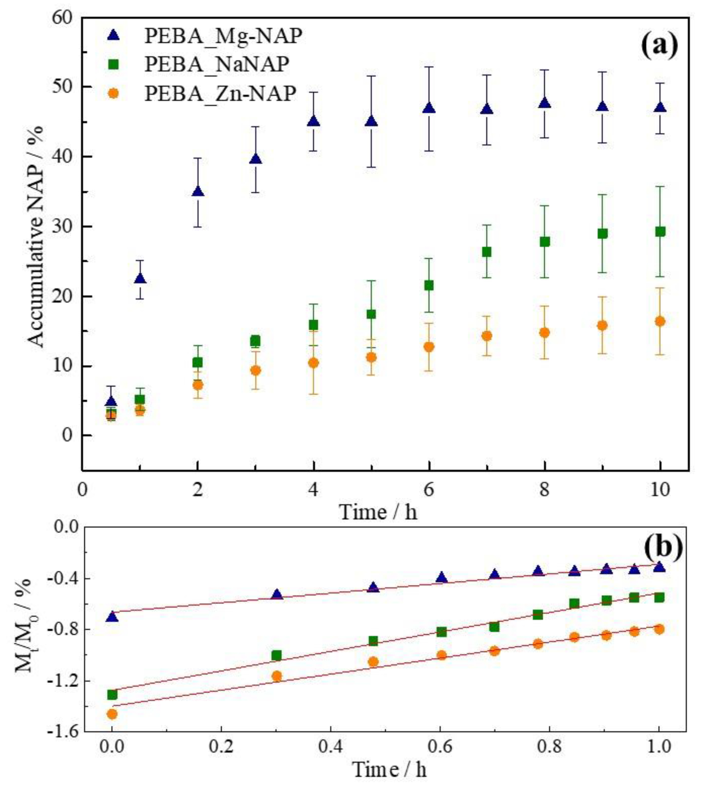
| Sample | k (h−n) | n | R2 |
|---|---|---|---|
| PEBA_Mg-NAP | 0.216 | 0.375 | 0.945 |
| PEBA_NaNAP | 0.053 | 0.762 | 0.985 |
| PEBA_Zn-NAP | 0.040 | 0.626 | 0.969 |
Publisher’s Note: MDPI stays neutral with regard to jurisdictional claims in published maps and institutional affiliations. |
© 2020 by the authors. Licensee MDPI, Basel, Switzerland. This article is an open access article distributed under the terms and conditions of the Creative Commons Attribution (CC BY) license (http://creativecommons.org/licenses/by/4.0/).
Share and Cite
Figueiredo, M.P.; Borrego-Sánchez, A.; García-Villén, F.; Miele, D.; Rossi, S.; Sandri, G.; Viseras, C.; Constantino, V.R.L. Polymer/Iron-Based Layered Double Hydroxides as Multifunctional Wound Dressings. Pharmaceutics 2020, 12, 1130. https://doi.org/10.3390/pharmaceutics12111130
Figueiredo MP, Borrego-Sánchez A, García-Villén F, Miele D, Rossi S, Sandri G, Viseras C, Constantino VRL. Polymer/Iron-Based Layered Double Hydroxides as Multifunctional Wound Dressings. Pharmaceutics. 2020; 12(11):1130. https://doi.org/10.3390/pharmaceutics12111130
Chicago/Turabian StyleFigueiredo, Mariana Pires, Ana Borrego-Sánchez, Fátima García-Villén, Dalila Miele, Silvia Rossi, Giuseppina Sandri, César Viseras, and Vera Regina Leopoldo Constantino. 2020. "Polymer/Iron-Based Layered Double Hydroxides as Multifunctional Wound Dressings" Pharmaceutics 12, no. 11: 1130. https://doi.org/10.3390/pharmaceutics12111130