Therapeutic Apheresis, Circulating PLD, and Mucocutaneous Toxicity: Our Clinical Experience through Four Years


Abstract
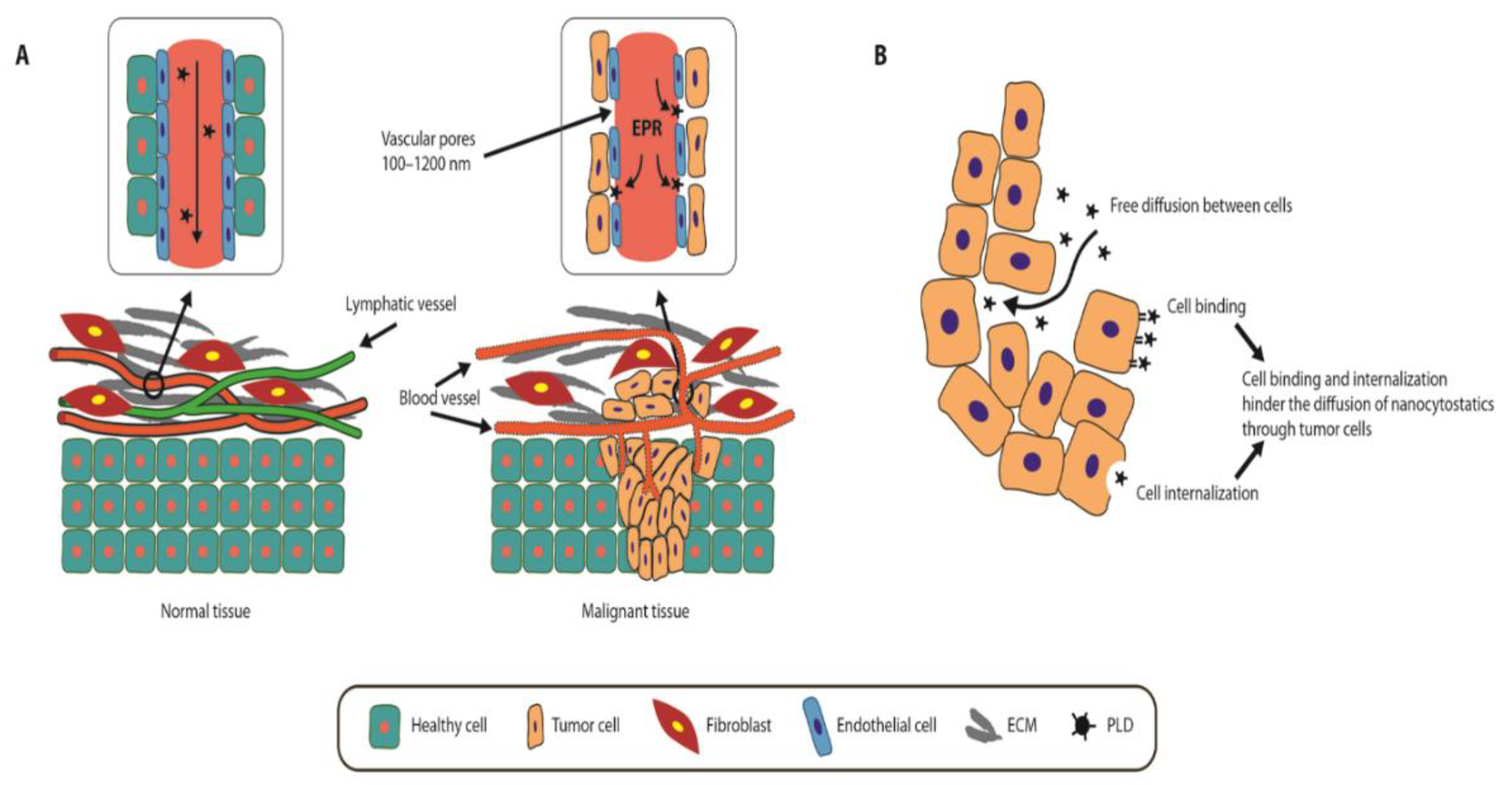
:1. Introduction
2. Pegylated Liposomal Doxorubicin (PLD)
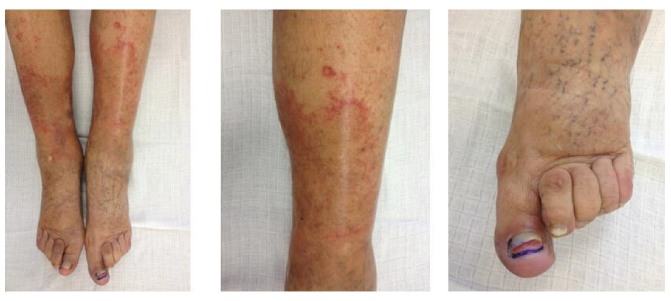
3. Side Effects of PLD Treatment
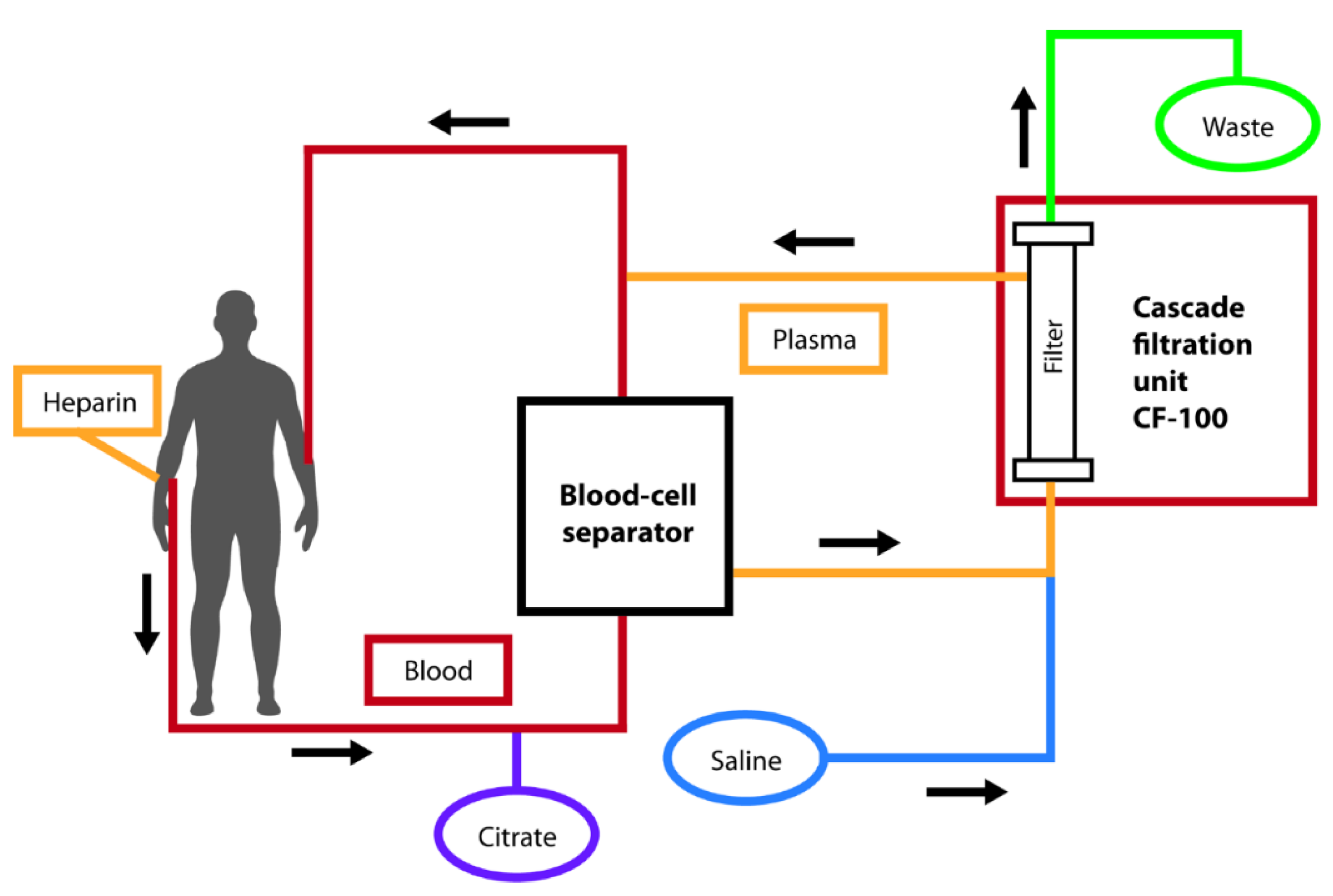
4. Therapeutic Removal of PLD through Double Plasma Filtration (DFPP)
5. Future Perspectives
Author Contributions
Funding
Conflicts of Interest
References
- Pütz, G.; Schmah, O.; Eckes, J.; Hug, M.J.; Winkler, K. Controlled application and scheduled removal of nanoparticle based chemotherapeutics (CARL) will reduce dose limiting adverse events in anticancer chemotherapy. Med. Hypotheses 2009, 72, 393–397. [Google Scholar] [CrossRef]
- Waite, C.L.; Roth, C.M. Nanoscale drug delivery systems for enhanced drug penetration into solid tumors: Current progress and opportunities. Crit. Rev. Biomed. Eng. 2012, 40, 21–41. [Google Scholar] [CrossRef]
- Zhang, R.X.; Ahmed, T.; Li, L.Y.; Li, J.; Abbasi, A.Z.; Wu, X.Y. Design of nanocarriers for nanoscale drug delivery to enhance cancer treatment using hybrid polymer and lipid building blocks. Nanoscale 2017, 9, 1334–1355. [Google Scholar] [CrossRef]
- Allen, T.M.; Cullis, P.R. Drug Delivery Systems: Entering the Mainstream. Science 2004, 303, 1818–1822. [Google Scholar] [CrossRef] [PubMed] [Green Version]
- Samad, A.; Sultana, Y.; Aqil, M. Liposomal drug delivery systems: An update review. Curr. Drug Deliv. 2007, 4, 297–305. [Google Scholar] [CrossRef] [PubMed]
- Fang, J.; Nakamura, H.; Maeda, H. The EPR effect: Unique features of tumor blood vessels for drug delivery, factors involved, and limitations and augmentation of the effect. Adv. Drug Deliv. Rev. 2011, 63, 136–151. [Google Scholar] [CrossRef]
- Millard, M.; Yakavets, I.; Zorin, V.; Kulmukhamedova, A.; Marchal, S.; Bezdetnaya, L. Drug delivery to solid tumors: The predictive value of the multicellular tumor spheroid model for nanomedicine screening. Int. J. Nanomed. 2017, 12, 7993–8007. [Google Scholar] [CrossRef] [PubMed] [Green Version]
- Gabizon, A.; Shmeeda, H.; Barenholz, Y. Pharmacokinetics of Pegylated Liposomal Doxorubicin. Clin. Pharmacokinet. 2003, 42, 419–436. [Google Scholar] [CrossRef] [PubMed]
- Matsumura, Y.; Maeda, H. A new concept for macromolecular therapeutics in cancer chemotherapy: Mechanism of tumoritropic accumulation of proteins and the antitumor agent smancs. Cancer Res. 1986, 46, 6387–6392. [Google Scholar]
- Maeda, H.; Wu, J.; Sawa, T.; Matsumura, Y.; Hori, K. Tumor vascular permeability and the EPR effect in macromolecular therapeutics: A review. J. Control. Release 2000, 65, 271–284. [Google Scholar] [CrossRef]
- Maeda, H. Polymer therapeutics and the EPR effect. J. Drug Target. 2017, 25, 781–785. [Google Scholar] [CrossRef] [PubMed]
- Natfji, A.A.; Ravishankar, D.; Osborn, H.M.I.; Greco, F. Parameters Affecting the Enhanced Permeability and Retention Effect: The Need for Patient Selection. J. Pharm. Sci. 2017, 106, 3179–3187. [Google Scholar] [CrossRef] [PubMed]
- Ye, H.; Shen, Z.; Li, Y.; Wei, M.; Li, Y. Manipulating nanoparticle transport within blood flow through external forces: An exemplar of mechanics in nanomedicine. Proc. Math. Phys. Eng. Sci. 2018, 474, 20170845. [Google Scholar] [CrossRef] [Green Version]
- Pütz, G.; Schmah, O.; Eckes, J.; Hug, M.J.; Winkler, K. Controlled application and removal of liposomal therapeutics: Effective elimination of pegylated liposomal doxorubicin by double-filtration plasmapheresis in vitro. J. Clin. Apher. 2010, 25, 54–62. [Google Scholar] [CrossRef] [PubMed]
- Kubecek, O.; Martínková, J.; Chladek, J.; Bláha, M.; Maláková, J.; Hodek, M.; Špaček, J.; Filip, S. Plasmafiltration as an effective method in the removal of circulating pegylated liposomal doxorubicin (PLD) and the reduction of mucocutaneous toxicity during the treatment of advanced platinum-resistant ovarian cancer. Cancer Chemother. Pharmacol. 2019, 85, 353–365. [Google Scholar] [CrossRef] [PubMed]
- Regev, R.; Yeheskely-Hayon, D.; Katzir, H.; Eytan, G.D. Transport of anthracyclines and mitoxantrone across membranes by a flip-flop mechanism. Biochem. Pharmacol. 2005, 70, 161–169. [Google Scholar] [CrossRef] [PubMed]
- Ngoune, R.; Peters, A.; Von Elverfeldt, D.; Winkler, K.; Pütz, G. Accumulating nanoparticles by EPR: A route of no return. J. Control Release 2016, 238, 58–70. [Google Scholar] [CrossRef] [Green Version]
- Yang, F.; Kemp, C.J.; Henikoff, S. Anthracyclines induce double-strand DNA breaks at active gene promoters. Mutat. Res. Mol. Mech. Mutagen. 2015, 773, 9–15. [Google Scholar] [CrossRef] [Green Version]
- Kotamraju, S.; Kalivendi, S.V.; Konorev, E.; Chitambar, C.R.; Joseph, J.; Kalyanaraman, B. Oxidant-Induced Iron Signaling in Doxorubicin-Mediated Apoptosis. In Enzyme Engineering and Evolution: General Methods; Elsevier: Amsterdam, The Netherlands, 2004; Volume 378, pp. 362–382. [Google Scholar]
- Minotti, G.; Menna, P.; Salvatorelli, E.; Cairo, G.; Gianni, L. Anthracyclines: Molecular Advances and Pharmacologic Developments in Antitumor Activity and Cardiotoxicity. Pharmacol. Rev. 2004, 56, 185–229. [Google Scholar] [CrossRef] [Green Version]
- Gabizon, A.A. Pegylated Liposomal Doxorubicin: Metamorphosis of an Old Drug into a New Form of Chemotherapy. Cancer Investig. 2001, 19, 424–436. [Google Scholar] [CrossRef]
- Eckes, J.; Schmah, O.; Siebers, J.W.; Groh, U.; Zschiedrich, S.; Rautenberg, B.; Hasenburg, A.; Jansen, M.; Hug, M.J.; Winkler, K.; et al. Kinetic Targeting of pegylated liposomal Doxorubicin: A new Approach to Reduce Toxicity during Chemotherapy (CARL-trial). BMC Cancer 2011, 11, 337. [Google Scholar] [CrossRef] [PubMed] [Green Version]
- Ngoune, R.; Contini, C.; Hoffmann, M.M.; Von Elverfeldt, D.; Winkler, K.; Putz, G. Optimizing Antitumor Efficacy and Adverse Effects of Pegylated Liposomal Doxorubicin by Scheduled Plasmapheresis: Impact of Timing and Dosing. Curr. Drug Deliv. 2018, 15, 1261–1270. [Google Scholar] [CrossRef] [PubMed]
- Gabizon, A.A.; Patil, Y.; La-Beck, N.M. New insights and evolving role of pegylated liposomal doxorubicin in cancer therapy. Drug Resist. Updates 2016, 29, 90–106. [Google Scholar] [CrossRef] [PubMed]
- Tahover, E.; Patil, Y.P.; Gabizon, A.A. Emerging delivery systems to reduce doxorubicin cardiotoxicity and improve therapeutic index. Anti-Cancer Drugs 2015, 26, 241–258. [Google Scholar] [CrossRef]
- Ichihara, M.; Shimizu, T.; Imoto, A.; Hashiguchi, Y.; Uehara, Y.; Ishida, T.; Kiwada, H. Anti-PEG IgM Response against PEGylated Liposomes in Mice and Rats. Pharmaceutics 2010, 3, 1–11. [Google Scholar] [CrossRef] [Green Version]
- Sousa, I.; Rodrigues, F.; Prazeres, H.; Lima, R.T.; Soares, P. Liposomal therapies in oncology: Does one size fit all? Cancer Chemother. Pharmacol. 2018, 82, 741–755. [Google Scholar] [CrossRef]
- O’Brien, M.E.R.; Wigler, N.; Inbar, M.; Rosso, R.; Grischke, E.; Santoro, A.; Catane, R.; Kieback, D.G.; Tomczak, P.; Ackland, S.P.; et al. Reduced cardiotoxicity and comparable efficacy in a phase IIItrial of pegylated liposomal doxorubicin HCl(CAELYX™/Doxil®) versus conventional doxorubicin forfirst-line treatment of metastatic breast cancer. Ann. Oncol. 2004, 15, 440–449. [Google Scholar] [CrossRef]
- Gandy, J.; How, C.; Harrold, K. Palmar–plantar erythrodysesthesia (PPE): A literature review with commentary on experience in a cancer centre. Eur. J. Oncol. Nurs. 2007, 11, 238–246. [Google Scholar] [CrossRef]
- Boers-Sonderen, M.J.; Van Herpen, C.M.L.; Van Der Graaf, W.T.A.; Desar, I.M.E.; Van Der Logt, M.G.W.A.; De Beer, Y.M.; Ottevanger, P.B.; Van Erp, N.P. Correlation of toxicity and efficacy with pharmacokinetics (PK) of pegylated liposomal doxorubicin (PLD) (Caelyx®). Cancer Chemother. Pharmacol. 2014, 74, 457–463. [Google Scholar] [CrossRef] [Green Version]
- Bun, S.; Yunokawa, M.; Tamaki, Y.; Shimomura, A.; Shimoi, T.; Kodaira, M.; Shimizu, C.; Yonemori, K.; Fujiwara, Y.; Makino, Y.; et al. Symptom management: The utility of regional cooling for hand-foot syndrome induced by pegylated liposomal doxorubicin in ovarian cancer. Support. Care Cancer 2018, 26, 2161–2166. [Google Scholar] [CrossRef]
- Solomon, R.; Gabizon, A.A. Clinical Pharmacology of Liposomal Anthracyclines: Focus on Pegylated Liposomal Doxorubicin. Clin. Lymphoma Myeloma 2008, 8, 21–32. [Google Scholar] [CrossRef] [PubMed]
- Lyass, O.; Uziely, B.; Ben-Yosef, R.; Tzemach, D.; Heshing, N.I.; Lotem, M.; Brufman, G.; Gabizon, A. Correlation of toxicity with pharmacokinetics of pegylated liposomal doxorubicin (Doxil) in metastatic breast carcinoma. Cancer 2000, 89, 1037–1047. [Google Scholar] [CrossRef]
- Minisini, A.M.; Andreetta, C.; Fasola, G.; Puglisi, F. Pegylated liposomal doxorubicin in elderly patients with metastatic breast cancer. Expert Rev. Anticancer. Ther. 2008, 8, 331–342. [Google Scholar] [CrossRef]
- Gordon, A.N.; Granai, C.; Rose, P.G.; Hainsworth, J.; Lopez, A.; Weissman, C.; Rosales, R.; Sharpington, T. Phase II Study of Liposomal Doxorubicin in Platinum- and Paclitaxel-Refractory Epithelial Ovarian Cancer. J. Clin. Oncol. 2000, 18, 3093–3100. [Google Scholar] [CrossRef] [PubMed]
- Gordon, A.N.; Fleagle, J.T.; Guthrie, D.; Parkin, D.E.; Gore, M.E.; Lacave, A.J. Recurrent Epithelial Ovarian Carcinoma: A Randomized Phase III Study of Pegylated Liposomal Doxorubicin Versus Topotecan. J. Clin. Oncol. 2001, 19, 3312–3322. [Google Scholar] [CrossRef]
- Colombo, N.; Kutarska, E.; Dimopoulos, M.; Bae, D.-S.; Rzepka-Gorska, I.; Bidzinski, M.; Scambia, G.; Engelholm, S.A.; Joly, F.; Weber, D.; et al. Randomized, Open-Label, Phase III Study Comparing Patupilone (EPO906) With Pegylated Liposomal Doxorubicin in Platinum-Refractory or -Resistant Patients With Recurrent Epithelial Ovarian, Primary Fallopian Tube, or Primary Peritoneal Cancer. J. Clin. Oncol. 2012, 30, 3841–3847. [Google Scholar] [CrossRef]
- Mayer, L.D.; Tai, L.C.; Ko, D.S.; Masin, D.; Ginsberg, R.S.; Cullis, P.R.; Bally, M.B. Influence of vesicle size, lipid composition, and drug-to-lipid ratio on the biological activity of liposomal doxorubicin in mice. Cancer Res. 1989, 49, 5922–5930. [Google Scholar]
- Blaha, M.; Martinkova, J.; Lanska, M.; Filip, S.; Malakova, J.; Kubecek, O.; Bezouška, J.; Spacek, J. Plasma filtration for the controlled removal of liposomal therapeutics—From the apheretic site of view. Atheroscler. Suppl. 2017, 30, 286–293. [Google Scholar] [CrossRef]
- Martinkova, J.; Bláha, M.; Kubecek, O.; Malakova, J.; Spacek, J.; Bezouška, J.; Krulichová, I.S.; Filip, S. Plasmafiltration as a possible contributor to kinetic targeting of pegylated liposomal doxorubicin (PLD) in order to prevent organ toxicity and immunosuppression. Cancer Chemother. Pharmacol. 2015, 77, 429–437. [Google Scholar] [CrossRef]
- Moss, D.M.; Siccardi, M. Optimizing nanomedicine pharmacokinetics using physiologically based pharmacokinetics modelling. Br. J. Pharmacol. 2014, 171, 3963–3979. [Google Scholar] [CrossRef]
- Schultink, A.H.M.D.V.; Suleiman, A.A.; Schellens, J.H.M.; Beijnen, J.H.; Huitema, A.D.R. Pharmacodynamic modeling of adverse effects of anti-cancer drug treatment. Eur. J. Clin. Pharmacol. 2016, 72, 645–653. [Google Scholar] [CrossRef] [PubMed] [Green Version]
- Ribba, B.; Holford, N.; Magni, P.; Trocóniz, I.F.; Gueorguieva, I.; Girard, P.; Sarr, C.; Elishmereni, M.; Kloft, C.; Friberg, L.E. A Review of Mixed-Effects Models of Tumor Growth and Effects of Anticancer Drug Treatment Used in Population Analysis. CPT: Pharmacometrics Syst. Pharmacol. 2014, 3, e113. [Google Scholar] [CrossRef] [PubMed]
- Jagetia, G.C.; Nayak, V. Effect of doxorubicin on cell survival and micronuclei formation in HeLa cells exposed to different doses of gamma-radiation. Strahlenther. Onkol. 2000, 176, 422–428. [Google Scholar] [CrossRef] [PubMed]
- Wu, S.; Chou, H.; Yuh, C.; Mekuria, S.L.; Kao, Y.; Tsai, H.-C. Radiation-Sensitive Dendrimer-Based Drug Delivery System. Adv. Sci. 2017, 5, 1700339. [Google Scholar] [CrossRef] [PubMed]
- Fiets, W.; Van Helvoirt, R.; Nortier, J.; Van Der Tweel, I.; Struikmans, H. Acute toxicity of concurrent adjuvant radiotherapy and chemotherapy (CMF or AC) in breast cancer patients. Eur. J. Cancer 2003, 39, 1081–1088. [Google Scholar] [CrossRef]
- Bahaj, W.; Ya’Qoub, L.; Toor, M.; Masood, A. Radiation Recall in a Patient with Intrahepatic Cholangiocarcinoma: Case Report and a Literature Review. Cureus 2019, 11, e5020. [Google Scholar] [CrossRef] [Green Version]
- Burris, H.A.; Hurtig, J. Radiation Recall with Anticancer Agents. Oncologist 2010, 15, 1227–1237. [Google Scholar] [CrossRef] [Green Version]
- Camidge, R.; Price, A. Characterizing the phenomenon of radiation recall dermatitis. Radiother. Oncol. 2001, 59, 237–245. [Google Scholar] [CrossRef]
- Wei, Q.; Xu, W.-H.; Han, M.; Dong, Q.; Fu, Z.-X.; Diao, Y.-Y.; Liu, H.; Xu, J.; Jiang, H.-L.; Zheng, S.; et al. Doxorubicin-mediated radiosensitivity in multicellular spheroids from a lung cancer cell line is enhanced by composite micelle encapsulation. Int. J. Nanomed. 2012, 7, 2661–2671. [Google Scholar] [CrossRef] [Green Version]
- Dicheva, B.M.; Koning, G.A. Targeted thermosensitive liposomes: An attractive novel approach for increased drug delivery to solid tumors. Expert Opin. Drug Deliv. 2013, 11, 83–100. [Google Scholar] [CrossRef]
- Lokerse, W.J.; Bolkestein, M.; Hagen, T.L.T.; De Jong, M.; Eggermont, A.M.; Grüll, H.; Koning, G.A. Investigation of Particle Accumulation, Chemosensitivity and Thermosensitivity for Effective Solid Tumor Therapy Using Thermosensitive Liposomes and Hyperthermia. Theranostics 2016, 6, 1717–1731. [Google Scholar] [CrossRef]
- Huang, S.K.; Stauffer, P.R.; Hong, K.; Guo, J.W.; Phillips, T.L.; Huang, A.; Papahadjopoulos, D. Liposomes and hyperthermia in mice: Increased tumor uptake and therapeutic efficacy of doxorubicin in sterically stabilized liposomes. Cancer Res. 1994, 54, 2186–2191. [Google Scholar]
- Willerding, L.; Limmer, S.; Hossann, M.; Zengerle, A.; Wachholz, K.; Hagen, T.L.T.; Koning, G.A.; Sroka, R.; Lindner, L.H.; Peller, M. Method of hyperthermia and tumor size influence effectiveness of doxorubicin release from thermosensitive liposomes in experimental tumors. J. Control. Release 2016, 222, 47–55. [Google Scholar] [CrossRef] [PubMed]
- Mi, Y.; Shao, Z.; Vang, J.; Kaidar-Person, O.; Wang, A.Z. Application of nanotechnology to cancer radiotherapy. Cancer Nanotechnol. 2016, 7, 511. [Google Scholar] [CrossRef] [PubMed] [Green Version]
- DuRoss, A.N.; Neufeld, M.J.; Rana, S.; Thomas, C.R.; Sun, C. Integrating nanomedicine into clinical radiotherapy regimens. Adv. Drug Deliv. Rev. 2019, 144, 35–56. [Google Scholar] [CrossRef] [PubMed]


| Toxicity | Gordon et al. (2000) [35] n = 89 * | Gordon et al. (2001) [36] n = 239 * | Colombo et al. (2012) [37] n = 409 * | Kubeček et al. (2020) [15] n = 15 ** |
|---|---|---|---|---|
| Anemia (Any Grade) | 35 (39.3%) | 85 (35.6%) | 88 (21.5%) | 5 (33.3%) |
| Grade 3 to 4 | 12 (13.5%) | 13 (5.4%) | 15 (3.7%) | 0 |
| Thrombocytopenia (Any Grade) | n/a | 31 (13.0%) | n/a | 2 (13.3%) |
| Grade 3 to 4 | n/a | 3 (1.3%) | n/a | 0 |
| Neutropenia (Any Grade) | 33 (37.1%) | 84 (35.1%) | 89 (21.8%) | 0 |
| Grade 3 to 4 | 14 (15.7%) | 29 (12.1%) | 41 (10.0%) | 0 |
| PPE (Hand-foot syndrome) | 37 (41.6%) | 117 (49.0%) | 171 (41.8%) | 1 (6.7%) |
| Grade 3 to 4 | 18 (20.2%) | 55 (23.0%) | 55 (13.4%) | 1 (6.7%) |
| Mucositis (Any Grade) | 31 (34.8%) | 95 (39.7%) | 176 (43.0%) | 1 (6.7%) |
| Grade 3 to 4 | 8 (9.0%) | 20 (8.4%) | 41 (10.0%) | 0 |
| Nausea (Any Grade) | 34 (38.2%) | n/a | 188 (46.0%) | 6 (40.0%) |
| Grade 3 to 4 | 6 (6.7%) | n/a | 24 (5.9%) | 1 (6.7%) |
| Vomiting (Any Grade) | 17 (19.1%) | n/a | 131 (32.0%) | 4 (26.7%) |
| Grade 3 to 4 | 4 (4.5%) | n/a | 24 (5.9%) | 0 |
| Constipation (Any Grade) | n/a | n/a | 143 (35.0%) | 3 (20.0%) |
| Grade 3 to 4 | n/a | n/a | 11 (2.7%) | 0 |
| Fatigue (Any Grade) | 37 (41.6%) | n/a | 190 (46.5%) | 2 (13.3%) |
| Grade 3 to 4 | 8 (9.0%) | n/a | 34 (8.3%) | 0 |
© 2020 by the authors. Licensee MDPI, Basel, Switzerland. This article is an open access article distributed under the terms and conditions of the Creative Commons Attribution (CC BY) license (http://creativecommons.org/licenses/by/4.0/).
Share and Cite
Filip, S.; Kubeček, O.; Špaček, J.; Lánská, M.; Bláha, M. Therapeutic Apheresis, Circulating PLD, and Mucocutaneous Toxicity: Our Clinical Experience through Four Years. Pharmaceutics 2020, 12, 940. https://doi.org/10.3390/pharmaceutics12100940
Filip S, Kubeček O, Špaček J, Lánská M, Bláha M. Therapeutic Apheresis, Circulating PLD, and Mucocutaneous Toxicity: Our Clinical Experience through Four Years. Pharmaceutics. 2020; 12(10):940. https://doi.org/10.3390/pharmaceutics12100940
Chicago/Turabian StyleFilip, Stanislav, Ondřej Kubeček, Jiří Špaček, Miriam Lánská, and Milan Bláha. 2020. "Therapeutic Apheresis, Circulating PLD, and Mucocutaneous Toxicity: Our Clinical Experience through Four Years" Pharmaceutics 12, no. 10: 940. https://doi.org/10.3390/pharmaceutics12100940