Article

Herpes Simplex Virus 1 Glycoprotein B from a Hyperfusogenic Virus Mediates Enhanced Cell–Cell Fusion

by
Katrina A. Gianopulos
1,2,3,
Albina O. Makio
1,3,
Suzanne M. Pritchard
1,
Cristina W. Cunha
1,4,
McKenna A. Hull
1 and
Anthony V. Nicola
1,*
1
Department of Veterinary Microbiology and Pathology, College of Veterinary Medicine, Washington State University, Pullman, WA 99164, USA
2
School of Molecular Biosciences, College of Veterinary Medicine, Washington State University, Pullman, WA 99164, USA
3
Protein Biotechnology Graduate Training Program, Washington State University, Pullman, WA 99164, USA
4
Animal Disease Research Unit, Agricultural Research Service, United States Department of Agriculture, Pullman, WA 99164, USA
*
Author to whom correspondence should be addressed.
Viruses 2024, 16(2), 251; https://doi.org/10.3390/v16020251
Submission received: 5 December 2023 / Revised: 29 January 2024 / Accepted: 30 January 2024 / Published: 4 February 2024
(This article belongs to the Special Issue Advances in HSV Research)

Abstract

:
Herpes simplex virus 1 (HSV-1) causes significant morbidity and death in humans worldwide. Herpes simplex virus 1 has a complex fusion mechanism that is incompletely understood. The HSV-1 strain ANG has notable fusion and entry activities that distinguish it from wild type. HSV-1 ANG virions fused with the Vero cell surface at 4 °C and also entered cells more efficiently at 15 °C, relative to wild type HSV-1 strain KOS virions, consistent with a hyperfusogenic phenotype. Understanding the molecular basis for the unique entry and fusion activities of HSV-1 strain ANG will help decipher the HSV fusion reaction and entry process. Sequencing of HSV-1 ANG genes revealed multiple changes in gB, gC, gD, gH, and gL proteins relative to wild type HSV-1 strains. The ANG UL45 gene sequence, which codes for a non-essential envelope protein, was identical to wild type KOS. HSV-1 ANG gB, gD, and gH/gL were necessary and sufficient to mediate cell–cell fusion in a virus-free reporter assay. ANG gB, when expressed with wild type KOS gD and gH/gL, increased membrane fusion, suggesting that ANG gB has hyperfusogenic cell–cell fusion activity. Replacing the KOS gD, gH, or gL with the corresponding ANG alleles did not enhance cell–cell fusion. The novel mutations in the ANG fusion and entry glycoproteins provide a platform for dissecting the cascade of interactions that culminate in HSV fusion and entry.

1. Introduction

Herpes viruses contain multi-component fusion complexes and commandeer multiple entry pathways to enter target cells [1,2,3,4]. HSV particles contain at least 12 different virus-encoded envelope proteins. Wild type HSV entry into all cells requires gB, gD, and gH/gL. Transient cell expression of these four glycoproteins is sufficient for cell–cell fusion [5]. During viral entry, virion gD binds to a host cell receptor, such as nectin-1 or nectin-2. Then, a cascade of interactions between gD, gH/gL, and gB are thought to occur prior to fusion. Host endosomal pH is proposed to be necessary for fusion during epithelial cell entry [6,7].
The HSV-1 ANG strain [8] and its mouse brain passaged derivative ANG path [9] exhibit unique functions in entry and fusion. HSV-1 ANG forms syncytia in culture instead of wild type plaques. ANG has the ability to trigger fusion-from-without (FFWO) [10,11,12], which is the induction of target cell fusion by high MOI addition of intact virions to the monolayer surface in the absence of viral protein expression. A change of alanine to valine at residue 855 in gB causes the ANG syncytial phenotype. FFWO is caused by the combination of an alanine at gB amino acid 553 and the syncytial mutation at 855 [12]. HSV-1 ANG gD contains amino acid changes at 25 and 27 that allow the virus to utilize the nectin-2 receptor for entry [13,14,15,16]. Most strains of HSV enter model CHO-receptor cells by endocytosis [3,7]. A notable exception is that the ANG viruses enter CHO-nectin-2 cells by fusing directly to the cell surface [17,18]. This unique phenotype is not determined solely by the ANG gD or gB alleles [17]. Thus, it cannot be assumed that all of ANG’s unusual fusion phenotypes are dictated by gB. Understanding the viral determinants of the unique entry and fusion activities of HSV-1 strain ANG will help decipher the HSV fusion reaction and entry process. Toward this end, we determined the fusion activities of ANG glycoproteins in a virus-free, cell–cell fusion assay.
In addition to known mutations in ANG gD and gB, sequencing revealed several other amino acid substitutions in the HSV-1 ANG entry glycoproteins, including gC, gH, and gL. Despite the many glycoprotein mutations and the highly fusogenic activities of HSV-1 ANG, the ANG gB, gD, and gH/gL alleles were necessary and sufficient for cell–cell fusion. ANG gB exhibited hyperfusogenic cell–cell fusion activity when co-expressed with wild type KOS gD, gH, and gL. HSV-1 ANG virions fused with the plasma membrane of Vero cells at 4 °C were consistent with enhanced fusion activity of HSV-1 ANG during viral entry.

2. Materials and Methods

2.1. Cells and Viruses

CHO-K1 cells (American Type Culture Collection (ATCC), Manassas, VA, USA) were propagated in Ham’s F12 nutrient mixture (Gibco/Life Technologies, Grand Island, NY, USA), supplemented with 10% fetal bovine serum (FBS) (Atlanta Biologicals, Atlanta, GA, USA). Vero cells (ATCC) were propagated in Dulbecco’s Modified Eagle’s Medium (Thermo Fisher Scientific, Waltham, MA, USA), supplemented with 10% FBS. CHO-HVEM cells [19] or CHO-nectin-2 cells [16] (obtained from G. Cohen and R. Eisenberg) were stably transformed to express the indicated human receptor, and were propagated in Ham’s F12 nutrient mixture, supplemented with 10% FBS, 150 ug/mL puromycin (Sigma-Aldrich, St. Louis, MO, USA), and 250 ug/mL G418 sulfate (Thermo Fisher Scientific). Cells were subcultured in nonselective medium prior to use in all experiments.
HSV-1 strain KOS was obtained from Priscilla Schaffer, Harvard University. HSV-1 KOSrid1 was obtained from Patricia Spear, Northwestern University. Rid1 is a KOS derivative with a Q27P mutation in gD [20]. HSV-1 strain ANG [8] was obtained from R. Eisenberg and G. Cohen. HSV-1 ANG path [9] was obtained from Thomas Holland, Wayne State University. All viruses were propagated and tittered on Vero cells.

2.2. Effect of Reduced Temperature on HSV Entry into Vero Cells

Five-fold dilutions of HSV-1 ANG, KOS, or rid1 (MOI of 1) were prepared in carbonate-free, serum-free DMEM, supplemented with 20 mM HEPES and 0.2% bovine serum albumin. Twenty-four well plate cultures of Vero cells and virus dilutions were equilibrated to 4 °C, 15 °C, or 37 °C for 15 min. The virus was added to cells, and cultures were incubated at 4 °C, 15 °C, or 37 °C for 2 h. Cells were treated with warmed sodium citrate buffer (pH 3.0) at 37 °C for 5 min to inactivate attached viruses that did not fuse with the Vero cell surface. At 18–24 h post-infection at 37 °C, virus titers were measured by plaque assay.

2.3. Sequencing Analysis

Total genomic DNA was extracted from HSV-1 ANG and ANG path virions using the QiaAmp Mini DNA Kit (Qiagen, Louisville, KY, USA). Full-length nucleotide sequences of UL44, UL27, US6, UL22, UL1, and UL45 genes, encoding gC, gB, gD, gH, gL, and UL45p, respectively, were obtained using flanking primers based on HSV-1 KOS and 17 strains. Internal primers based on the generated sequences were also used to obtain the full-length gene when necessary. Sequencing was performed using the Sanger method (Eurofins Genomic LLC, Loisville, KY, USA). Sequences were proofread and assembled using Vector NTI (Invitrogen, Carlsbad, CA, USA) and/or SnapGene (Dotmatics, Boston, MA, USA). Nucleotide and amino acid sequences were compared using Blast software and databases (NCBI, Bethesda, MD, USA).
The DNA sequences of HSV-1 ANG gB, gD, gH, gL, gC, and UL45 and HSV-1 ANG path gH, gL, gC, and UL45 were deposited to GenBank under the respective accession numbers, which are as follows: OQ263027, OQ263028, OQ759608, OQ263025, OQ263031, OQ759609, OQ263026, OQ263030, and OQ263032.

2.4. Construction of Expression Vectors

HSV-1 ANG or ANG path DNA and PCR mutagenesis was used to generate an upstream NsiI and downstream EcoRI site for gB, and an upstream NsiI and downstream SacI site for gC, gD, gH, and gL. For the NsiI site, the primer pairs consisted of sense primer 5′-AGT TAT GCA TTC ACA GGT CGT CCT CG-3′ and antisense primer 5′-CGC AGA ATT CAT GCG CCA GGG C-3′ (gB); sense primer 5′-CAC TAT GCA TTT ACC GCC GAT GAC GC-3′ and antisense primer 5′-ATT AGA GCT C AT GGC CCC GGG G-3′ (gC); sense primer 5′-CAC AAT GCA TAT CTA GTA AAA CAA GGG CTG GT-3′ and antisense primer 5′-TAT AGA GCT CAT GGG GGG GGC T-5′ (gD); sense primer 5′-CAC TAT GCA TCC CTT TAT TCG CGT CTC-3′ and antisense primer 5′-GAG ACG CGA ATA AAG AGG TGC ATA GTG-3′ (gH); and sense primer 5′-AGG TAT GCA TTT AGA TGC GCC GGG A-3′ and antisense primer 5′-T CCC GGC GCA TCT AAA TGC ATA CCT-3′ (gL).
The HSV-1 ANG gB PCR product was first subcloned into the pCR4-TOPO vector (Invitrogen) using TA cloning. The resulting plasmid was digested with EcoRI and NsiI. The HSV-1 ANG gC, gD, gH, and gL PCR products were digested with SacI and NsiI. pCAGGS/MCS [21,22] was digested with SacI and NsiI (or EcoRI and NsiI for gB). The gB gene fragment was gel purified using a QiaQuick Gel Extraction Kit (Qiagen). The glycoprotein gene fragments and pCAGGS/MCS DNAs were ligated using Instant Sticky-End Ligase (New England Biolabs, Ipswich, MA, USA) and transformed into competent E. coli TOP10 cells (Thermo Fisher), generating pSP1 (ANG gB), pSP3 (ANG gC), pSP4 (ANG gD), pSP5 (ANG gH), and pSP2 (ANG gL). All restriction enzymes were from New England Biolabs. All plasmids were sequence-verified.

2.5. Antibodies

Anti-HSV-1 gB mouse monoclonal antibodies H126 [23], H1359, and H1817 [24] were purchased from Virusys, Taneytown, MD, USA. The anti-HSV-1 gC mouse monoclonal antibodies used were T96 (Thermo Scientific), H1413 (Virusys), 3G9 (Virusys), 1C8 [25], and rabbit polyclonal antibody to gC R47 [26] from Roselyn Eisenberg and Gary Cohen. Anti-gD mouse monoclonal antibody DL6 [27] was a gift from G. Cohen and R. Eisenberg, University of Pennsylvania. Anti-HSV-1 gH mouse monoclonal antibody BBH1 was purchased from Abcam, and rabbit polyclonal antibody to gH (R137) was from R. Eisenberg and G. Cohen.

2.6. SDS-PAGE and Western Blot

Transfected cell lysates in SDS sample buffer containing 200 mM dithiothreitol were heated to 85 °C for 5 min. Proteins were separated on 4–20% Tris-glycine gels (Invitrogen, Carlsbad, CA, USA). Gels were transferred to a nitrocellulose membrane and then blocked with 5% milk in PBS-0.2% Tween 20 for 20 min. Membranes were probed overnight with primary antibody to HSV-1 protein gB (H1359), gC (R47), gD (DL6), or gH/gL (R137). The appropriate fluorescent-conjugated secondary antibody was added for 20 min. Images were obtained using an Azure Biosystems imager.

2.7. CELISA

CHO-K1 cells in 96 well plates were transfected with Lipofectamine 3000 (Invitrogen, Carlsbad, CA, USA) and plasmids encoding individual ANG or wild type KOS glycoproteins. Cells were cultured for 18 h, and then fixed in 4% paraformaldehyde. Fixed cells were blocked with 3% BSA in PBS for 2 h. Specific monoclonal antibodies in 3% BSA in PBS were added overnight at 4 °C. Protein A conjugated to horseradish peroxidase (Invitrogen) was added for 2 h at room temperature. Substrate 2,2′-Azinobis [3-ethylbenzothiazoline-6-sulfonic acid]-diammonium salt (ABTS; Thermo Fisher Scientific) was added, and absorbance was measured at 405 nm using a BioTek microplate reader.

2.8. Virus-Free Luciferase Reporter Assay for Cell–Cell Fusion

CHO-K1 (effector) cells were transfected with plasmids encoding T7 RNA polymerase (pCAGT7), HSV-1 wild type plasmids pPEP98 (KOS gB), pPEP99 (KOS gD), pPEP100 (KOS gH), and pPEP101 (KOS gL) [28], all obtained from P. Spear, Northwestern University, or HSV-1 ANG plasmids pSP1 (ANG gB), pSP2 (ANG gC), pSP3 (ANG gD), pSP4 (ANG gH), and pSP5 (ANG gL). Transfections were performed using the Lipofectamine 3000 kit (Invitrogen). Target cells (CHO-HVEM, CHO-nectin-1, or CHO-nectin-2) were transfected with plasmid encoding the firefly luciferase gene under control of the T7 promoter (pT7EMCLuc). Following 6 h incubation in OptiMEM (ThermoFisher Scientific) at 37 °C, target cells were added to effector cells and co-cultured in Ham’s F12 medium for 18 h at 37 °C. Using the ProMega Luciferase Assay System, cells were lysed and lysates were frozen and thawed. Substrate was added to cell lysates and immediately assayed for light output (luciferase activity; fusion) using a BioTek Synergy Neo microplate luminometer.

3. Results

3.1. HSV-1 ANG Enters Vero Cells at 4 °C

The membrane fusion reaction mediated by viral glycoproteins is temperature-dependent. Wild type HSV-1 enters cells rapidly, with a t1/2 of 8–10 min [29,30]. The entry kinetics of HSV-1 ANG at 37 °C are similar to wild type strains [17]. To probe deeper into the ability of HSV-1 virions to fuse with the plasma membrane during entry, plaque formation by HSV-1 KOS, rid1, or ANG was determined at 4, 15, or 37 °C. Vero cells support HSV entry by a pH-independent, direct fusion pathway. HSV-1 ANG virions entered Vero cells at 4 °C (Figure 1). HSV-1 rid1 and KOS exhibited no evidence of entry at 4 °C under the conditions tested (Figure 1). ANG was more effective at Vero cell entry at 15 °C than the other viruses (Figure 1). HSV rid1 can utilize the nectin-2 receptor efficiently due to a Q27P substitution in gD. Likewise HSV-1 ANG interacts with nectin-2 by virtue of amino acid changes in the ANG gD N-terminus [16]. The failure of HSV-1 rid1 to penetrate at 4 °C indicates that nectin-2 receptor utilization is not sufficient for the low temperature fusion activity of ANG. Low temperature virion-cell fusion during entry is another hyperfusogenic feature of the ANG strain.

3.2. HSV-1 ANG and Its Derivative ANG Path Harbor Mutations in Entry and Fusion Glycoproteins

The entry-associated glycoprotein genes UL44 (gC), UL27 (gB), US6 (gD), UL22 (gH), and UL1 (gL) from strain ANG were sequenced. Table 1 indicates the amino acid substitutions in ANG glycoproteins relative to their counterparts in other wild type strains. We confirmed previously reported changes in gB (residue 855) and gD (residues 25 and 27). Relative to other wild type HSV-1s, ANG gC and gH/gL had five and nine changes, respectively (Table 1). gC is not strictly required for HSV entry, but mediates cell attachment [31] and influences gB conformational changes during endosomal entry [32]. gC itself also undergoes pH-triggered conformational changes [33]. The ANG UL45 gene was sequenced as a control and, interestingly, was identical to other wild type strains (Table 1). The UL45 gene is adjacent to UL44 (gC) and encodes the HSV envelope protein UL45p, which has no known role in entry [34,35]. The UL44 (gC), UL22 (gH), UL1 (gL), and UL45 genes from HSV-1 strain ANG path were also sequenced. These ANG path sequences were identical to the corresponding ANG genes (submitted to GenBank).

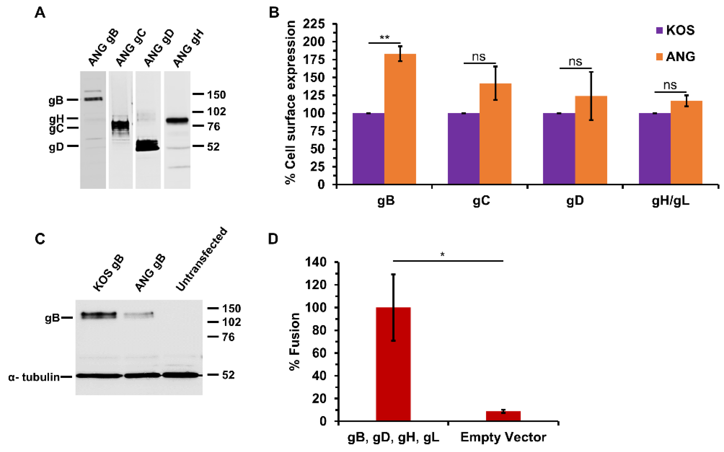
3.3. HSV-1 ANG gB, gD, gH, and gL Are Sufficient for Cell–Cell Fusion

The HSV-1 ANG glycoproteins were each cloned into the pCAGGS plasmid expression vector. CHO-K1 cells were transiently transfected to confirm ANG glycoprotein expression. SDS-PAGE and western blot analysis of transfected cell lysates indicated protein expression of ANG gB, gC, gD, and gH at appropriate molecular weights (Figure 2A). Cell surface expression of the ANG glycoproteins relative to their KOS counterparts was evaluated by an ELISA assay on fixed transfected cells (CELISA) (Figure 2B). ANG gB, gC, gD, and gH/gL were expressed on the CHO cell surface. ANG gB exhibited increased surface expression relative to KOS gB. Protein expression of ANG gB in transfected CHO cells was decreased relative to KOS gB (Figure 2C). The virus-free reporter assay for transfected cell fusion was employed to assess the fusion activity of the ANG glycoproteins. Effector CHO cells transiently expressing ANG gB, gD, gH, and gL mediated fusion with CHO-nectin-2 target cells (Figure 2D). Omitting ANG gB resulted in background levels of fusion (Figure 3B). These results suggest that despite the multiple amino acid substitutions in the ANG glycoproteins, ANG gB, gD, and gH/gL mediate HSV fusion.

3.4. ANG gB Exhibits Hyperfusogenic Activity for Cell–Cell Fusion When Co-Expressed with Wild Type gD and gH/gL

The ANG gB allele is sufficient for ANG virions to induce fusion-from-without (FFWO) in a KOS background [12]. Likewise, ANG syncytium formation is determined by ANG gB, specifically by the valine at position 855 of gB. Results from the virus-free reporter assay for cell–cell fusion have been central to current models of the HSV fusion mechanism. We used this luciferase reporter assay to determine the ability of ANG gB to mediate fusion together with KOS gD and gH/gL. A FFWO allele of gB has not previously been evaluated for cell–cell fusion activity.
Effector cells expressing KOS gB, gD, gH, and gL are necessary and sufficient for fusion with target cells that express HVEM or nectin-1 (Figure 3A,B) [5,28]. Omission of any one of the four proteins results in low levels of luciferase activity, representing assay background. When KOS gB was replaced with ANG gB, there was enhanced HVEM-mediated fusion (Figure 3A) and enhanced nectin-1-mediated fusion (Figure 3B). This enhancement is at least partly explained by the increased surface expression of ANG gB compared to KOS gB (Figure 2B). Expression of KOS gB with ANG gD and ANG gH/gL elicited significantly less fusion than the four ANG glycoproteins together. This fusion was comparable to that detected with the four wild type KOS glycoproteins. Together, the results suggest that HSV-1 ANG glycoprotein mediated cell–cell fusion is enhanced relative to KOS, and that ANG gB determines this hyperfusogenicity.

3.5. HSV-1 ANG Alleles of gD or gH/gL Do Not Alter Cell–Cell Fusion

Given the numerous amino acid substitutions in ANG entry glycoproteins other than gB (Table 1), we determined whether ANG gD, gH, or gL altered fusion activity. To evaluate the cell–cell fusion potential of ANG gD, nectin-1 was expressed in target cells, since ANG gD and KOS gD both bind to nectin-1. Expression of ANG gD with KOS gB and KOS gH/gL resulted in similar fusion to ANG gD and the ANG glycoproteins (Figure 4A). Expression of KOS gD with ANG gB and ANG gH/gL elicited similar fusion to ANG gD and the ANG glycoproteins (Figure 4A). These results suggest that ANG gD itself does not influence the level of cell–cell fusion mediated by nectin-1.
To assess the cell–cell fusion potential of ANG gH/gL, they were expressed together with KOS gB and gD. This resulted in similar fusion to ANG gH/gL and the ANG glycoproteins, regardless of whether nectin-1 (Figure 4B) or HVEM (Figure 4C) were present in the target cells. Expression of ANG gH or ANG gL with the appropriate three remaining KOS glycoproteins also elicited similar fusion to KOS gB, gD, and gH/gL (Figure 4B,C). These results suggest that ANG gH/gL and their amino acid changes do not influence the level of cell–cell fusion. KOS gH and gL, expressed together or individually with the appropriate remaining ANG glycoproteins, yielded fusion similar to ANG gB, gD, and gH/gL (Figure 4B). This suggests that among the ANG glycoproteins required for cell–cell fusion, gB is the only one with enhanced fusion activity. Together, the results further suggest that despite the nine amino acid changes in ANG gH/gL (Table 1), ANG gH is functionally compatible with wild type KOS gL for fusion, and that ANG gL is similarly compatible with KOS gH.

4. Discussion

The HSV-1 ANG strain has several fusion and entry activities that are distinct from other wild type strains. Results presented here suggest that HSV-1 ANG particles fused with the Vero cell plasma membrane at 4 °C (Figure 1). Thus, HSV-1 ANG has elevated virus-cell fusion activity during viral entry. Entry of HSV-1 into keratinocytes by non-endocytic mechanisms has been detected at 7 °C [36,37], suggesting that host cell factors may also play a role in determining low temperature fusion. The gD alleles from ANG and rid1 viruses both enable virus entry via the nectin-2 receptor [16]. However, rid1 failed to fuse with the Vero cell surface at reduced temperatures (Figure 1), suggesting that ANG gD is not sufficient for the 4 °C fusion activity. Sequencing revealed numerous substitutions in ANG glycoproteins, including gC and gH/gL, that are distinct from other wild type strains (Table 1). Future research will determine whether the newly described substitutions in ANG glycoproteins include determinants of low temperature fusion.
ANG gB, gD, and gH/gL mediated cell–cell fusion in a virus-free luciferase reporter assay that is used widely to construct models of HSV membrane fusion (Figure 2). ANG gB mediated enhanced cell–cell fusion relative to its KOS counterpart, and ANG gD or gH/gL did not (Figure 3 and Figure 4). This is consistent with the FFWO phenotype of ANG. It was demonstrated previously that the ANG gB ectodomain mutation 553, together with a cytoplasmic tail mutation at 855, were responsible for FFWO in a KOS background. Thus, the combination of the rate of entry mutation at gB (residue 553) with the syncytial mutation resulted in hyperfusogenic gB FFWO activity [12] The results for ANG gB and cell–cell fusion are consistent with the findings of Saharkhiz-Langroodi et al. in that ANG gB determines both ANG’s enhanced cell–cell fusion activity and its FFWO activity. In addition to valine at gB position 855, truncations, substitutions of other amino acids at 855, or single amino acid mutations at other residues in the gB cytoplasmic tail can determine syncytium formation in culture [38,39,40,41,42,43]. In addition to ANG gB, gB mutants that are responsible for syncytium formation cause enhanced cell–cell fusion [44,45]. Our results do not address the involvement of specific ANG gB residues, but the syncytial mutation at ANG gB residue 855 is likely critical for ANG’s cell–cell hyperfusogenic activity.
HSV-1 ANG gB has decreased reactivity with monoclonal antibody H126 to the gB fusion domain [18], indicating that it is antigenically distinct from wild type KOS gB. However, ANG gB undergoes fusion-associated conformational changes in gB Domains I and V that are similar to those detected in KOS gB [35,46,47]. Thus, although ANG gB may have a distinct conformation, there is no evidence that ANG gB is more prone to conformational changes than other wild type strains. HSV-1 gC optimizes fusion-associated conformational changes in gB [32]. Virion gC also protects gB from neutralizing antibodies [48]. HSV-1 ANG gC has five amino acid substitutions relative to wild type (Table 1). Its effect on cell–cell fusion mediated by gB, gD, and gH/gL remains to be determined. gK has been reported to play a role in HSV entry via direct penetration at the plasma membrane [49,50]. HSV-1 ANG gK has not yet been sequenced or evaluated for its impact on membrane fusion events.
HSV-1 ANG enters CHO-nectin-2 cells by direct fusion at the cell-surface. This entry pathway for HSV is distinct from the endocytic entry that occurs with all other combinations of virus strain and CHO-receptor cells. Importantly, the ANG gB or gD alleles alone are insufficient to mediate the surface fusion of virions with CHO-nectin-2 cells [17,18]. Thus, there are non-gB determinants of ANG that contribute to its unique virus-cell fusion activity during entry. Future investigation will assess whether the newly described substitutions in ANG glycoproteins, e.g., gH/gL, are determinants of the nectin-2-mediated entry at the plasma membrane. HSV-1 gB, gD, gH, and gL are necessary for viral entry and sufficient for cell–cell fusion. Interactions between gB, gC, gD, and gH/gL have been detected prior to fusion [51,52,53,54]. The fusion cascade is initiated by the binding of gD to a receptor such as nectin-1 or HVEM, resulting in the movement of the C-terminal extension, revealing receptor contact sites on the gD core [55,56,57]. This conformational change is proposed to transmit a signal to gH [58]. Interactions between gH and gB ectodomains are thought to be critical for fusion activity. Endosomal low pH has been proposed to induce fusion-activating changes in gB in a cell-specific manner [59,60]. The gH cytoplasmic tail is thought to function as a wedge that is inserted into a pocket on the gB cytoplasmic tail prior to fusion [43,61]. The fusion reaction culminates in gB refolding, and the merger of viral and host membranes forming a fusion pore. Defining a more detailed HSV fusion mechanism remains a priority and a target for novel interventions [1,2,3,4]. Elucidating the molecular determinants of the altered entry and fusion phenotypes of the HSV-1 ANG strain will lead to better understanding of the wild type HSV processes.

Author Contributions

Conceptualization, K.A.G., S.M.P. and A.V.N.; methodology, K.A.G., A.O.M., S.M.P., C.W.C., M.A.H. and A.V.N.; formal analysis, K.A.G., A.O.M., S.M.P., C.W.C. and A.V.N.; investigation, K.A.G., A.O.M., S.M.P. and C.W.C.; writing—original draft, K.A.G. and A.V.N.; writing—review and editing, K.A.G., A.O.M., S.M.P., C.W.C., M.A.H. and A.V.N.; supervision S.M.P., M.A.H. and A.V.N.; project administration, A.V.N.; funding acquisition, A.V.N. All authors have read and agreed to the published version of the manuscript.

Funding

This study was funded by National Institutes of Health grants R01GM152745, R56AI119159, and T32GM008336.

Institutional Review Board Statement

Not applicable.

Informed Consent Statement

Not applicable.

Data Availability Statement

All data reported in the manuscript will be made available upon request to the corresponding author.

Acknowledgments

We thank Gary Cohen, Roselyn Eisenberg, Thomas Holland, Priscilla Schaffer, and Patricia Spear for providing reagents.

Conflicts of Interest

The authors declare no conflicts of interest.

References

  1. Connolly, S.A.; Jardetzky, T.S.; Longnecker, R. The structural basis of herpesvirus entry. Nat. Rev. Microbiol. 2021, 19, 110–121. [Google Scholar] [CrossRef] [PubMed]
  2. Krummenacher, C.; Carfi, A.; Eisenberg, R.J.; Cohen, G.H. Entry of herpesviruses into cells: The enigma variations. Adv. Exp. Med. Biol. 2013, 790, 178–195. [Google Scholar] [CrossRef] [PubMed]
  3. Nicola, A.V. Herpesvirus Entry into Host Cells Mediated by Endosomal Low pH. Traffic 2016, 17, 965–975. [Google Scholar] [CrossRef] [PubMed]
  4. Weed, D.J.; Nicola, A.V. Herpes simplex virus Membrane Fusion. Adv. Anat. Embryol. Cell Biol. 2017, 223, 29–47. [Google Scholar] [CrossRef] [PubMed]
  5. Turner, A.; Bruun, B.; Minson, T.; Browne, H. Glycoproteins gB, gD, and gHgL of herpes simplex virus type 1 are necessary and sufficient to mediate membrane fusion in a Cos cell transfection system. J. Virol. 1998, 72, 873–875. [Google Scholar] [CrossRef]
  6. Nicola, A.V.; Hou, J.; Major, E.O.; Straus, S.E. Herpes simplex virus type 1 enters human epidermal keratinocytes, but not neurons, via a pH-dependent endocytic pathway. J. Virol. 2005, 79, 7609–7616. [Google Scholar] [CrossRef]
  7. Nicola, A.V.; McEvoy, A.M.; Straus, S.E. Roles for endocytosis and low pH in herpes simplex virus entry into HeLa and Chinese hamster ovary cells. J. Virol. 2003, 77, 5324–5332. [Google Scholar] [CrossRef]
  8. Munk, K.; Donner, D. Cytopathischer effekt und plaque-morphologie verschiedener herpes-simplex-virus-stämme. Arch. Für Die Gesamte Virusforsch 1963, 13, 529–540. [Google Scholar] [CrossRef]
  9. Kaerner, H.C.; Schroder, C.H.; Ott-Hartmann, A.; Kumel, G.; Kirchner, H. Genetic variability of herpes simplex virus: Development of a pathogenic variant during passaging of a nonpathogenic herpes simplex virus type 1 virus strain in mouse brain. J. Virol. 1983, 46, 83–93. [Google Scholar] [CrossRef] [PubMed]
  10. Falke, D.; Knoblich, A.; Muller, S. Fusion from without induced by herpes simplex virus type 1. Intervirology 1985, 24, 211–219. [Google Scholar] [CrossRef] [PubMed]
  11. Lingen, M.; Seck, T.; Weise, K.; Falke, D. Single amino acid substitutions in the glycoprotein B carboxy terminus influence the fusion from without property of herpes simplex virus type 1. J. Gen. Virol. 1995, 76 Pt 7, 1843–1849. [Google Scholar] [CrossRef]
  12. Saharkhiz-Langroodi, A.; Holland, T.C. Identification of the fusion-from-without determinants of herpes simplex virus type 1 glycoprotein B. Virology 1997, 227, 153–159. [Google Scholar] [CrossRef]
  13. Dean, H.J.; Warner, M.S.; Terhune, S.S.; Johnson, R.M.; Spear, P.G. Viral determinants of the variable sensitivity of herpes simplex virus strains to gD-mediated interference. J. Virol. 1995, 69, 5171–5176. [Google Scholar] [CrossRef]
  14. Izumi, K.M.; Stevens, J.G. Molecular and biological characterization of a herpes simplex virus type 1 (HSV-1) neuroinvasiveness gene. J. Exp. Med. 1990, 172, 487–496. [Google Scholar] [CrossRef]
  15. Lopez, M.; Cocchi, F.; Menotti, L.; Avitabile, E.; Dubreuil, P.; Campadelli-Fiume, G. Nectin2alpha (PRR2alpha or HveB) and nectin2delta are low-efficiency mediators for entry of herpes simplex virus mutants carrying the Leu25Pro substitution in glycoprotein D. J. Virol. 2000, 74, 1267–1274. [Google Scholar] [CrossRef]
  16. Warner, M.S.; Geraghty, R.J.; Martinez, W.M.; Montgomery, R.I.; Whitbeck, J.C.; Xu, R.; Eisenberg, R.J.; Cohen, G.H.; Spear, P.G. A cell surface protein with herpesvirus entry activity (HveB) confers susceptibility to infection by mutants of herpes simplex virus type 1, herpes simplex virus type 2, and pseudorabies virus. Virology 1998, 246, 179–189. [Google Scholar] [CrossRef]
  17. Delboy, M.G.; Patterson, J.L.; Hollander, A.M.; Nicola, A.V. Nectin-2-mediated entry of a syncytial strain of herpes simplex virus via pH-independent fusion with the plasma membrane of Chinese hamster ovary cells. Virol. J. 2006, 3, 105. [Google Scholar] [CrossRef] [PubMed]
  18. Roller, D.G.; Dollery, S.J.; Doyle, J.L.; Nicola, A.V. Structure-function analysis of herpes simplex virus glycoprotein B with fusion-from-without activity. Virology 2008, 382, 207–216. [Google Scholar] [CrossRef] [PubMed]
  19. Nicola, A.V.; De Leon, M.P.; Xu, R.; Hou, W.; Whitbeck, J.C.; Krummenacher, C.; Montgomery, R.I.; Spear, P.G.; Eisenberg, R.J.; Cohen, G.H. Monoclonal antibodies to distinct sites on herpes simplex virus (HSV) glycoprotein D block HSV binding to HVEM. J. Virol. 1998, 72, 3595–3601. [Google Scholar] [CrossRef] [PubMed]
  20. Dean, H.J.; Terhune, S.S.; Shieh, M.-T.; Susmarski, N.; Spear, P.G. Single Amino Acid Substitutions in gD of Herpes Simplex Virus I Confer Resistance to gD-Mediated Interference and Cause Cell-Type-Dependent Alterations in Infectivity. Virology 1994, 199, 67–80. [Google Scholar] [CrossRef] [PubMed]
  21. Hitoshi, N.; Ken-ichi, Y.; Jun-ichi, M. Efficient selection for high-expression transfectants with a novel eukaryotic vector. Gene 1991, 108, 193–199. [Google Scholar] [CrossRef] [PubMed]
  22. Kobasa, D.; Rodgers, M.E.; Wells, K.; Kawaoka, Y. Neuraminidase hemadsorption activity, conserved in avian influenza A viruses, does not influence viral replication in ducks. J. Virol. 1997, 71, 6706–6713. [Google Scholar] [CrossRef]
  23. Kousoulas, K.G.; Pellett, P.E.; Pereira, L.; Roizman, B. Mutations affecting conformation or sequence of neutralizing epitopes identified by reactivity of viable plaques segregate from syn and ts domains of HSV-1(F) gB gene. Virology 1984, 135, 379–394. [Google Scholar] [CrossRef] [PubMed]
  24. Pereira, L.; Ali, M.; Kousoulas, K.; Huo, B.; Banks, T. Domain structure of herpes simplex virus 1 glycoprotein B: Neutralizing epitopes map in regions of continuous and discontinuous residues. Virology 1989, 172, 11–24. [Google Scholar] [CrossRef]
  25. Friedman, H.M.; Cohen, G.H.; Eisenberg, R.J.; Seidel, C.A.; Cines, D.B. Glycoprotein C of herpes simplex virus 1 acts as a receptor for the C3b complement component on infected cells. Nature 1984, 309, 633–635. [Google Scholar] [CrossRef]
  26. Eisenberg, R.J.; Ponce de Leon, M.; Friedman, H.M.; Fries, L.F.; Frank, M.M.; Hastings, J.C.; Cohen, G.H. Complement component C3b binds directly to purified glycoprotein C of herpes simplex virus types 1 and 2. Microb. Pathog. 1987, 3, 423–435. [Google Scholar] [CrossRef]
  27. Eisenberg, R.J.; Long, D.; Ponce de Leon, M.; Matthews, J.T.; Spear, P.G.; Gibson, M.G.; Lasky, L.A.; Berman, P.; Golub, E.; Cohen, G.H. Localization of epitopes of herpes simplex virus type 1 glycoprotein D. J. Virol. 1985, 53, 634–644. [Google Scholar] [CrossRef]
  28. Pertel, P.E.; Fridberg, A.; Parish, M.L.; Spear, P.G. Cell fusion induced by herpes simplex virus glycoproteins gB, gD, and gH-gL requires a gD receptor but not necessarily heparan sulfate. Virology 2001, 279, 313–324. [Google Scholar] [CrossRef] [PubMed]
  29. Sodeik, B.; Ebersold, M.W.; Helenius, A. Microtubule-mediated transport of incoming herpes simplex virus 1 capsids to the nucleus. J. Cell Biol. 1997, 136, 1007–1021. [Google Scholar] [CrossRef]
  30. Nicola, A.V.; Straus, S.E. Cellular and viral requirements for rapid endocytic entry of herpes simplex virus. J. Virol. 2004, 78, 7508–7517. [Google Scholar] [CrossRef]
  31. Herold, B.C.; WuDUNN, D.; Soltys, N.; Spear, P.G. Glycoprotein C of herpes simplex virus type 1 plays a principal role in the adsorption of virus to cells and in infectivity. J. Virol. 1991, 65, 1090–1098. [Google Scholar] [CrossRef]
  32. Sari, T.K.; Gianopulos, K.A.; Weed, D.J.; Schneider, S.M.; Pritchard, S.M.; Nicola, A.V. Herpes Simplex Virus Glycoprotein C Regulates Low-pH Entry. Msphere 2020, 5, e00826-19. [Google Scholar] [CrossRef]
  33. Gianopulos, K.A.; Komala Sari, T.; Weed, D.J.; Pritchard, S.M.; Nicola, A.V. Conformational Changes in Herpes Simplex Virus Glycoprotein C. J. Virol. 2022, 96, e00163–22. [Google Scholar] [CrossRef] [PubMed]
  34. Dollery, S.J.; Lane, K.D.; Delboy, M.G.; Roller, D.G.; Nicola, A.V. Role of the UL45 protein in herpes simplex virus entry via low pH-dependent endocytosis and its relationship to the conformation and function of glycoprotein B. Virus Res. 2010, 149, 115–118. [Google Scholar] [CrossRef] [PubMed]
  35. Siekavizza-Robles, C.R.; Dollery, S.J.; Nicola, A.V. Reversible conformational change in herpes simplex virus glycoprotein B with fusion-from-without activity is triggered by mildly acidic pH. Virol. J. 2010, 7, 352. [Google Scholar] [CrossRef]
  36. Sayers, C.L.; Elliott, G. Herpes Simplex Virus 1 Enters Human Keratinocytes by a Nectin-1-Dependent, Rapid Plasma Membrane Fusion Pathway That Functions at Low Temperature. J. Virol. 2016, 90, 10379–10389. [Google Scholar] [CrossRef] [PubMed]
  37. De La Cruz, N.; Knebel-Morsdorf, D. Endocytic Internalization of Herpes Simplex Virus 1 in Human Keratinocytes at Low Temperature. J. Virol. 2021, 95, e02195-20. [Google Scholar] [CrossRef] [PubMed]
  38. Bzik, D.J.; Fox, B.A.; DeLuca, N.A.; Person, S. Nucleotide sequence of a region of the herpes simplex virus type 1 gB glycoprotein gene: Mutations affecting rate of virus entry and cell fusion. Virology 1984, 137, 185–190. [Google Scholar] [CrossRef] [PubMed]
  39. Cai, W.H.; Gu, B.; Person, S. Role of glycoprotein B of herpes simplex virus type 1 in viral entry and cell fusion. J. Virol. 1988, 62, 2596–2604. [Google Scholar] [CrossRef] [PubMed]
  40. Cai, W.Z.; Person, S.; DebRoy, C.; Gu, B.H. Functional regions and structural features of the gB glycoprotein of herpes simplex virus type 1. An analysis of linker insertion mutants. J. Mol. Biol. 1988, 201, 575–588. [Google Scholar] [CrossRef]
  41. Gage, P.J.; Levine, M.; Glorioso, J.C. Syncytium-inducing mutations localize to two discrete regions within the cytoplasmic domain of herpes simplex virus type 1 glycoprotein B. J. Virol. 1993, 67, 2191–2201. [Google Scholar] [CrossRef]
  42. Engel, J.P.; Boyer, E.P.; Goodman, J.L. Two novel single amino acid syncytial mutations in the carboxy terminus of glycoprotein B of herpes simplex virus type 1 confer a unique pathogenic phenotype. Virology 1993, 192, 112–120. [Google Scholar] [CrossRef] [PubMed]
  43. Pataki, Z.; Sanders, E.K.; Heldwein, E.E. A surface pocket in the cytoplasmic domain of the herpes simplex virus fusogen gB controls membrane fusion. PLoS Pathog. 2022, 18, e1010435. [Google Scholar] [CrossRef] [PubMed]
  44. Ruel, N.; Zago, A.; Spear, P.G. Alanine substitution of conserved residues in the cytoplasmic tail of herpes simplex virus gB can enhance or abolish cell fusion activity and viral entry. Virology 2006, 346, 229–237. [Google Scholar] [CrossRef] [PubMed]
  45. Silverman, J.L.; Greene, N.G.; King, D.S.; Heldwein, E.E. Membrane requirement for folding of the herpes simplex virus 1 gB cytodomain suggests a unique mechanism of fusion regulation. J. Virol. 2012, 86, 8171–8184. [Google Scholar] [CrossRef] [PubMed]
  46. Weed, D.J.; Dollery, S.J.; Komala Sari, T.; Nicola, A.V. Acidic pH Mediates Changes in Antigenic and Oligomeric Conformation of Herpes Simplex Virus gB and Is a Determinant of Cell-Specific Entry. J. Virol. 2018, 92, e01034-18. [Google Scholar] [CrossRef] [PubMed]
  47. Dollery, S.J.; Delboy, M.G.; Nicola, A.V. Low pH-induced conformational change in herpes simplex virus glycoprotein B. J. Virol. 2010, 84, 3759–3766. [Google Scholar] [CrossRef]
  48. Komala Sari, T.; Gianopulos, K.A.; Nicola, A.V. Glycoprotein C of herpes simplex virus 1 shields glycoprotein B from antibody neutralization. J. Virol. 2020, 94, e01852-19. [Google Scholar] [CrossRef] [PubMed]
  49. David, A.T.; Saied, A.; Charles, A.; Subramanian, R.; Chouljenko, V.N.; Kousoulas, K.G. A herpes simplex virus 1 (McKrae) mutant lacking the glycoprotein K gene is unable to infect via neuronal axons and egress from neuronal cell bodies. MBio 2012, 3, e00144-12. [Google Scholar] [CrossRef]
  50. Chowdhury, S.; Chouljenko, V.N.; Naderi, M.; Kousoulas, K.G. The amino terminus of herpes simplex virus 1 glycoprotein K is required for virion entry via the paired immunoglobulin-like type-2 receptor alpha. J. Virol. 2013, 87, 3305–3313. [Google Scholar] [CrossRef]
  51. Atanasiu, D.; Whitbeck, J.C.; Cairns, T.M.; Reilly, B.; Cohen, G.H.; Eisenberg, R.J. Bimolecular complementation reveals that glycoproteins gB and gH/gL of herpes simplex virus interact with each other during cell fusion. Proc. Natl. Acad. Sci. USA 2007, 104, 18718–18723. [Google Scholar] [CrossRef] [PubMed]
  52. Atanasiu, D.; Saw, W.T.; Cohen, G.H.; Eisenberg, R.J. Cascade of events governing cell-cell fusion induced by herpes simplex virus glycoproteins gD, gH/gL, and gB. J. Virol. 2010, 84, 12292–12299. [Google Scholar] [CrossRef] [PubMed]
  53. Avitabile, E.; Forghieri, C.; Campadelli-Fiume, G. Complexes between herpes simplex virus glycoproteins gD, gB, and gH detected in cells by complementation of split enhanced green fluorescent protein. J. Virol. 2007, 81, 11532–11537. [Google Scholar] [CrossRef] [PubMed]
  54. Avitabile, E.; Forghieri, C.; Campadelli-Fiume, G. Cross talk among the glycoproteins involved in herpes simplex virus entry and fusion: The interaction between gB and gH/gL does not necessarily require gD. J. Virol. 2009, 83, 10752–10760. [Google Scholar] [CrossRef] [PubMed]
  55. Cocchi, F.; Fusco, D.; Menotti, L.; Gianni, T.; Eisenberg, R.J.; Cohen, G.H.; Campadelli-Fiume, G. The soluble ectodomain of herpes simplex virus gD contains a membrane-proximal pro-fusion domain and suffices to mediate virus entry. Proc. Natl. Acad. Sci. USA 2004, 101, 7445–7450. [Google Scholar] [CrossRef] [PubMed]
  56. Krummenacher, C.; Supekar, V.M.; Whitbeck, J.C.; Lazear, E.; Connolly, S.A.; Eisenberg, R.J.; Cohen, G.H.; Wiley, D.C.; Carfi, A. Structure of unliganded HSV gD reveals a mechanism for receptor-mediated activation of virus entry. EMBO J. 2005, 24, 4144–4153. [Google Scholar] [CrossRef] [PubMed]
  57. Gallagher, J.R.; Saw, W.T.; Atanasiu, D.; Lou, H.; Eisenberg, R.J.; Cohen, G.H. Displacement of the C terminus of herpes simplex virus gD is sufficient to expose the fusion-activating interfaces on gD. J. Virol. 2013, 87, 12656–12666. [Google Scholar] [CrossRef] [PubMed]
  58. Atanasiu, D.; Cairns, T.M.; Whitbeck, J.C.; Saw, W.T.; Rao, S.; Eisenberg, R.J.; Cohen, G.H. Regulation of herpes simplex virus gB-induced cell-cell fusion by mutant forms of gH/gL in the absence of gD and cellular receptors. MBio 2013, 4, e00046-13. [Google Scholar] [CrossRef]
  59. Dollery, S.J.; Wright, C.C.; Johnson, D.C.; Nicola, A.V. Low-pH-dependent changes in the conformation and oligomeric state of the prefusion form of herpes simplex virus glycoprotein B are separable from fusion activity. J. Virol. 2011, 85, 9964–9973. [Google Scholar] [CrossRef]
  60. Weed, D.J.; Pritchard, S.M.; Gonzalez, F.; Aguilar, H.C.; Nicola, A.V. Mildly Acidic pH Triggers an Irreversible Conformational Change in the Fusion Domain of Herpes Simplex Virus 1 Glycoprotein B and Inactivation of Viral Entry. J. Virol. 2017, 91, e02123-16. [Google Scholar] [CrossRef]
  61. Fan, Q.; Hippler, D.P.; Yang, Y.; Longnecker, R.; Connolly, S.A. Multiple Sites on Glycoprotein H (gH) Functionally Interact with the gB Fusion Protein to Promote Fusion during Herpes Simplex Virus (HSV) Entry. MBio 2023, 14, e03368-22. [Google Scholar] [CrossRef] [PubMed]
Figure 1. Entry of HSV-1 ANG, KOS, and rid1 at 4 °C, 15 °C, or 37 °C. Five-fold dilutions of HSV-1 ANG, KOS, or rid1 (MOI of 1) were added to Vero cells at 4 °C, 15 °C, or 37 °C for 2 h; then, after attached virus inactivation, cells were incubated at 37 °C for 18–24 h. Virus titers were measured by plaque assay. Titers determined at 37 °C were normalized to 1.0.
Viruses 16 00251 g001
Figure 2. HSV-1 ANG glycoprotein cell surface expression and glycoprotein-mediated cell–cell fusion. (A) CHO-K1 cells were transfected with plasmids encoding HSV-1 ANG glycoproteins gB, gC, gD, or gH and gL. Lysates were resolved by SDS-PAGE. Western blots were probed with anti-gB, gC, gD, or gH antibodies. Molecular weight standards (in kilodaltons) are shown to the right. (B) CHO-K1 cells were transfected with plasmids encoding HSV-1 KOS or ANG glycoproteins for 24 h. Cells were fixed with paraformaldehyde, and then incubated with antibodies specific for gB (cocktail of H126, H1359, and H1817), gC (cocktail of 1C8, 3G9, H1413, and T96), gD (DL6), or gH (BBH1). HRP-conjugated Protein A was added, followed by ABTS substrate. OD values ranged from 0.59 to 1.73 for KOS glycoproteins and 0.17 to 0.26 for empty vector. (C) CHO-K1 cells were transfected with plasmids encoding HSV-1 KOS gB or HSV-1 ANG gB. Lysates were resolved by SDS-PAGE, and the western blot was probed with anti-gB and anti-α-tubulin antibodies (H1359 and DM1A, respectively). Molecular weight standards (in kilodaltons) are shown to the right. (D) CHO-K1 effector cells transiently expressing HSV-1 ANG gB, gD, gH, gL, and T7 polymerase were co-cultured for 18 h with CHO-nectin-2 cells transiently transfected with a plasmid coding luciferase under control of the T7 promoter, and fusion was quantitated by luciferase-induced luminescence. Results are the mean of three independent experiments. *, p < 0.05; **, p < 0.01; ns, not significant; Student’s t test.
Viruses 16 00251 g002
Figure 3. HSV-1 ANG glycoprotein B enhances cell–cell fusion. CHO-K1 effector cells expressing either HSV-1 KOS or ANG gB, gD, gH, gL, and T7 polymerase were co-cultured for 18 h with (A) CHO-HVEM cells or (B) CHO-K1 cells expressing nectin-1 transfected with the luciferase plasmid. Fusion was quantitated by luciferase-induced luminescence. Luciferase activity of KOS gD, gB, gH, and gL was set to 100%. Results are the means of at least two independent experiments. *, p < 0.05, Student’s t test.
Viruses 16 00251 g003
Figure 4. Effect of ANG gD and gH/gL on cell–cell fusion. CHO-K1 effector cells expressing either HSV-1 KOS or ANG gB, gD, gH, gL, and T7 polymerase were co-cultured for 18 h with (A,B) CHO-K1 cells expressing nectin-1 or (C) CHO-HVEM cells transfected with the luciferase plasmid. Fusion was quantitated by luciferase-induced luminescence. Luciferase activity of KOS gD, gB, gH, and gL was set to 100%. Results are the mean of three independent experiments. ns, not significant (Student’s t test).
Viruses 16 00251 g004
Table 1. HSV-1 ANG envelope protein amino acid substitutions. Amino acid substitutions in HSV-1 ANG glycoproteins relative to HSV-1 strains KOS, F, 17, and Patton are shown. ANG and ANG path nucleotide and amino acid sequences were identical for gC, gH, gL, and UL45.
HSV-1 (ANG) Envelope ProteinAmino Acid Substitutions Relative to HSV-1 (WT)
gBD285N, D329N, V553A, E830K, A855V
gDL25P, Q27R, T230I, A346G
gHA138L, S138L, T150V, A150V, E304K, E459K
gLK90R, V100G, N115D, P168L, P196S
gCR151H, M163T, I182V, T238I, V300D
UL45None
Disclaimer/Publisher’s Note: The statements, opinions and data contained in all publications are solely those of the individual author(s) and contributor(s) and not of MDPI and/or the editor(s). MDPI and/or the editor(s) disclaim responsibility for any injury to people or property resulting from any ideas, methods, instructions or products referred to in the content.

