Article

Discovery of a Novel Intron in US10/US11/US12 of HSV-1 Strain 17

Laboratory of Human Retrovirology and Lmmunoinformatics, Frederick National Laboratory for Cancer Research, Frederick, MD 21702, USA
*
Author to whom correspondence should be addressed.
Viruses 2023, 15(11), 2144; https://doi.org/10.3390/v15112144
Submission received: 13 September 2023 / Revised: 23 October 2023 / Accepted: 23 October 2023 / Published: 25 October 2023
(This article belongs to the Special Issue Advances in HSV Research)

Abstract

:
Herpes Simplex Virus type 1 (HSV-1) infects humans and causes a variety of clinical manifestations. Many HSV-1 genomes have been sequenced with high-throughput sequencing technologies and the annotation of these genome sequences heavily relies on the known genes in reference strains. Consequently, the accuracy of reference strain annotation is critical for future research and treatment of HSV-1 infection. In this study, we analyzed RNA-Seq data of HSV-1 from NCBI databases and discovered a novel intron in the overlapping coding sequence (CDS) of US10 and US11, and the 3′ UTR of US12 in strain 17, a commonly used HSV-1 reference strain. To comprehensively understand the shared US10/US11/US12 intron structure, we used US11 as a representative and surveyed all US11 gene sequences from the NCBI nt/nr database. A total of 193 high-quality US11 sequences were obtained, of which 186 sequences have a domain of uninterrupted tandemly repeated RXP (Arg-X-Pro) in the C-terminus half of the protein. In total, 97 of the 186 sequences encode US11 protein with the same length of the mature US11 in strain 17:26 of them have the same structure of US11 and can be spliced as in strain 17; 71 of them have transcripts that are the same as mature US11 mRNA in strain 17. In total, 76 US11 gene sequences have either canonical or known noncanonical intron border sequences and may be spliced like strain 17 and obtain mature US11 CDS with the same length. If not spliced, they will have extra RXP repeats. A tandemly repeated RXP domain was proposed to be essential for US11 to bind with RNA and other host factors. US10 protein sequences from the same strains have also been studied. The results of this study show that even a frequently used reference organism may have errors in widely used databases. This study provides accurate annotation of the US10, US11, and US12 gene structure, which will build a more solid foundation to study expression regulation of the function of these genes.

1. Introduction

Herpes Simplex Virus type 1 (HSV-1) is a double-stranded DNA (~152 Kb) virus belonging to the Alphaherpesvirinae subfamily of the Orthoherpesviridae family [1,2]. HSV-1 infects humans and causes a variety of clinical manifestations, including neonatal herpes, corneal blindness, herpetic whitlow, meningitis, encephalitis, and genital herpes [3,4]. With the advance of high-throughput sequencing technologies, an increased number of HSV-1 strains have been sequenced. The annotation of these genome sequences heavily relies on the known genes in reference strains. Therefore, the accuracy of reference strain annotation is critical for the field. Strain 17 is a commonly used HSV-1 reference strain, which has 56 protein coding genes in the long unique region (UL) [5,6], 2 protein coding genes in the two inverted long repeated regions (TRL and IRL) [6,7], 12 protein coding genes in the short unique region (US) [6,8], and 1 protein coding gene in the two inverted short repeat regions (IRS and TRS) [6,9].
Next Generation Sequencing (NGS) technologies have accelerated the advances in all biological and medical research in the past two decades [10,11,12,13]. RNA-sequencing (RNA-Seq) uses NGS to determine the presence of all RNA molecules and their quality in a biological sample and directly compares these results between experiments [14]. RNA-Seq, using the Illumina platform, has been used to study the repertoire of human fibroblast circular RNAs associated with cellular responses to HSV-1 strain 17 infection [15]. Using the data from this study, we evaluated the transcripts annotated in the HSV-1 strain 17 genome sequence and discovered a 27 nt novel intron in overlapping coding regions in US10 and US11, and the 3′ UTR of US12. The results were further validated with poly-A RNA-Seq data using the PacBio platform from another group [16,17]. The genome sequences in the same region of other HSV-1 strain genomes were evaluated to obtain the full picture of this region in circulating HSV-1 strains. Although the function of US10 is unclear [18], US11 has been studied and reported by several groups during the past several decades. US11 encodes a late-expressed, double-stranded RNA binding protein [19,20]. It is a virion component and associates with the 60S subunit of ribosomes [20,21]. Like HIV (human immunodeficiency virus) Rev and HLTV-1 (human T-lymphotropic virus type 1) Rex, US11 is involved in post-transcriptional regulation of gene expression [22]. US11 interacts with protein kinase R (PKR), blocks the phosphorylation of eIF-2alpha by activated PKR and prevents the cessation of protein synthesis [23]. US11 also interacts with other host factors, including Nucleolin, which is involved in US11 trafficking [24]; PAT1, which is important for the intracellular movement of viral components [25]; and Nucleophosmin, which is implicated in multiple stages of viral infection for different viruses [26]. In virus-infected cells, the US11 protein drastically reduces the formation of autophagosomes by disrupting the TRIM23-TBK1 Complex, which regulates the innate immune response in the infected cells [27]. An accurate HSV-1 US10/US11/US12 structure and annotation will provide a more solid foundation to study expression regulation and the function of these genes and may help to control HSV-1 infection.

2. Materials and Methods

2.1. Virus Genome Sequence and RNA-Seq Data

All data used in this study were downloaded from NCBI. HSV-1 genome sequences from GenBank: strain 17 (NC_001806.2), F (GU734771.1), McKrae (MN136524.1) and KOS (JQ780693.1), whole genome sequencing data of HSV-1 strain 17 (NCBI SRA #: ERR3278848). RNA-Seq data from SRA using SRA-Tools (v.3.0.3): Illumina total RNA-Seq data of HSV-1 strain 17-infected human fibroblast KMB17 cells (NCBI SRA #: SRR6029569, SRR6029570, SRR6029571) [15], strain F, McKrae and KOS infected neuron (NCBI SRA #: SRR10885165 and SRR10885180 for strain F, SRR10885168 and SRR10885184 for McKrae, SRR10885177 and SRR10885186 for KOS) [28]; PacBio poly-A RNA-Seq data of HSV-1 strain 17-infected Vero cell culture (NCBI BioProject #: PRJNA382882) [16,17].

2.2. Analysis of US10/US11/US12 Gene and Protein

To study the transcriptome of HSV-1, the Illumina RNA-Seq sequencing reads were quality trimmed with Trimmomatic [29] with a quality score > 30 within a sliding window size of 20 and minimum length of 100. The trimmed reads were then aligned to virus genome sequences using STAR (V. 2.7.10b) [30] and indexed with Samtools [31]. For the PacBio poly-A RNA-Seq data, the sequencing reads were aligned to the genome sequence using minimap2 (v2.24) [32]; the SAM file was then converted to BAM, and sorted and indexed with Samtools. The indexed alignment was visualized and investigated using IGV (V. 2.13.2).
Since the novel intron is shared by US10/US11/US12, we only need to use one as a representative to understand the intron structure. US11 sequences were obtained by BLAST searching the NCBI nr/nt database with the coding region of the HSV-1 strain 17 US11 gene (searched on 28 June 2023). Duplicated sequences of the same strains, the sequence of recombinant genomes and the sequences containing ambiguous bases (N), which indicated a low quality of the sequence, were removed. The remaining sequences were grouped based on the lengths of US11 nucleotide sequences of its coding region. The sequences in the same group were then aligned with a multiple sequence aligner, Clustal Omega (V.2.1) [33], in the EBI website (https://www.ebi.ac.uk/Tools/msa/clustalo/, accessed April–September 2023). The US10 protein sequences in the same strains were retrieved from GenBank and followed by the same analyses. The US12 protein structure was not studied because the novel intron is located in the 3′ UTR of US12.

2.3. Phylogenetic Analysis of HSV-1 Strains with Different US11 Gene Length

To analyze the relationship between HSV-1 strains with different US10/US11/US12 gene lengths, we performed a phylogenetic analysis as previously described [34]. Briefly, we used the genome sequences with TRL (~9 K) and TRS (~7 K) trimmed to perform the phylogenetic analysis since TRL and IRL, and TRS and IRS, are two pairs of inverted repeat sequences. These trimmed non-redundant sequences were aligned using ClustalW with standard parameters (v2.1) [35]. The alignment file was then converted to mega format with the MEGAX software package (v11.0.10) [36]. A maximum-likelihood tree was generated using MEGAX software, with the general time-reversible nucleotide substitution model with 5 gamma categories, 1000 bootstrap replicates, and complete deletion of alignment gaps, giving a total of 125,368 positions in the data set.

3. Results

3.1. Discovery of Novel Intron in US10/US11/US12 of HSV-1 Strain 17

To evaluate the annotation of the genome sequence, the Illumina total RNA-Seq sequencing reads from HSV-1 strain 17-infected fibroblast KMB14 were obtained from NCBI (NCBI SRA #: SRR6029571), quality trimmed with Trimmomatic (V 0.39) [29], and aligned with the HSV-1 strain 17 genome sequence (NCBI Accession #: NC_001806.2) using STAR (V 2.7.10b) [37]. The alignments were visualized and evaluated using IGV together with the annotation file. A 27 nt gap was present (upper part in Figure 1A) in the overlapped US10 and US11 coding sequence, which also overlapped with the 3′ UTR of US12. The genome sequence used here can be validated by aligning with the Illumina genome sequencing reads (NCBI SRA #: ERR3278848) (lower part of Figure 1A). With proper shifting of bases of RNA-Seq sequencing reads aligned to the gap region, it becomes clear that this 27 nt gap is a complete intron structure with the border sequence GT–CG (Figure 1B). The intron boundary sequence GT-CG was previously observed [38,39]. The well-known intron in US12 [40,41,42] was observed in the same alignment (upper part of Figure 1A and Figure S1), which validated that the sequencing reads are RNA-Seq reads. The novel intron in US10/US11/US12 was further verified in two other samples, NCBI SRA #: SRR6029569 (Figure S2A,B) and SRR6029570 (Figure S2C,D), showing the reproducibility of the results. We could not find intron-unprocessed RNAs of US10/US11/US12 in these RNA-Seq sequencing reads with total RNA. Most likely, the portion of unprocessed pre-mRNA is too small and needs higher depth to be detected. We further validated our novel discovery with the data from a different platform: combined RNA-Seq sequencing reads with the PacBio SMRT platform from six HSV-1 strain 17-infected Vero cell culture samples (NCBI BioProject #: PRJNA382882) were aligned with the same HSV-1 strain 17 genome sequence. We could identify the same newly discovered intron structure for US10, US11, and US12 (Figure S3). With this novel intron spliced (Strain_17_s), the HSV-1 strain 17 US10 and US11 protein have the same length as other laboratory HSV-1 strains, including F, McKrae, and MacIntyre (303 aa for US10 and 152 aa for US11); is nine amino acids shorter than the US10 and US11 annotated in GenBank (NCBI Accession: NC_001806.2, Strain_17_g); and is three amino acids longer than strain KOS (300 for US10 and 149 aa for US11) (Figure 2A,B). The alignment of publicly available RNA-Seq sequencing reads of strain F (NCBI SRA #: SRR10885165 and SRR10885180), McKrae (NCBI SRA #: SRR10885168 and SRR10885184), and KOS (NCBI SRA #: SRR10885177 and SRR10885186) to their genomic sequences (NCBI ACC #: F, GU734771.1; McKrae, MN136524.1; and KOS, JQ780693.1) showed that these strains also contain the known intron within the US12 transcript [40,41,42], but do not have the intron in the US10/US11/US12 overlapping region (Figure 2C–E and Figure S4A–C).

3.2. Survey of the Intron Region of US11/US10/US12 in HSV-1 Strains

At the nucleotide level, the intron structure for US10, US11, and US12 is identical. Therefore, we used the US11 sequence to comprehensively survey the structure around the newly discovered intron. A total of 408 full-length US11 sequences were obtained by BLAST searching the NCBI nr/nt database with the HSV-1 strain 17 genome sequence of the US11 coding region (searched on 28 June 2023). In total, 241 of these remained after removing duplicated sequences of the same strains or recombinant genomes and 193 sequences were left after removing the sequences containing ambiguous bases (Ns), which indicated low quality. More than half of the sequences were the same length as the HSV-1 strain 17 genome sequence covering US11 coding sequences (486 bp, 26 strains; Figure 3A, Supplemental Data S1) or the same length as the mature HSV-1 strain 17 US11 coding sequence (459 bp, 71 strains; Figure 3B, Supplemental Data S2). Alignment of the 26 US11 genomic sequences of length 486 bp (Supplemental Data S1) showed 9 of them with canonical intron border sequences GT-AG [43] and 17 of them with known noncanonical intron border sequences: 15 with GT-CG [38,39] and 1 with GG-CA [43]. The remaining one GG-CG had known 5′ splicing donor site GG [44] and 3′ splicing acceptor site CG [38,39]. Therefore, the 27 nt sequence within the US11 gene in these strains can be spliced out like strain 17, and they form the same length of mature US11 protein (152 aa) as strain 17.
Most of the remaining US11 gene sequences of the other strains are also a multiple of 9 nt different from the HSV-1 strain 17 US11 gene: 7 sequences are 9 nt shorter (Figure 3C, Supplemental Data S3); 66 sequences are 18 nt shorter (Figure 3D, Supplemental Data S4); 3 sequences are 9 nt (GenBank ACC #: OP297870.1), 18 nt (GenBank ACC #: OQ102003.1), and 27 nt (GenBank ACC #: HM585510.2) longer than HSV-1 strain 17 US11 (Figure 3E, Supplemental Data S5), respectively. All of these sequences (76 total) have either canonical or noncanonical intron donor and acceptor sequences. Therefore, mature US11 in these strains can possibly have the same length as that of US11 in strain 17. However, we do not have access to transcript data for these strains to evaluate this possibility. US11 in 13 strains is shorter than mature US11 in HSV-1 strain 17: 10 sequences are 36 nt shorter than the US11 gene in strain 17 including the frequently used strain KOS (Figure 3G, JQ780693.1) and 3 sequences are 45 nt shorter than the US11 gene in strain 17 (Figure 3F).
Only seven sequences were not a multiple of 9 nt different from US11 of strain 17. Three are 21 nt and one is 24 nt shorter than the US11 gene of strain 17 (Supplemental Data S6), and three sequences are not a multiple of 3 nt (22, 23, and 25) shorter compared with the HSV-1 strain 17 US11 gene (Supplemental Data S7). US11 in these three strains will have a frameshift at the C-terminus. It is worth noting that these three genome sequences contain many ambiguous nucleotides, indicating that these non-multiples of three nt deletions may be due to sequencing errors.
In summary, 186 out of 193 HSV-1 US11 sequences surveyed have either the same mature length as the coding sequence of US11 in strain 17 or a multiple of 9 nt difference from the coding sequence of US11 in strain 17. All of these differences are around the region aligned to the novel intron and are present in US10 and US12.

3.3. HSV-1 US11 and US10 Protein Structure

As described above, US11 in more than half of the strains that either have the 27 nt novel intron or the same length of the mature US11 in strain 17 (Figure 3A,B) will have the US11 protein of 152 aa (Figure 2A). There are a total of 20 tandem RXP repeats located in the C-terminus half of the US11 protein strain 17, as reported by Rixon and McGeoch [45]. The only difference is that they had three extra RXP repeats from the intron, which was unknown at the time. US10 in these strains will also have the same length (Figure 2B). As described above, 76 of these US11 gene sequences are longer than mature US11 in strain 17 and have canonical or known noncanonical splicing sites (Figure 3C–E). If spliced, the US11 protein in these strains may have the same length of mature US11 in strain 17. If these sequences are not spliced, the retained sequence would be translated to one or multiple copies of RXP, and the tandemly repeated RXP in these US11 proteins would still be uninterrupted (Figure 4A) except for strain OQ102003.1, which had a Serine in the Proline position (highlighted in green). Since US11 genes with a length that is 9 or 18 nt shorter than that of US11 in strain 17 have the same sequence within the intron splicing border, we used strain ES (NCBI ACC #: OQ658624) and HSV-N-7 (NCBI ACC #: KY922719.1) to represent each group in this protein alignment (Figure 4A and Figure S5A). Ten US11 genes, which are 36 nt shorter than the US11 gene in strain 17 (Figure 3G), have one RXP repeat less than the mature US11 protein in strain 17 (Figure 4B). Although 45 nt deletions in three US11 genes are aligned to different locations of the US11 gene of strain 17 (Figure 3F), the two RXP deletions are aligned to the same location due to the tandem repeat feature (Figure 4C). In summary, the uninterrupted RXP repeat domain might be essential for the function of US11, but its length is not stringent.
The proposed intron sequences within US11 in different strains are located in the overlapping coding sequence of US10 and US11. US10 and US11 are in different reading frames and US10 has several short PGX (Proline-Glycine-X) repeat regions. If the proposed non-27-nt introns could not be spliced, the US10 protein would retain an extra peptide in the proposed intron regions (Figure S5A). The US10 sequences with a length 9 nt and 18 nt shorter than US10 in HSV-1 strain 17 represented by ES (NCBI ACC #: OQ658624) and HSV-N-7 (NCBI ACC #: KY922719.1) would have an extra six amino acids (PGLPGS) or three amino acids (PGL) in the short tandem PGX repeat domain if the intron could not be spliced. The US10 sequences with a length 36 nt shorter than US10 in HSV-1 strain 17 represented by KOS (NCBI ACC #: JQ780693.1) had lost a PGS repeat in the short tandem PGX repeat domain (Figure S5B). We noticed that US10 proteins in three strains with a length 45 nt shorter than US11 in strain 17 (HSV-H12118—NCBI ACC #: MH999847.1, 1974-HTZ—NCBI ACC #: ON960059.1, and 2158_2007—NCBI ACC #: LT594106.1) have significantly different sequences around the region aligned to the newly discovered intron in US10 in strain 17 (Figure S5C), suggesting that the region is flexible for US10 function. For the three strains that have a longer proposed US10/US11/US12 intron than strain 17 (OP297870.1, OQ102003.1, and HM585510.2), US10 would also retain a significantly different peptide sequence if the intron could not be spliced (Figure S5A).

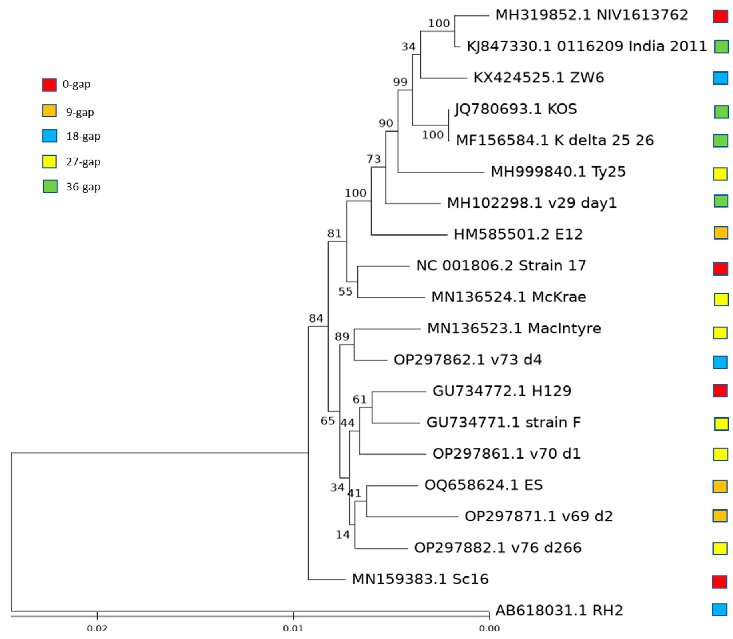
3.4. Evolution of HSV-1 with Different Length of US10/US11 Gene

To determine if the strains with different lengths of US10 and US11 genes have an evolutionary relationship based on the length, we performed a phylogenetic analysis with 20 strains including 5 strains commonly used in HSV-1 research as references: strains 17, F, McKrae, MacIntyre, and KOS, and 15 randomly selected strains with different lengths: 3 from the groups with common strains and 4 from the groups that do not have commonly used strains, to ensure that every group has a balanced number of strains. The phylogenetic tree shows that the strains with the same US10/US11 gene length were not necessarily clustered together (Figure 5), suggesting that strains with the same US10/US11 gene length evolved independently.

4. Discussion

In this work, we discovered a 27 nt novel intron in commonly used HSV-1 reference strain 17, which is located within an overlapping CDS of US10/US11 and the 3′ UTR of US12, using data from the publicly available NCBI databases. The intron is located within the tandem RXP repeat region in US11 with the 27 nt intron representing 3 RXP repeats in the annotation from GenBank in NCBI (NC_001806.2). A total of 20 RXP repeats can be found from position 86 to 145 in mature US11 of strain 17 (Figure 2A). This region is critical for US11 to bind with RNA and associate with ribosomes [20]. It is also the region interacting with PKR [23], Nucleolin [24], PAT1 [25], and Nucleophosmin [26].
A total of 193 US11 gene sequences were surveyed in this study, revealing different lengths of US11. A total of 71 strains have US11 gene sequences with the same length of mature HSV-1 strain 17 US11 without the intron (Figure 3B, Supplemental Data S2). A total of 26 strains have gene sequences with the same length and structure of US11 in strain 17 and can be spliced as US11 in strain 17 (Figure 3A, Supplemental Data S1). Together, more than half of the investigated strains (97 out of 193) have the same US11 protein structure (152 aa). A total of 89 out of the remaining US11 genome sequences are a multiple of 9 nt different from HSV-1 strain 17 US11. A total of 76 of them are longer than mature HSV-1 US11 in strain 17 (Figure 3C–E, and Supplemental Data S3–S5). All 76 of these US11 gene sequences have either canonical or known noncanonical intron donor and acceptor sequences. Therefore, they could have a mature US11 protein of the same length as the strain 17 mature US11 protein, which needs to be further investigated by researchers with access to those strains. If these US11 sequences cannot be spliced, they would have longer lengths than US11 in strain 17 with extra RXP repeats (Figure 4A, shaded with red). Thirteen US11 gene sequences are shorter than the mature US11 in HSV-1 strain 17. Of these, 10 sequences are 36 nt shorter than the US11 gene in strain 17 (Figure 3G) and 3 amino acids (one RXP) shorter than mature US11 in strain 17, like strain KOS (Figure 4B); 3 sequences are 45 nt shorter than the US11 gene in strain 17 (Figure 3F) and 6 amino acids (two RXP) shorter than mature US11 in strain 17 (Figure 4C). Only seven sequences were not a multiple of 9 nt different from US11 of strain 17. A total of 186 out of 193 surveyed US11 amino sequences have uninterrupted RXP repeats, suggesting its possible role in the function of US11. It is proposed that the tandem RXP repeat can form poly-L-proline II helices, which have a strong electrostatic polarity [20,46]. The ring atoms of the proline residues form the uncharged and relatively hydrophobic face. The side chains of the repeating arginine residues form the second, highly positively charged face, which may stabilize binding to RNA by electrostatic interactions with phosphates. The third face is formed with a variety of hydrophobic, uncharged polar, and acidic side chains, which can provide the specificity of binding by US11 [20]. Poly-L-proline II helices are not stabilized with intrachain hydrogen bonds, which is consistent with our predicted structure of mature US11 protein in HSV-1 strain 17 using secondary and tertiary structural prediction tools (Supplemental Methods, Figure S6).
In contrast, the difference in US10 from three strains that are 45 nt shorter than the US10 gene sequence of strain 17 is significantly different in the nearby region from the strain 17 mature US10 protein because the US10 and US11 genes are in different reading frames (Figure S4C), suggesting that the region is not important for the function of US10. In fact, we could not find any substantial reports on the function of US10. For the three strains, which have longer US10 gene sequences than that of strain 17, the US10 protein sequences are the same as mature US10 in strain 17 if the proposed intron was spliced. They would be significantly different with the proposed intron from the strain 17 mature US10 if the proposed intron cannot be spliced (Figure S5A, shaded with red in OP297870.1, OQ102003.1, and HM58551.2).
As described above, US10/US11 in HSV-1 strains have different lengths of gene sequences. Most of them could have mature US10 and US11 of the same length as that of strain 17. We found that not all strains with the same gene lengths evolved from the same ancestor strains using a phylogenetic analysis (Figure 5). How this novel intron is acquired or lost in US10/US11/US12 and the effect of the introns with different lengths on the regulation of gene expression need to be further investigated. On the other hand, the RXP tandem repeat might be a strict restraint for HSV-1 evolution and 9 nt is the insertion/deletion module in the corresponding region of the intron we discovered in strain 17.
The organization of US10, US11, and US12 in the HSV-1 genome is very interesting: they start from a different position but end at the same position. More interestingly, US12 has a very long 3′ UTR, which covers the overlapping full US10 and US11 coding sequences [47]. Since this intron is located within the 3′ UTR of US12, we do not expect any effect on the function of US12. However, the 3′ UTR has been proven to play important roles in the regulation of mRNA including stability, localization, and translation of the mRNA [48]. The function of the 3′ UTR of US12 and how the intron affects this function need to be further investigated.

5. Conclusions

In summary, we discovered a novel intron in the overlapping coding sequences of US10 and US11, which overlaps the 3′ UTR of US12 in the frequently used HSV-1 reference strain 17. The intron structure remains intact in many other strains and these strains have mature US10 and US11 of the same length. This study shows that even sequence information of frequently used model organisms can contain errors and accumulated publicly available data can be useful to correct them. The correction of US10/US11/US12 transcript annotation in HSV-1 strains offers the opportunity to study the function of this intron in the regulation of these genes and could help to better understand HSV-1 infection and its treatment in the future.

Supplementary Materials

The following supporting information can be downloaded at: https://www.mdpi.com/article/10.3390/v15112144/s1, Supplemental Materials: Supplemental Methods with seven references [49,50,51,52,53,54,55], and Figures; Supplemental Data S1: Alignment of the US11 Gene with the Same Gene Length of US11 Gene in HSV-1 Strain 17; Supplemental Data S2: Alignment of the HSV-1 US11 Gene without Intron; Supplemental Data S3: Alignment of the HSV-1 US11 Gene 9 nt Shorter Than HSV-1 Strain 17; Supplemental Data S4: Alignment of the HSV-1 US11 Gene 18 nt Shorter Than HSV-1 Strain 17; Supplemental Data S5: Alignment of the HSV-1 US11 Gene with the Length of Multiple-9-nt Longer Than HSV-1 Strain 17; Supplemental Data S6: Alignment of the HSV-1 US11 Gene Non-Multiple-9-nt shorter Than US11 Gene in HSV-1 Strain 17; Supplemental Data S7: Alignment of the HSV-1 US11 Gene Non-Multiple-3-nt shorter Than US11 Gene in HSV-1 Strain 17.

Author Contributions

Conceptualization, W.C. and T.I.; data analysis, W.C., M.H., J.Q. and B.T.S.; analysis system setting up, B.T.S. and W.C.; writing—original draft preparation, W.C.; review and editing, W.C., M.H., J.Q., B.T.S. and T.I.; supervision, W.C. and T.I.; project administration, W.C. and T.I. All authors have read and agreed to the published version of the manuscript.

Funding

This project was funded in whole or in part with federal funds from the National Cancer Institute, National Institutes of Health, under Contract No. HHSN261200800001E. The content of this publication does not necessarily reflect the views or policies of the Department of Health and Human Services, nor does mention of trade names, commercial products, or organizations imply endorsement by the U.S. Government. This research was supported (in part) by the National Institute of Allergy and Infectious Disease.

Institutional Review Board Statement

Not applicable.

Informed Consent Statement

Not applicable.

Data Availability Statement

All data are acquired from NCBI and EBI using the information in the Materials and Methods section.

Acknowledgments

This work utilized the computational resources of the NIH HPC Biowulf cluster (http://hpc.nih.gov) and the Office of Cyber Infrastructure and Computational Biology (OCICB) High Performance Computing (HPC) cluster at the National Institute of Allergy and Infectious Diseases (NIAID), Bethesda, MD. We thank staff in both facilities for their strong support!

Conflicts of Interest

The authors declare no conflict of interest.

References

  1. Whitley, R.J.; Kimberlin, D.W.; Roizman, B. Herpes simplex viruses. Clin. Infect. Dis. 1998, 26, 541–553. [Google Scholar] [CrossRef] [PubMed]
  2. Gatherer, D.; Depledge, D.P.; Hartley, C.A.; Szpara, M.L.; Vaz, P.K.; Benkő, M.; Brandt, C.R.; Bryant, N.A.; Dastjerdi, A.; Doszpoly, A.; et al. ICTV Virus Taxonomy Profile: Herpesviridae 2021. J. Gen. Virol. 2021, 102, 001673. [Google Scholar] [CrossRef] [PubMed]
  3. Bernstein, D.I.; Bellamy, A.R.; Hook, E.W., 3rd; Levin, M.J.; Wald, A.; Ewell, M.G.; Wolff, P.A.; Deal, C.D.; Heineman, T.C.; Dubin, G.; et al. Epidemiology, clinical presentation, and antibody response to primary infection with herpes simplex virus type 1 and type 2 in young women. Clin. Infect. Dis. 2013, 56, 344–351. [Google Scholar] [CrossRef] [PubMed]
  4. Gnann, J.W., Jr.; Whitley, R.J. Clinical Practice. Genital Herpes. N. Engl. J. Med. 2016, 375, 666–674. [Google Scholar] [CrossRef] [PubMed]
  5. McGeoch, D.J.; Dalrymple, M.A.; Davison, A.J.; Dolan, A.; Frame, M.C.; McNab, D.; Perry, L.J.; Scott, J.E.; Taylor, P. The complete DNA sequence of the long unique region in the genome of herpes simplex virus type 1. J. Gen. Virol. 1988, 69 Pt 7, 1531–1574. [Google Scholar] [CrossRef] [PubMed]
  6. Davison, A.J. Evolution of sexually transmitted and sexually transmissible human herpesviruses. Ann. N. Y. Acad. Sci. 2011, 1230, E37–E49. [Google Scholar] [CrossRef]
  7. Perry, L.J.; McGeoch, D.J. The DNA sequences of the long repeat region and adjoining parts of the long unique region in the genome of herpes simplex virus type 1. J. Gen. Virol. 1988, 69 Pt 11, 2831–2846. [Google Scholar] [CrossRef]
  8. McGeoch, D.J.; Dolan, A.; Donald, S.; Rixon, F.J. Sequence determination and genetic content of the short unique region in the genome of herpes simplex virus type 1. J. Mol. Biol. 1985, 181, 1–13. [Google Scholar] [CrossRef]
  9. McGeoch, D.J.; Dolan, A.; Donald, S.; Brauer, D.H. Complete DNA sequence of the short repeat region in the genome of herpes simplex virus type 1. Nucleic Acids Res. 1986, 14, 1727–1745. [Google Scholar] [CrossRef]
  10. Mardis, E.R. The impact of next-generation sequencing technology on genetics. Trends Genet. 2008, 24, 133–141. [Google Scholar] [CrossRef]
  11. Oehler, J.B.; Wright, H.; Stark, Z.; Mallett, A.J.; Schmitz, U. The application of long-read sequencing in clinical settings. Hum. Genom. 2023, 17, 73. [Google Scholar] [CrossRef] [PubMed]
  12. Enko, D.; Michaelis, S.; Schneider, C.; Schaflinger, E.; Baranyi, A.; Schnedl, W.J.; Muller, D.J. The Use of Next-Generation Sequencing in Pharmacogenomics. Clin. Lab. 2023, 69. [Google Scholar] [CrossRef] [PubMed]
  13. Carangelo, G.; Magi, A.; Semeraro, R. From multitude to singularity: An up-to-date overview of scRNA-seq data generation and analysis. Front. Genet. 2022, 13, 994069. [Google Scholar] [CrossRef] [PubMed]
  14. Wang, Z.; Gerstein, M.; Snyder, M. RNA-Seq: A revolutionary tool for transcriptomics. Nat. Rev. Genet. 2009, 10, 57–63. [Google Scholar] [CrossRef] [PubMed]
  15. Shi, J.; Hu, N.; Mo, L.; Zeng, Z.; Sun, J.; Hu, Y. Deep RNA Sequencing Reveals a Repertoire of Human Fibroblast Circular RNAs Associated with Cellular Responses to Herpes Simplex Virus 1 Infection. Cell. Physiol. Biochem. 2018, 47, 2031–2045. [Google Scholar] [CrossRef]
  16. Tombácz, D.; Csabai, Z.; Szűcs, A.; Balázs, Z.; Moldován, N.; Sharon, D.; Snyder, M.; Boldogkői, Z. Long-Read Isoform Sequencing Reveals a Hidden Complexity of the Transcriptional Landscape of Herpes Simplex Virus Type 1. Front. Microbiol. 2017, 8, 1079. [Google Scholar] [CrossRef]
  17. Tombácz, D.; Moldován, N.; Balázs, Z.; Gulyás, G.; Csabai, Z.; Boldogkői, M.; Snyder, M.; Boldogkői, Z. Multiple Long-Read Sequencing Survey of Herpes Simplex Virus Dynamic Transcriptome. Front. Genet. 2019, 10, 834. [Google Scholar] [CrossRef]
  18. Yamada, H.; Daikoku, T.; Yamashita, Y.; Jiang, Y.M.; Tsurumi, T.; Nishiyama, Y. The product of the US10 gene of herpes simplex virus type 1 is a capsid/tegument-associated phosphoprotein which copurifies with the nuclear matrix. J. Gen. Virol. 1997, 78 Pt 11, 2923–2931. [Google Scholar] [CrossRef]
  19. Johnson, P.A.; MacLean, C.; Marsden, H.S.; Dalziel, R.G.; Everett, R.D. The product of gene US11 of herpes simplex virus type 1 is expressed as a true late gene. J. Gen. Virol. 1986, 67 Pt 5, 871–883. [Google Scholar] [CrossRef]
  20. Roller, R.J.; Monk, L.L.; Stuart, D.; Roizman, B. Structure and function in the herpes simplex virus 1 RNA-binding protein U(s)11: Mapping of the domain required for ribosomal and nucleolar association and RNA binding in vitro. J. Virol. 1996, 70, 2842–2851. [Google Scholar] [CrossRef]
  21. Roller, R.J.; Roizman, B. The herpes simplex virus 1 RNA binding protein US11 is a virion component and associates with ribosomal 60S subunits. J. Virol. 1992, 66, 3624–3632. [Google Scholar] [CrossRef] [PubMed]
  22. Diaz, J.J.; Dodon, M.D.; Schaerer-Uthurralt, N.; Simonin, D.; Kindbeiter, K.; Gazzolo, L.; Madjar, J.J. Post-transcriptional transactivation of human retroviral envelope glycoprotein expression by herpes simplex virus Us11 protein. Nature 1996, 379, 273–277. [Google Scholar] [CrossRef] [PubMed]
  23. Cassady, K.A.; Gross, M.; Roizman, B. The herpes simplex virus US11 protein effectively compensates for the gamma1 (34.5) gene if present before activation of protein kinase R by precluding its phosphorylation and that of the alpha subunit of eukaryotic translation initiation factor 2. J. Virol. 1998, 72, 8620–8626. [Google Scholar] [CrossRef] [PubMed]
  24. Greco, A.; Arata, L.; Soler, E.; Gaume, X.; Couté, Y.; Hacot, S.; Callé, A.; Monier, K.; Epstein, A.L.; Sanchez, J.C.; et al. Nucleolin interacts with US11 protein of herpes simplex virus 1 and is involved in its trafficking. J. Virol. 2012, 86, 1449–1457. [Google Scholar] [CrossRef] [PubMed]
  25. Benboudjema, L.; Mulvey, M.; Gao, Y.; Pimplikar, S.W.; Mohr, I. Association of the herpes simplex virus type 1 Us11 gene product with the cellular kinesin light-chain-related protein PAT1 results in the redistribution of both polypeptides. J. Virol. 2003, 77, 9192–9203. [Google Scholar] [CrossRef]
  26. Nouri, K.; Moll, J.M.; Milroy, L.G.; Hain, A.; Dvorsky, R.; Amin, E.; Lenders, M.; Nagel-Steger, L.; Howe, S.; Smits, S.H.; et al. Biophysical Characterization of Nucleophosmin Interactions with Human Immunodeficiency Virus Rev and Herpes Simplex Virus US11. PLoS ONE 2015, 10, e0143634. [Google Scholar] [CrossRef]
  27. Liu, X.; Matrenec, R.; Gack, M.U.; He, B. Disassembly of the TRIM23-TBK1 Complex by the Us11 Protein of Herpes Simplex Virus 1 Impairs Autophagy. J. Virol. 2019, 93, e00497-19. [Google Scholar] [CrossRef]
  28. Mangold, C.A.; Rathbun, M.M.; Renner, D.W.; Kuny, C.V.; Szpara, M.L. Viral infection of human neurons triggers strain-specific differences in host neuronal and viral transcriptomes. PLoS Pathog. 2021, 17, e1009441. [Google Scholar] [CrossRef]
  29. Bolger, A.M.; Lohse, M.; Usadel, B. Trimmomatic: A flexible trimmer for Illumina sequence data. Bioinformatics 2014, 30, 2114–2120. [Google Scholar] [CrossRef]
  30. Gale, C.V.; Myers, R.; Tedder, R.S.; Williams, I.G.; Kellam, P. Development of a novel human immunodeficiency virus type 1 subtyping tool, Subtype Analyzer (STAR): Analysis of subtype distribution in London. AIDS Res. Hum. Retroviruses 2004, 20, 457–464. [Google Scholar] [CrossRef]
  31. Li, H.; Handsaker, B.; Wysoker, A.; Fennell, T.; Ruan, J.; Homer, N.; Marth, G.; Abecasis, G.; Durbin, R. The Sequence Alignment/Map format and SAMtools. Bioinformatics 2009, 25, 2078–2079. [Google Scholar] [CrossRef] [PubMed]
  32. Li, H. Minimap2: Pairwise alignment for nucleotide sequences. Bioinformatics 2018, 34, 3094–3100. [Google Scholar] [CrossRef] [PubMed]
  33. Sievers, F.; Wilm, A.; Dineen, D.; Gibson, T.J.; Karplus, K.; Li, W.; Lopez, R.; McWilliam, H.; Remmert, M.; Söding, J.; et al. Fast, scalable generation of high-quality protein multiple sequence alignments using Clustal Omega. Mol. Syst. Biol. 2011, 7, 539. [Google Scholar] [CrossRef] [PubMed]
  34. Chang, W.; Jiao, X.; Sui, H.; Goswami, S.; Sherman, B.T.; Fromont, C.; Caravaca, J.M.; Tran, B.; Imamichi, T. Complete Genome Sequence of Herpes Simplex Virus 2 Strain G. Viruses 2022, 14, 536. [Google Scholar] [CrossRef] [PubMed]
  35. Larkin, M.A.; Blackshields, G.; Brown, N.P.; Chenna, R.; McGettigan, P.A.; McWilliam, H.; Valentin, F.; Wallace, I.M.; Wilm, A.; Lopez, R.; et al. Clustal W and Clustal X version 2.0. Bioinformatics 2007, 23, 2947–2948. [Google Scholar] [CrossRef]
  36. Kumar, S.; Stecher, G.; Li, M.; Knyaz, C.; Tamura, K. MEGA X: Molecular Evolutionary Genetics Analysis across Computing Platforms. Mol. Biol. Evol. 2018, 35, 1547–1549. [Google Scholar] [CrossRef]
  37. Dobin, A.; Davis, C.A.; Schlesinger, F.; Drenkow, J.; Zaleski, C.; Jha, S.; Batut, P.; Chaisson, M.; Gingeras, T.R. STAR: Ultrafast universal RNA-seq aligner. Bioinformatics 2013, 29, 15–21. [Google Scholar] [CrossRef]
  38. Gilson, P.R.; Su, V.; Slamovits, C.H.; Reith, M.E.; Keeling, P.J.; McFadden, G.I. Complete nucleotide sequence of the chlorarachniophyte nucleomorph: Nature’s smallest nucleus. Proc. Natl. Acad. Sci. USA 2006, 103, 9566–9571. [Google Scholar] [CrossRef]
  39. Jackson, I.J. A reappraisal of non-consensus mRNA splice sites. Nucleic Acids Res. 1991, 19, 3795–3798. [Google Scholar] [CrossRef]
  40. Todo, T.; Martuza, R.L.; Rabkin, S.D.; Johnson, P.A. Oncolytic herpes simplex virus vector with enhanced MHC class I presentation and tumor cell killing. Proc. Natl. Acad. Sci. USA 2001, 98, 6396–6401. [Google Scholar] [CrossRef]
  41. Umene, K.; Kawana, T. Divergence of reiterated sequences in a series of genital isolates of herpes simplex virus type 1 from individual patients. J. Gen. Virol. 2003, 84 Pt 4, 917–923. [Google Scholar] [CrossRef] [PubMed]
  42. Umene, K.; Yoshida, M. Reiterated sequences of herpes simplex virus type 1 (HSV-1) genome can serve as physical markers for the differentiation of HSV-1 strains. Arch. Virol. 1989, 106, 281–299. [Google Scholar] [CrossRef] [PubMed]
  43. Burset, M.; Seledtsov, I.A.; Solovyev, V.V. Analysis of canonical and non-canonical splice sites in mammalian genomes. Nucleic Acids Res. 2000, 28, 4364–4375. [Google Scholar] [CrossRef] [PubMed]
  44. Robertson, H.M. Noncanonical GA and GG 5′ Intron Donor Splice Sites Are Common in the Copepod Eurytemora affinis. G3 2017, 7, 3967–3969. [Google Scholar] [CrossRef]
  45. Rixon, F.J.; McGeoch, D.J. A 3′ co-terminal family of mRNAs from the herpes simplex virus type 1 short region: Two overlapping reading frames encode unrelated polypeptide one of which has highly reiterated amino acid sequence. Nucleic Acids Res. 1984, 12, 2473–2487. [Google Scholar] [CrossRef]
  46. Schaerer-Uthurralt, N.; Erard, M.; Kindbeiter, K.; Madjar, J.J.; Diaz, J.J. Distinct domains in herpes simplex virus type 1 US11 protein mediate post-transcriptional transactivation of human T-lymphotropic virus type I envelope glycoprotein gene expression and specific binding to the Rex responsive element. J. Gen. Virol. 1998, 79 Pt 7, 1593–1602. [Google Scholar] [CrossRef]
  47. Nishiyama, Y.; Kurachi, R.; Daikoku, T.; Umene, K. The US 9, 10, 11, and 12 genes of herpes simplex virus type 1 are of no importance for its neurovirulence and latency in mice. Virology 1993, 194, 419–423. [Google Scholar] [CrossRef]
  48. Mayr, C. What Are 3′ UTRs Doing? Cold Spring Harb. Perspect. Biol. 2019, 11, a034728. [Google Scholar] [CrossRef]
  49. McGuffin, L.J.; Bryson, K.; Jones, D.T. The PSIPRED protein structure prediction server. Bioinformatics 2000, 16, 404–405. [Google Scholar] [CrossRef]
  50. Montgomerie, S.; Cruz, J.A.; Shrivastava, S.; Arndt, D.; Berjanskii, M.; Wishart, D.S. PROTEUS2: A web server for comprehensive protein structure prediction and structure-based annotation. Nucleic Acids Res. 2008, 36 (Suppl 2), W202–W209. [Google Scholar] [CrossRef]
  51. Høie, M.H.; Kiehl, E.N.; Petersen, B.; Nielsen, M.; Winther, O.; Nielsen, H.; Hallgren, J.; Marcatili, P. NetSurfP-3.0: Accurate and fast prediction of protein structural features by protein language models and deep learning. Nucleic Acids Res. 2022, 50, W510–W515. [Google Scholar] [CrossRef] [PubMed]
  52. Du, Z.; Su, H.; Wang, W.; Ye, L.; Wei, H.; Peng, Z.; Anishchenko, I.; Baker, D.; Yang, J. The trRosetta server for fast and accurate protein structure prediction. Nat. Protoc. 2021, 16, 5634–5651. [Google Scholar] [CrossRef] [PubMed]
  53. Kim, D.E.; Chivian, D.; Baker, D. Protein structure prediction and analysis using the Robetta server. Nucleic Acids Res. 2004, 32 Suppl. 2), W526–W531. [Google Scholar] [CrossRef]
  54. Xu, D.; Zhang, Y. Ab initio protein structure assembly using continuous structure fragments and optimized knowledge-based force field. Proteins 2012, 80, 1715–1735. [Google Scholar] [CrossRef] [PubMed]
  55. Waterhouse, A.; Bertoni, M.; Bienert, S.; Studer, G.; Tauriello, G.; Gumienny, R.; Heer, F.T.; de Beer, T.A.P.; Rempfer, C.; Bordoli, L.; et al. SWISS-MODEL: Homology modelling of protein structures and complexes. Nucleic Acids Res. 2018, 46, W296–W303. [Google Scholar] [CrossRef] [PubMed]
Figure 1. Identification of a novel intron in US10/US11/US12 genes of HSV-1 strain 17: (A) the RNA-Seq sequencing reads from an HSV-1 strain 17-infected human fibroblast KMB17 cell culture sample (NCBI SRA #: SRR6029571) and the genome sequencing reads of HSV-1 strain 17 BAC (NCBI SRA #: ERR3278848) were aligned with the reference sequence, HSV-1 strain 17 genome sequence (NCBI AC #: NC_001806.2), and are shown with light gray and dark gray color, respectively. The novel intron in US10/US11/US12 and well-known US12 intron are marked with a red bracket. The annotated gene structure of US10, US11, and US12 in the reference is shown at the bottom of the panel. Thick bar: coding sequence, narrow bar: untranslated exon sequence, line: intron. These genes are on the complement strand of the reference sequence. (B) A zoomed-in view of the region of a newly identified novel intron. After we move base “C” on right side of gap to left side, the splicing donor site GT (complement of AC) and acceptor site CG (complement of CG) are shown at the border of the novel intron. Some read sequences within the intron could be moved properly as shown in the figure, consistent with the intron structure. US12 intron zoomed-in view is shown in Figure S1.
Viruses 15 02144 g001
Figure 2. No new intron identified in US10/US11/US12 genes in HSV-1 strain F, McKrae, and KOS. Alignment of annotated US11 (A) and US10 (B) protein sequence in HSV-1 strain 17 in reference sequence (Strain_17_g, NCBI ACC #: NC_001806.2) and mature form with 27 nt intron spliced out (Strain_17_s), together with the annotated US11 and US10 from HSV-1 strain F, McKrae, MacItyre, and KOS. (C) The alignment of the RNA-Seq sequencing reads from an HSV-1 strain F-infected human neuron cell culture sample (NCBI SRA #: SRR10885165) with its genome sequence (NCBI ACC #: GU734771.1). (D) The alignment of the RNA-Seq sequencing reads from the HSV-1 strain McKrae-infected human neuron cell culture sample (NCBI SRA #: SRR10885168) with its genome sequence (NCBI ACC #: MN136524.1). (E) The alignment of the RNA-Seq sequencing reads from the HSV-1 strain KOS-infected human neuron cell culture sample (NCBI SRA #: SRR10885177) with its DNA genome sequence (NCBI ACC #: JQ780693.1). The blue bars at the bottom of panels (CE) are the ORFs of US10, US11, and US12. * annotate the homologous position based on the alignment.
Viruses 15 02144 g002
Figure 3. The alignment of US11 gene sequences with different lengths compared to the US11 gene in strain 17. (A) The same length; (B) 27 nt shorter, the same length of mature HSV-1 US11 in HSV-1 strain 17; (C) 9 nt shorter; (D) 18 nt shorter; (E) 9 nt, 18 nt, and 27 nt longer; (F) 45 nt shorter; (G) 36 nt shorter. Splicing donor and acceptor sites are noted with bold font in US11 sequence of strain 17. Bold letters (GT, CG) represent intron border sequences; * represent the homologous position based on the alignment.
Viruses 15 02144 g003
Figure 4. The alignment of US11 amino acid sequences with different lengths compared to mature US11 protein in strain 17. (A) Alignment of the US11 amino acid sequence of different lengths with mature US11 in HSV-1 strain 17: HSV-N-7 (representative of 18-nt-shorter group), ES (representative of 9-nt-shorter group), OP297870.1 (9 nt longer), OQ102003.1 (18 nt longer), and HM585510.2 (27 nt longer). The peptide shaded with red represents the intron region if the introns were not spliced. If the proposed introns were spliced, these peptide sequences would not be present and US11 protein is of the same length as US11 in HSV-1 strain 17. (B) Alignment of the US11 amino acid sequences of 36-nt-shorter group with mature US11 in HSV-1 strain 17. Amino acids in red represent peptide translated from the proposed intron region if the intron is not spliced out. Green “S” annotated Serion occupies Proline position in RXP repeat. (C) Alignment of the US11 amino acid sequences of 45-nt-shorter group with mature US11 in HSV-1 strain 17. * represents homologous position based on the alignment.
Viruses 15 02144 g004
Figure 5. Phylogenetic analysis of HSV-1 genome sequences from the groups with different US10/US11 gene length. Red square: same length of US10/US11 gene in strain 17; brown square: 9 nt shorter than US10/US11 gene in strain 17; blue square: 18 nt shorter than US10/US11 gene in strain 17; yellow square: 27 nt shorter than US10/US11 gene in strain 17; green square: 36 nt shorter than US10/US11 gene in strain 17.
Viruses 15 02144 g005
Disclaimer/Publisher’s Note: The statements, opinions and data contained in all publications are solely those of the individual author(s) and contributor(s) and not of MDPI and/or the editor(s). MDPI and/or the editor(s) disclaim responsibility for any injury to people or property resulting from any ideas, methods, instructions or products referred to in the content.

