Review

Cellular Processes Induced by HSV-1 Infections in Vestibular Neuritis

by
Zhengdong Zhao
1,†,
Xiaozhou Liu
1,†,
Yanjun Zong
1,
Xinyu Shi
1 and
Yu Sun
1,2,*
1
Department of Otorhinolaryngology, Union Hospital, Tongji Medical College, Huazhong University of Science and Technology, Wuhan 430022, China
2
Hubei Province Key Laboratory of Oral and Maxillofacial Development and Regeneration, Wuhan 430022, China
*
Author to whom correspondence should be addressed.
These authors contributed equally to this work.
Viruses 2024, 16(1), 12; https://doi.org/10.3390/v16010012
Submission received: 29 November 2023 / Revised: 18 December 2023 / Accepted: 19 December 2023 / Published: 20 December 2023
(This article belongs to the Special Issue Herpesvirus Manipulation of Cellular Processes 2.0)

Abstract

:
Herpesvirus is a prevalent pathogen that primarily infects human epithelial cells and has the ability to reside in neurons. In the field of otolaryngology, herpesvirus infection primarily leads to hearing loss and vestibular neuritis and is considered the primary hypothesis regarding the pathogenesis of vestibular neuritis. In this review, we provide a summary of the effects of the herpes virus on cellular processes in both host cells and immune cells, with a focus on HSV-1 as illustrative examples.

1. Introduction

Human herpesviruses are large, enveloped, double-stranded DNA viruses that cause a variety of diseases and establish lifelong latent infections in the majority of the global population. The Herpesviridae family comprises nine viruses that are capable of causing human infections and is divided into three subfamilies: Alphaherpesvirinae; Betaherpesvirinae; and Gammaherpesvirinae. Alphaherpesvirinae consists of herpes simplex virus I (HSV-1), HSV-2, and varicella zoster virus (VZV). Betaherpesvirinae includes human cytomegalovirus (HCMV), human herpesvirus 6A (HHV-6A), HHV-6B, and HHV-7. Epstein–Barr virus (EBV) and Kaposi’s sarcoma herpesvirus (KSHV) belong to Gammaherpesvirinae.
Vestibular neuritis (VN) is a clinical condition in the field of otolaryngology characterized by acute and persistent peripheral vertigo caused by unilateral vestibular afferent nerve block. The main symptoms include acute/subacute persistent severe vertigo, accompanied by spontaneous horizontal nystagmus, unsteady posture, and nausea, but without associated auditory dysfunction. The cause of VN is still unknown, but the most common hypothesis is viral infection or reactivation, particularly by HSV (Table 1). Several researchers have reported herpes virus infection in the vestibular ganglion of patients with vestibular neuritis [1,2,3,4,5]. Arbusow et al. reported the presence of HSV-1 DNA in both the human vestibular ganglion and vestibular nuclei [6], suggesting the potential migration of the virus to the human vestibular labyrinth [4]. Furthermore, HSV-1 DNA or HSV latency-related transcripts have been detected in the vestibular ganglia removed during surgery in patients with Meniere’s disease [3,7]. Herpesviruses have a tendency to invade sensory neurons, establish a latency period, and can be reactivated to cause disease. The initial infection or reactivation of the herpesvirus can profoundly affect cellular processes in the host. Mice inoculated with HSV-1 and HSV-2 into the middle ear exhibited hearing loss and vestibular dysfunction. HSV infection was observed in columnar epithelial cells of the infected mice in the stria vascularis, leading to apoptosis in a portion of the infected cells, while many uninfected cells in the spiral organ of Corti also underwent apoptosis. While vestibular ganglion cells did not undergo apoptosis, some of the cells experienced functional loss [8]. Mice inoculated with HSV-1 after auricle also exhibited vestibular dysfunction along with the death of vestibular ganglion cells [9]. In addition to the damage caused to cells by virus infection, the killing of host cells by immune cells may also contribute to the pathogenesis of vestibular neuritis. The coexistence of CD8+ cells and HSV-1 has been observed in the vestibular ganglion cells of patients with vestibular neuritis.
This article provides a summary of the alterations in cellular processes post-infection, using HSV-1 as an exemplars, with the aspiration that this knowledge will aid in the treatment of VN and other diseases instigated by human herpesviruses.

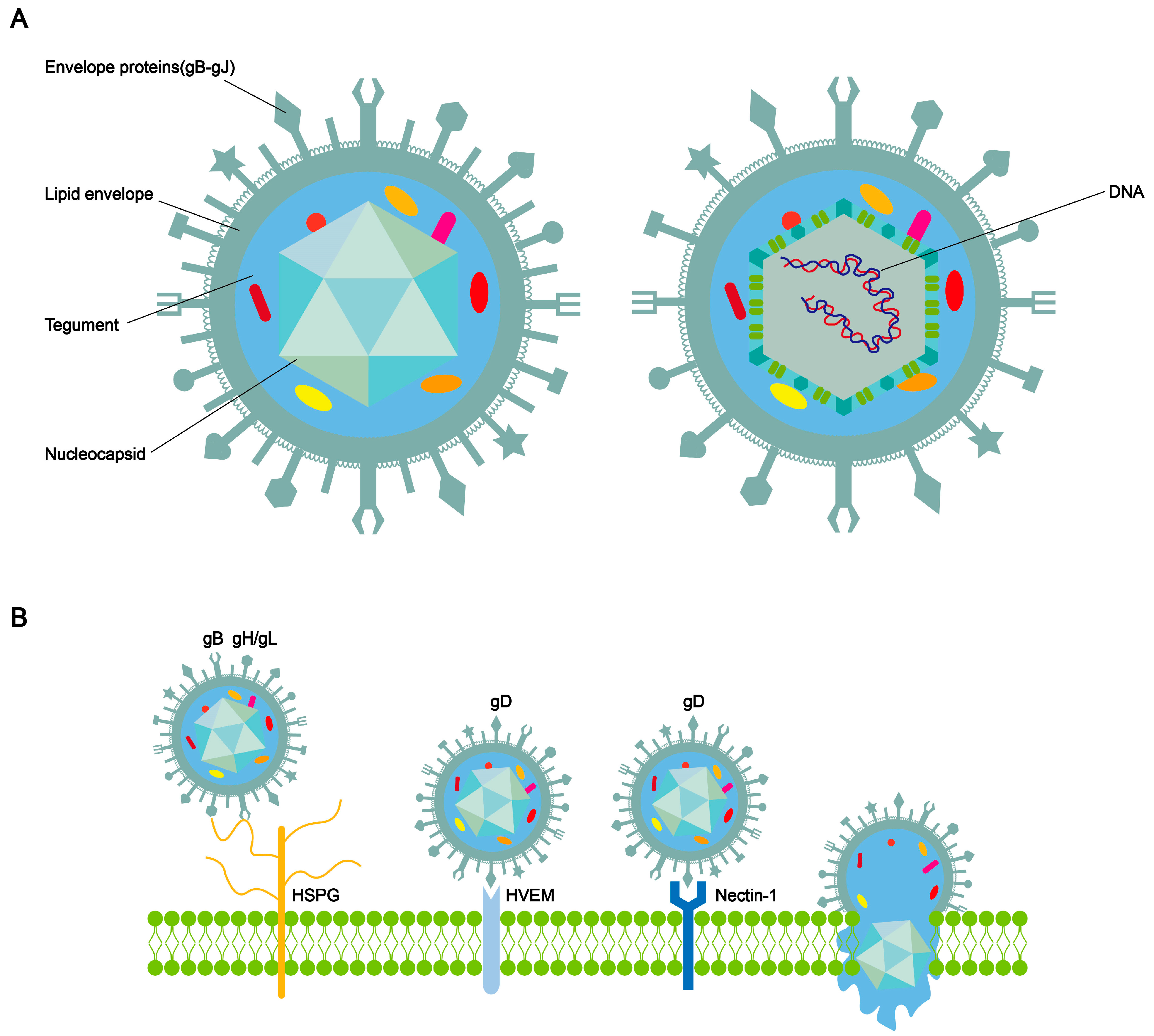
2. The Structure of HSV-1 and the Process of Entry into Cells

HSV-1 has a spherical shape [10]. The complete HSV-1 virus comprises double-stranded DNA, a nucleocapsid, teguments, envelope proteins, and a lipid envelope (Figure 1A) [11,12,13]. The nucleocapsid shell exhibits a symmetrical three-dimensional icosahedral structure. HSV-1 is primarily transmitted through close contact. The entry of herpesvirus into human cells for receptor binding and membrane fusion requires the involvement of multifunctional viral glycoproteins on its surface [14,15,16]. HSV-1 carries a minimum of 12 different glycoproteins. There existed four membrane glycoproteins required for HSV entry into cells: the glycoprotein D (gD)-binding receptor; glycoprotein B (gB); and glycoprotein H/glycoprotein L (gH/gL) constitute the core fusion mechanism [17]. Briefly, when HSV is adsorbed on the cell membrane surface, the initial non-specific binding between glycoprotein gC and/or gB and the heparan sulfate mucin (HSPG) on the cell surface reduces the spatial distance between the viral envelope and the cell membrane. gD can specifically bind to herpes virus entry mediator (HVEM), nectin-1, nectin-2, or 3-O-sulfated heparan sulfate (3-OS-HS). This binding further initiates gH and gL. Subsequently, gH-gL transmits signals to gB [18]. gB undergoes a conformational change, inserts into the host cell membrane, and then refolds to fuse the cellular and viral membranes together (Figure 1B). The refolding of multiple gB trimers creates pores in the membrane, initiating the fusion process between the viral envelope and cell membrane. This process may enable the viral nucleocapsid and DNA to enter the cytoplasm and translocate to the nucleus [19].

3. The Association between HSV-1 Infection and Vestibular Neuritis

The vestibular ganglion is one of the important sensory nerve structures in the human body, which consists of the superior vestibular nerve and the inferior vestibular nerve. The upper portion of bipolar neurons forms the anterior vestibular nerve, supplying the anterior semicircular canals, otoliths, and part of the utricle. The lower portion of the ganglion cells forms the posterior vestibular nerve, receiving signals from the utricle and posterior semicircular canals. Vestibular neuritis is a type of partial rather than complete paralysis, primarily involving the anterior semicircular canal, horizontal semicircular canal, and utricle [20]. When vestibular neuritis occurs, it can result in complete paralysis of the anterior and horizontal semicircular canals, as well as partial paralysis of the otolith and utricle function. Inflammation that affects the entire vestibular ganglion can lead to complete loss of vestibular function on one side by ocular tilt reaction and spontaneous nystagmus towards the unaffected ear. A case has been reported in which herpes zoster oticus caused facial nerve paralysis, vertigo, and hearing loss on the right side. With the assistance of MRI, the symptoms of the patient are believed to be associated with viral inflammation affecting the right vestibular nerve [21]. Hirata et al. inoculated HSV-1 into the mouse pinna to establish an animal model of vestibular neuritis. Some of the mice infected with HSV-1 showed vestibular dysfunction, and pathological examination revealed degeneration of Scarpa’s ganglion [22]. After inoculation of HSV-1 in the middle ear of mice, all mice showed hearing loss and vestibular dysfunction, suggesting an association between VN and HSV-1 infection [8]. Furuta et al. examined theular ganglia of autopsied adults, and HSV-DNA was detected in 6 out of 10 vestibular ganglia specimens [3]. Arbusow et al. also reported the presence of HSV-1 DNA in human vestibular ganglia and nucleus vestibularis, suggesting that the virus may migrate to the human vestibular labyrinth and cause inflammation [23]. The inflammation in VN patients may lead to acute unilateral vestibular deafferentation and benign paroxysmal positional vertigo. Pollak et al. reported serological evidence indicating a higher prevalence of HSV-1 exposure in patients with VN, which can serve as evidence for HSV-1 infection or reactivation as one of the causes of VN [24]. The vestibular dysfunction observed in the herpes simplex virus labyrinthitis mouse model and the detection of HSV-1 DNA in human vestibular ganglion specimens suggest that vestibular neuritis may be caused by HSV-1 infection or reactivation of latent HSV-1. In fact, it has been demonstrated in vitro that vestibular ganglion neurons can be infected with HSV1 in both a lytic and latent manner [25]. Rujescu et al. reported that genome-wide association analysis revealed an association between the polymorphism rs12979860 and the severity of VN. This rs12979860 locus is associated with the recurrence of herpes simplex and the clearance and treatment outcomes, as well as the severity of the hepatitis C virus [26].
When patients are infected with diseases such as herpes simplex virus (HSV-1) causing herpetic gingivostomatitis, the infected HSV-1 can retrogradely transport along the trigeminal nerve to the vestibular ganglion. The research conducted by Himmelein and colleagues indicates that in 65% of vestibular neuritis, the inferior vestibular nerve was found to travel through two distinct bony canals, unlike the superior vestibular nerve (SVN). Additionally, connections between the facial and vestibulocochlear nerves occurred more frequently with the SVN than with the IVN. This might explain why the superior vestibular is more commonly impacted than the inferior vestibular nerve [2]. When the reactivation of latent HSV-1 in neurons occurs, it leads to selective inflammation of the superior vestibular nerve and typical dysfunction of the vestibular semicircular canals (vestibular paralysis). Moreover, HSV-1 in VN patients may further trigger herpes encephalitis [27].

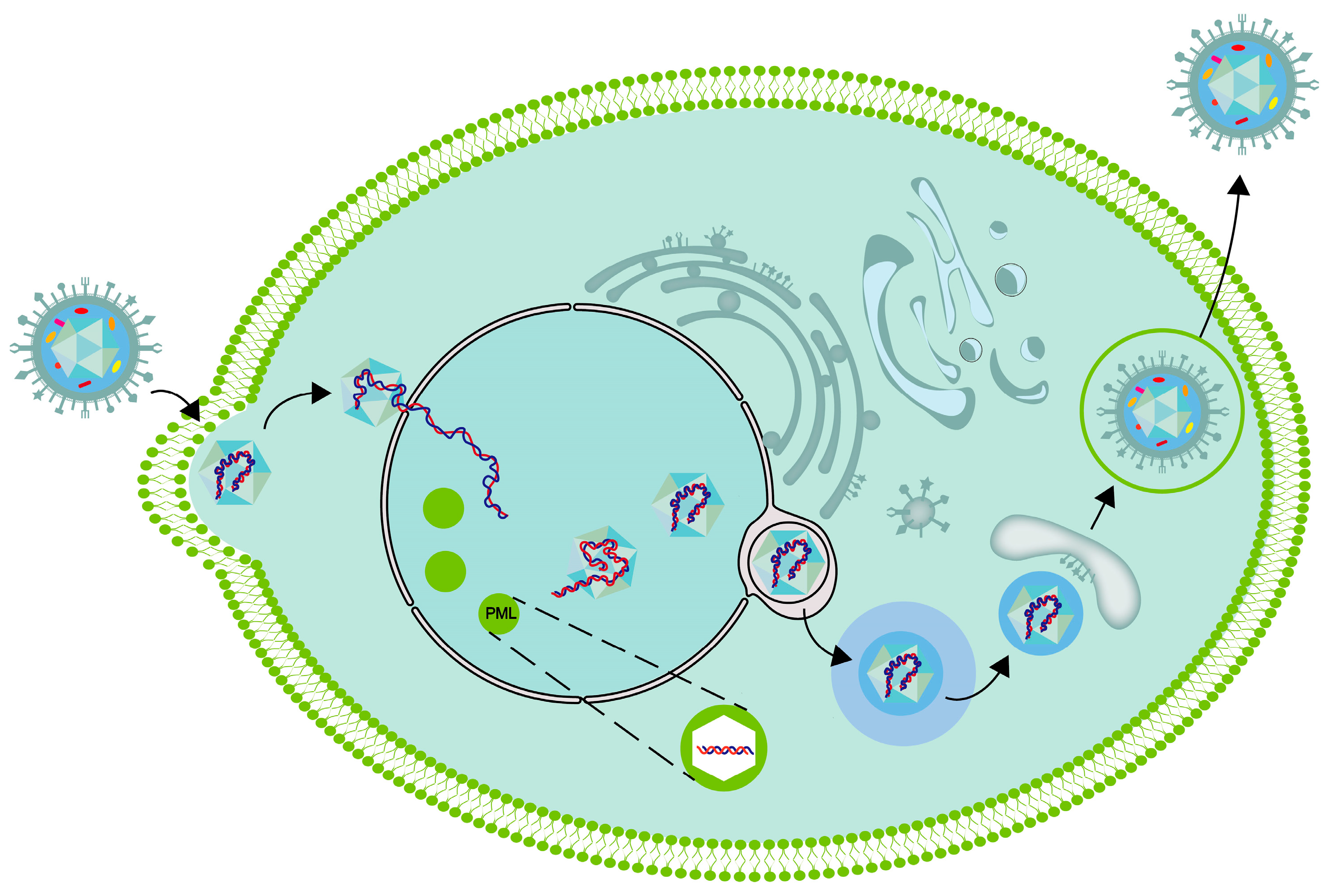
4. Transportation of HSV-1 in Vestibular Ganglion

The transportation of viral capsids and vesicles carrying viral glycoproteins in the cytoplasm is closely linked to microtubules, and their translocation along axons depends on microtubules [28,29,30,31]. HSV-I utilizes microtubules and actin to enter cells retrogradely along axons and undergoes paracrine transport during viral assembly and efflux [28,30,32]. There are two types of axonal transport—fast and slow. Fast axonal transport occurs in both cis and retrograde directions, transporting mitochondria, neurotransmitters, channel proteins, and more [33,34]. In contrast, slow axonal transport occurs in a paracrine direction, transporting cytoskeletal components, such as neurofilaments, microtubule proteins, and actin [35,36]. HSV-1 is actively directed to spread from neurons through the axonal cytoskeleton and molecular motors. Studies using time-lapse microscopy have shown that HSV-1 undergoes rapid axonal flow in both directions [37,38].

5. The Replication Process of the Virus within the Cell

After the nucleocapsid is transported to the surrounding area of the nucleus, it can interact with the nuclear pore complex, and then, dsDNA is injected into the nucleus through the nuclear pore [39]. DNA viruses, such as herpesviruses, replicate in specific inclusions within the nucleus. These inclusions, referred to as viral replication compartments (VRCs), are the sites where viral DNA replication, viral transcription, and virion assembly take place [40,41,42]. Compartmentalization is an essential feature of living organisms. Cellular organisms typically utilize cell membranes to partition cells into compartments. Moreover, eukaryotic cells possess membrane-free compartments, such as stress granules and P-bodies [43,44]. Certain compartments exhibit liquid properties and are formed through a process known as liquid–liquid phase separation (LLPS), analogous to the formation of oil droplets in water [45]. There is a hypothesis suggesting that the nuclear VRCs of DNA viruses, such as HSV-1, are also phase-separated condensates [46,47]. Seyffert et al. demonstrated that the HSV-1 transcription factor ICP4 has the ability to induce protein condensation, thereby imparting liquid-like properties to the VRC [48].
After the HSV-1 virus DNA enters the cell nucleus, it is mediated by the host cell RNA polymerase II complex and initiated by the virus capsid protein VP16 to transcribe the viral DNA and initiate the process of viral replication [49,50,51]. VP16, as a capsid protein and a structure of the virus, plays a role in the structural components of the virus once HSV-1 enters the cell. Some cell protein molecules, such as host cell factor 1 (HCF-1) and octamer binding protein-1 (Oct-1), can form multiprotein complexes with viral proteins, binding to the promoter region of the alpha genes in the herpes virus genome and activate the transcription of five alpha genes, namely, ICP0, ICP4, ICP22, ICP27, and ICP47, in a cis-activation manner, thereby initiating the process of linear transcription of the viral genome [52,53,54,55]. Subsequently, beta gene products, such as viral DNA replication, begin to be produced, and the viral genomic DNA molecules start to replicate [55,56,57,58]. When gamma gene products (mostly structural proteins) are sequentially transcribed and translated, the complete replicated gene molecules can be used as the viral genome of progeny viruses for assembly and synthesis [55,59,60].
The process of mature capsids containing viral DNA leaving the nucleus needs the extracellular output complex formed by pUL31 and pUL34. Subsequently, it crosses the nuclear membrane and enters the cytoplasm (Figure 2). During this process, the virus is initially coated with an envelope, which may originate from the inner membrane of the nuclear envelope. Subsequently, the viruses lose the initial envelope through fusion with the outer nuclear membrane and are released into the cytoplasm without an envelope. Upon arrival in the cytoplasm, the capsid is subsequently reenveloped within an intracellular organelle, where it acquires its mature envelope and completes tegumentation. The capsid undergoes secondary envelopment before being released from the cell. During this process, the nucleocapsid, which is now associated with tegument proteins, buds into the membrane of a cytoplasmic organelle, resulting in the formation of an enveloped virion inside a vesicle. The synthesized HSV-1 capsid acquires inner tegument proteins in the cytoplasm, and the outer tegument proteins and viral membrane are obtained through vesicles. The origin of organelle membranes in the secondary envelope is still controversial, with some suggesting that they may originate from membrane tubes derived from recycled endosomes or vesicles from the trans-Golgi network. PUL36 and pUL37 guide the outer shell to move toward the position of the secondary envelope on the microtubule through interaction with the microtubule and also play important roles in the transport of HSV from the nucleus to the periphery by binding to motor proteins. The vesicles contained in gD can effectively transport through axons. Viruses that have completed the secondary envelopment are released through exocytosis.

6. The Latency and Reactivation of HSV-1

HSV often results in either lytic or latent infection. After primary infection, HSV-1 can establish a latent state within the neurons of the human sensory ganglia after initial infection. The trigeminal ganglia (TG) serve as the site of latency for HSV-1 in the human body, although the vestibular ganglia, geniculate ganglia, spiral ganglia, and sacral ganglia can also harbor latent viruses [61,62]. During the latency period, the activity of the HSV-1 virus is restricted, only the latency-associated transcripts (LAT) being abundantly expressed. LATs are approximately 8.3 kb long non-coding RNAs expressed in the nucleus of latent infected cells. LAT is involved in establishing latent infection and reactivation processes while also promoting neuronal survival after HSV-1 infection by silencing immediate early gene expression and reducing apoptosis [63,64,65,66,67]. Reactivation can occur spontaneously and be induced by various stimuli. Several viral factors, such as VP16, that may play a role, have been identified; however, the theories and mechanisms underlying latent infection and reactivation are still in need of refinement [68]. The low levels of envelope proteins VP16 and ICP0 may lead to insufficient gene expression, resulting in the latency of HSV-1 in neurons [69]. Infection of neurons by HSV typically results in the production of new infectious viral particles within the cell, whereas infections on axons rarely lead to the synthesis of infectious particles, especially when the viral titer of the infected neurons is low. However, the addition of VP16 to the cell body region of ax infections can result in the production of infectious viruses, indicating that VP16 plays a critical role in the reactivation of the virus [70].
HSV-1 mutants without VP16 transcriptional activation characteristics cannot be effectively reactivated in mice [68,71]. Epigenetic modifications have also been shown to regulate virus latency and reactivation. The DNA of the herpes simplex virus (HSV) lacks histones in its original state. Once it enters the host cell, histone modifications occur on the HSV gene, thereby limiting the expression of important genes [72]. Rujescu et al. identified genome-wide associations with vestibular neuritis in four regions containing protein-coding genes that can be grouped into two functional categories, virus hypothesis and insulin metabolism, through a genome-wide association study [73].
It has been found that certain groups of sensory neurons are more susceptible to latent infection. All neuronal populations in the mouse trigeminal ganglion can be infected by HSV-1; however, neurons positive for KH10 are more susceptible to infection; those expressing the glycosphingolipid-associated embryonic antigen 3 (SSEA3) exhibit a lower rate of infection [74,75]. Similarly, the sensitivity to infection is higher in A5-positive trigeminal and dorsal root ganglion neurons compared to those that are KH10-positive. Flowerdew et al. utilized in situ hybridization to detect latency-associated transcripts in neurons expressing each of the marker proteins [75,76]. The study assessed the frequency of positively labeled neurons and the average size of neurons. Among them, TrkA-positive neurons had the highest frequency, while Ret-positive neurons had the lowest. RT97-positive neurons were the largest in size, while peripherin-positive neurons were the smallest [77].

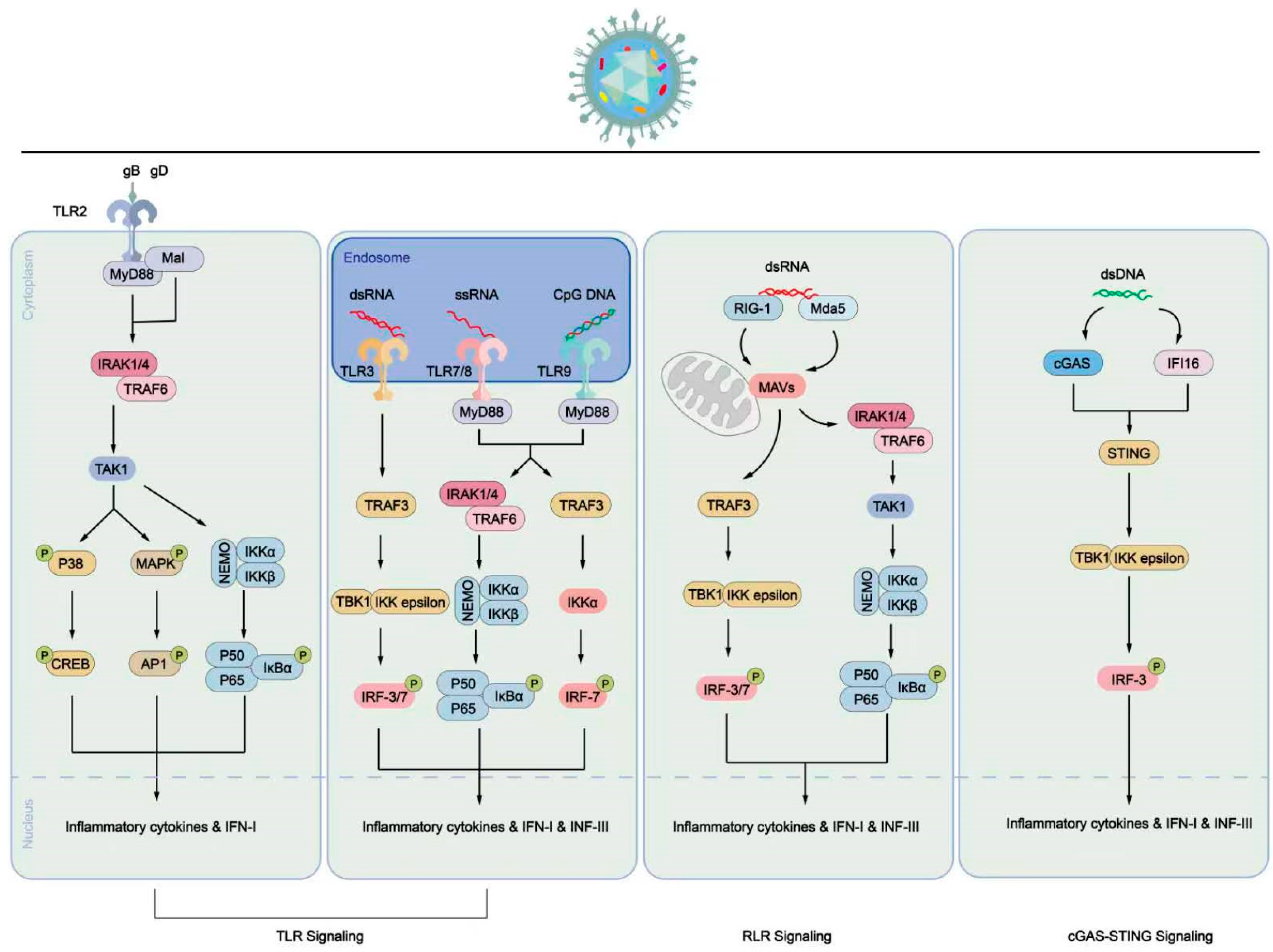
7. Host Cell Processes Caused by HSV-1 Infection

HSV-1 belongs to the lysogenic family of viruses, and its lytic replication results in the destruction of host cells. Cell aggregation is observed almost immediately after cells are infected with HSV-1, and the severity tends to increase with the number of infections [78]. According to Roizman et al. [78], herpes simplex virus infection can lead to the production of multinucleated cells, which result from the fusion of functional cells with different phenotypic characteristics. In HSV-1-infected cells, Avitabile et al. [79] found that microtubules are partially broken, especially at the cell periphery, where the connection between the microtubule network and the plasma membrane appears to be lost. Subsequently, the microtubules form bundles around the nucleus, resulting in a near-spherical shape of the cells [79]. Heeg et al. observed that infection with high doses of various strains of HSV-1 for two and a half hours resulted in cell rounding, accompanied by the breakdown of actin-containing microfilaments and the appearance of knob-like protuberances containing actin at the cell periphery [80]. Hampar et al. reported that HSV-1 infection of cells causes chromosome breaks, translocations, and fusions [81]. Roizman et al. reported that protein synthesis must precede viral DNA synthesis in the early stages of HSV-infected cells [82]. Both functional and structural proteins required for viral proliferation are produced by the host cell’s translation system. HSV has been observed to decrease protein synthesis and mRNA levels in host cells, with the expression level of viral proteins rapidly increasing, accompanied by the rapid degradation of previously existing polyribosomes and some host cell mRNA [83]. Aubert et al. summarize that the manifestations of HSV-1 infection include (i) the loss of matrix binding proteins on the cell surface, leading to detachment; (ii) modifications of membranes; (iii) cytoskeletal destabilizations; (iv) nucleolar alterations; and (v) chromatin margination and aggregation or damage, as well as (vi) a decrease in cellular macromolecular synthesis [78,79,80,81,83,84].
Cellular autophagy, apoptosis, and necrosis pathways are crucial cellular processes that are interconnected to restrict the spread of pathogens by eliminating infected cells [85]. Viral proteins can interact with these signaling molecules, disrupting downstream signal transduction and promoting viral replication and spread. Dufour et al. demonstrated that the ribonucleotide reductase R1 subunit of HSV inhibits Caspase8, thereby protecting cells from apoptosis induced by tumor necrosis factor (TNF) α and Fas ligand [86]. Furthermore, the research group demonstrated that this HSV protein disrupts the structural domain interactions of the Toll interleukin (IL)-1 receptor, thereby inhibiting poly I:C-induced apoptosis in HeLa cells [87]. Moreover, in addition to inducing the formation of filopodia in infected cells to facilitate viral transmission through cell-to-cell contact [88], Us3 proteins disable Bad by inhibiting its phosphorylation [89], thereby safeguarding the cell against DNA fragmentation, nuclear disintegration, and apoptosis [88,90].
Autophagy is a crucial cellular process that involves the self-degradation and recycling of cellular components, including the cell membrane, cytoplasm, and organelles. It plays a role in eliminating misfolded proteins, damaged organelles, and intracellular pathogens. However, certain HSV proteins, such as US11 and ICP34.5, interfere with cellular autophagy. US11 is a ribosome-associated double-stranded RNA-binding protein that directly interacts with PKR [89]. On the other hand, ICP34.5 consists of a C-terminal structural domain and an N-terminal structural domain. The C-terminal domain recruits protein phosphatase 1 (PP-1) to inhibit PKR-mediated phosphorylation of eLF2α [91], while the N-terminal domain directly interacts with Beclin-1 to block autophagy [92]. In summary, there are multiple pathways through which the host induces apoptosis and autophagy in infected cells, and HSV employs its own proteins to interfere with certain steps in these pathways to protect the survival of infected cells and facilitate its own replication and dissemination.

8. Immune Cell Process Caused by HSV-1 Infection

The intrinsic and innate immune responses serve as the first line of defense against viral infections, including HSV. They work together to limit the spread of viral replication until the body develops an adaptive immune response. Intrinsic immunity is directly mediated by host cell restriction factors such as promyelocytic leukemia nuclear bodies constituent proteins (PML-NBs) to control viral expression [93]. The innate immune response is initiated through the cellular expression of pattern recognition receptors (PRRs), which detect pathogen-associated molecular patterns (PAMPs) and damage-associated molecular patterns [94]. This recognition stimulates the secretion of interferon (IFN) α, β, or γ, along with other cytokines [95,96,97]. These cytokines can act in an autocrine and paracrine manner and play a crucial role in controlling HSV infection and coordinating innate and adaptive immune responses. Among the PRRs, Toll-like receptors (TLRs) are involved in detecting HSV nucleic acids and proteins (Figure 3). TLR2 interacts with gH/gL on the viral envelope and signals through myeloid differentiation factor 88 (MyD88) [98,99]. TLR2 activation promotes the expression of pro-inflammatory cytokines, exerting antiviral effects. However, studies on TLR2-deficient mice infected with HSV have shown that these mice exhibit fewer symptoms and longer survival than wild-type mice, suggesting that TLR2 activation may have harmful effects on the host [100,101]. TLR3 recognizes dsDNA and induces nuclear factor-kappaB (NF-κB) activation and IFN production to exert antiviral effects [102]. Herpes simplex virus encephalitis can be caused by defects in the TLR 3 pathway [103]. Mouse experiments suggest that astrocytes rely on TLR3 to mediate resistance to HSV infection [104]. However, another study demonstrated that TLR3-deficient neurons and oligodendrocytes were more susceptible to HSV-1 infection compared to control cells, indicating the importance of TLR3 in protecting neuronal cells from HSV infection [105]. TLR9 recognizes HSV DNA and is significant for certain cell types, such as plasmacytoid dendritic cells (pDC), where the absence of TLR9 results in impaired IFN responses [95,106]. The HSV DNA within the cytoplasm is encapsulated by capsid proteins, thereby being protected. However, a mechanism observed in macrophages involves the degradation of the HSV capsid, resulting in the release of HSV DNA into the cytoplasm. Consequently, there are pathways in the cell for the detection of HSV-1 DNA. Cyclic GMP-AMP synthase (cGAS) and IFI16 can detect the released HSV DNA in the cell and activate STING, leading to the recruitment of TBK1, activation of IRF3, and induction of IFN [107,108,109,110].
The adaptive immune response plays a crucial role in managing HSV infection and reactivation. Cell-mediated immunity, particularly involving T cells, is a key component of the adaptive immune response. After viral infection, cells present antigens to CD8+ T cells through surface major histocompatibility complex (MHC) class 1 molecules. This triggers the elimination of infected cells, limiting viral spread. T cells have been found to play a major role in the adaptive immune response to HSV. Specific T cells have been identified in the sensory ganglia of infected individuals and in active and latent lesions of patients [111,112,113,114]. Following acute HSV infection, the percentage of blood-specific T cells is lower in infected individuals [115,116]. HSV-specific CD8+ T cells in the blood express high levels of cytolytic molecules when re-exposed to viral antigens [117]. CD4+ T cells recognize HSV-1 proteins and express cytokines associated with helper T cell type 1 (Th1)/Th0-like responses with cytolytic potential [116,118].
HSV-1 is capable of establishing a latency period in the dorsal root ganglia (DRG) of severely combined immunodeficient mice, even when CD8+ memory T cells are transplanted prior to infection. However, the presence of T cells reduces the number of infected DRG neurons, potentially limiting HSV-1 reactivation [119,120]. In mouse models, the rate of in vitro reactivation of trigeminal ganglia (TG) is directly correlated with viral ganglionic load rather than the number of specific CD8+ T cells [121]. Specific CD8+ and CD4+ T cells are also present in the TG following human HSV-1 infection [111,112]. The infiltrating T cells in human-infected TGs are characterized as memory effector T cells and surround the cell bodies and axons of neurons [111,122]. In mouse models, memory CD8+ T cells express interferon-gamma (IFN-γ), which prevents HSV replication in neurons and inhibits neuronal apoptosis, potentially promoting the survival of neurons and HSV-1 silencing and latency [123,124,125]. The mechanism of CD4+ and CD8+ T cell recognition of latently infected neurons is not fully understood. It is possible that there may be limited viral gene expression that can be recognized by T cells, allowing for the CD8+ T cell recognition and reactivation, along with potentially low levels of neuronal MHC class I molecule expression [126]. Additionally, satellite cells can act as antigen-presenting cells and express T-cell suppressor molecules to control HSV-1 latency without damaging neurons [127]. HSV also employs various strategies to inhibit antigen presentation and modulate adaptive immune responses. For example, the viral protein ICP47 blocks antigen presentation, and ICP34.5 inhibits autophagy, which is involved in antigen presentation [128]. Furthermore, HSV can inhibit antibody responses by interacting with antibodies and complement components, inhibiting antibody-dependent cell-mediated cytotoxicity [129]. These mechanisms suggest that HSV can modulate the adaptive immune response and influence the pathogenesis of the infection.

9. Conclusions

The presence of herpesviruses has been detected in surgically removed tissues from patients with Meniere’s disease, and viral infection is considered a leading hypothesis for the development of vestibular neuritis. However, the exact role of herpesviruses in the pathogenesis of vestibular neuritis is still not fully understood. In this paper, we aimed to summarize the effects of herpesvirus infections, particularly HSV-1, on host cells and immune system processes. This information can be valuable in predicting neuronal cell damage, as well as the infiltration and killing of immune cells following viral infection. We hope that this review will stimulate further work and efforts to advance the prevention and treatment of diseases like vestibular neuritis that are potentially caused by viral infections.

Author Contributions

Y.S. conceived and designed the manuscript; Z.Z. and X.L. wrote the manuscript; Y.Z. and X.S. drew a schematic diagram; Y.S. reviewed and edited the manuscript. All authors have read and agreed to the published version of the manuscript.

Funding

This research was funded by the Innovative Research Groups of Hubei Province (No. 2023AFA038), the National Natural Science Foundation of China (No. 82071058), and the National Key Research and Development Program of China (No. 2021YFF0702303).

Data Availability Statement

Not applicable.

Conflicts of Interest

The authors declare no conflict of interest.

References

  1. Theil, D.; Arbusow, V.; Derfuss, T.; Strupp, M.; Pfeiffer, M.; Mascolo, A.; Brandt, T. Prevalence of HSV-1 LAT in human trigeminal, geniculate, and vestibular ganglia and its implication for cranial nerve syndromes. Brain Pathol. 2001, 11, 408–413. [Google Scholar] [CrossRef] [PubMed]
  2. Arbusow, V.; Schulz, P.; Strupp, M.; Dieterich, M.; von Reinhardstoettner, A.; Rauch, E.; Brandt, T. Distribution of herpes simplex virus type 1 in human geniculate and vestibular ganglia: Implications for vestibular neuritis. Ann. Neurol. 1999, 46, 416–419. [Google Scholar] [CrossRef] [PubMed]
  3. Furuta, Y.; Takasu, T.; Fukuda, S.; Inuyama, Y.; Sato, K.C.; Nagashima, K. Latent herpes simplex virus type 1 in human vestibular ganglia. Acta Otolaryngol. Suppl. 1993, 503, 85–89. [Google Scholar] [CrossRef] [PubMed]
  4. Schulz, P.; Arbusow, V.; Strupp, M.; Dieterich, M.; Rauch, E.; Brandt, T. Highly variable distribution of HSV-1-specific DNA in human geniculate, vestibular and spiral ganglia. Neurosci. Lett. 1998, 252, 139–142. [Google Scholar] [CrossRef] [PubMed]
  5. Morgenstein, K.M.; Seung, H.I. Vestibular neuronitis. Laryngoscope 1971, 81, 131–139. [Google Scholar] [CrossRef] [PubMed]
  6. Arbusow, V.; Strupp, M.; Wasicky, R.; Horn, A.K.; Schulz, P.; Brandt, T. Detection of herpes simplex virus type 1 in human vestibular nuclei. Neurology 2000, 55, 880–882. [Google Scholar] [CrossRef]
  7. Suzuki, S. [Detection of latent herpes simplex virus in human vestibular ganglia]. Hokkaido Igaku Zasshi 1996, 71, 561–571. [Google Scholar]
  8. Esaki, S.; Goshima, F.; Kimura, H.; Ikeda, S.; Katsumi, S.; Kabaya, K.; Watanabe, N.; Hashiba, M.; Nishiyama, Y.; Murakami, S. Auditory and vestibular defects induced by experimental labyrinthitis following herpes simplex virus in mice. Acta Otolaryngol. 2011, 131, 684–691. [Google Scholar] [CrossRef]
  9. Hirata, Y. An experimental herpes simplex virus infection in the vestibular nerve. Nihon Jibiinkoka Gakkai Kaiho 1994, 97, 1191–1199. [Google Scholar] [CrossRef]
  10. Grünewald, K.; Desai, P.; Winkler, D.C.; Heymann, J.B.; Belnap, D.M.; Baumeister, W.; Steven, A.C. Three-dimensional structure of herpes simplex virus from cryo-electron tomography. Science 2003, 302, 1396–1398. [Google Scholar] [CrossRef]
  11. Zhou, Z.H.; Chen, D.H.; Jakana, J.; Rixon, F.J.; Chiu, W. Visualization of tegument-capsid interactions and DNA in intact herpes simplex virus type 1 virions. J. Virol. 1999, 73, 3210–3218. [Google Scholar] [CrossRef]
  12. Schrag, J.D.; Prasad, B.V.; Rixon, F.J.; Chiu, W. Three-dimensional structure of the HSV1 nucleocapsid. Cell 1989, 56, 651–660. [Google Scholar] [CrossRef] [PubMed]
  13. Loret, S.; Guay, G.; Lippé, R. Comprehensive characterization of extracellular herpes simplex virus type 1 virions. J. Virol. 2008, 82, 8605–8618. [Google Scholar] [CrossRef] [PubMed]
  14. Vallbracht, M.; Backovic, M.; Klupp, B.G.; Rey, F.A.; Mettenleiter, T.C. Common characteristics and unique features: A comparison of the fusion machinery of the alphaherpesviruses Pseudorabies virus and Herpes simplex virus. Adv. Virus Res. 2019, 104, 225–281. [Google Scholar] [PubMed]
  15. Möhl, B.S.; Chen, J.; Longnecker, R. Gammaherpesvirus entry and fusion: A tale how two human pathogenic viruses enter their host cells. Adv. Virus Res. 2019, 104, 313–343. [Google Scholar] [PubMed]
  16. Nishimura, M.; Mori, Y. Entry of betaherpesviruses. Adv. Virus Res. 2019, 104, 283–312. [Google Scholar] [PubMed]
  17. Atanasiu, D.; Whitbeck, J.C.; de Leon, M.P.; Lou, H.; Hannah, B.P.; Cohen, G.H.; Eisenberg, R.J. Bimolecular complementation defines functional regions of Herpes simplex virus gB that are involved with gH/gL as a necessary step leading to cell fusion. J. Virol. 2010, 84, 3825–3834. [Google Scholar] [CrossRef]
  18. Atanasiu, D.; Saw, W.T.; Cohen, G.H.; Eisenberg, R.J. Cascade of events governing cell-cell fusion induced by herpes simplex virus glycoproteins gD, gH/gL, and gB. J. Virol. 2010, 84, 12292–12299. [Google Scholar] [CrossRef]
  19. Fontana, J.; Atanasiu, D.; Saw, W.T.; Gallagher, J.R.; Cox, R.G.; Whitbeck, J.C.; Brown, L.M.; Eisenberg, R.J.; Cohen, G.H. The Fusion Loops of the Initial Prefusion Conformation of Herpes Simplex Virus 1 Fusion Protein Point Toward the Membrane. mBio 2017, 8, e01268-17. [Google Scholar] [CrossRef]
  20. Büchele, W.; Brandt, T. Vestibular neuritis—A horizontal semicircular canal paresis? Adv. Otorhinolaryngol. 1988, 42, 157–161. [Google Scholar]
  21. Arbusow, V.; Dieterich, M.; Strupp, M.; Dreher, A.; Jäger, L.; Brandt, T. Herpes zoster neuritis involving superior and inferior parts of the vestibular nerve causes ocular tilt reaction. Neuro Ophthalmol. 2009, 19, 17–22. [Google Scholar] [CrossRef]
  22. Hirata, Y.; Gyo, K.; Yanagihara, N. Herpetic vestibular neuritis: An experimental study. Acta Otolaryngol. Suppl. 1995, 519, 93–96. [Google Scholar] [CrossRef] [PubMed]
  23. Theil, D.; Horn, A.K.; Derfuss, T.; Strupp, M.; Arbusow, V.; Brandt, T. Prevalence and distribution of HSV-1, VZV, and HHV-6 in human cranial nerve nuclei III, IV, VI, VII, and XII. J. Med. Virol. 2004, 74, 102–106. [Google Scholar] [CrossRef] [PubMed]
  24. Pollak, L.; Book, M.; Smetana, Z.; Alkin, M.; Soupayev, Z.; Mendelson, E. Herpes simplex virus type 1 in saliva of patients with vestibular neuronitis: A preliminary study. Neurologist 2011, 17, 330–332. [Google Scholar] [CrossRef] [PubMed]
  25. Roehm, P.C.; Camarena, V.; Nayak, S.; Gardner, J.B.; Wilson, A.; Mohr, I.; Chao, M.V. Cultured vestibular ganglion neurons demonstrate latent HSV1 reactivation. Laryngoscope 2011, 121, 2268–2275. [Google Scholar] [CrossRef] [PubMed]
  26. Rujescu, D.; Herrling, M.; Hartmann, A.M.; Maul, S.; Giegling, I.; Konte, B.; Strupp, M. High-risk Allele for Herpes Labialis Severity at the IFNL3/4 Locus is Associated With Vestibular Neuritis. Front. Neurol. 2020, 11, 570638. [Google Scholar] [CrossRef]
  27. Philpot, S.J.; Archer, J.S. Herpes encephalitis preceded by ipsilateral vestibular neuronitis. J. Clin. Neurosci. 2005, 12, 958–959. [Google Scholar] [CrossRef] [PubMed]
  28. Lee, G.E.; Murray, J.W.; Wolkoff, A.W.; Wilson, D.W. Reconstitution of herpes simplex virus microtubule-dependent trafficking in vitro. J. Virol. 2006, 80, 4264–4275. [Google Scholar] [CrossRef]
  29. Kharkwal, H.; Smith, C.G.; Wilson, D.W. Blocking ESCRT-mediated envelopment inhibits microtubule-dependent trafficking of alphaherpesviruses in vitro. J. Virol. 2014, 88, 14467–14478. [Google Scholar] [CrossRef]
  30. Sodeik, B.; Ebersold, M.W.; Helenius, A. Microtubule-mediated transport of incoming herpes simplex virus 1 capsids to the nucleus. J. Cell Biol. 1997, 136, 1007–1021. [Google Scholar] [CrossRef]
  31. Saksena, M.M.; Wakisaka, H.; Tijono, B.; Boadle, R.A.; Rixon, F.; Takahashi, H.; Cunningham, A.L. Herpes simplex virus type 1 accumulation, envelopment, and exit in growth cones and varicosities in mid-distal regions of axons. J. Virol. 2006, 80, 3592–3606. [Google Scholar] [CrossRef] [PubMed]
  32. Miranda-Saksena, M.; Armati, P.; Boadle, R.A.; Holland, D.J.; Cunningham, A.L. Anterograde transport of herpes simplex virus type 1 in cultured, dissociated human and rat dorsal root ganglion neurons. J. Virol. 2000, 74, 1827–1839. [Google Scholar] [CrossRef] [PubMed]
  33. Falzone, T.L.; Stokin, G.B.; Lillo, C.; Rodrigues, E.M.; Westerman, E.L.; Williams, D.S.; Goldstein, L.S. Axonal stress kinase activation and tau misbehavior induced by kinesin-1 transport defects. J. Neurosci. 2009, 29, 5758–5767. [Google Scholar] [CrossRef] [PubMed]
  34. Kaether, C.; Skehel, P.; Dotti, C.G. Axonal membrane proteins are transported in distinct carriers: A two-color video microscopy study in cultured hippocampal neurons. Mol. Biol. Cell 2000, 11, 1213–1224. [Google Scholar] [CrossRef] [PubMed]
  35. Li, Y.; Jung, P.; Brown, A. Axonal transport of neurofilaments: A single population of intermittently moving polymers. J. Neurosci. 2012, 32, 746–758. [Google Scholar] [CrossRef] [PubMed]
  36. Scott, D.A.; Das, U.; Tang, Y.; Roy, S. Mechanistic logic underlying the axonal transport of cytosolic proteins. Neuron 2011, 70, 441–454. [Google Scholar] [CrossRef]
  37. Antinone, S.E.; Zaichick, S.V.; Smith, G.A. Resolving the assembly state of herpes simplex virus during axon transport by live-cell imaging. J. Virol. 2010, 84, 13019–13030. [Google Scholar] [CrossRef] [PubMed]
  38. Smith, G.A.; Gross, S.P.; Enquist, L.W. Herpesviruses use bidirectional fast-axonal transport to spread in sensory neurons. Proc. Natl. Acad. Sci. USA 2001, 98, 3466–3470. [Google Scholar] [CrossRef]
  39. Villanueva-Valencia, J.R.; Tsimtsirakis, E.; Evilevitch, A. Role of HSV-1 Capsid Vertex-Specific Component (CVSC) and Viral Terminal DNA in Capsid Docking at the Nuclear Pore. Viruses 2021, 13, 2515. [Google Scholar] [CrossRef]
  40. Quinlan, M.P.; Chen, L.B.; Knipe, D.M. The intranuclear location of a herpes simplex virus DNA-binding protein is determined by the status of viral DNA replication. Cell 1984, 36, 857–868. [Google Scholar] [CrossRef]
  41. de Bruyn Kops, A.; Knipe, D.M. Formation of DNA replication structures in herpes virus-infected cells requires a viral DNA binding protein. Cell 1988, 55, 857–868. [Google Scholar] [CrossRef] [PubMed]
  42. Charman, M.; Weitzman, M.D. Replication Compartments of DNA Viruses in the Nucleus: Location, Location, Location. Viruses 2020, 12, 151. [Google Scholar] [CrossRef] [PubMed]
  43. Feric, M.; Vaidya, N.; Harmon, T.S.; Mitrea, D.M.; Zhu, L.; Richardson, T.M.; Kriwacki, R.W.; Pappu, R.V.; Brangwynne, C.P. Coexisting Liquid Phases Underlie Nucleolar Subcompartments. Cell 2016, 165, 1686–1697. [Google Scholar] [CrossRef] [PubMed]
  44. Brangwynne, C.P.; Eckmann, C.R.; Courson, D.S.; Rybarska, A.; Hoege, C.; Gharakhani, J.; Jülicher, F.; Hyman, A.A. Germline P granules are liquid droplets that localize by controlled dissolution/condensation. Science 2009, 324, 1729–1732. [Google Scholar] [CrossRef] [PubMed]
  45. Katzir, I.; Haimov, E.; Lampel, A. Tuning the Dynamics of Viral-Factories-Inspired Compartments Formed by Peptide-RNA Liquid-Liquid Phase Separation. Adv. Mater. 2022, 34, e2206371. [Google Scholar] [CrossRef] [PubMed]
  46. Subedi, S.; Nag, N.; Shukla, H.; Padhi, A.K.; Tripathi, T. Comprehensive analysis of liquid-liquid phase separation propensities of HSV-1 proteins and their interaction with host factors. J. Cell Biochem. 2023. online ahead of print. [Google Scholar] [CrossRef] [PubMed]
  47. Liu, D.; Lum, K.K.; Treen, N.; Núñez, C.T.; Yang, J.; Howard, T.R.; Levine, M.; Cristea, I.M. IFI16 phase separation via multi-phosphorylation drives innate immune signaling. Nucleic Acids Res. 2023, 51, 6819–6840. [Google Scholar] [CrossRef] [PubMed]
  48. Seyffert, M.; Georgi, F.; Tobler, K.; Bourqui, L.; Anfossi, M.; Michaelsen, K.; Vogt, B.; Greber, U.F.; Fraefel, C. The HSV-1 Transcription Factor ICP4 Confers Liquid-Like Properties to Viral Replication Compartments. Int. J. Mol. Sci. 2021, 22, 4447. [Google Scholar] [CrossRef]
  49. Birkenheuer, C.H.; Danko, C.G.; Baines, J.D. Herpes Simplex Virus 1 Dramatically Alters Loading and Positioning of RNA Polymerase II on Host Genes Early in Infection. J. Virol. 2018, 92, e02184-17. [Google Scholar] [CrossRef]
  50. Goodrich, J.A.; Hoey, T.; Thut, C.J.; Admon, A.; Tjian, R. Drosophila TAFII40 interacts with both a VP16 activation domain and the basal transcription factor TFIIB. Cell 1993, 75, 519–530. [Google Scholar] [CrossRef]
  51. Ingles, C.J.; Shales, M.; Cress, W.D.; Triezenberg, S.J.; Greenblatt, J. Reduced binding of TFIID to transcriptionally compromised mutants of VP16. Nature 1991, 351, 588–590. [Google Scholar] [CrossRef] [PubMed]
  52. Suk, H.; Knipe, D.M. Proteomic analysis of the herpes simplex virus 1 virion protein 16 transactivator protein in infected cells. Proteomics 2015, 15, 1957–1967. [Google Scholar] [PubMed]
  53. Babb, R.; Huang, C.C.; Aufiero, D.J.; Herr, W. DNA recognition by the herpes simplex virus transactivator VP16: A novel DNA-binding structure. Mol. Cell Biol. 2001, 21, 4700–4712. [Google Scholar] [CrossRef]
  54. Weir, J.P. Regulation of herpes simplex virus gene expression. Gene 2001, 271, 117–130. [Google Scholar] [CrossRef] [PubMed]
  55. Rajcáni, J.; Andrea, V.; Ingeborg, R. Peculiarities of herpes simplex virus (HSV) transcription: An overview. Virus Genes 2004, 28, 293–310. [Google Scholar] [CrossRef] [PubMed]
  56. Rodríguez, M.C.; Dybas, J.M.; Hughes, J.; Weitzman, M.D.; Boutell, C. The HSV-1 ubiquitin ligase ICP0: Modifying the cellular proteome to promote infection. Virus Res. 2020, 285, 198015. [Google Scholar] [CrossRef]
  57. Pande, N.T.; Petroski, M.D.; Wagner, E.K. Functional modules important for activated expression of early genes of herpes simplex virus type 1 are clustered upstream of the TATA box. Virology 1998, 246, 145–157. [Google Scholar] [CrossRef]
  58. Carrozza, M.J.; DeLuca, N.A. Interaction of the viral activator protein ICP4 with TFIID through TAF250. Mol. Cell Biol. 1996, 16, 3085–3093. [Google Scholar] [CrossRef]
  59. Cai, W.; Schaffer, P.A. Herpes simplex virus type 1 ICP0 regulates expression of immediate-early, early, and late genes in productively infected cells. J. Virol. 1992, 66, 2904–2915. [Google Scholar] [CrossRef]
  60. Chen, J.; Silverstein, S. Herpes simplex viruses with mutations in the gene encoding ICP0 are defective in gene expression. J. Virol. 1992, 66, 2916–2927. [Google Scholar] [CrossRef]
  61. Wilson, A.C.; Mohr, I. A cultured affair: HSV latency and reactivation in neurons. Trends Microbiol. 2012, 20, 604–611. [Google Scholar] [CrossRef] [PubMed]
  62. Held, K.; Derfuss, T. Control of HSV-1 latency in human trigeminal ganglia--current overview. J. Neurovirol. 2011, 17, 518–527. [Google Scholar] [CrossRef] [PubMed]
  63. Thompson, R.L.; Sawtell, N.M. The herpes simplex virus type 1 latency-associated transcript gene regulates the establishment of latency. J. Virol. 1997, 71, 5432–5440. [Google Scholar] [CrossRef] [PubMed]
  64. Perng, G.C.; Slanina, S.M.; Yukht, A.; Ghiasi, H.; Nesburn, A.B.; Wechsler, S.L. The latency-associated transcript gene enhances establishment of herpes simplex virus type 1 latency in rabbits. J. Virol. 2000, 74, 1885–1891. [Google Scholar] [CrossRef] [PubMed]
  65. Hill, J.M.; Sedarati, F.; Javier, R.T.; Wagner, E.K.; Stevens, J.G. Herpes simplex virus latent phase transcription facilitates in vivo reactivation. Virology 1990, 174, 117–125. [Google Scholar] [CrossRef] [PubMed]
  66. Perng, G.C.; Dunkel, E.C.; Geary, P.A.; Slanina, S.M.; Ghiasi, H.; Kaiwar, R.; Nesburn, A.B.; Wechsler, S.L. The latency-associated transcript gene of herpes simplex virus type 1 (HSV-1) is required for efficient in vivo spontaneous reactivation of HSV-1 from latency. J. Virol. 1994, 68, 8045–8055. [Google Scholar] [CrossRef] [PubMed]
  67. Perng, G.C.; Jones, C.; Ciacci-Zanella, J.; Stone, M.; Henderson, G.; Yukht, A.; Slanina, S.M.; Hofman, F.M.; Ghiasi, H.; Nesburn, A.B.; et al. Virus-induced neuronal apoptosis blocked by the herpes simplex virus latency-associated transcript. Science 2000, 287, 1500–1503. [Google Scholar] [CrossRef]
  68. Thompson, R.L.; Preston, C.M.; Sawtell, N.M. De novo synthesis of VP16 coordinates the exit from HSV latency in vivo. PLoS Pathog. 2009, 5, e1000352. [Google Scholar] [CrossRef]
  69. Zhu, S.; Viejo-Borbolla, A. Pathogenesis and virulence of herpes simplex virus. Virulence 2021, 12, 2670–2702. [Google Scholar] [CrossRef] [PubMed]
  70. Hafezi, W.; Lorentzen, E.U.; Eing, B.R.; Müller, M.; King, N.J.; Klupp, B.; Mettenleiter, T.C.; Kühn, J.E. Entry of herpes simplex virus type 1 (HSV-1) into the distal axons of trigeminal neurons favors the onset of nonproductive, silent infection. PLoS Pathog. 2012, 8, e1002679. [Google Scholar] [CrossRef]
  71. Sawtell, N.M.; Triezenberg, S.J.; Thompson, R.L. VP16 serine 375 is a critical determinant of herpes simplex virus exit from latency in vivo. J. Neurovirol. 2011, 17, 546–551. [Google Scholar] [CrossRef] [PubMed]
  72. Knipe, D.M.; Cliffe, A. Chromatin control of herpes simplex virus lytic and latent infection. Nat. Rev. Microbiol. 2008, 6, 211–221. [Google Scholar] [CrossRef]
  73. Rujescu, D.; Hartmann, A.M.; Giegling, I.; Konte, B.; Herrling, M.; Himmelein, S.; Strupp, M. Genome-Wide Association Study in Vestibular Neuritis: Involvement of the Host Factor for HSV-1 Replication. Front. Neurol. 2018, 9, 591. [Google Scholar] [CrossRef] [PubMed]
  74. Yang, L.; Voytek, C.C.; Margolis, T.P. Immunohistochemical analysis of primary sensory neurons latently infected with herpes simplex virus type 1. J. Virol. 2000, 74, 209–217. [Google Scholar] [CrossRef] [PubMed]
  75. Margolis, T.P.; Dawson, C.R.; LaVail, J.H. Herpes simplex viral infection of the mouse trigeminal ganglion. Immunohistochemical analysis of cell populations. Investig. Ophthalmol. Vis. Sci. 1992, 33, 259–267. [Google Scholar]
  76. Bertke, A.S.; Swanson, S.M.; Chen, J.; Imai, Y.; Kinchington, P.R.; Margolis, T.P. A5-positive primary sensory neurons are nonpermissive for productive infection with herpes simplex virus 1 in vitro. J. Virol. 2011, 85, 6669–6677. [Google Scholar] [CrossRef] [PubMed]
  77. Flowerdew, S.E.; Wick, D.; Himmelein, S.; Horn, A.K.; Sinicina, I.; Strupp, M.; Brandt, T.; Theil, D.; Hüfner, K. Characterization of neuronal populations in the human trigeminal ganglion and their association with latent herpes simplex virus-1 infection. PLoS ONE 2013, 8, e83603. [Google Scholar] [CrossRef] [PubMed]
  78. Roizman, B. Polykaryocytosis induced by viruses. Proc. Natl. Acad. Sci. USA 1962, 48, 228–234. [Google Scholar] [CrossRef]
  79. Avitabile, E.; Di Gaeta, S.; Torrisi, M.R.; Ward, P.L.; Roizman, B.; Campadelli-Fiume, G. Redistribution of microtubules and Golgi apparatus in herpes simplex virus-infected cells and their role in viral exocytosis. J. Virol. 1995, 69, 7472–7482. [Google Scholar] [CrossRef]
  80. Heeg, U.; Dienes, H.P.; Müller, S.; Falke, D. Involvement of actin-containing microfilaments in HSV-induced cytopathology and the influence of inhibitors of glycosylation. Arch. Virol. 1986, 91, 257–270. [Google Scholar] [CrossRef]
  81. Hampar, B.; Ellison, S.A. Chromosomal aberrations induced by an animal virus. Nature 1961, 192, 145–147. [Google Scholar] [CrossRef] [PubMed]
  82. Roizman, B. The programming of herpes virus multiplication in doubly-infected and in puromycin-treated cells. Proc. Natl. Acad. Sci. USA 1963, 49, 165–171. [Google Scholar] [CrossRef] [PubMed]
  83. Roizman, B.; Roane, P.R., Jr. The multiplication of herpes simplex virus. II. the relation between protein synthesis and the duplication of viral dna in infected HEp-2 cells. Virology 1964, 22, 262–269. [Google Scholar] [CrossRef] [PubMed]
  84. Aubert, M.; Blaho, J.A. The herpes simplex virus type 1 regulatory protein ICP27 is required for the prevention of apoptosis in infected human cells. J. Virol. 1999, 73, 2803–2813. [Google Scholar] [CrossRef] [PubMed]
  85. Finlay, D.; Cantrell, D.A. Metabolism, migration and memory in cytotoxic T cells. Nat. Rev. Immunol. 2011, 11, 109–117. [Google Scholar] [CrossRef]
  86. Dufour, F.; Sasseville, A.M.; Chabaud, S.; Massie, B.; Siegel, R.M.; Langelier, Y. The ribonucleotide reductase R1 subunits of herpes simplex virus types 1 and 2 protect cells against TNFα- and FasL-induced apoptosis by interacting with caspase-8. Apoptosis 2011, 16, 256–271. [Google Scholar] [CrossRef]
  87. Dufour, F.; Bertrand, L.; Pearson, A.; Grandvaux, N.; Langelier, Y. The ribonucleotide reductase R1 subunits of herpes simplex virus 1 and 2 protect cells against poly(I · C)-induced apoptosis. J. Virol. 2011, 85, 8689–8701. [Google Scholar] [CrossRef]
  88. Finnen, R.L.; Johnston, S.M.; Neron, C.E.; Banfield, B.W. Nucleocytoplasmic shuttling of the HSV-2 serine/threonine kinase Us3. Virology 2011, 417, 229–237. [Google Scholar] [CrossRef]
  89. Cartier, A.; Komai, T.; Masucci, M.G. The Us3 protein kinase of herpes simplex virus 1 blocks apoptosis and induces phosporylation of the Bcl-2 family member Bad. Exp. Cell Res. 2003, 291, 242–250. [Google Scholar] [CrossRef]
  90. Benetti, L.; Roizman, B. Herpes simplex virus protein kinase US3 activates and functionally overlaps protein kinase A to block apoptosis. Proc. Natl. Acad. Sci. USA 2004, 101, 9411–9416. [Google Scholar] [CrossRef]
  91. Li, Y.; Zhang, C.; Chen, X.; Yu, J.; Wang, Y.; Yang, Y.; Du, M.; Jin, H.; Ma, Y.; He, B.; et al. ICP34.5 protein of herpes simplex virus facilitates the initiation of protein translation by bridging eukaryotic initiation factor 2alpha (eIF2alpha) and protein phosphatase 1. J. Biol. Chem. 2011, 286, 24785–24792. [Google Scholar] [CrossRef] [PubMed]
  92. Orvedahl, A.; Alexander, D.; Tallóczy, Z.; Sun, Q.; Wei, Y.; Zhang, W.; Burns, D.; Leib, D.A.; Levine, B. HSV-1 ICP34.5 confers neurovirulence by targeting the Beclin 1 autophagy protein. Cell Host Microbe 2007, 1, 23–35. [Google Scholar] [CrossRef] [PubMed]
  93. Everett, R.D.; Sourvinos, G.; Leiper, C.; Clements, J.B.; Orr, A. Formation of nuclear foci of the herpes simplex virus type 1 regulatory protein ICP4 at early times of infection: Localization, dynamics, recruitment of ICP27, and evidence for the de novo induction of ND10-like complexes. J. Virol. 2004, 78, 1903–1917. [Google Scholar] [CrossRef]
  94. Takeuchi, O.; Akira, S. Pattern recognition receptors and inflammation. Cell 2010, 140, 805–820. [Google Scholar] [CrossRef] [PubMed]
  95. Krug, A.; Luker, G.D.; Barchet, W.; Leib, D.A.; Akira, S.; Colonna, M. Herpes simplex virus type 1 activates murine natural interferon-producing cells through toll-like receptor 9. Blood 2004, 103, 1433–1437. [Google Scholar] [CrossRef]
  96. Imanishi, T.; Hara, H.; Suzuki, S.; Suzuki, N.; Akira, S.; Saito, T. Cutting edge: TLR2 directly triggers Th1 effector functions. J. Immunol. 2007, 178, 6715–6719. [Google Scholar] [CrossRef] [PubMed]
  97. Palmer, P.; Tovey, M.G.; Raschilas, F.; Brassart, L.; Meritet, J.F.; Porcher, R.; Lebon, P. Type I interferon subtypes produced by human peripheral mononuclear cells from one normal donor stimulated by viral and non-viral inducing factors. Eur. Cytokine Netw. 2007, 18, 108–114. [Google Scholar] [PubMed]
  98. Leoni, V.; Gianni, T.; Salvioli, S.; Campadelli-Fiume, G. Herpes simplex virus glycoproteins gH/gL and gB bind Toll-like receptor 2, and soluble gH/gL is sufficient to activate NF-κB. J. Virol. 2012, 86, 6555–6562. [Google Scholar] [CrossRef]
  99. Cai, M.; Li, M.; Wang, K.; Wang, S.; Lu, Q.; Yan, J.; Mossman, K.L.; Lin, R.; Zheng, C. The herpes simplex virus 1-encoded envelope glycoprotein B activates NF-κB through the Toll-like receptor 2 and MyD88/TRAF6-dependent signaling pathway. PLoS ONE 2013, 8, e54586. [Google Scholar] [CrossRef]
  100. Kurt-Jones, E.A.; Chan, M.; Zhou, S.; Wang, J.; Reed, G.; Bronson, R.; Arnold, M.M.; Knipe, D.M.; Finberg, R.W. Herpes simplex virus 1 interaction with Toll-like receptor 2 contributes to lethal encephalitis. Proc. Natl. Acad. Sci. USA 2004, 101, 1315–1320. [Google Scholar] [CrossRef]
  101. Marino, A.; Pergolizzi, S.; Cimino, F.; Lauriano, E.R.; Speciale, A.; D’Angelo, V.; Sicurella, M.; Argnani, R.; Manservigi, R.; Marconi, P. Role of Herpes Simplex Envelope Glycoprotein B and Toll-Like Receptor 2 in Ocular Inflammation: An ex vivo Organotypic Rabbit Corneal Model. Viruses 2019, 11, 819. [Google Scholar] [CrossRef] [PubMed]
  102. Alexopoulou, L.; Holt, A.C.; Medzhitov, R.; Flavell, R.A. Recognition of double-stranded RNA and activation of NF-kappaB by Toll-like receptor 3. Nature 2001, 413, 732–738. [Google Scholar] [CrossRef] [PubMed]
  103. Gao, D.; Ciancanelli, M.J.; Zhang, P.; Harschnitz, O.; Bondet, V.; Hasek, M.; Chen, J.; Mu, X.; Itan, Y.; Cobat, A.; et al. TLR3 controls constitutive IFN-β antiviral immunity in human fibroblasts and cortical neurons. J. Clin. Investig. 2021, 131, e134529. [Google Scholar] [CrossRef] [PubMed]
  104. Reinert, L.S.; Harder, L.; Holm, C.K.; Iversen, M.B.; Horan, K.A.; Dagnæs-Hansen, F.; Ulhøi, B.P.; Holm, T.H.; Mogensen, T.H.; Owens, T.; et al. TLR3 deficiency renders astrocytes permissive to herpes simplex virus infection and facilitates establishment of CNS infection in mice. J. Clin. Investig. 2012, 122, 1368–1376. [Google Scholar] [CrossRef] [PubMed]
  105. Leib, D.A. Herpes simplex virus encephalitis: Toll-free access to the brain. Cell Host Microbe 2012, 12, 731–732. [Google Scholar] [CrossRef] [PubMed]
  106. Lund, J.; Sato, A.; Akira, S.; Medzhitov, R.; Iwasaki, A. Toll-like receptor 9-mediated recognition of Herpes simplex virus-2 by plasmacytoid dendritic cells. J. Exp. Med. 2003, 198, 513–520. [Google Scholar] [CrossRef] [PubMed]
  107. Eriksson, K.; Svensson, A.; Hait, A.S.; Schlüter, K.; Tunbäck, P.; Nordström, I.; Padyukov, L.; Liljeqvist, J.; Mogensen, T.H.; Paludan, S.R. Cutting Edge: Genetic Association between IFI16 Single Nucleotide Polymorphisms and Resistance to Genital Herpes Correlates with IFI16 Expression Levels and HSV-2-Induced IFN-β Expression. J. Immunol. 2017, 199, 2613–2617. [Google Scholar] [CrossRef]
  108. Orzalli, M.H.; DeLuca, N.A.; Knipe, D.M. Nuclear IFI16 induction of IRF-3 signaling during herpesviral infection and degradation of IFI16 by the viral ICP0 protein. Proc. Natl. Acad. Sci. USA 2012, 109, E3008–E3017. [Google Scholar] [CrossRef]
  109. Conrady, C.D.; Zheng, M.; Fitzgerald, K.A.; Liu, C.; Carr, D.J. Resistance to HSV-1 infection in the epithelium resides with the novel innate sensor, IFI-16. Mucosal Immunol. 2012, 5, 173–183. [Google Scholar] [CrossRef]
  110. Iqbal, J.; Ansari, M.A.; Kumar, B.; Dutta, D.; Roy, A.; Chikoti, L.; Pisano, G.; Dutta, S.; Vahedi, S.; Veettil, M.V.; et al. Histone H2B-IFI16 Recognition of Nuclear Herpesviral Genome Induces Cytoplasmic Interferon-β Responses. PLoS Pathog. 2016, 12, e1005967. [Google Scholar] [CrossRef]
  111. Verjans, G.M.; Hintzen, R.Q.; van Dun, J.M.; Poot, A.; Milikan, J.C.; Laman, J.D.; Langerak, A.W.; Kinchington, P.R.; Osterhaus, A.D. Selective retention of herpes simplex virus-specific T cells in latently infected human trigeminal ganglia. Proc. Natl. Acad. Sci. USA 2007, 104, 3496–3501. [Google Scholar] [CrossRef] [PubMed]
  112. van Velzen, M.; Jing, L.; Osterhaus, A.D.; Sette, A.; Koelle, D.M.; Verjans, G.M. Local CD4 and CD8 T-cell reactivity to HSV-1 antigens documents broad viral protein expression and immune competence in latently infected human trigeminal ganglia. PLoS Pathog. 2013, 9, e1003547. [Google Scholar] [CrossRef] [PubMed]
  113. Zhu, J.; Koelle, D.M.; Cao, J.; Vazquez, J.; Huang, M.L.; Hladik, F.; Wald, A.; Corey, L. Virus-specific CD8+ T cells accumulate near sensory nerve endings in genital skin during subclinical HSV-2 reactivation. J. Exp. Med. 2007, 204, 595–603. [Google Scholar] [CrossRef] [PubMed]
  114. Zhu, J.; Peng, T.; Johnston, C.; Phasouk, K.; Kask, A.S.; Klock, A.; Jin, L.; Diem, K.; Koelle, D.M.; Wald, A.; et al. Immune surveillance by CD8αα+ skin-resident T cells in human herpes virus infection. Nature 2013, 497, 494–497. [Google Scholar] [CrossRef]
  115. Jing, L.; Haas, J.; Chong, T.M.; Bruckner, J.J.; Dann, G.C.; Dong, L.; Marshak, J.O.; McClurkan, C.L.; Yamamoto, T.N.; Bailer, S.M.; et al. Cross-presentation and genome-wide screening reveal candidate T cells antigens for a herpes simplex virus type 1 vaccine. J. Clin. Investig. 2012, 122, 654–673. [Google Scholar] [CrossRef] [PubMed]
  116. Moss, N.J.; Magaret, A.; Laing, K.J.; Kask, A.S.; Wang, M.; Mark, K.E.; Schiffer, J.T.; Wald, A.; Koelle, D.M. Peripheral blood CD4 T-cell and plasmacytoid dendritic cell (pDC) reactivity to herpes simplex virus 2 and pDC number do not correlate with the clinical or virologic severity of recurrent genital herpes. J. Virol. 2012, 86, 9952–9963. [Google Scholar] [CrossRef]
  117. Laing, K.J.; Magaret, A.S.; Mueller, D.E.; Zhao, L.; Johnston, C.; De Rosa, S.C.; Koelle, D.M.; Wald, A.; Corey, L. Diversity in CD8(+) T cell function and epitope breadth among persons with genital herpes. J. Clin. Immunol. 2010, 30, 703–722. [Google Scholar] [CrossRef]
  118. Koelle, D.M.; Corey, L.; Burke, R.L.; Eisenberg, R.J.; Cohen, G.H.; Pichyangkura, R.; Triezenberg, S.J. Antigenic specificities of human CD4+ T-cell clones recovered from recurrent genital herpes simplex virus type 2 lesions. J. Virol. 1994, 68, 2803–2810. [Google Scholar] [CrossRef]
  119. Valyi-Nagy, T.; Deshmane, S.L.; Raengsakulrach, B.; Nicosia, M.; Gesser, R.M.; Wysocka, M.; Dillner, A.; Fraser, N.W. Herpes simplex virus type 1 mutant strain in1814 establishes a unique, slowly progressing infection in SCID mice. J. Virol. 1992, 66, 7336–7345. [Google Scholar] [CrossRef]
  120. Wakim, L.M.; Jones, C.M.; Gebhardt, T.; Preston, C.M.; Carbone, F.R. CD8(+) T-cell attenuation of cutaneous herpes simplex virus infection reduces the average viral copy number of the ensuing latent infection. Immunol. Cell Biol. 2008, 86, 666–675. [Google Scholar] [CrossRef]
  121. Hoshino, Y.; Pesnicak, L.; Cohen, J.I.; Straus, S.E. Rates of reactivation of latent herpes simplex virus from mouse trigeminal ganglia ex vivo correlate directly with viral load and inversely with number of infiltrating CD8+ T cells. J. Virol. 2007, 81, 8157–8164. [Google Scholar] [CrossRef] [PubMed]
  122. Derfuss, T.; Segerer, S.; Herberger, S.; Sinicina, I.; Hüfner, K.; Ebelt, K.; Knaus, H.G.; Steiner, I.; Meinl, E.; Dornmair, K.; et al. Presence of HSV-1 immediate early genes and clonally expanded T-cells with a memory effector phenotype in human trigeminal ganglia. Brain Pathol. 2007, 17, 389–398. [Google Scholar] [CrossRef] [PubMed]
  123. Geiger, K.D.; Nash, T.C.; Sawyer, S.; Krahl, T.; Patstone, G.; Reed, J.C.; Krajewski, S.; Dalton, D.; Buchmeier, M.J.; Sarvetnick, N. Interferon-gamma protects against herpes simplex virus type 1-mediated neuronal death. Virology 1997, 238, 189–197. [Google Scholar] [CrossRef]
  124. Knickelbein, J.E.; Khanna, K.M.; Yee, M.B.; Baty, C.J.; Kinchington, P.R.; Hendricks, R.L. Noncytotoxic lytic granule-mediated CD8+ T cell inhibition of HSV-1 reactivation from neuronal latency. Science 2008, 322, 268–271. [Google Scholar] [CrossRef]
  125. Frank, G.M.; Lepisto, A.J.; Freeman, M.L.; Sheridan, B.S.; Cherpes, T.L.; Hendricks, R.L. Early CD4(+) T cell help prevents partial CD8(+) T cell exhaustion and promotes maintenance of Herpes Simplex Virus 1 latency. J. Immunol. 2010, 184, 277–286. [Google Scholar] [CrossRef] [PubMed]
  126. Liu, T.; Khanna, K.M.; Chen, X.; Fink, D.J.; Hendricks, R.L. CD8(+) T cells can block herpes simplex virus type 1 (HSV-1) reactivation from latency in sensory neurons. J. Exp. Med. 2000, 191, 1459–1466. [Google Scholar] [CrossRef] [PubMed]
  127. van Velzen, M.; Laman, J.D.; Kleinjan, A.; Poot, A.; Osterhaus, A.D.; Verjans, G.M. Neuron-interacting satellite glial cells in human trigeminal ganglia have an APC phenotype. J. Immunol. 2009, 183, 2456–2461. [Google Scholar] [CrossRef]
  128. Gobeil, P.A.; Leib, D.A. Herpes simplex virus γ34.5 interferes with autophagosome maturation and antigen presentation in dendritic cells. mBio 2012, 3, e00267-12. [Google Scholar] [CrossRef]
  129. Lubinski, J.M.; Jiang, M.; Hook, L.; Chang, Y.; Sarver, C.; Mastellos, D.; Lambris, J.D.; Cohen, G.H.; Eisenberg, R.J.; Friedman, H.M. Herpes simplex virus type 1 evades the effects of antibody and complement in vivo. J. Virol. 2002, 76, 9232–9241. [Google Scholar] [CrossRef]
Figure 1. (A) Schematic diagram of the structure of HSV-1. HSV-1 is composed of double-stranded DNA, nucleocapsid, tegument, envelope proteins, and a lipid envelope. (B) Schematic diagram of HSV-1 entry into cells via envelope proteins. While the types and composition of envelope proteins may vary among different viruses in the sporozoan virus family, the fusion of the viral envelope with the host cell membrane and the entry of the nucleocapsid rely on the binding of multiple envelope proteins to receptors on the host cell membrane and conformational changes.
Viruses 16 00012 g001
Figure 2. Schematic diagram of intracellular cycle of HSV-1.
Viruses 16 00012 g002
Figure 3. Pattern diagram of immune cell response process triggered by PRR signal triggered by HSV-1 infection. Inducing the secretion of inflammatory cytokines or IFN through the TLR signaling pathway (left, middle) or RLR signaling pathway (right). In the TLR signaling pathway, TLR2 recognizes signals induced by HSV-1 envelope proteins, such as gB or gD. The signal is transmitted to the cytoplasm, where MyD88 binds to the cytoplasmic domain of TLR2, leading to the activation of transcription factors like NF-κB. This activation promotes the translocation of P50/P65 into the nucleus and increases the expression of inflammatory cytokines and IFN-1. Additionally, TLR3, TLR7/8, and TLR9 signaling are activated by dsRNA, ssRNA, or CpG DNA, respectively, in endosomes. These signals activate IRF-3, IRF-7, and NF-κB, ultimately resulting in increased expression of inflammatory cytokines, IFN-1, IFN-III, and interferon-stimulated genes (ISGs). In the RLR signaling pathway, RIG-I and MDA5, which contain N-terminal caspase activation and recruitment domains, recruit and activate the mitochondrial antiviral signaling (MAVS) protein to mediate signal transduction. The activated MAVS protein further activates downstream signaling, promoting the expression of inflammatory cytokines and IFN. Both pathways contribute to the immune response against HSV-1 infection by triggering the production of inflammatory cytokines and interferons, which play crucial roles in controlling viral replication and coordinating innate and adaptive immune responses.
Viruses 16 00012 g003
Table 1. Viruses associated with VN.
Viruses Associated with VNFamily of Viruses
Herpes simplex virusAlphaherpesvirinae
Varicella-zoster virus
Human cytomegalovirusBetaherpesvirinae
Epstein–Barr virusGammaherpesvirinae
Influenza virus ANon-herpesvirus family
Influenza virus B
Adenoviruses
Rubella virus
Parainfluenza virus
Disclaimer/Publisher’s Note: The statements, opinions and data contained in all publications are solely those of the individual author(s) and contributor(s) and not of MDPI and/or the editor(s). MDPI and/or the editor(s) disclaim responsibility for any injury to people or property resulting from any ideas, methods, instructions or products referred to in the content.

