

Structural and Synthetic Aspects of Small Ring Oxa- and Aza-Heterocyclic Ring Systems as Antiviral Activities
 ,
,
Abstract
:1. Introduction
2. Overview of the Viral Diseases
2.1. Human Immunodeficiency Virus (HIV)
2.2. Hepatitis C Virus (HCV)
2.3. Hepatitis B Virus (HBV)
2.4. Respiratory Syncytial Virus (RSV)
2.5. Human Cytomegalovirus (HCMV)
2.6. Herpes Simplex Virus (HSV)
2.7. Ebola Virus (EBOV)
2.8. Severe Acute Respiratory Syndrome CoV-2 (SARS-CoV-2) Virus
2.9. The Human Papillomavirus (HPV)
2.10. Rabies Virus
2.11. Zika Virus
2.12. The Poliovirus
2.13. West Nile Virus
2.14. The Chickenpox Virus
2.15. The Influenza Virus
2.16. Yellow Fever
3. Overview of the Antiviral Drugs
3.1. Representative Antiviral Drug Candidates
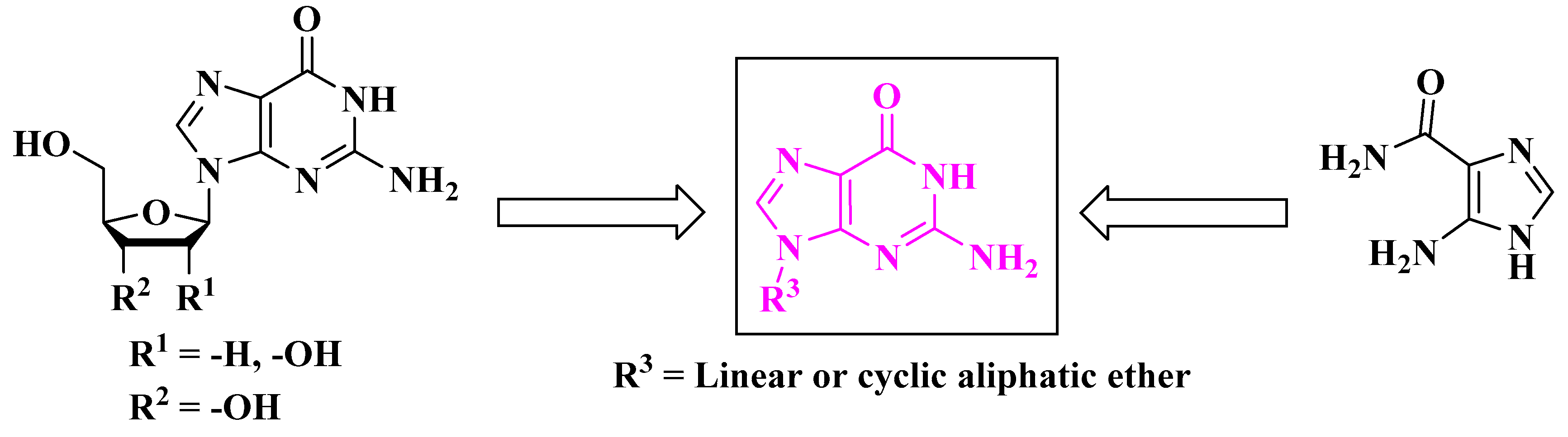
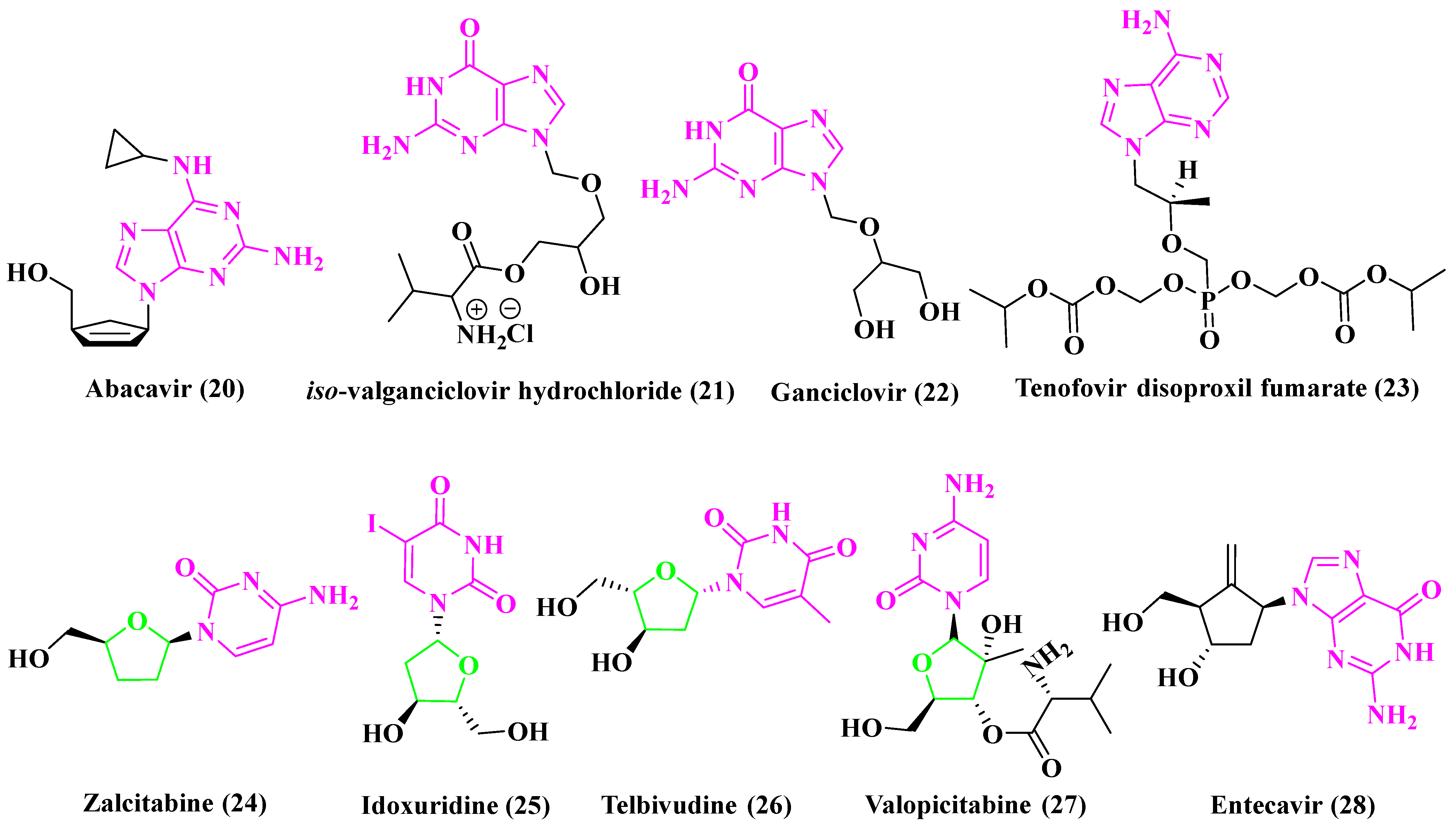
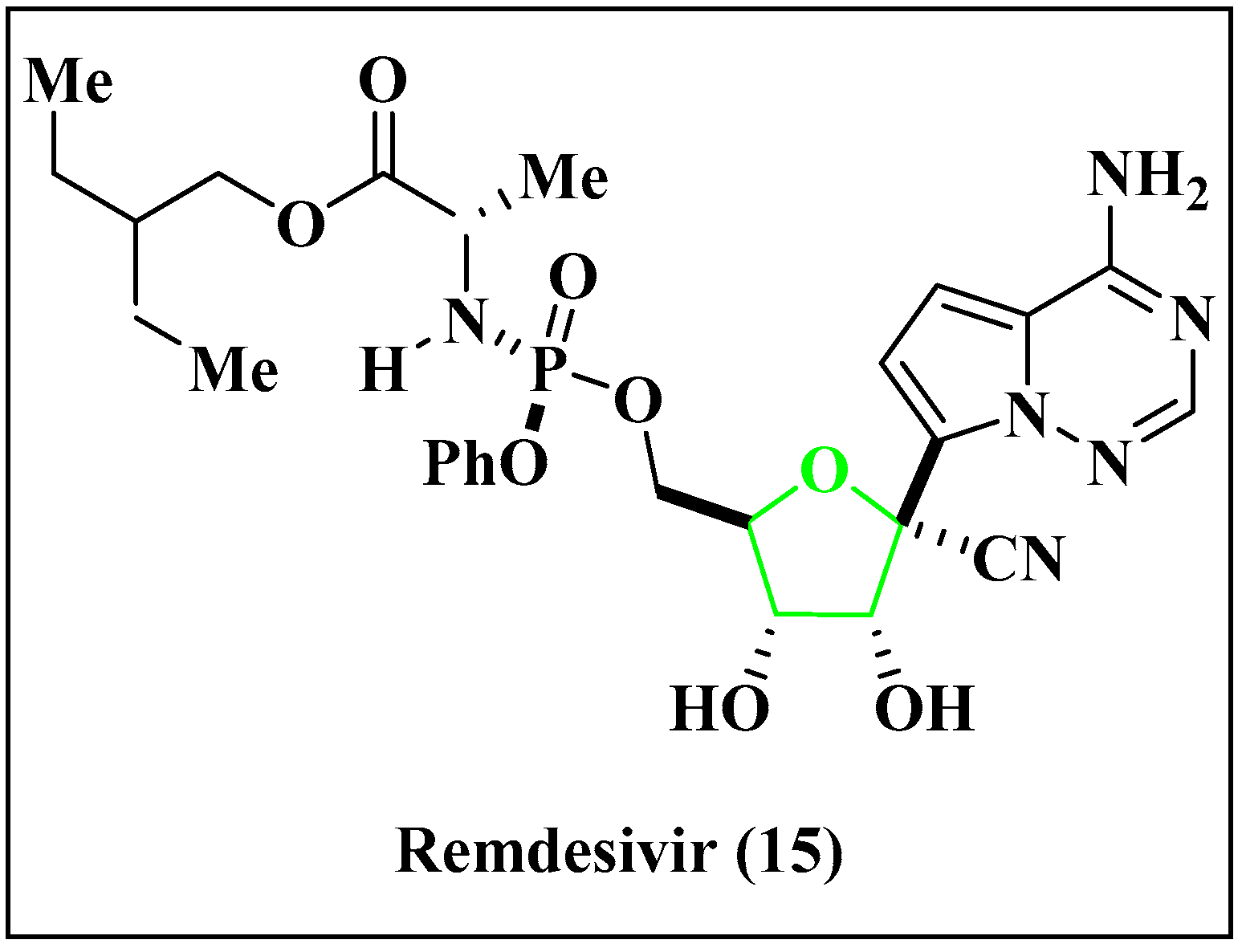
3.2. Antiviral Drugs Containing Nucleoside Subunit
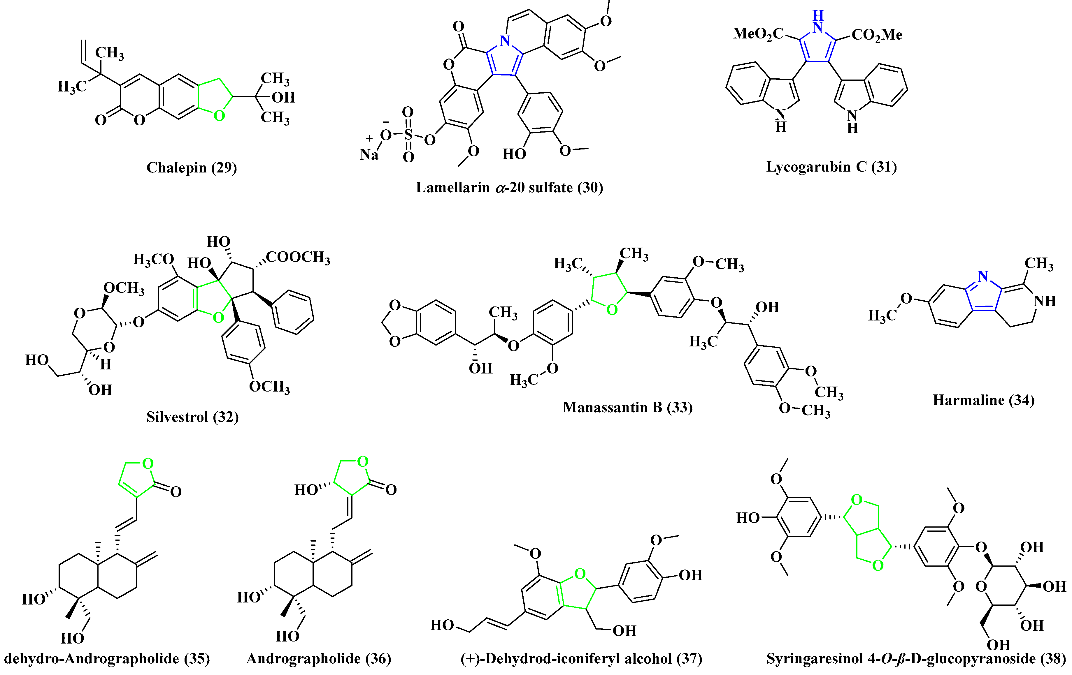
3.3. Examples of Natural Products with Antiviral Properties
- Chalepin (29), from a Ruta angustifolia species plant, shows a good inhibitory effect against HCV [99].
- Lamellarin α-20 sulfate (30) is an alkaloid found in marine Lamellarins [100], and is responsible for inhibiting the integration of HIV-1 replication in its very early stages.
- Lycogarubins (A, B and C) are isolated from fruit bodies of Myxomycetes Lycogala epidendnrm, and contain two indole groups connected with dimethyl pyrrole-dicarboxylate, in which Lycogarubin C (31) shows activity against HSV [101].
- Silvestrol (32), from the bark of the Aglaia foveolate type of plants, contains a substituted dioxane and acts as a potent inhibitor of the Ebola virus [102].
- Manassantin B (33), extracted from Saururus chinensis Baill plants, shows inhibitory properties against the Epstein–Barr virus [103].
- Harmaline (34) is an indole alkaloid from Peganum harmala, and shows antiviral properties against HSV-1 [104].
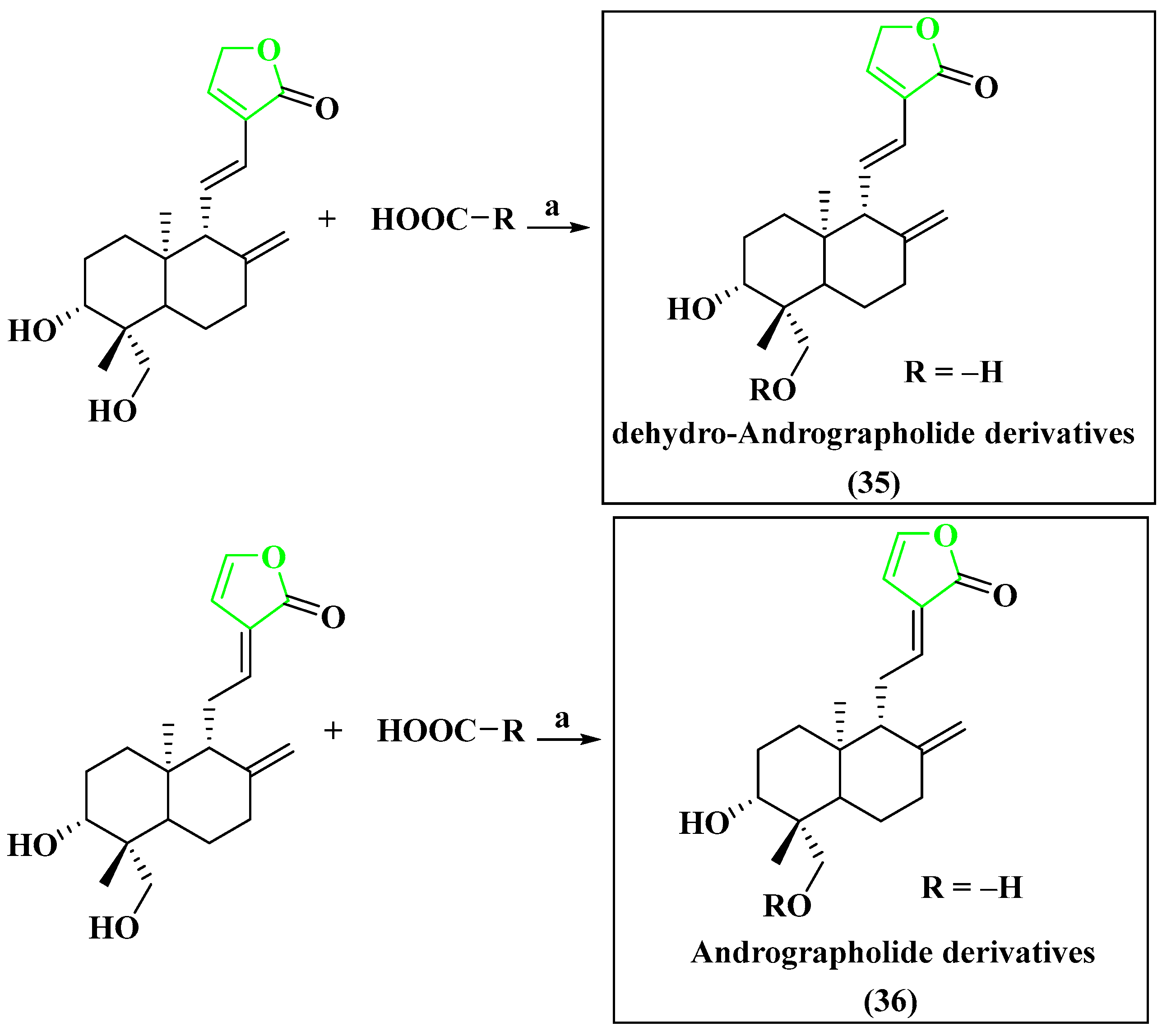
- Dehydro-Andrographolide (35) and Andrographolide (36) are two types of natural diterpenoids that have been extracted from Andrographis paniculata. These compounds have demonstrated the ability to inhibit the replication of HBV DNA [105].
- (+)-Dehydrod-iconiferyl alcohol (37) that has been isolated from Swertia patens shows inhibitory activities on the secretion of HBsAg, with IC50 value of 1.94 mM [106].
- Syringaresinol 4″-O-β-D-glucopyranoside (38), which was extracted from Swertia chirayita, exhibited an inhibitory effect on the secretion of HBsAg, with IC50 values of 1.49 ± 0.033 mM [107].
4. Importance of Heterocyclic Ring Systems as Antiviral Agents
5. Synthetic Outlines of Representative Antiviral Drug Candidates
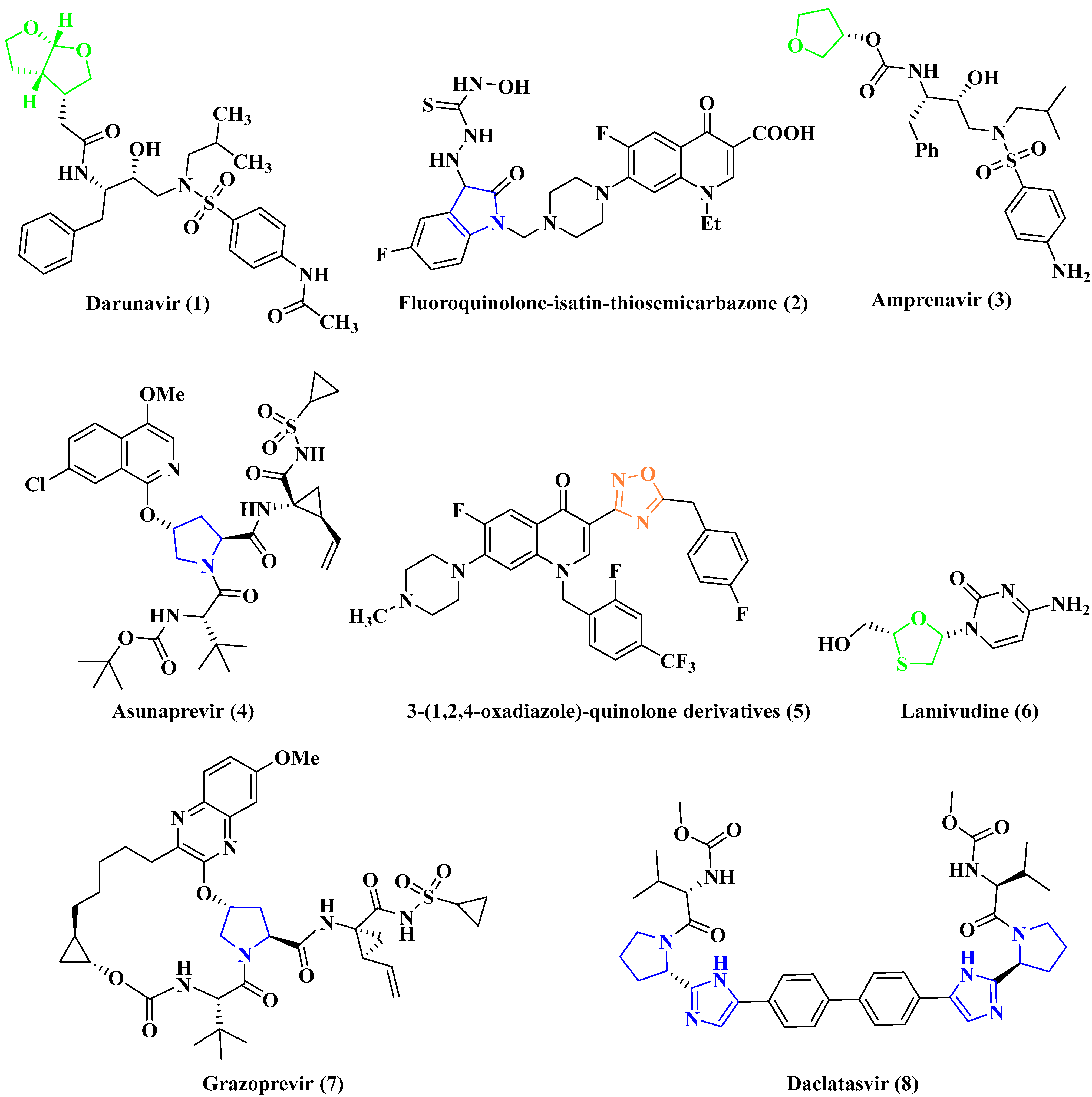
5.1. Anti-HIV Agent
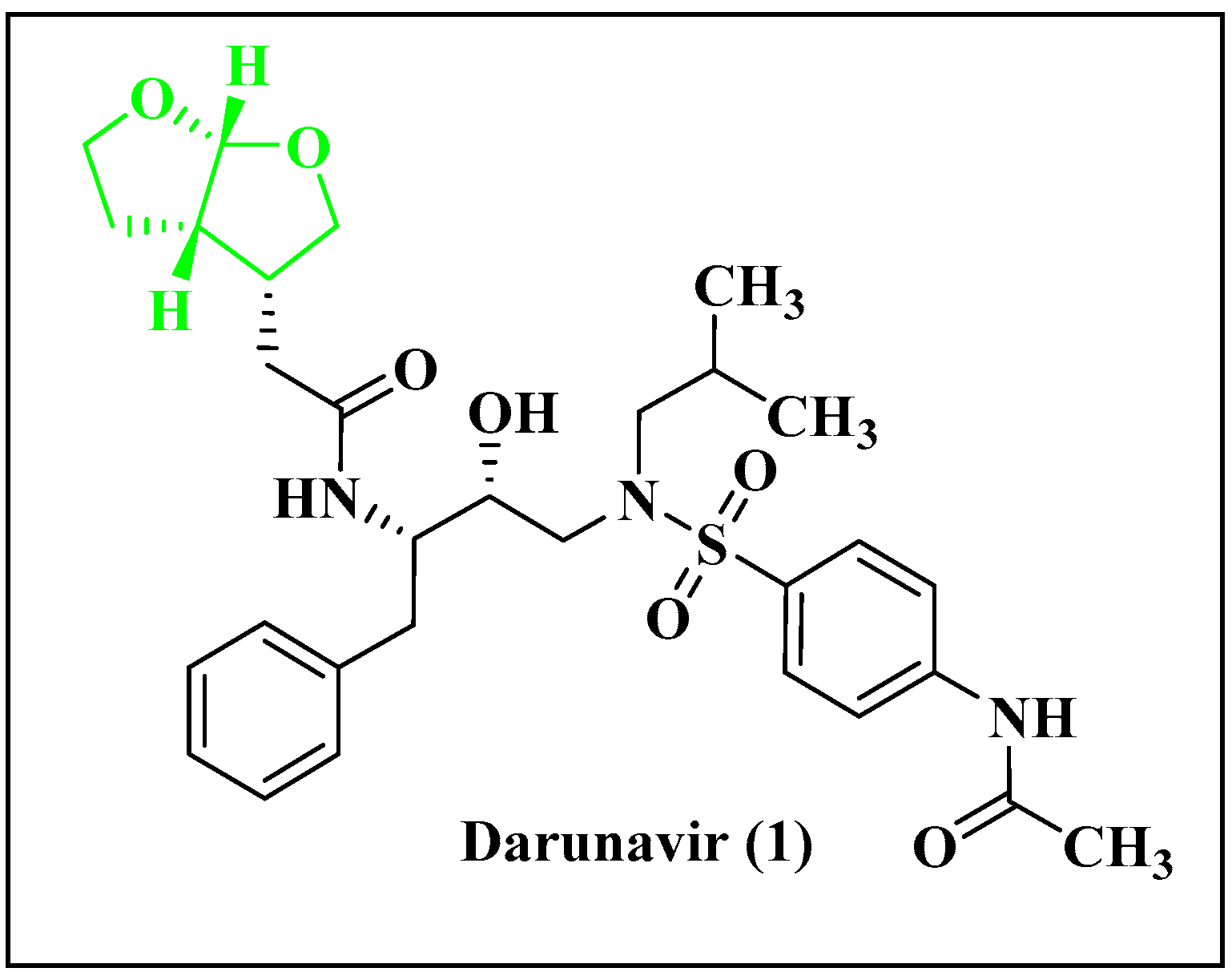
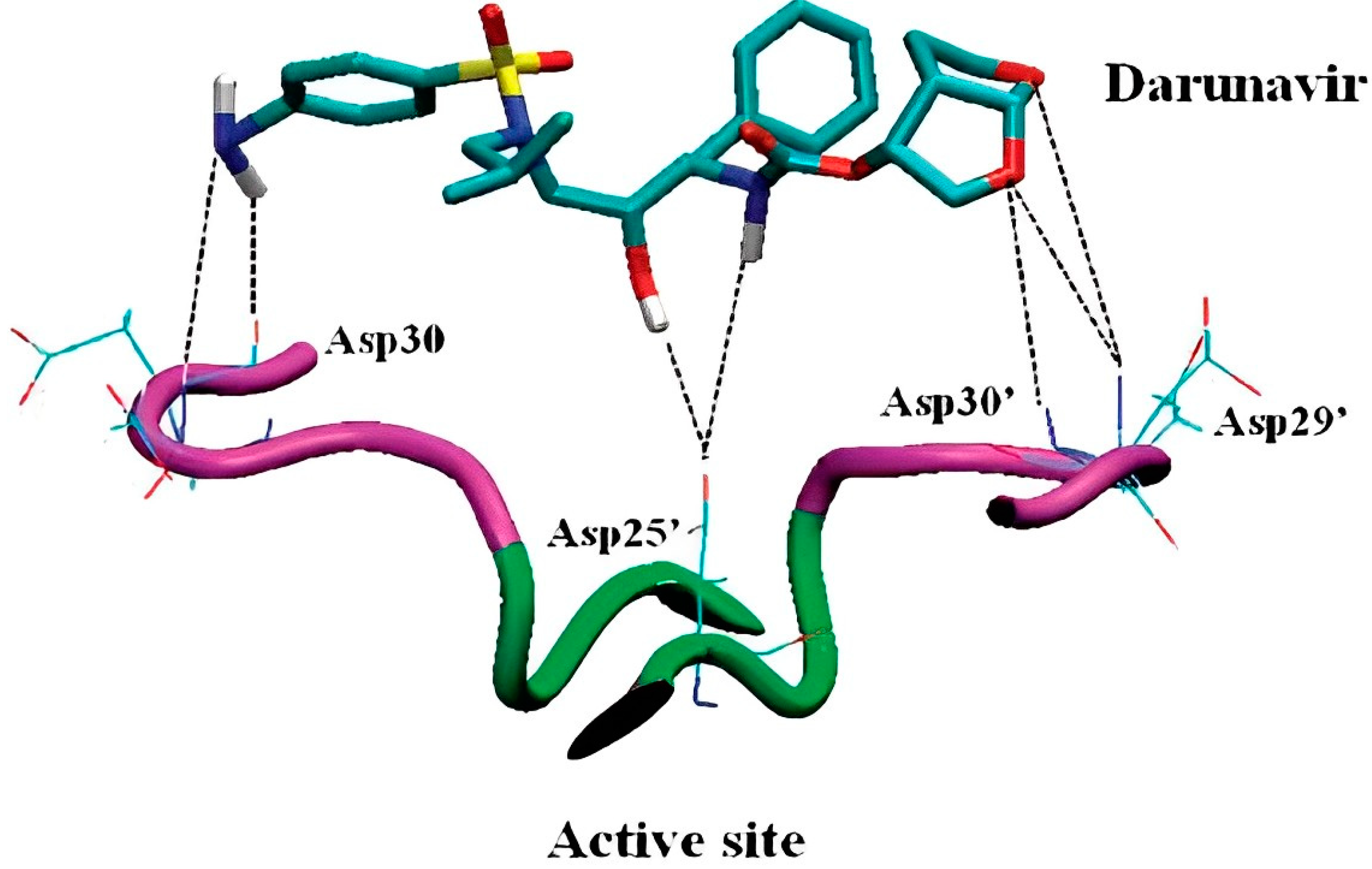
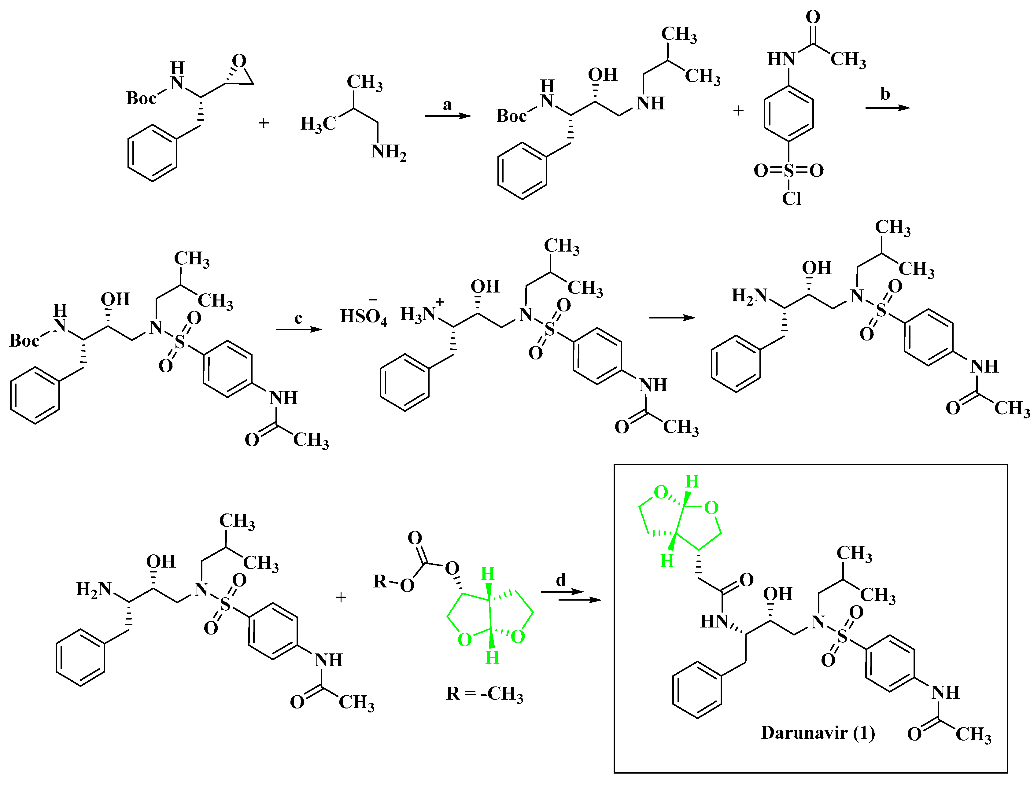
5.1.1. Anti-HIV Agent Darunavir
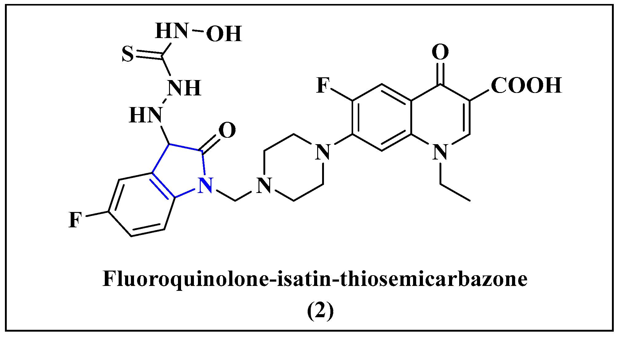
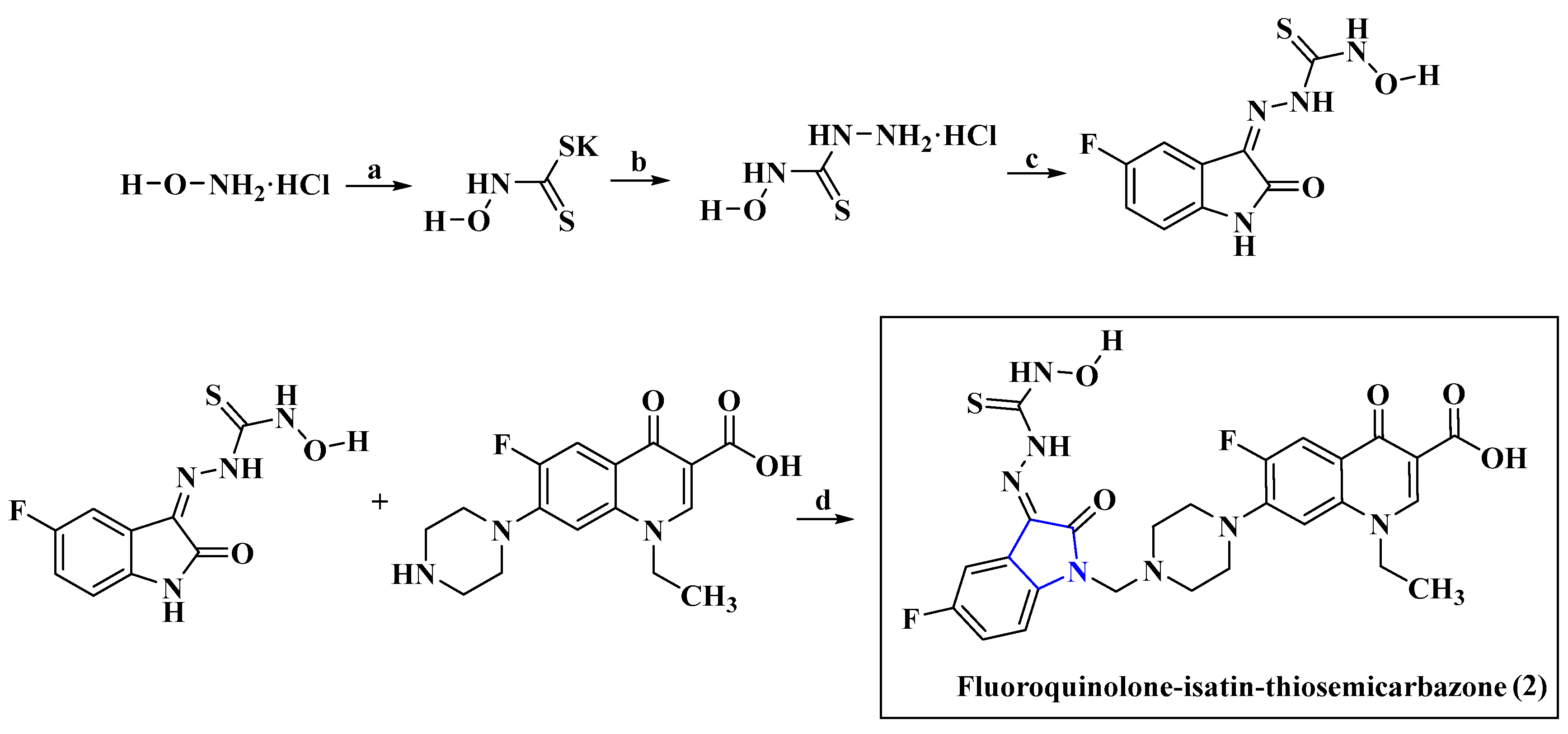
5.1.2. Anti-HIV Agent Fluoroquinolone-Isatin-Thiosemicarbazone Hybrids
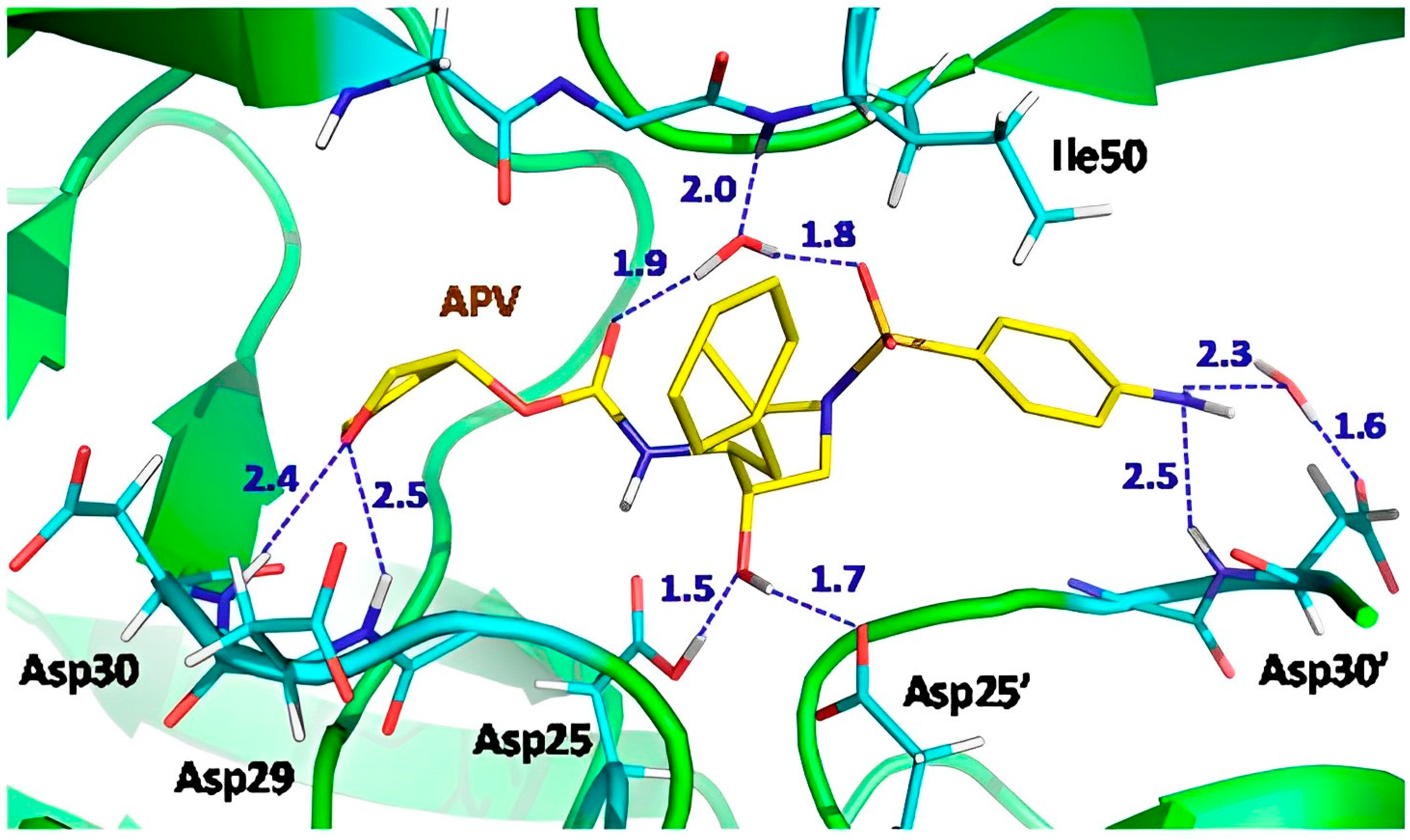
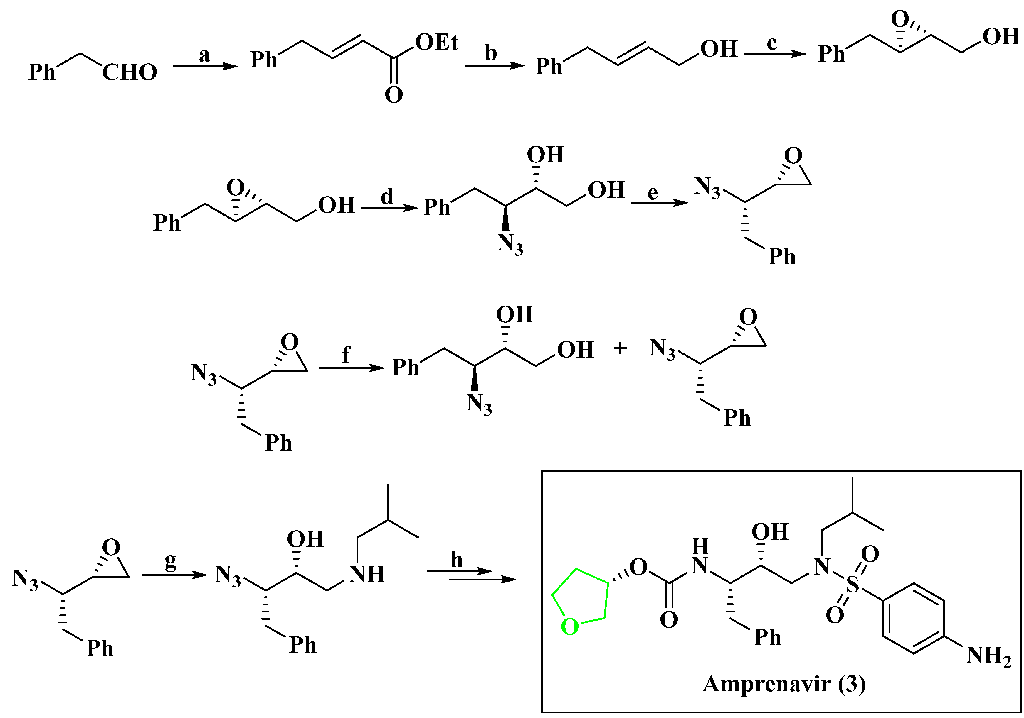
5.1.3. Anti-HIV Agent Amprenavir
5.2. Anti-HCV Agent
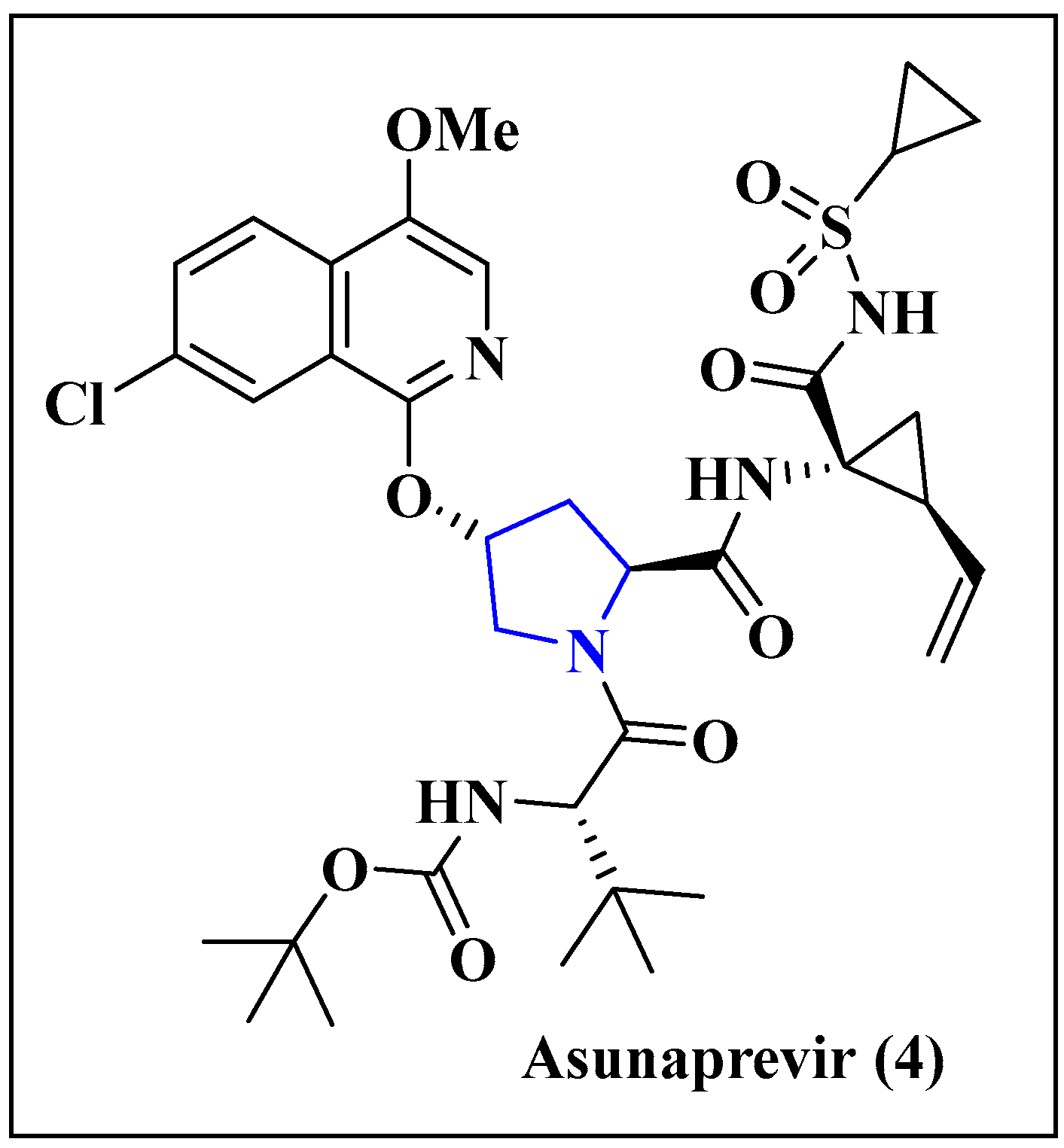
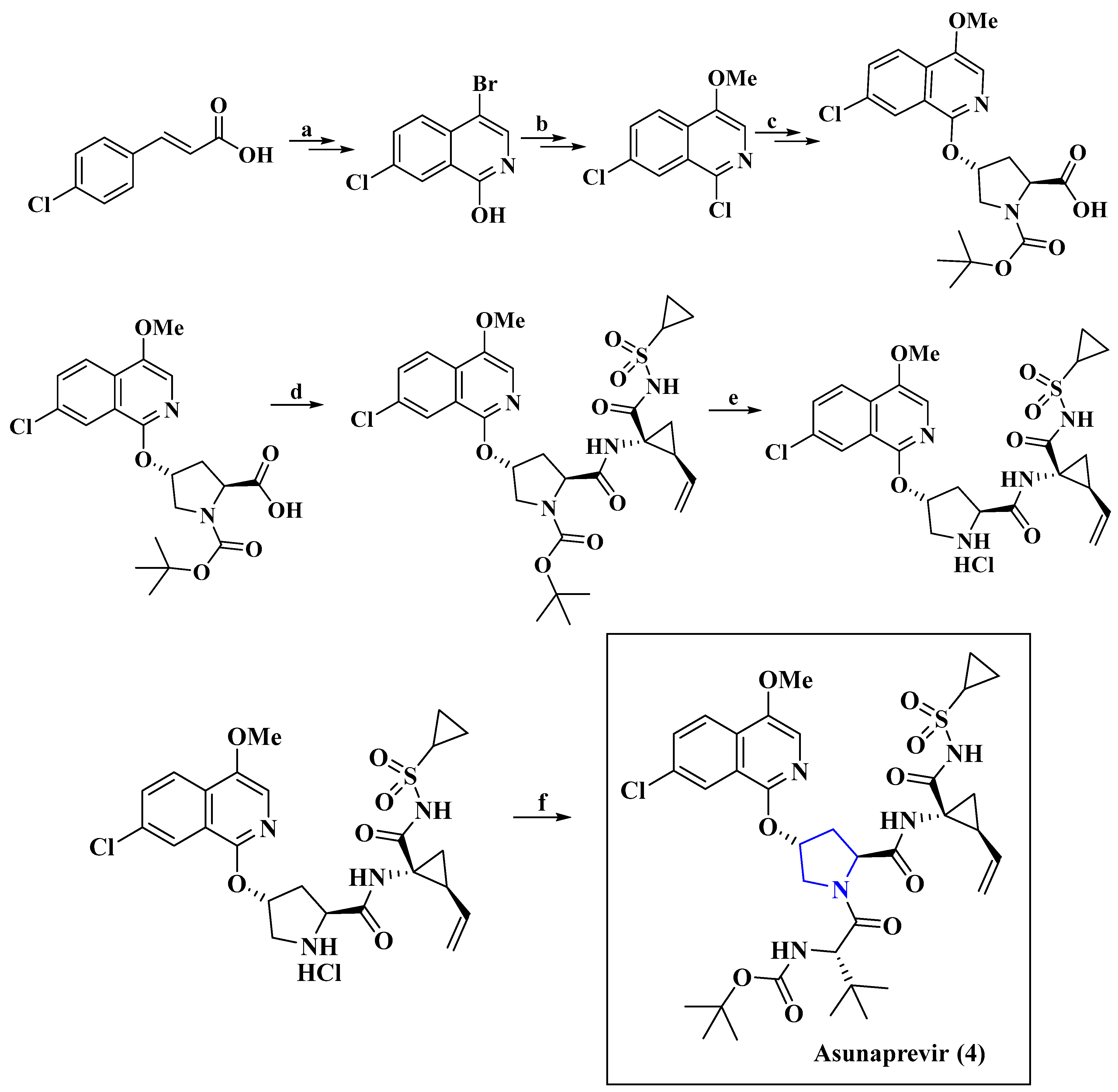
5.2.1. Anti-HCV Agent Asunaprevir
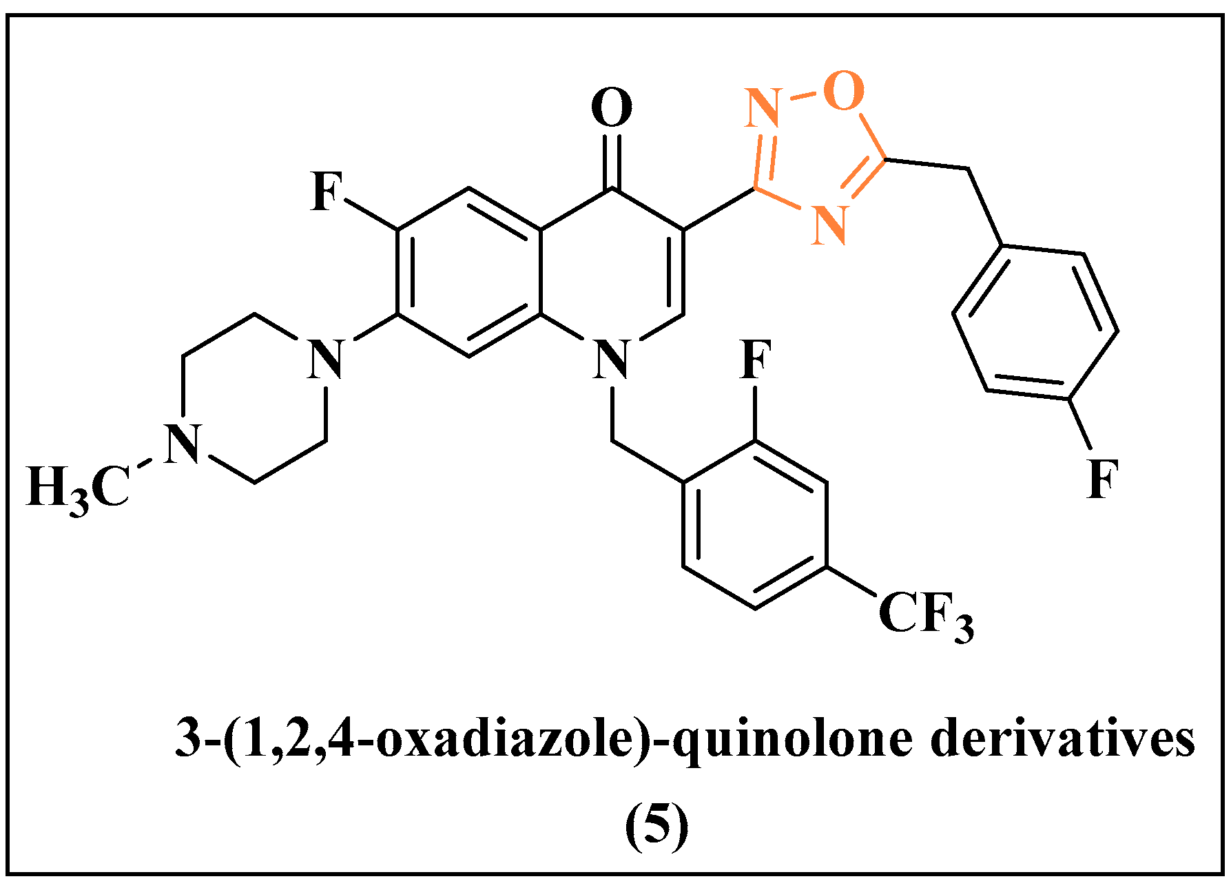
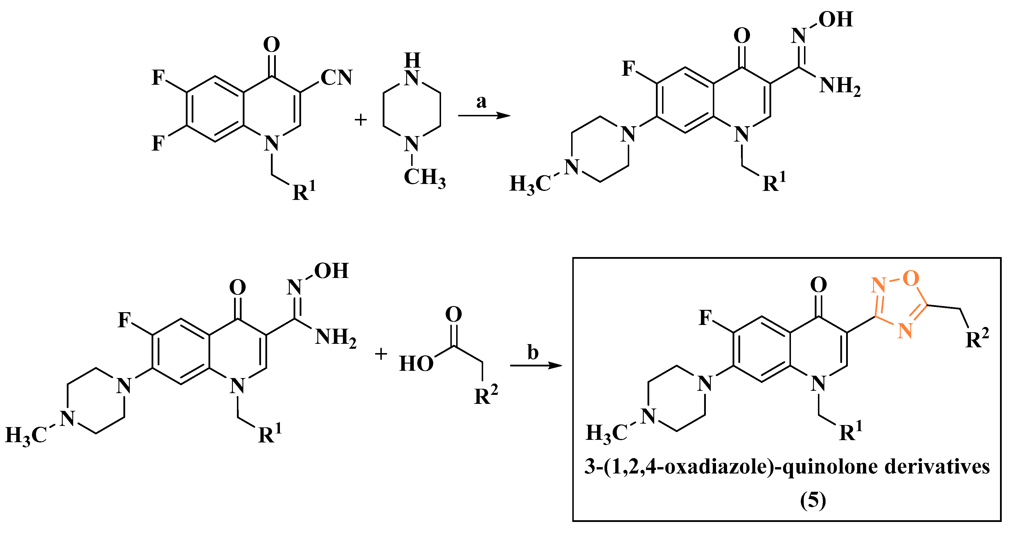
5.2.2. Anti-HCV Agent 3-(1,2,4-oxadiazole)-quinolone
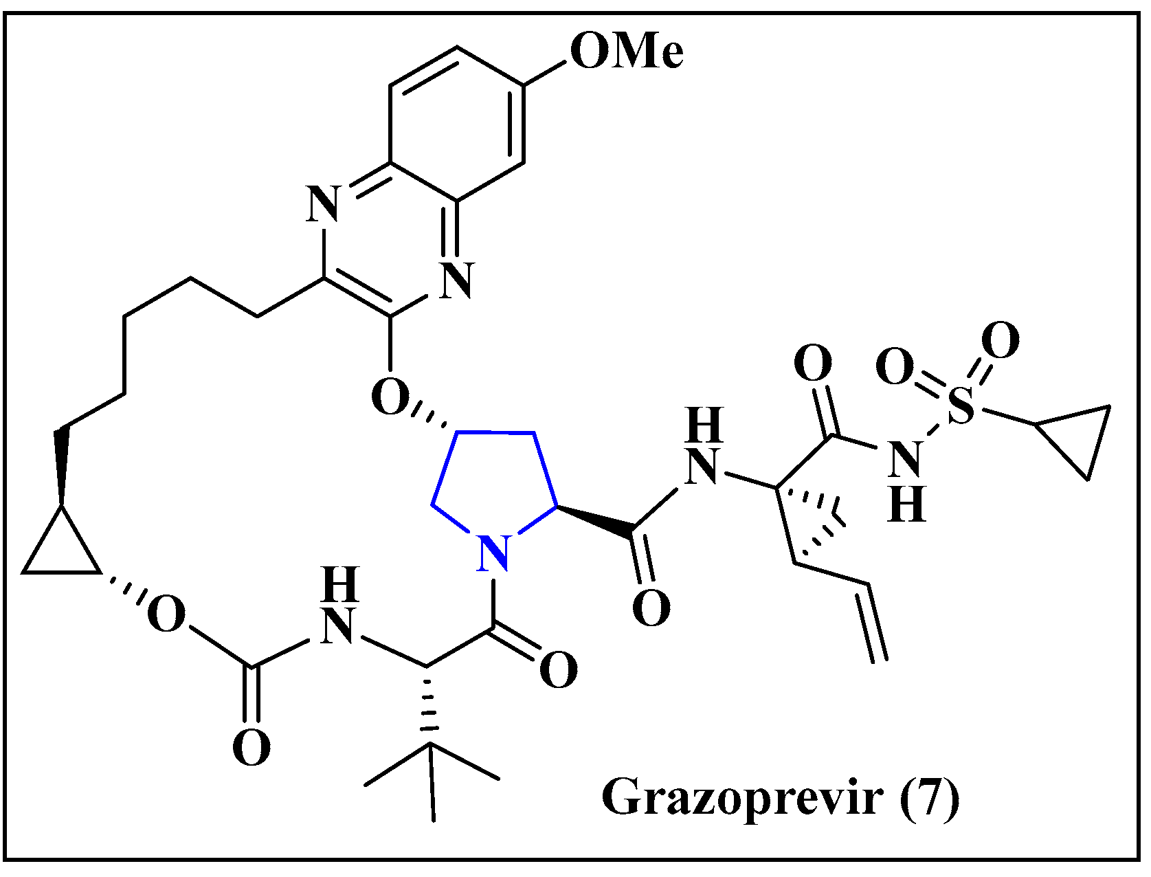
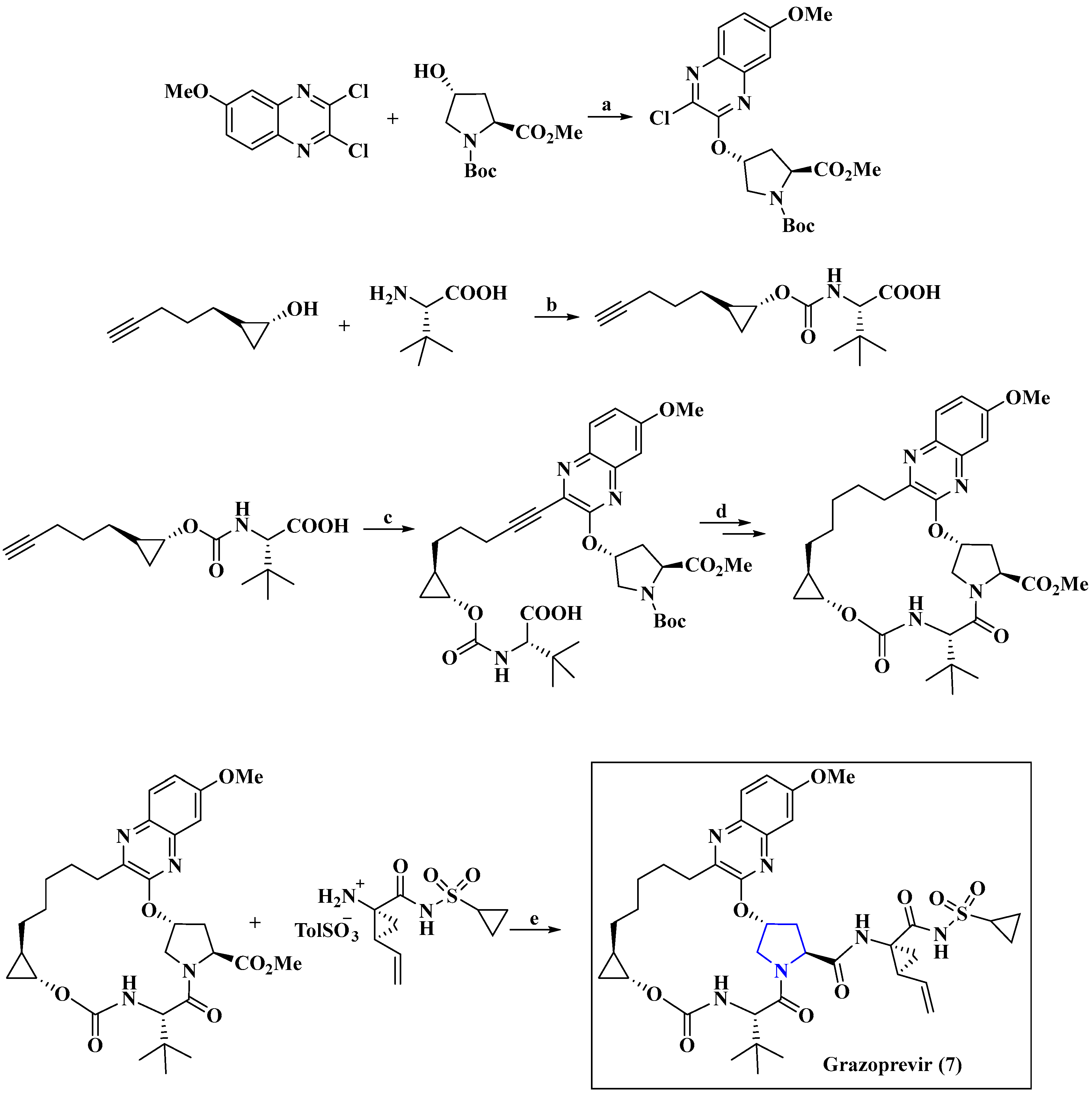
5.2.3. Anti-HCV Agent Grazoprevir
5.3. Anti-HBV Agent
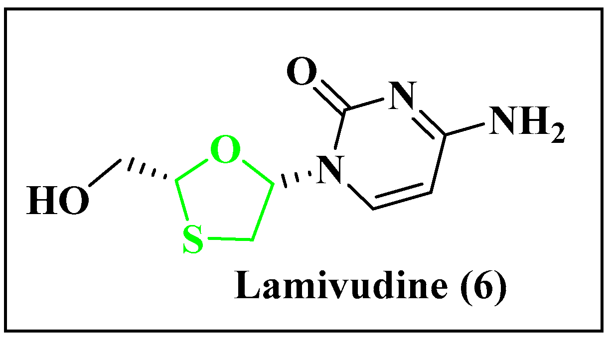
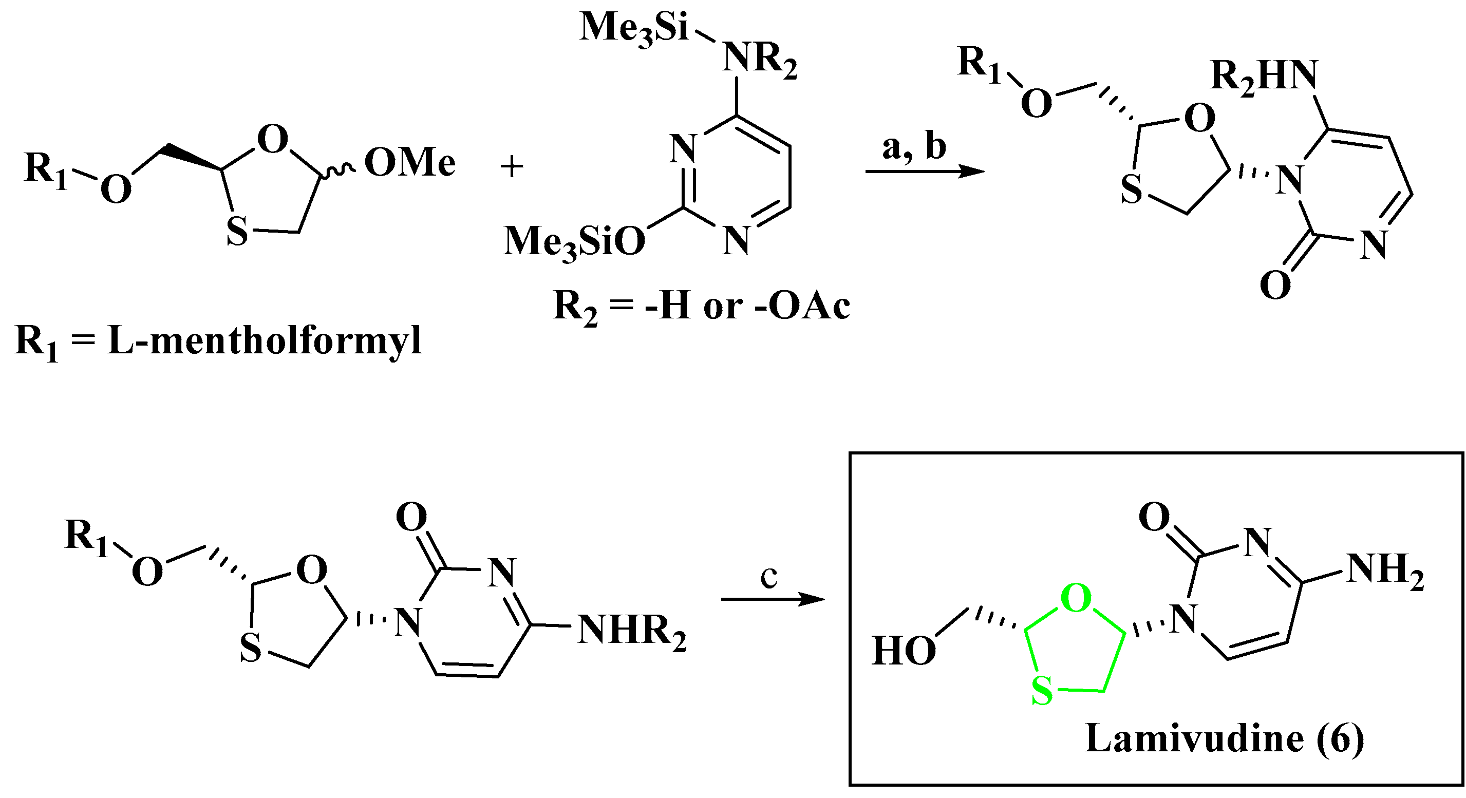
5.3.1. Anti-HBV Agent Lamivudine
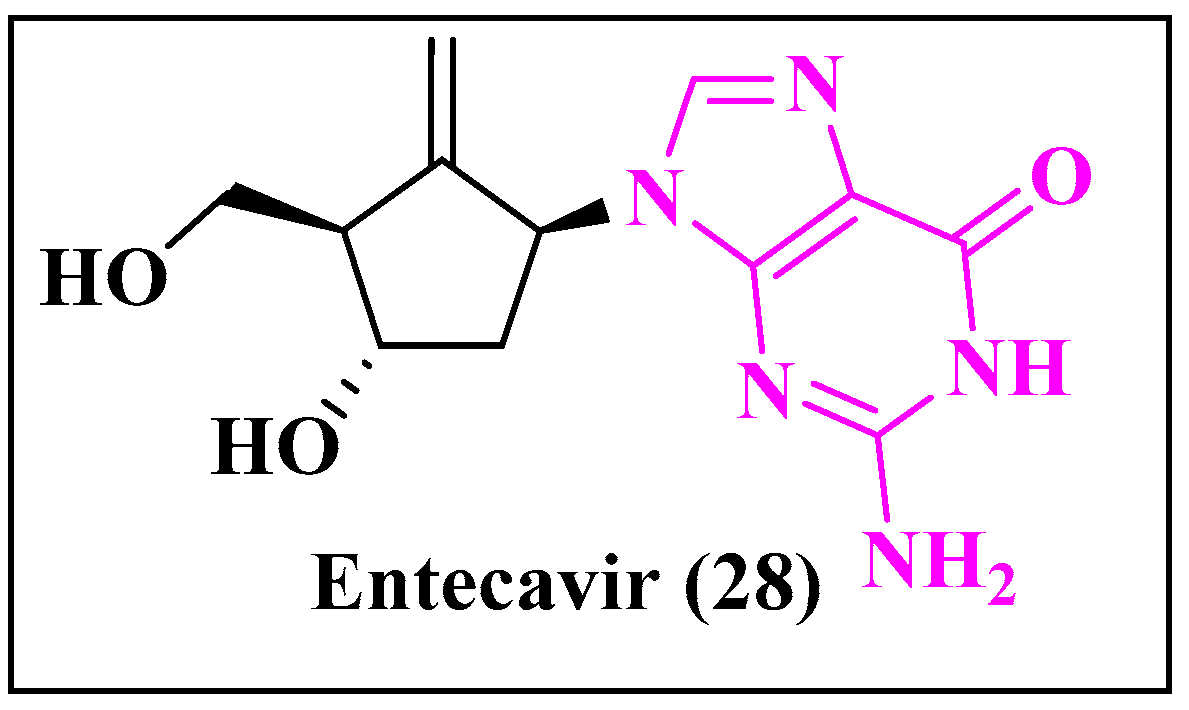
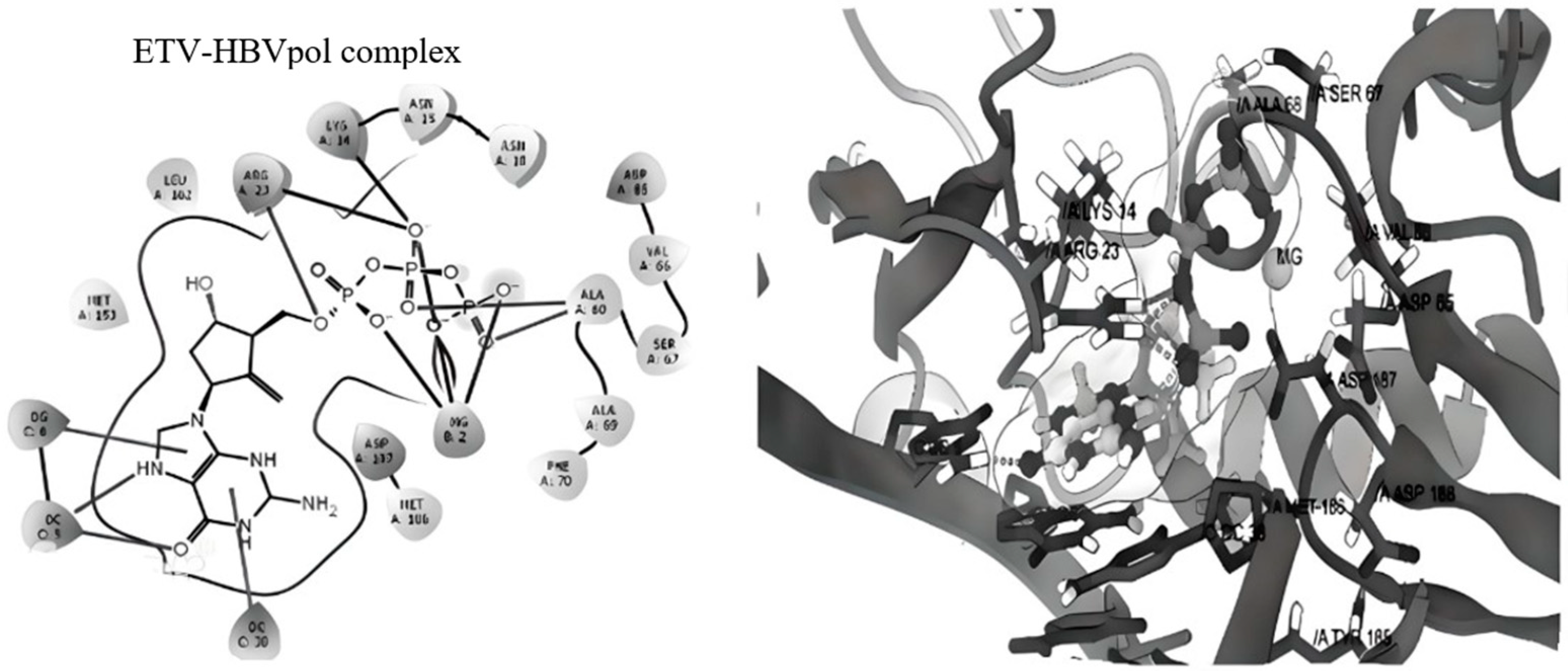
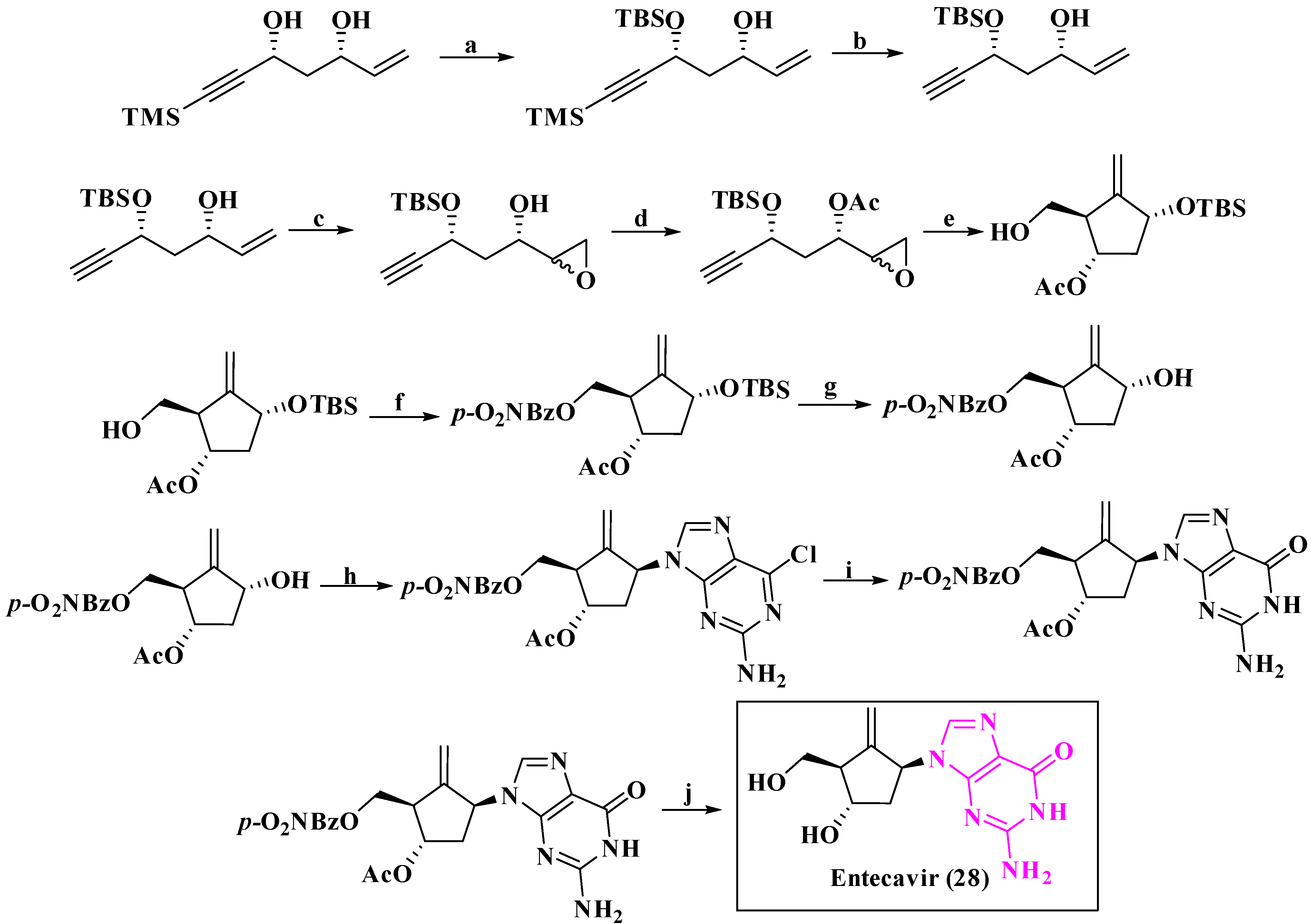
5.3.2. Anti-HBV Agent Entecavir
5.3.3. Anti-HBV Agent Dehydro-Andrographolide and Andrographolide Derivatives

| Sl. No. | Antiviral Agent | Drug Target | Activity |
|---|---|---|---|
| 1. |  | HBV [189] | Capsid assembly modulators (CpAMs) are antiviral compounds that target the core protein of the hepatitis B virus (HBV) to disrupt assembly. In HepG2.2.15 cells, which express HBV, these compounds inhibit HBV replication by interfering with capsid protein assembly with EC50 = 511 nM. |
| 2. |  | HBV [190] | The anti-HCV activities were tested in the Huh-Luc/neo cell line and cytotoxicity of the test compound was determined on both MT-2 cell lines with EC50 = 10 µM. |
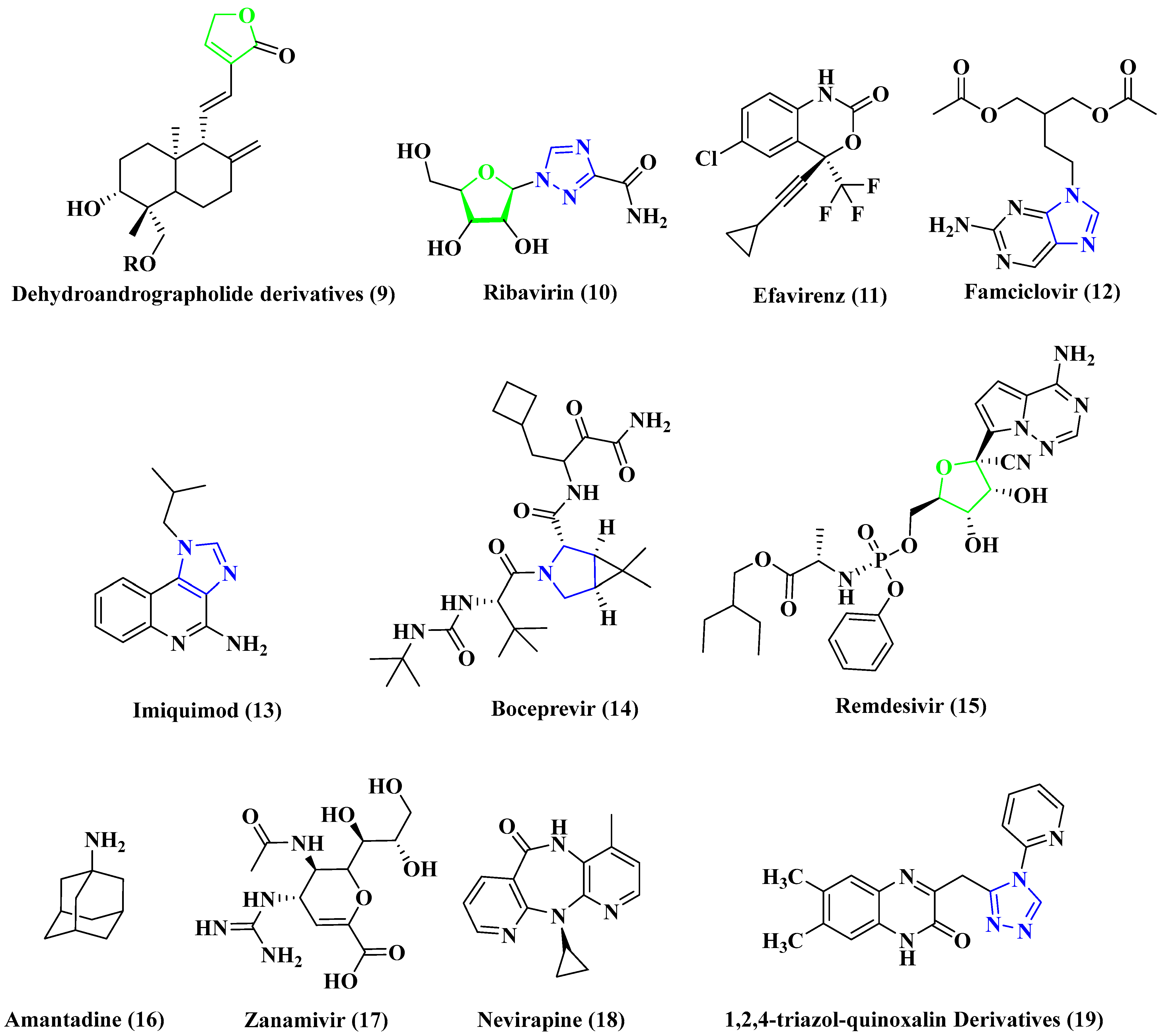
5.4. Anti-RSV Agent
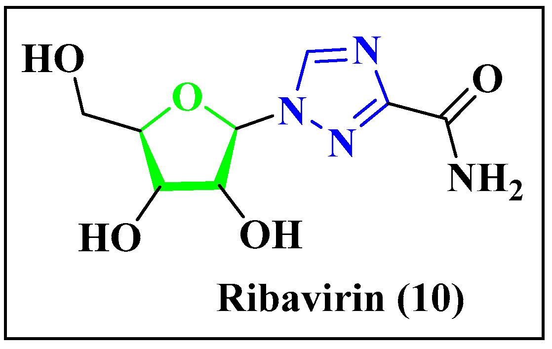
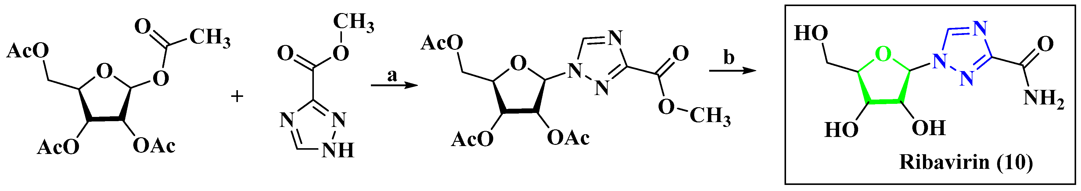
Anti-RSV Agent Ribavirin
5.5. Anti-HCMV Agent
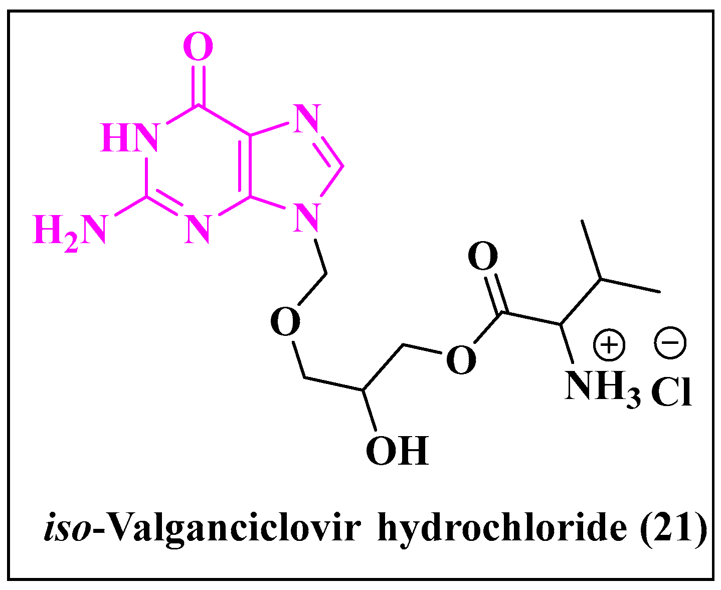
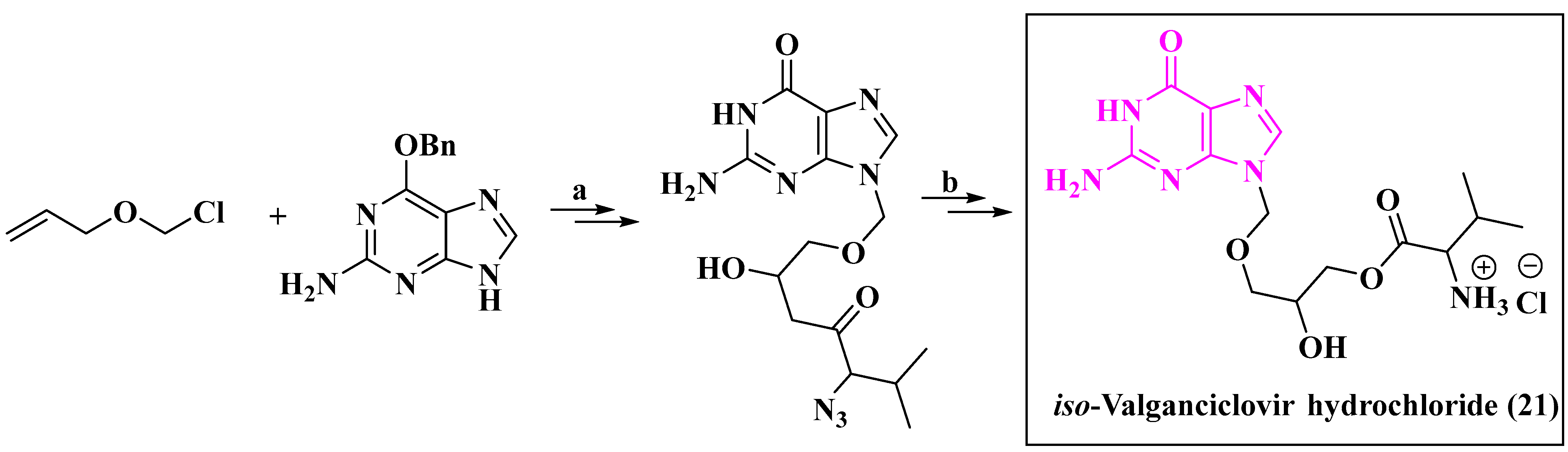
5.5.1. Anti-HCMV Agent Iso-Valganciclovir Hydrochloride
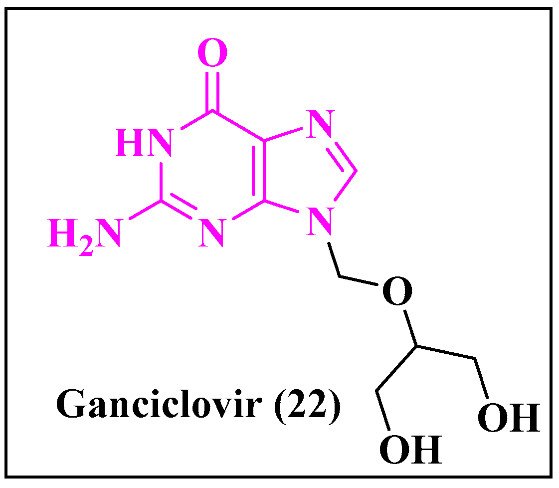
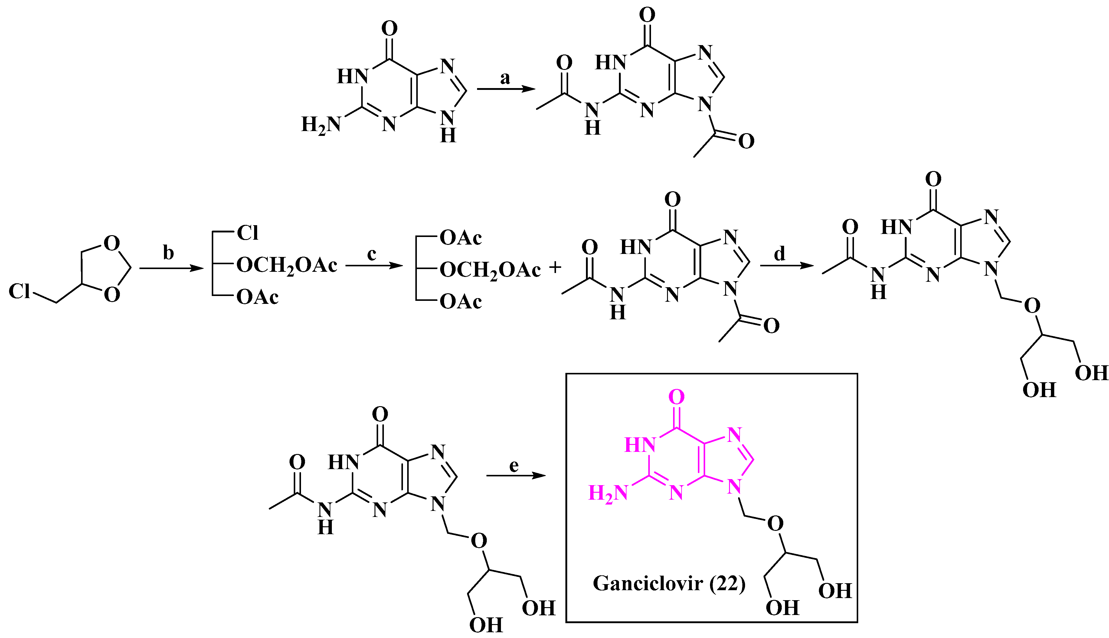
5.5.2. Anti-HCMV Agent Ganciclovir
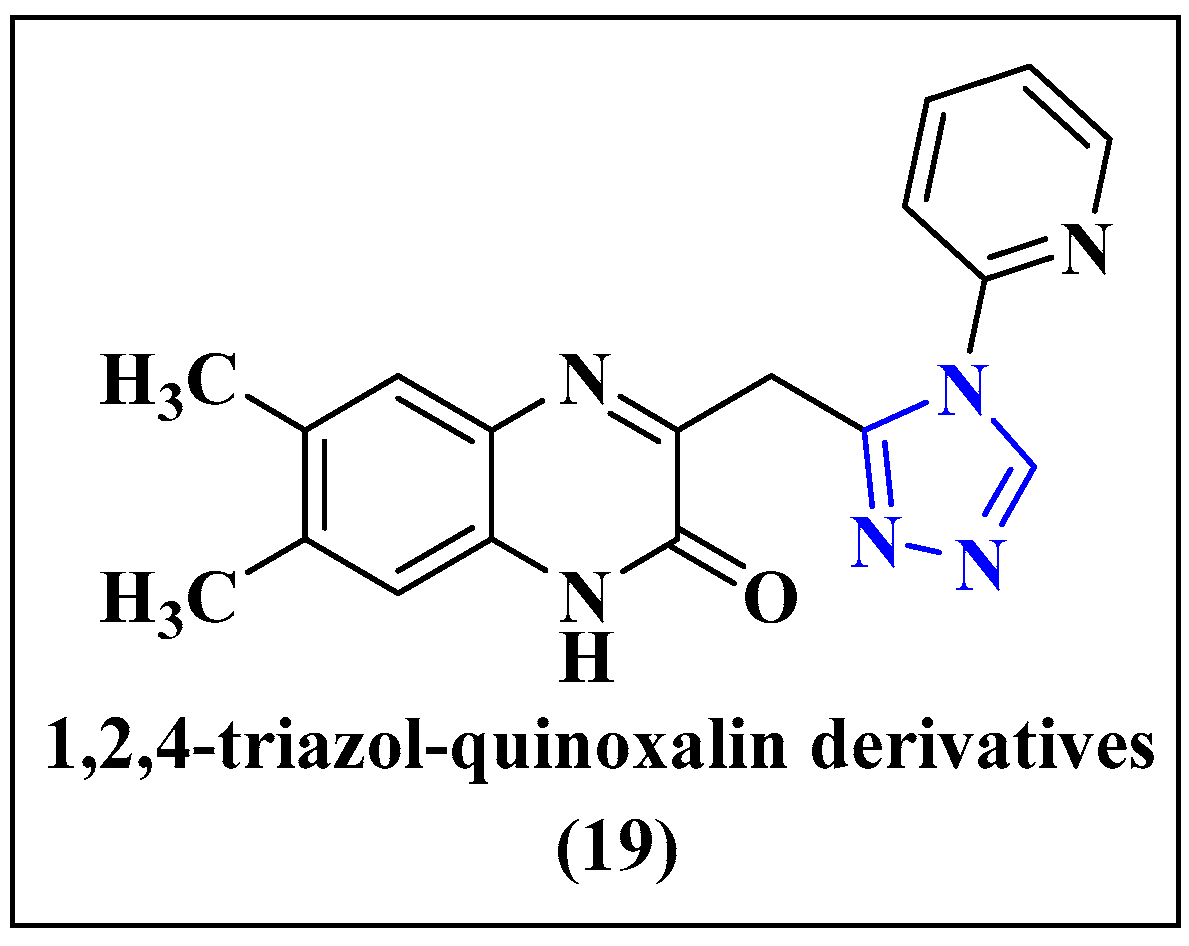
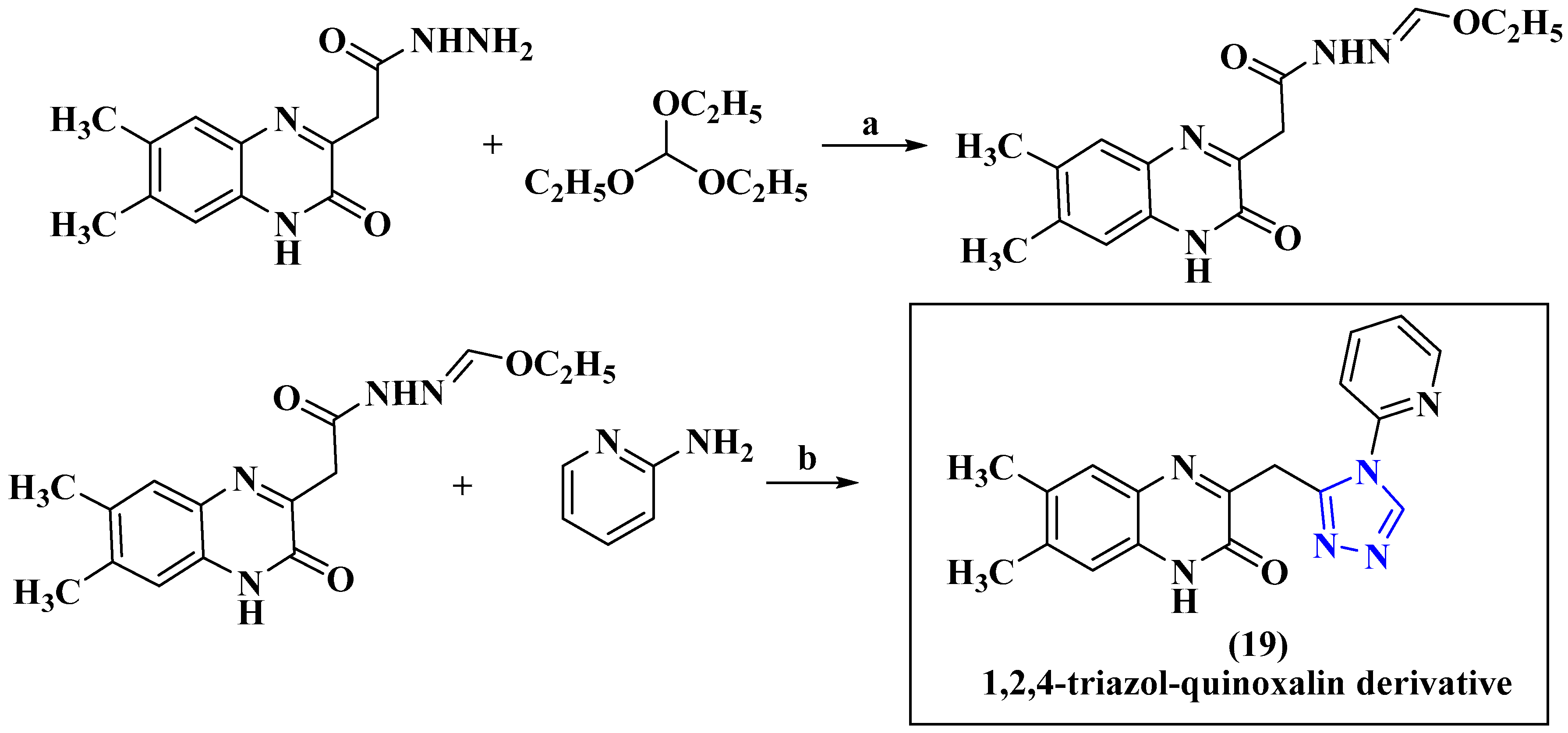
5.5.3. Anti-HCMV Agent 1,2,4-Triazol-Quinoxalin Derivative
5.6. Anti-HSV Agent
5.7. Anti-Ebola Agent
5.8. Anti-SARS-COV-2 Agent
- Comirnaty vaccine by Pfizer/BioNTech, approved 31 December 2020.
- SII/COVISHIELD and AstraZeneca/AZD1222 vaccines, approved 16 February 2021.
- Janssen/Ad26.COV 2.S vaccine developed by Johnson & Johnson, approved 12 March 2021.
- Moderna COVID-19 vaccine (mRNA 1273), approved 30 April 2021.
- Sinopharm COVID-19 vaccine, approved 7 May 2021.
- Sinovac-CoronaVac vaccine, approved 1 June 2021.
- Bharat Biotech BBV152 COVAXIN vaccine, approved 3 November 2021.
- Covovax (NVX-CoV2373) vaccine, approved 17 December 2021.
- Nuvaxovid (NVX-CoV2373) vaccine, approved 20 December 2021.
| Sl. No. | Drug Name | Drug Target | Mechanism of Action | Ways of Use | Side Effect | Brand Name |
|---|---|---|---|---|---|---|
| 1. |  | SARS-CoV-2 [239] | Nirmatrelvir inhibits cysteine residue in the 3C-like protease (3CLPRO) of SARS-CoV-2 | Oral | There is no such side effect observed | Paxlovid |
| 2. |  | COVID-19 [240] | Baricitinib inhibits the activity of JAK proteins and modulates the signaling pathway of various interleukins, interferons | Oral | There is no such side effect observed | Olumiant |
5.9. Anti-HPV Agent
5.10. Anti-Rabies Agent
5.11. Anti-Zika Agent
5.12. Anti-Polio Agent
5.13. Anti-West Nile Agent
5.14. Anti-Chickenpox Agent
5.15. Anti-Influenza Agent
5.16. Anti-Yellow Fever Agent
6. Conclusions
7. Scope, Limitation and the Presentation of the Future Trend of Antiviral Drugs
Author Contributions
Funding
Acknowledgments
Conflicts of Interest
Abbreviations
| Abbreviation | Full Name |
| DNA | Deoxyribonucleic Acid |
| RNA | Ribonucleic Acid |
| AIDS | Acquired Immune Deficiency Syndrome |
| HIV | Human Immunodeficiency Virus |
| HCV | Hepatitis C Virus |
| HBV | Hepatitis B Virus |
| RSV | Respiratory Syncytial Virus |
| HCMV | Human Cytomegalovirus |
| HSV | Herpes Simplex Virus |
| EBOV | Ebola Virus |
| SARS-CoV-2 | Severe Acute Respiratory Syndrome CoV-2 |
| PNBA | Para Nitro Benzoic Acid |
| GP120-CCR5 | Beta chemokine receptors |
| dATP | Deoxyadenosine triphosphate |
| mRNA | Messenger Ribonucleic Acid |
| eIF4A | Eukaryotic initiation factor 4A |
| ToS | Toluenesulfonyl |
| OiPr | Isopropoxide |
| OSBT | O-(Tert-Butyldimethylsilyl)hydroxylamine |
| Boc | tert-butoxycarbonyl |
| PMP | Polymethylpentene |
| OBn | Benzyl group |
| mCPBA | meta-chloroperoxybenzoic acid |
| DMSO | Dimethyldioxirane |
| TFAA | Trifluoroacetic anhydride |
| TFA | Trifluoroacetic acid |
| DSC | N,N′-Disuccinimidyl carbonate |
| DEC | Diethylcarbamazine |
| DMAP | 4-Dimethylaminopyridine |
| LDA | Lithium diisopropylamide |
| DDQ | 2,3-Dichloro-5,6-Dicyanobenzoquinone |
| THF | Tetrahydrofuran |
| NBS | N-Bromosuccinimide |
| DPPA | Diphenylphosphoryl azide |
| HATU | Hexafluorophosphate Azabenzotriazole Tetramethyl Uronium |
| DCM | Dichloromethane |
| DMAc | N,N-Dimethylacetamide |
| DBU | 1,8-Diazabicyclo(5.4.0)undec-7-ene |
| CDI | Carbonyldiimidazole |
| CPME | Cyclopentyl methyl ether |
| DIAD | Diisopropyl azodicarboxylate |
| IPAc | Isopropyl acetate |
| DMF | Dimethylformamide |
| DCC | N,N′-Dicyclohexylcarbodiimide |
| DEAD | Diethyl azodicarboxylate |
| TBAF | Tetra-n-butylammonium fluoride |
| 4Å MS | 4Å Molecular Sieve |
References
- Parvez, M.K.; Parveen, S. Evolution and Emergence of Pathogenic Viruses: Past, Present, and Future. Intervirology 2017, 60, 1–7. [Google Scholar] [CrossRef]
- Gaba, A.; Ayalew, L.E.; Tikoo, S.K. Animal Adenoviruses. In Recent Advances in Animal Virology; Malik, Y.S., Singh, R.K., Yadav, M.P., Eds.; Springer: Singapore, 2019; ISBN 978-981-13-9072-2. [Google Scholar] [CrossRef]
- Zhu, Z.; Lian, X.; Su, X.; Wu, W.; Marraro, G.A.; Zeng, Y. From SARS and MERS to COVID-19: A brief summary and comparison of severe acute respiratory infections caused by threeighly pathogenic human coronaviruses. Respir. Res. 2020, 21, 224. [Google Scholar] [CrossRef] [PubMed]
- Wirth, T. Biodiversity and Evolution; Elsevier: Amsterdam, The Netherlands, 2018; Volume 3, pp. 123–137. [Google Scholar] [CrossRef]
- Morens, D.M.; Fauci, A.S. Emerging Pandemic Diseases: How We Got to COVID-19. Cell 2020, 182, 1077–1092. [Google Scholar] [CrossRef] [PubMed]
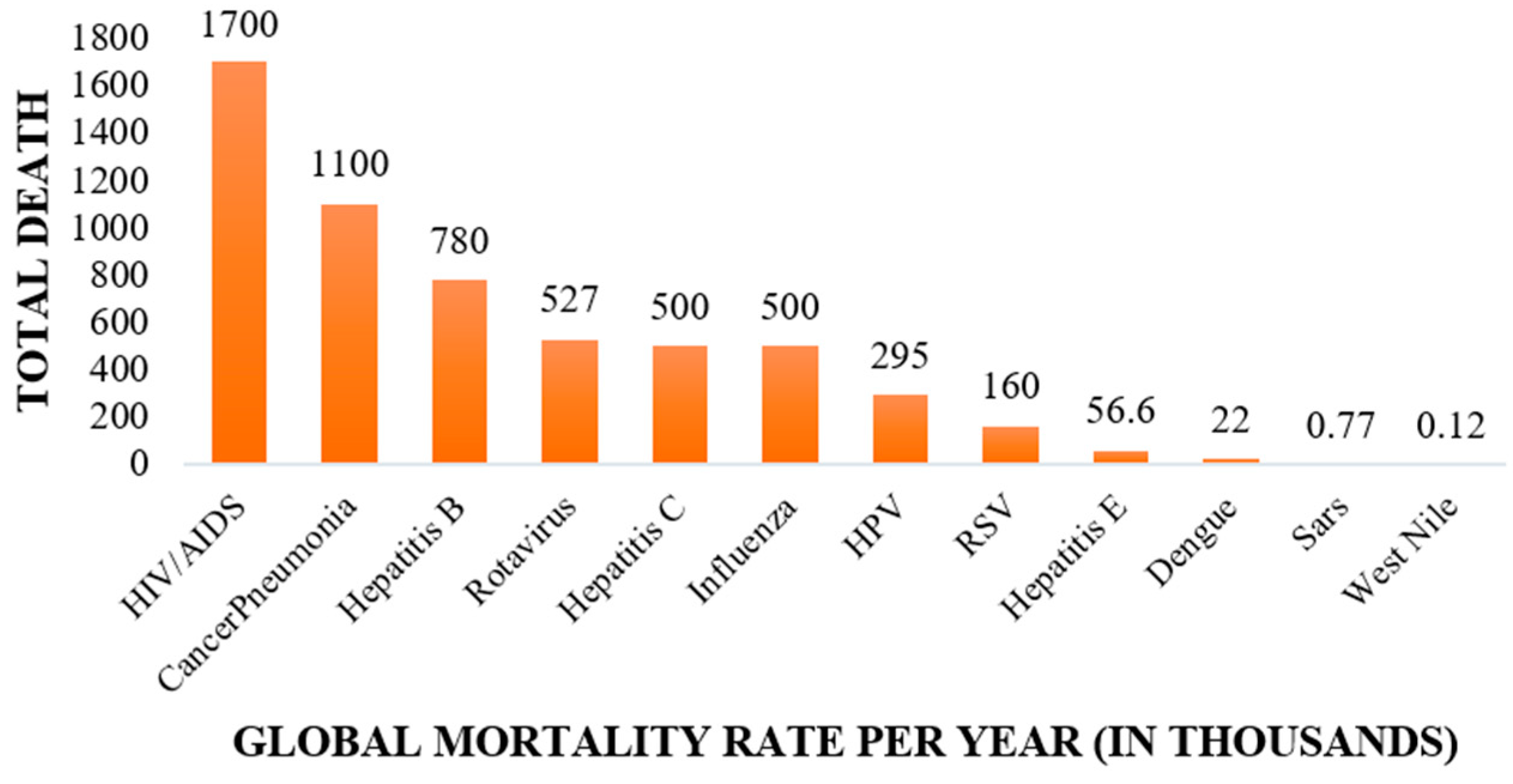
- Pagidipati, N.J.; Gaziano, T.A. Estimating Deaths From Cardiovascular Disease: A Review of Global Methodologies of Mortality Measurement. Circulation 2013, 127, 749–756. [Google Scholar] [CrossRef] [PubMed]
- Joint United Nations Programme on HIV/AIDS (UNAIDS). 2006 Report on the Global AIDS Epidemic; UNAIDS: Geneva, Switzerland, 2006. [Google Scholar]
- Mathers, C.D.; Boerma, T.; Ma Fat, D. Global and regional causes of death. Br. Med. Bull. 2009, 92, 7–32. [Google Scholar] [CrossRef] [PubMed]
- World Health Organization. Division of Emerging and Other Communicable Diseases Surveillance and Control. World Survey of Rabies: No. 30: For the Year 1994; World Health Organization: Geneva, Switzerland, 1996. [Google Scholar]
- Piot, P.; Muyembe, J.J.; Edmunds, W.J. Ebola in west Africa: From disease outbreak to humanitarian crisis. Lancet Infect. Dis. 2014, 14, 1034–1035. [Google Scholar] [CrossRef]
- Gostin, L.O.; Lucey, D.; Phelan, A. The Ebola Epidemic A Global Health Emergency. JAMA J. Am. Med. Assoc. 2014, 312, 1095–1096. [Google Scholar] [CrossRef]
- Meyers, L.; Frawley, T.; Goss, S.; Kang, C. Ebola Virus Outbreak 2014: Clinical Review for Emergency Physicians. Ann. Emerg. Med. 2015, 65, 101–108. [Google Scholar] [CrossRef]
- Gogineni, V.; Schinazi, R.F.; Hamann, M.T. Role of Marine Natural Products in the Genesis of Antiviral Agents. Chem. Rev. 2015, 115, 9655–9706. [Google Scholar] [CrossRef]
- More, A.F.; Loveluck, C.P.; Clifford, H.; Handley, M.J.; Korotkikh, E.V.; Kurbatov, A.V.; McCormick, M.; Mayewski, P.A. The health benefit of physical exercise on COVID-19 pandemic: Evidence from mainland China. GeoHealth 2020, 4, 277–284. [Google Scholar] [CrossRef]
- Mammas, I.; Spandidos, D.A. [Comment] COVID-19 threat and the 1918 Spanish flu outbreak: The following day. Exp. Ther. Med. 2019, 20, 292. [Google Scholar] [CrossRef]
- Taubenberger, J.K.; Morens, D.M. 1918 Influenza: The mother of all pandemics. Emerg. Infect. Dis. 2006, 12, 15–22. [Google Scholar] [CrossRef] [PubMed]
- Trilla, A.; Trilla, G.; Daer, C. The 1918 “Spanish Flu” in Spain. Clin. Infect. Dis. 2008, 47, 668–673. [Google Scholar] [CrossRef] [PubMed]
- Li, L.; Huang, T.; Wang, Y.; Wang, Z.; Liang, Y.; Huang, T.; Zhang, H.; Sun, W.; Wang, Y.J. COVID-19 patients’ clinical characteristics, discharge rate, and fatality rate of meta-analysis. Med. Virol. 2020, 92, 577–583. [Google Scholar] [CrossRef] [PubMed]
- Dagan, N.; Barda, N.; Kepten, E.; Miron, O.; Perchik, S.; Katz, M.A.; Hernán, M.A.; Lipsitch, M.; Reis, B.; Balicer, R.D. BNT162b2 mRNA COVID-19 Vaccine in a Nationwide Mass Vaccination Setting. N. Engl. J. Med. 2021, 384, 1412–1423. [Google Scholar] [CrossRef] [PubMed]
- Haynes, B.F.; Corey, L.; Fernandes, P.; Gilbert, P.B.; Hotez, P.J.; Rao, S.; Santos, M.R.; Schuitemaker, H.; Watson, M.; Arvin, A. Prospects for a safe COVID-19 vaccine. Sci. Transl. Med. 2020, 12, eabe0948. [Google Scholar] [CrossRef] [PubMed]
- Corey, L.; Mascola, J.R.; Fauci, A.S.; Collins, F.S. A strategic approach to COVID-19 vaccine R&D. Science 2020, 368, 948–950. [Google Scholar] [CrossRef]
- Kim, J.H.; Marks, F.; Clemens, J.D. Looking beyond COVID-19 vaccine phase 3 trials. Nat. Med. 2021, 27, 205–211. [Google Scholar] [CrossRef]
- Cao, Y.; Deng, Q.; Dai, S. Remdesivir for severe acute respiratory syndrome coronavirus 2 causing COVID-19: An evaluation of the evidence. Travel Med. Infect. Dis. 2020, 35, 101647–101677. [Google Scholar] [CrossRef]
- World Health Organization. Newsroom: Coronavirus Disease (COVID-19): Vaccines. Available online: https://www.who.int/news-room/q-a-detail/coronavirus-disease-(COVID-19)-vaccines (accessed on 15 August 2021).
- Onnuswamy, M.N.; Gromiha, M.M.; Sony, S.M.M.; Saraboji, K. QSAR and Molecular Modeling Studies in Heterocyclic Drugs I; Springer: Berlin/Heidelberg, Germany, 2006; Volume 3, pp. 81–147. [Google Scholar] [CrossRef]
- Sliwoski, G.R.; Meiler, J.; Lowe, E.W. Computational methods in drug discovery. Comput. Methods Drug Discov. 2014, 66, 334–395. [Google Scholar] [CrossRef]
- Jambhekar, S.S.; Breen, P.J. Drug dissolution: Significance of physicochemical properties and physiological conditions. Drug Discov. Today 2013, 18, 1173–1184. [Google Scholar] [CrossRef] [PubMed]
- Syam, Y.; Kamel, M. Structure and physicochemical properties in relation to drug action. Egypt. Pharm. J. 2013, 12, 95–108. [Google Scholar] [CrossRef]
- Martin-Benlloch, X.; Haid, S.; Novodomska, A.; Rominger, F.; Pietschmann, T.; Davioud-Charvet, E.; Elhabiri, M. Physicochemical properties govern the activity of potent antiviral flavones. ACS Omega 2019, 4, 4871–4887. [Google Scholar] [CrossRef]
- Klimenko, K.; Marcou, G.; Horvath, D.; Varnek, A.J. Chemical Space Mapping and Structure–Activity Analysis of the ChEMBL Antiviral Compound Set. Chem. Inf. Model. 2016, 56, 1438–1454. [Google Scholar] [CrossRef]
- Prashantha Kumar, B.R.; Soni, M.; Bharvi Bhikhalal, U.; Kakkot, I.R.; Jagadeesh, M.; Bommu, P.; Nanjan, M.J. Analysis of physicochemical properties for drugs from nature. Nat. Med. Chem. Res. 2010, 19, 984–992. [Google Scholar] [CrossRef]
- Evstigneev, M.P. Physicochemical mechanisms of synergistic biological action of combinations of aromatic heterocyclic compounds. Org. Chem. Int. 2013, 2013, 278143. [Google Scholar] [CrossRef]
- Kerru, N.; Gummidi, L.; Maddila, S.; Gangu, K.K.; Jonnalagadda, S.B. A review on recent advances in nitrogen-containing molecules and their biological applications. Molecules 2020, 25, 1909. [Google Scholar] [CrossRef]
- Gomtsyan, AHeterocycles in drugs and drug discovery. Chem. Heterocycl. Compd. 2012, 48, 7–10. [CrossRef]
- De Vivo, M.; Masetti, M.; Bottegoni, G.; Cavalli, A. Role of molecular dynamics and related methods in drug discovery. J. Med. Chem. 2016, 59, 4035–4061. [Google Scholar] [CrossRef]
- Pastorino, B.; Nougairède, A.; Wurtz, N.; Gould, E.; de Lamballerie, X. Role of host cell factors in flavivirus infection: Implications for pathogenesis and development of antiviral drugs. Antivir. Res. 2010, 87, 281–294. [Google Scholar] [CrossRef]
- Monto, A.S. Vaccines and antiviral drugs in pandemic preparedness. Emerg. Infect. Dis. 2006, 12, 55–60. [Google Scholar] [CrossRef]
- Schmid, M.; Speiseder, T.; Dobner, T.; Gonzalez, R.A. DNA virus replication compartments. J. Virol. 2014, 88, 1404–1420. [Google Scholar] [CrossRef]
- Schwartz, M.; Chen, J.; Janda, M.; Sullivan, M.; Den Boon, J.; Ahlquist, P. A positive-strand RNA virus replication complex parallels form and function of retrovirus capsids. Mol. Cell 2002, 9, 505–514. [Google Scholar] [CrossRef]
- Vahlne, A. A historical reflection on the discovery of human retroviruses. Retrovirology 2009, 6, 40. [Google Scholar] [CrossRef]
- Levin, J.G.; Mitra, M.; Mascarenhas, A.; Musier-Forsyth, K. Role of HIV-1 nucleocapsid protein in HIV-1 reverse transcription. RNA Biol. 2010, 7, 754–774. [Google Scholar] [CrossRef]
- Gallo, R.C.; Montagnier, L. The discovery of HIV as the cause of AIDS. N. Engl. J. Med. 2003, 349, 2283–2285. [Google Scholar] [CrossRef]
- World Health Organization. Newsroom: HIV and AIDS. Available online: https://www.who.int/news-room/fact-sheets/detail/hiv-aids (accessed on 4 May 2023).
- Ghosh, A.K.; Fyvie, W.S.; Brindisi, M.; Steffey, M.; Agniswamy, J.; Wang, Y.F.; Aoki, M.; Amano, M.; Weber, I.T.; Mitsuya, H. Design, Synthesis, Biological Evaluation, and X-ray Studies of HIV-1 Protease Inhibitors with Modified P2′ Ligands of Darunavir. ChemMedChem 2017, 12, 1942–1952. [Google Scholar] [CrossRef]
- Du, L.; Jia, J.; Ge, P.; Jin, Y. Self-assemblies of 5′-cholesteryl-ethyl-phosphoryl zidovudine. Colloids Surf. B. 2016, 148, 385–391. [Google Scholar] [CrossRef]
- Belk, D.; Belk, P. The Great American Healthcare Scam: How Kickbacks, Collusion and Propaganda Have Exploded Healthcare Costs in the United States Paperback; David Belk: Greensboro, NC, USA, 2020; ISBN 13: 9781734709018. [Google Scholar]
- Vasilyeva, S.V.; Shtil, A.A.; Petrova, A.S.; Balakhnin, S.M.; Achigecheva, P.Y.; Stetsenko, D.A.; Silnikov, V.N. Conjugates of phosphorylated zalcitabine and lamivudine with SiO2 nanoparticles: Synthesis by CuAAC click chemistry and preliminary assessment of anti-HIV and antiproliferative activity. Bioorg. Med. Chem. 2017, 25, 1696–1702. [Google Scholar] [CrossRef]
- Paton, N.I.; Musaazi, J.; Kityo, C.; Walimbwa, S.; Hoppe, A.; Balyegisawa, A.; Kaimal, A.; Mirembe, G.; Tukamushabe, P.; Ategeka, G. Dolutegravir or darunavir in combination with zidovudine or tenofovir to treat HIV. N. Engl. J. Med. 2021, 385, 330–341. [Google Scholar] [CrossRef]
- Mayer, K.H.; Molina, J.-M.; Thompson, M.A.; Anderson, P.L.; Mounzer, K.C.; De Wet, J.J.; DeJesus, E.; Jessen, H.; Grant, R.M.; Ruane, P.J. Emtricitabine and tenofovir alafenamide vs emtricitabine and tenofovir disoproxil fumarate for HIV pre-exposure prophylaxis (DISCOVER): Primary results from a randomised, double-blind, multicentre, active-controlled, phase 3, non-inferiority trial. Lancet 2020, 396, 239–254. [Google Scholar] [CrossRef]
- Alter, M.J. Epidemiology of hepatitis C virus infection. World J. Gastroenterol. 2007, 13, 2436–2441. [Google Scholar] [CrossRef]
- Massengill, M.T.; Park, J.C.; McAnany, J.J.; Hyde, R.A. Occult retinopathy following treatment of Hepatitis C with glecaprevir/pibrentasvir (Mavyret). Doc. Ophthalmol. 2023, 146, 191–197. [Google Scholar] [CrossRef]
- Gentile, I.; Buonomo, A.R.; Zappulo, E.; Minei, G.; Morisco, F.; Borrelli, F.; Coppola, N.; Borgia, G. Asunaprevir, a protease inhibitor for the treatment of hepatitis C infection. Ther. Clin. Risk Manag. 2014, 10, 493–504. [Google Scholar] [CrossRef]
- Chang, M.H.; Gordon, L.A.; Fung, H.B. Infection of common marmosets with GB virus B chimeric virus encoding the major nonstructural proteins NS2 to NS4A of hepatitis C virus. Clin. Ther. 2012, 34, 2021–2038. [Google Scholar] [CrossRef]
- Kiang, T.K.L. Clinical pharmacokinetics and drug–drug interactions of elbasvir/grazoprevir. Eur. J. Drug Metab. Pharmacokinet. 2018, 43, 509–531. [Google Scholar] [CrossRef]
- Blumberg, B.S. Hepatitis B virus, the vaccine, and the control of primary cancer of the liver. Proc. Natl. Acad. Sci. USA 1997, 94, 7121–7125. [Google Scholar] [CrossRef]
- Liu, J.; Li, T.; Zhang, L.; Xu, A. The role of hepatitis B surface antigen in nucleos (t) ide analogues cessation among asian patients with chronic hepatitis B: A systematic review. Hepatology 2019, 70, 1045–1055. [Google Scholar] [CrossRef]
- Sulkowski, M.S.; Agarwal, K.; Ma, X.; Nguyen, T.T.; Schiff, E.R.; Hann, H.W.L.; Dieterich, D.T.; Nahass, R.G.; Park, J.S.; Chan, S.J. Safety and efficacy of vebicorvir administered with entecavir in treatment-naïve patients with chronic hepatitis B virus infection. Hepatol. 2022, 77, 1265–1275. [Google Scholar] [CrossRef]
- Zenchenko, A.A.; Drenichev, M.S.; Il’icheva, I.A.; Mikhailov, S.N. Antiviral and antimicrobial nucleoside derivatives: Structural features and mechanisms of action. Mol. Biol. 2021, 55, 786–812. [Google Scholar] [CrossRef]
- Menéndez-Arias, L.; Álvarez, M.; Pacheco, B. Nucleoside/nucleotide analog inhibitors of hepatitis B virus polymerase: Mechanism of action and resistance. Curr. Opin. Virol. 2014, 8, 1–9. [Google Scholar] [CrossRef] [PubMed]
- Acosta, P.L.; Caballero, M.T.; Polack, F.P. Brief history and characterization of enhanced respiratory syncytial virus disease. Clin. Vaccine Immunol. 2016, 23, 189–195. [Google Scholar] [CrossRef] [PubMed]
- Li, Y.; Wang, X.; Blau, D.M.; Caballero, M.T.; Feikin, D.R.; Gill, C.J.; Madhi, S.A.; Omer, S.B.; Simões, E.A.F.; Campbell, H. Brief history and characterization of enhanced respiratory syncytial virus disease. Lancet 2022, 399, 2047–2064. [Google Scholar] [CrossRef] [PubMed]
- Shi, T.; Vennard, S.; Mahdy, S.; Nair, H.J. Risk factors for RSV associated acute lower respiratory infection poor outcome and mortality in young children: A systematic review and meta-analysis. Infect. Dis. 2022, 226, S10–S16. [Google Scholar] [CrossRef]
- Nair, H.; Nokes, D.J.; Gessner, B.D.; Dherani, M.; Madhi, S.A.; Singleton, R.J.; O’Brien, K.L.; Roca, A.; Wright, P.F.; Bruce, N.; et al. Global burden of acute lower respiratory infections due to respiratory syncytial virus in young children: A systematic review and meta-analysis. Lancet 2010, 375, 1545–1555. [Google Scholar] [CrossRef] [PubMed]
- Wang, Y.Q.; Zhao, X.Y. Human cytomegalovirus primary infection and reactivation: Insights from virion-carried molecules. Front. Microbiol. 2020, 11, 1511. [Google Scholar] [CrossRef]
- Stoelben, S.; Arns, W.; Renders, L.; Hummel, J.; Mühlfeld, A.; Stangl, M.; Fischereder, M.; Gwinner, W.; Suwelack, B.; Witzke, O.; et al. Preemptive treatment of Cytomegalovirus infection in kidney transplant recipients with letermovir: Results of a Phase 2a study. Transpl. Int. 2014, 27, 77–86. [Google Scholar] [CrossRef]
- Biron, K.K. Antiviral drugs for cytomegalovirus diseases. Antivir. Res. 2006, 71, 154–163. [Google Scholar] [CrossRef]
- Corey, L.; Sper, P.G. Infections with herpes simplex viruses. N. Engl. J. Med. 1986, 314, 686–691. [Google Scholar] [CrossRef]
- Gottlieb, S.L.; Giersing, B.K.; Hickling, J.; Jones, R.; Deal, C.; Kaslow, D.C. Meeting report: Initial World Health Organization consultation on herpes simplex virus (HSV) vaccine preferred product characteristics, March 2017. Vaccine 2019, 37, 7408–7418. [Google Scholar] [CrossRef]
- Feldmann, H.; Jones, S.; Klenk, H.-D.; Schnittler, H.-J. Ebola virus: From discovery to vaccine. Nat. Rev. Immunol. 2003, 3, 677–685. [Google Scholar] [CrossRef] [PubMed]
- Mirza, M.U.; Vanmeert, M.; Ali, A.; Iman, K.; Froeyen, M.; Idrees, M. Perspectives towards antiviral drug discovery against Ebola virus. J. Med. Virol. 2019, 91, 2029–2048. [Google Scholar] [CrossRef] [PubMed]
- Grellet, E.; Goulet, A.; Imbert, I. Replication of the Coronavirus Genome: A Paradox among Positive-Strand RNA Viruses. J. Biol. Chem. 2022, 5, 101923–101938. [Google Scholar] [CrossRef]
- Tyrrell, D.A.J.; Bynoe, M.L. Cultivation of viruses from a high proportion of patients with colds. Lancet 1966, 287, 76–77. [Google Scholar] [CrossRef] [PubMed]
- Kahn, J.S.; McIntosh, K. History and Recent Advances in Coronavirus Discovery. Pediatr. Infect. Dis. J. 2005, 24, S223–S227. [Google Scholar] [CrossRef] [PubMed]
- Hu, B.; Guo, H.; Zhou, P.; Shi, Z.L. Characteristics of SARS-CoV-2 and COVID-19. Nat. Rev. Microbiol. 2021, 19, 141–154. [Google Scholar] [CrossRef]
- Chang, F.; Syrjänen, S.; Kellokoski, J.; Syrjänen, K. Human Papillomavirus (HPV) Infections and Their Associations with Oral Disease. J. Oral Pathol. Med. 1991, 20, 305–317. [Google Scholar] [CrossRef]
- World Health Organization. Newsroom: Fact Sheets. Cervical Cancer. Available online: https://www.who.int/news-room/fact-sheets/detail/cervical-cancer (accessed on 1 May 2023).
- Fekadu, M.; Shaddock, J.H.; Chandler, F.W.; Baer, G.M. Rabies Virus in the Tonsils of a Carrier Dog. Arch. Virol. 1983, 78, 37–47. [Google Scholar] [CrossRef]
- Dick, G.W.A.; Kitchen, S.F.; Haddow, A.J. zika virus (i). Isolations and serological specificity. Trans. R. Soc. Trop. Med. Hyg. 1952, 46, 509–520. [Google Scholar] [CrossRef]
- Posen, H.J.; Keystone, J.S.; Gubbay, J.B.; Morris, S.K. Epidemiology of Zika Virus, 1947–2007. BMJ Glob. Health 2016, 1, e000087. [Google Scholar] [CrossRef]
- Chen, H.L.; Tang, R.B.J. Why Zika virus infection has become a public health concern? Chin. Med. Assoc. 2016, 79, 174–178. [Google Scholar] [CrossRef] [PubMed]
- Gorshkov, K.; Shiryaev, S.A.; Fertel, S.; Lin, Y.W.; Huang, C.T.; Pinto, A.; Farhy, C.; Strongin, A.Y.; Zheng, W.; Terskikh, A.V. Transdermal permeation of bacteriophage particles by choline oleate: Potential for treatment of soft-tissue infections. Front. Microbiol. 2019, 9, 3252–3296. [Google Scholar] [CrossRef] [PubMed]
- Skern, T. 100 years poliovirus: From discovery to eradication. A meeting report. Arch. Virol. 2010, 155, 1371–1381. [Google Scholar] [CrossRef] [PubMed]
- Melnick, J.L. Current Status of Poliovirus Infections. Clin. Microbiol. Rev. 1996, 9, 293–300. [Google Scholar] [CrossRef] [PubMed]
- Dowdle, W.R.; Birmingham, M.E. The Biologic Principles of Poliovirus Eradication. J. Infect. Dis. 1997, 175, S286–S292. [Google Scholar] [CrossRef]
- World Health Organization. Health Topics: Poliomyelitis. Available online: https://www.who.int/health-topics/poliomyelitis (accessed on 1 May 2023).
- Smithburn, K.C.; Hughes, T.P.; Burke, A.W.; Paul, J.H. A neurotropic virus isolated from the blood of a native of Uganda. Am. J. Trop. Med. 1940, 20, 471–472. [Google Scholar] [CrossRef]
- World Health Organization. Newsroom: Fact Sheets. West Nile Virus. Available online: https://www.who.int/news-room/fact-sheets/detail/west-nile-virus (accessed on 1 May 2023).
- Takahashi, M. Chickenpox Virus. Adv. Virus Res. 1983, 28, 285–356. [Google Scholar] [CrossRef]
- Hutchinson, E.C. Influenza Virus. Trends Microbiol. 2018, 26, 809–810. [Google Scholar] [CrossRef]
- World Health Organization. Newsroom: Fact Sheets. Detail. Influenza (Seasonal). Available online: https://www.who.int/news-room/fact-sheets/detail/influenza-(seasonal) (accessed on 1 May 2023).
- Monath, T.P.; Vasconcelos, P.F.C. Yellow Fever. J. Clin. Virol. 2015, 64, 160–173. [Google Scholar] [CrossRef]
- World Health Organization. Health Topics: Yellow-Fever. Available online: https://www.who.int/health-topics/yellow-fever (accessed on 1 May 2023).
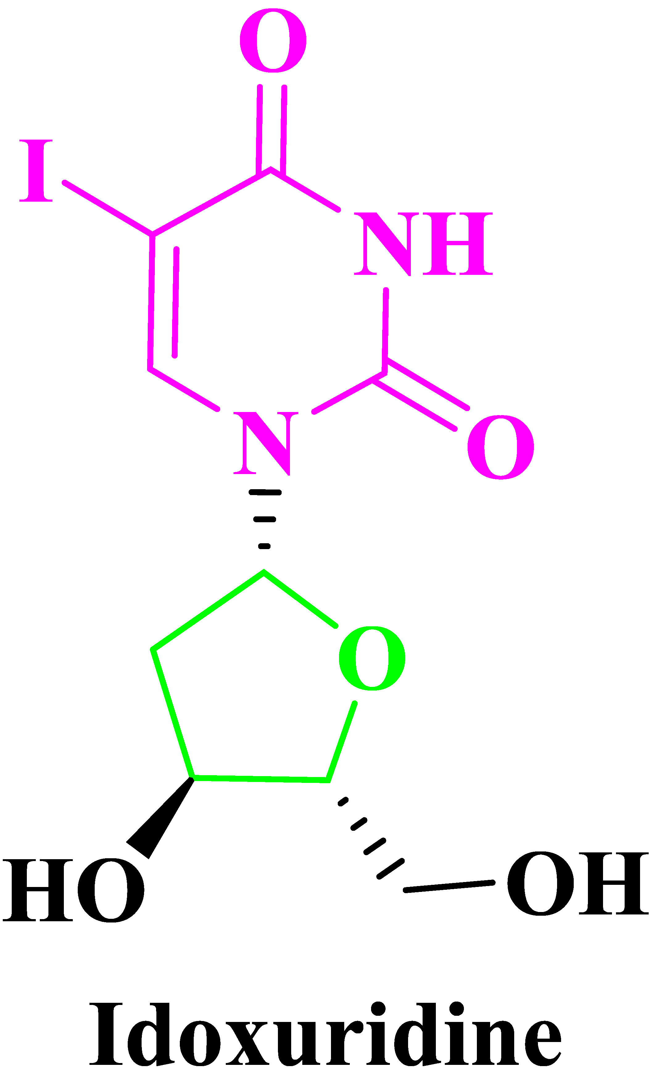
- Prusoff, W.H. Synthesis and biological activities of iododeoxyuridine, an analog of thymidine. BBA—Biochim. Biophys. Acta 1959, 32, 295–296. [Google Scholar] [CrossRef]
- Cheng, Y.; Prusoff, W.H. Relationship between the inhibition constant (&) and the concentration of inhibitor which causes 50 per cent inhibition (iso) of an enzymatic reaction. Biochem. Pharmacol. 1973, 22, 3099–3108. [Google Scholar] [CrossRef] [PubMed]
- Kaufman, H.E.; Martola, E.-L.; Dohlman, C. Use of 5-Iodo-2’-Deoxyuridine(IDU) in Treatment of Herpes Simplex Keratitis. Arch. Ophthalmol. 1962, 68, 235–239. [Google Scholar] [CrossRef] [PubMed]
- De Clercq, E.; Li, G. Approved Antiviral Drugs over the Past 50 Years. Clin. Microbiol. Rev. 2016, 29, 695–747. [Google Scholar] [CrossRef] [PubMed]
- Tchesnokov, E.P.; Feng, J.Y.; Porter, D.P.; Götte, M. Mechanism of Inhibition of Ebola Virus RNA-Dependent RNA Polymerase by Remdesivir. Viruses 2019, 11, 326. [Google Scholar] [CrossRef]
- Bergmann, W.; Feeney, R.J.J. The isolation of a new thymine pentoside from sponges. J. Am. Chem. Soc. 1950, 72, 2809–2810. [Google Scholar] [CrossRef]
- Wahyuni, T.S.; Widyawaruyanti, A.; Lusida, M.I.; Fuad, A.; Soetjipto; Fuchino, H.; Kawahara, N.; Hayashi, Y.; Aoki, C.; Hotta, H. Inhibition of hepatitis C virus replication by chalepin and pseudane IX isolated from Ruta angustifolia leaves. Fitoterapia 2014, 99, 276–283. [Google Scholar] [CrossRef]
- Andersen, R.J.; Faulkner, D.J.; He, C.H.; Van Duyne, G.D.; Clardy, J.J. Metabolites of the Marine Prosobranch Mollusc Lamellaria sp. Am. Chem. Soc. 1985, 107, 5492–5495. [Google Scholar] [CrossRef]
- Hashimoto, T.; Akiyo, Y.; Akazawa, K.; Takaoka, S.; Tori, M.; Asakawa, Y. Three novel dimethyl pyrroledicarboxylate, lycogarubins A C, from the myxomycetes lycogala epidendrum. Tetrahedron Lett. 1994, 35, 2559–2560. [Google Scholar] [CrossRef]
- Biedenkopf, N.; Lange-Grünweller, K.; Schulte, F.W.; Weiber, A.; Muller, C.; Becker, D.; Becker, S.; Hartmann, R.K.; Grünweller, A. The natural compound silvestrol is a potent inhibitor of Ebola virus replication. Antivir. Res. 2017, 137, 76–81. [Google Scholar] [CrossRef]
- Cui, H.; Xu, B.; Wu, T.; Xu, J.; Yuan, Y.; Gu, Q.J. Potential Antiviral Lignans from the Roots of Saururus chinensis with Activity against Epstein−Barr Virus Lytic Replication. Nat. Prod. 2014, 77, 100–110. [Google Scholar] [CrossRef]
- Perez, R.M. Antiviral Activity of Compounds Isolated From Plants. Pharm. Biol. 2003, 41, 107–157. [Google Scholar] [CrossRef]
- Chen, H.; Ma, Y.B.; Huang, X.Y.; Geng, C.A.; Zhao, Y.; Wang, L.J.; Guo, R.H.; Liang, W.J.; Zhang, X.M.; Chen, J.J. Synthesis, structure–activity relationships and biological evaluation of dehydroandrographolide and andrographolide derivatives as novel anti-hepatitis B virus agents. Bioorg. Med. Chem. Lett. 2014, 24, 2353–2359. [Google Scholar] [CrossRef] [PubMed]
- Geng, C.A.; Chen, J.J. The Progress of Anti-HBV Constituents from Medicinal Plants in China. Nat. Prod. Bioprospect. 2018, 8, 227–244. [Google Scholar] [CrossRef]
- Zhou, N.J.; Geng, C.A.; Huang, X.Y.; Ma, Y.-B.; Zhang, X.M.; Wang, J.L.; Chen, J.J. Anti-Hepatitis B Virus Active Constituents from Swertia Chirayita. Fitoterapia 2015, 100, 27–34. [Google Scholar] [CrossRef]
- Baumann, M.; Baxendale, I.R.; Ley, S.V.; Nikbin, N. An overview of the key routes to the best selling 5-membered ring heterocyclic pharmaceuticals. Beilstein J. Org. Chem. 2011, 7, 442–495. [Google Scholar] [CrossRef]
- Vitaku, E.; Smith, D.T.; Njardarson, J.T. Analysis of the Structural Diversity, Substitution Patterns, and Frequency of Nitrogen Heterocycles among U.S. FDA Approved Pharmaceuticals. J. Med. Chem. 2014, 57, 10257–10274. [Google Scholar] [CrossRef] [PubMed]
- Delost, M.D.; Smith, D.T.; Anderson, B.J.; Njardarson, J.T. From Oxiranes to Oligomers: Architectures of U.S. FDA Approved Pharmaceuticals Containing Oxygen Heterocycles. J. Med. Chem. 2018, 61, 10996–11020. [Google Scholar] [CrossRef] [PubMed]
- De Clercq, E. Fifty Years in Search of Selective Antiviral Drugs. J. Med. Chem. 2019, 62, 7322–7339. [Google Scholar] [CrossRef]
- McKeage, K.; Perry, C.M.; Keam, S.J. Darunavir: A review of its use in the management of HIV infection in adults. Drugs 2009, 69, 477–503. [Google Scholar] [CrossRef]
- Kevin, M.; BelykHenry, G.; MorrisonAmar, J.; MahajanDaniel, J. KumkeHsien-Hsin TungLawrence WaiVanessa Pruzinsky. Potassium Salt of an HIV Integrase Inhibitor. U.S. Patent No.: US 7,754,731 B2, 8 July 2006. [Google Scholar]
- Ray, P.C.; Tummanapalli, J.M.C.; Gorantla, S.R. Process for the Largescale Production of Stavudine. U.S. Patent No.: US 8,026,356 B2, 31 May 2007. [Google Scholar]
- Leonis, G.; Czyżnikowska, Ż.; Megariotis, G.; Reis, H.; Papadopoulos, M.G.J. Computational Studies of Darunavir into HIV-1 Protease and DMPC Bilayer: Necessary Conditions for Effective Binding and the Role of the Flaps. Chem. Inf. Model. 2012, 52, 1542–1558. [Google Scholar] [CrossRef]
- Vellanki, S.R.P.; Sahu, A.; Katukuri, A.K.; Vanama, V.; Kothari, S.; Ponnekanti, V.S.; Datta, D. Process for the Preparation of Darunavir. U.S. Patent No. US8703980B2, 17 September 2019. [Google Scholar]
- Ghosh, A.K.; Martyr, C.D. Darunavir (Prezista): A HIV-1 Protease Inhibitor for Treatment of Multidrug-Resistant HIV. In Modern Drug Synthesis; John Wiley & Sons, Inc.: Hoboken, NJ, USA, 2010; pp. 29–44. [Google Scholar] [CrossRef]
- Koh, Y.; Matsumi, S.; Das, D.; Amano, M.; Davis, D.A.; Li, J.F.; Leschenko, S.; Baldridge, A.; Shioda, T.; Yarchoan, R.; et al. Potent Inhibition of HIV-1 Replication by Novel Non-peptidyl Small Molecule Inhibitors of Protease Dimerization. J. Biol Chem. 2007, 282, 28709–28720. [Google Scholar] [CrossRef] [PubMed]
- Fujimoto, H.; Higuchi, M.; Watanabe, H.; Koh, Y.; Ghosh, A.K.; Mitsuya, H.; Tanoue, N.; Hamada, A.; Saito, H. P-Glycoprotein Mediates Efflux Transport of Darunavir in Human Intestinal Caco-2 and ABCB1 Gene-Transfected Renal LLC-PK1 Cell Lines. Biol. Pharm. Bull. 2009, 32, 1588–1593. [Google Scholar] [CrossRef] [PubMed]
- Koh, Y.; Nakata, H.; Maeda, K.; Ogata, H.; Bilcer, G.; Devasamudram, T.; Kincaid, J.F.; Boross, P.; Wang, Y.F.; Tie, Y.; et al. Novel bis-Tetrahydrofuranylurethane-Containing Nonpeptidic Protease Inhibitor (PI) UIC-94017 (TMC114) with Potent Activity against Multi-PI-Resistant Human Immunodeficiency Virus In Vitro. Antimicrob. Agents Chemother. 2003, 47, 3123–3129. [Google Scholar] [CrossRef]
- Ghosh, A.K.; Sridhar, P.R.; Leshchenko, S.; Hussain, A.K.; Li, J.; Kovalevsky, A.Y.; Walters, D.E.; Wedekind, J.E.; Grum-Tokars, V.; Das, D.; et al. Structure-Based Design of Novel HIV-1 Protease Inhibitors To Combat Drug Resistance. J. Med. Chem. 2006, 49, 5252–5261. [Google Scholar] [CrossRef]
- Davis, D.A.; Soule, E.E.; Davidoff, K.S.; Daniels, S.I.; Naiman, N.E.; Yarchoan, R. Activity of Human Immunodeficiency Virus Type 1 Protease Inhibitors against the Initial Autocleavage in Gag-Pol Polyprotein Processing. Antimicrob. Agents Chemother. 2012, 56, 3620–3628. [Google Scholar] [CrossRef]
- Modh, R.P.; De Clercq, E.; Pannecouque, C.; Chikhalia, K.H.J. Design, synthesis, antimicrobial activity and anti-HIV activity evaluation of novel hybrid quinazoline–triazine derivatives. Enzym. Inhib. Med. Chem. 2014, 29, 100–108. [Google Scholar] [CrossRef] [PubMed]
- Xu, Z.; Zhao, S.J.; Lv, Z.S.; Gao, F.; Wang, Y.; Zhang, F.; Bai, L.; Deng, J.L. Fluoroquinolone-isatin hybrids and their biological activities. Eur. J. Med. Chem. 2019, 162, 396–406. [Google Scholar] [CrossRef]
- Banerjee, D.; Yogeeswari, P.; Bhat, P.; Thomas, A.; Srividya, M.; Sriram, D. Novel isatinyl thiosemicarbazones derivatives as potential molecule to combat HIV-TB co-infection. Eur. J. Med. Chem. 2011, 46, 106–121. [Google Scholar] [CrossRef] [PubMed]
- Pandeya, S.N.; Sriram, D.; Nath, G.; DeClercq, E. Synthesis, antibacterial, antifungal and anti-HIV activities of Schiff and Mannich bases derived from isatin derivatives and N-[4-(49-chlorophenyl)thiazol-2-yl] thiosemicarbazide. Eur. J. Pharm. Sci. 1999, 9, 25–31. [Google Scholar] [CrossRef]
- Bal, T.R.; Anand, B.; Yogeeswari, P.; Sriram, D. Synthesis and evaluation of anti-HIV activity of isatin b-thiosemicarbazone derivatives. Bioorg. Med. Chem. Lett. 2005, 15, 4451–4455. [Google Scholar] [CrossRef] [PubMed]
- Shen, C.-H.; Wang, Y.-F.; Kovalevsky, A.Y.; Harrison, R.W.; Weber, I.T. Amprenavir complexes with HIV-1 protease and its drug-resistant mutants altering hydrophobic clusters. FEBS J. 2010, 277, 3699–3714. [Google Scholar] [CrossRef] [PubMed]
- Weber, I.T.; Waltman, M.J.; Mustyakimov, M.; Blakeley, M.P.; Keen, D.A.; Ghosh, A.K.; Langan, P.; Kovalevsky, A.Y. Joint X-ray/Neutron Crystallographic Study of HIV-1 Protease with Clinical Inhibitor Amprenavir: Insights for Drug Design. J. Med. Chem. 2013, 56, 5631–5635. [Google Scholar] [CrossRef] [PubMed]
- Gadakh, S.K.; Santhosh Reddy, R.; Sudalai, A. Enantioselective synthesis of HIV protease inhibitor amprenavir via Co-catalyzed HKR of 2-(1-azido-2-phenylethyl)oxirane. Tetrahedron Asymmetry 2012, 23, 898–903. [Google Scholar] [CrossRef]
- Fan, L.L.; Liu, W.Q.; Xu, H.; Yang, L.M.; Lv, M.; Zheng, Y.T. Anti Human Immunodeficiency Virus-1 (HIV-1) Agents 3. Synthesis and in Vitro Anti-HIV-1 Activity of Some N-Arylsulfonylindoles. Chem. Pharm. Bull. 2009, 57, 797–800. [Google Scholar] [CrossRef] [PubMed]
- Ali, A.; Reddy, G.S.K.K.; Cao, H.; Anjum, S.G.; Nalam, M.N.L.; Schiffer, C.A.; Rana, T.M. Discovery of HIV-1 Protease Inhibitors with Picomolar Affinities Incorporating N-Aryl-oxazolidinone-5-carboxamides as Novel P2 Ligands. J. Med. Chem. 2006, 49, 7342–7356. [Google Scholar] [CrossRef] [PubMed]
- Yan, J.; Huang, N.; Li, S.; Yang, L.M.; Xing, W.; Zheng, Y.T.; Hu, Y. Synthesis and biological evaluation of novel amprenavir-based P1-substituted bi-aryl derivatives as ultra-potent HIV-1 protease inhibitors. Bioorg. Med. Chem. Lett. 2012, 22, 1976–1979. [Google Scholar] [CrossRef]
- Tie, Y.; Boross, P.I.; Wang, Y.F.; Gaddis, L.; Hussain, A.K.; Leshchenko, S.; Ghosh, A.K.; Louis, J.M.; Harrison, R.W.; Weber, I.T.J. High Resolution Crystal Structures of HIV-1 Protease with a Potent Non-peptide Inhibitor (UIC-94017) Active Against Multi-drug-resistant Clinical Strains. Mol. Biol. 2004, 338, 341–352. [Google Scholar] [CrossRef]
- Shahabadi, N.; Abbasi, A.R.; Moshtkob, A.; Shiri, F.J. DNA-binding studies of a new Cu(II) complex containing reverse transcriptase inhibitor and anti-HIV drug zalcitabine. Coord. Chem. 2019, 72, 1957–1972. [Google Scholar] [CrossRef]
- Brower, E.T.; Bacha, U.M.; Kawasaki, Y.; Freire, E. Title of article. Chem. Biol. Drug Des. 2008, 71, 298–305. [Google Scholar] [CrossRef]
- Gulick, R.M.; Lalezari, J.; Goodrich, J.; Clumeck, N.; DeJesus, E.; Horban, A.; Nadler, J.; Clotet, B.; Karlsson, A.; Wohlfeiler, M.; et al. Maraviroc for Previously Treated Patients with R5 HIV-1 Infection. N. Engl. J. Med. 2008, 359, 1429–1441. [Google Scholar] [CrossRef] [PubMed]
- De Clercq, E. Where rilpivirine meets with tenofovir, the start of a new anti-HIV drug combination era. Biochem. Pharmacol. 2012, 84, 241–248. [Google Scholar] [CrossRef] [PubMed]
- Orkin, C.; Llibre, J.M.; Gallien, S.; Antinori, A.; Behrens, G.M.N.; Carr, A. Nucleoside reverse transcriptase inhibitor-reducing strategies in HIV treatment: Assessing the evidence. HIV Med. 2018, 19, 18–32. [Google Scholar] [CrossRef] [PubMed]
- Clement, M.E.; Kofron, R.; Landovitz, R.J. Long-acting injectable cabotegravir for the prevention of HIV infection. Curr. Opin. HIV AIDS 2020, 15, 19. [Google Scholar] [CrossRef] [PubMed]
- Molina, J.-M.; Segal-Maurer, S.; Stellbrink, H.J.; Castagna, A.; Berhe, M.; Richmond, G.J.; Ruane, P.J.; Sinclair, G.I.; Siripassorn, K.; Wang, H.J.; et al. Efficacy and Safety of Long-Acting Subcutaneous Lenacapavir in Phase 2/3 in Heavily Treatment-Experienced People with HIV: Week 26 Results (Capella Study). Available online: https://theprogramme.ias2021.org/Abstract/Abstract/2605 (accessed on 29 November 2021).
- Curreli, F.; Choudhury, S.; Pyatkin, I.; Zagorodnikov, V.P.; Bulay, A.K.; Altieri, A.; Kwon, Y.D.; Kwong, P.D.; Debnath, A.K.J. Design, Synthesis, and Antiviral Activity of Entry Inhibitors That Target the CD4-Binding Site of HIV-1. Med. Chem. 2012, 55, 4764–4775. [Google Scholar] [CrossRef] [PubMed]
- Lobatón, E.; Rodríguez-Barrios, F.; Gago, F.; Pérez-Pérez, M.-J.; De Clercq, E.; Balzarini, J.; Camarasa, M.-J.; Velázquez, S. Synthesis of 3′′-Substituted TSAO Derivatives with Anti-HIV-1 and Anti-HIV-2 Activity through an Efficient Palladium-Catalyzed Cross-Coupling Approach. J. Med. Chem. 2002, 45, 3934–3945. [Google Scholar] [CrossRef]
- Kageyama, M.; Nagasawa, T.; Yoshida, M.; Ohrui, H.; Kuwahara, S. Enantioselective Total Synthesis of the Potent Anti-HIV Nucleoside EFdA. Org. Lett. 2011, 13, 5264–5266. [Google Scholar] [CrossRef]
- Wu, T.; Froeyen, M.; Kempeneers, V.; Pannecouque, C.; Wang, J.; Busson, R.; De Clercq, E.; Herdewijn, P.J. Deoxythreosyl Phosphonate Nucleosides as Selective Anti-HIV Agents. Am. Chem. Soc. 2005, 127, 5056–5065. [Google Scholar] [CrossRef]
- Ali, A.; Ghosh, A.; Nathans, R.S.; Sharova, N.; O’Brien, S.; Cao, H.; Stevenson, M.; Rana, T.M. Identification of Flavopiridol Analogues that Selectively Inhibit Positive Transcription Elongation Factor (P-TEFb) and Block HIV-1 Replication. ChemBioChem 2009, 10, 2072–2080. [Google Scholar] [CrossRef]
- Suzuki, Y.; Ikeda, K.; Suzuki, F.; Toyota, J.; Karino, Y.; Chayama, K.; Kawakami, Y.; Ishikawa, H.; Watanabe, H.; Hu, W.; et al. Dual oral therapy with daclatasvir and asunaprevir for patients with HCV genotype 1b infection and limited treatment options. J. Hepatol. 2013, 58, 655–662. [Google Scholar] [CrossRef]
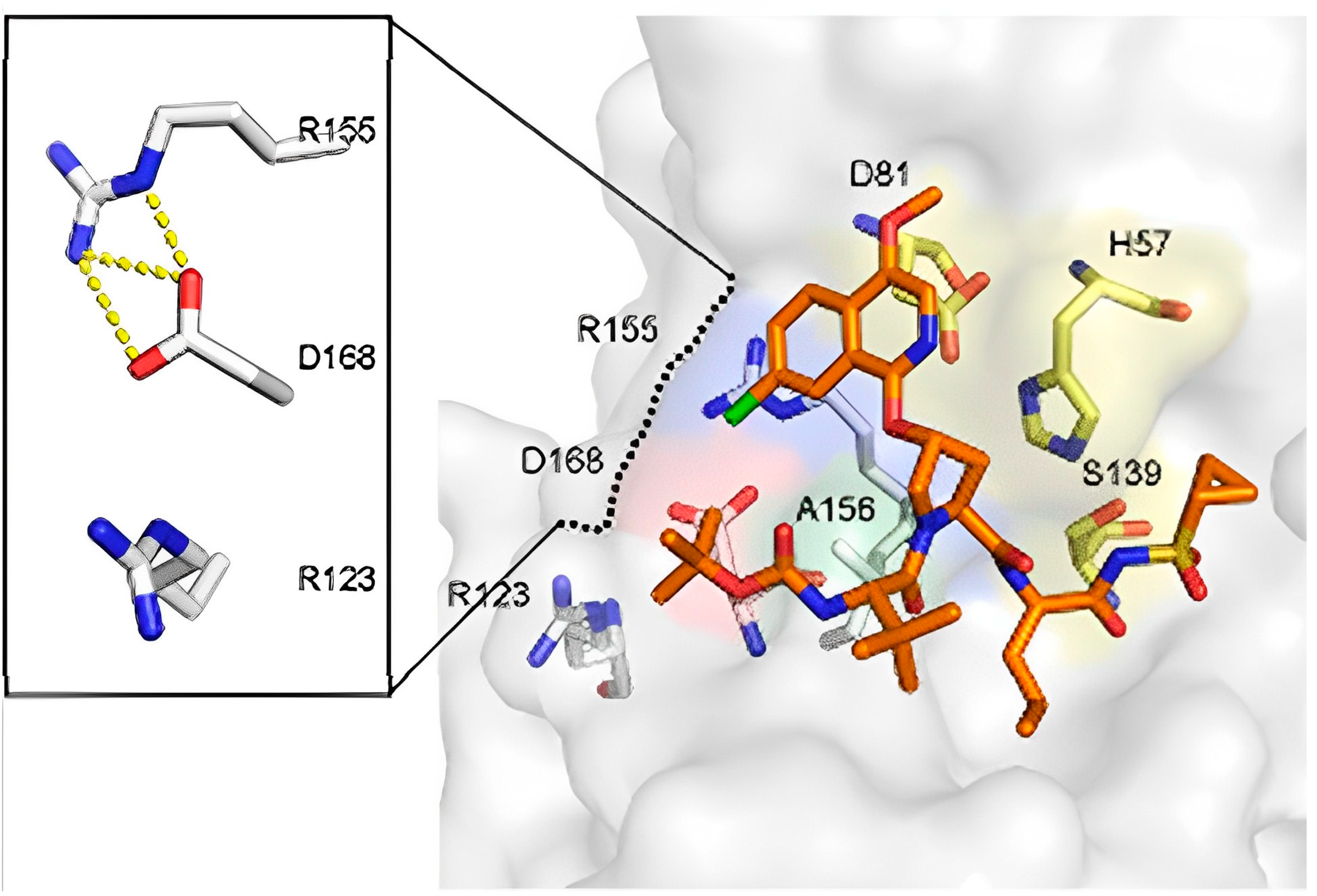
- Soumana, D.I.; Ali, A.; Schiffer, C.A. Structural Analysis of Asunaprevir Resistance in HCV NS3/4A Protease. ACS Chem. Biol. 2014, 9, 2485–2490. [Google Scholar] [CrossRef]
- Scola, P.M.; Sun, L.Q.; Wang, A.X.; Chen, J.; Sin, N.; Venables, B.L.; Sit, S.Y.; Chen, Y.; Cocuzza, A.; Bilder, D.M.; et al. The Discovery of Asunaprevir (BMS-650032), An Orally Efficacious NS3 Protease Inhibitor for the Treatment of Hepatitis C Virus Infection. J. Med. Chem. 2014, 57, 1730–1752. [Google Scholar] [CrossRef]
- Lok, A.S.; Gardiner, D.F.; Lawitz, E.; Martorell, C.; Everson, G.T.; Ghalib, R.; Reindollar, R.; Rustgi, V.; McPhee, F.; Wind-Rotolo, M. Preliminary Study of Two Antiviral Agents for Hepatitis C Genotype 1. N. Engl. J. Med. 2012, 366, 216–224. [Google Scholar] [CrossRef]
- Kumar, D.V.; Rai, R.; Brameld, K.A.; Riggs, J.; Somoza, J.R.; Rajagopalan, R.; Janc, J.W.; Xia, Y.M.; Ton, T.L.; Hu, H.; et al. 3-Heterocyclyl quinolone inhibitors of the HCV NS5B polymerase. Bioorg. Med. Chem. Lett. 2012, 22, 300–304. [Google Scholar] [CrossRef]
- McKercher, G.; Beaulieu, P.L.; Lamarre, D.; LaPlante, S.; Lefebvre, S.; Pellerin, C.; Thauvette, L.; Kukolj, G. Specific inhibitors of HCV polymerase identified using an NS5B with lower affinity for template/primer substrate. Nucleic Acids Res. 2004, 32, 422–431. [Google Scholar] [CrossRef]
- Kumar, D.V.; Rai, R.; Brameld, K.A.; Somoza, J.R.; Rajagopalan, R.; Janc, J.W.; Xia, Y.M.; Ton, T.L.; Shaghafi, M.B.; Hu, H. Quinolones as HCV NS5B polymerase inhibitors. Bioorg. Med. Chem. Lett. 2011, 21, 82–87. [Google Scholar] [CrossRef]
- Han, J.; Lee, M.K.; Jang, Y.; Cho, W.J.; Kim, M. Repurposing of cyclophilin A inhibitors as broad-spectrum antiviral agents. Drug Discov. Today. 2022, 27, 1895–1912. [Google Scholar] [CrossRef]
- El Kassas, M.; Elbaz, T.; Abd El Latif, Y.; Esmat, G. Elbasvir and grazoprevir for chronic hepatitis C genotypes 1 and 4. Expert Rev. Clin. Pharmacol. 2016, 9, 1413–1421. [Google Scholar] [CrossRef]
- Sofia, M.J.; Bao, D.; Chang, W.; Du, J.; Nagarathnam, D.; Rachakonda, S.; Reddy, P.G.; Ross, B.S.; Wang, P.; Zhang, H.R.; et al. Discovery of a β-D-20 -Deoxy-20 -r-fluoro-20 -β-C-methyluridine Nucleotide Prodrug (PSI-7977) for the Treatment of Hepatitis C Virus. J. Med. Chem. 2010, 53, 7202–7218. [Google Scholar] [CrossRef]
- Link, J.O.; Taylor, J.G.; Xu, L.; Mitchell, M.; Guo, H.; Liu, H.; Kato, D.; Kirschberg, T.; Sun, J.; Squires, N.; et al. Discovery of Ledipasvir (GS-5885): A Potent, Once-Daily Oral NS5A Inhibitor for the Treatment of Hepatitis C Virus Infection. J. Med. Chem. 2014, 57, 2033–2046. [Google Scholar] [CrossRef]
- Znabet, A.; Polak, M.M.; Janssen, E.; De Kanter, F.J.J.; Turner, N.J.; Orru, R.V.A.; Ruijter, E. A highly efficient synthesis of telaprevir by strategic use of biocatalysis and multicomponent reactions. Chem. Commun. 2010, 46, 7918–7920. [Google Scholar] [CrossRef]
- Xu, F.; Kim, J.; Waldman, J.; Wang, T.; Devine, P. Synthesis of Grazoprevir, a Potent NS3/4a Protease Inhibitor for the Treatment of Hepatitis C Virus. Org. Lett. 2018, 20, 7261–7265. [Google Scholar] [CrossRef]
- Rusere, L.N.; Matthew, A.N.; Lockbaum, G.J.; Jahangir, M.; Newton, A.; Petropoulos, C.J.; Huang, W.; Kurt Yilmaz, N.; Schiffer, C.A.; Ali, A. Quinoxaline-Based Linear HCV NS3/4A Protease Inhibitors Exhibit Potent Activity Against Drug Resistant Variants. ACS Med. Chem. Lett. 2018, 9, 691–696. [Google Scholar] [CrossRef] [PubMed]
- Matthew, A.N.; Zephyr, J.; Hill, C.J.; Jahangir, M.; Newton, A.; Petropoulos, C.J.; Huang, W.; Kurt-Yilmaz, N.; Schiffer, C.A.; Ali, A. Hepatitis C Virus NS3/4A Protease Inhibitors Incorporating Flexible P2 Quinoxalines Target Drug Resistant Viral Variants. J. Med. Chem. 2017, 60, 5699–5716. [Google Scholar] [CrossRef] [PubMed]
- Roth, D.; Nelson, D.R.; Bruchfeld, A.; Liapakis, A.; Silva, M.; Monsour, H.; Martin, P.; Pol, S.; Londoño, M.C.; Hassanein, T. Grazoprevir plus elbasvir in treatment-naive and treatment-experienced patients with hepatitis C virus genotype 1 infection and stage 4–5 chronic kidney disease (the C-SURFER study): A combination phase 3 study. Lancet 2015, 386, 1537–1545. [Google Scholar] [CrossRef] [PubMed]
- De Clercq, E. Current race in the development of DAAs (direct-acting antivirals) against HCV. Biochem. Pharmacol. 2014, 89, 441–452. [Google Scholar] [CrossRef]
- DeCarolis, D.D.; Chen, Y.C.; Westanmo, A.D.; Conley, C.; Gravely, A.A.; Khan, F.B. Decreased warfarin sensitivity among patients treated with elbasvir and grazoprevir for hepatitis C infection. Am. J. Health Pharm. 2019, 76, 1273–1280. [Google Scholar] [CrossRef]
- Keating, G.M.; Vaidya, A. Sofosbuvir: First Global Approval. Drugs 2014, 74, 273–282. [Google Scholar] [CrossRef]
- Lee, C. Daclatasvir: Potential role in hepatitis C. Drug Des. Dev. Ther. 2013, 7, 1223–1233. [Google Scholar] [CrossRef]
- Shannon, A.; Fattorini, V.; Sama, B.; Selisko, B.; Feracci, M.; Falcou, C.; Gauffre, P.; El Kazzi, P.; Delpal, A.; Decroly, E.; et al. A dual mechanism of action of AT-527 against SARS-CoV-2 polymerase. Nat. Commun. 2022, 13, 621–630. [Google Scholar] [CrossRef]
- Abdel-Magid, A.F. Fatty Acid Synthase Inhibitors as Possible Treatment for Cancer. ACS Med. Chem. Lett. 2012, 3, 612–613. [Google Scholar] [CrossRef] [PubMed]
- Oslob, J.D.; Johnson, R.J.; Cai, H.; Feng, S.Q.; Hu, L.; Kosaka, Y.; Lai, J.; Sivaraja, M.; Tep, S.; Yang, H.; et al. Imidazopyridine-Based Fatty Acid Synthase Inhibitors That Show Anti-HCV Activity and in Vivo Target Modulation. ACS Med. Chem. Lett. 2013, 4, 113–117. [Google Scholar] [CrossRef] [PubMed]
- Lohmann, V.; Korner, F.; Koch, J.; Herian, U.; Theilmann, L.; Bartenschlager, R. Replication of Subgenomic Hepatitis C Virus RNAs in a Hepatoma Cell Line. Science 1999, 285, 110–113. [Google Scholar] [CrossRef] [PubMed]
- Wang, N.Y.; Xu, Y.; Zuo, W.Q.; Xiao, K.J.; Liu, L.; Zeng, X.X.; You, X.Y.; Zhang, L.D.; Gao, C.; Liu, Z.H.; et al. Discovery of imidazo[2,1-b]thiazole HCV NS4B inhibitors exhibiting synergistic effect with other direct-acting antiviral agents. J. Med. Chem. 2015, 58, 2764–2778. [Google Scholar] [CrossRef] [PubMed]
- Matthews, G.V.; Seaberg, E.; Dore, G.J.; Bowden, S.; Lewin, S.R.; Sasadeusz, J.; Marks, P.; Goodman, Z.; Philp, F.H.; Tang, Y. Combination HBV therapy is linked to greater HBV DNA suppression in a cohort of lamivudineexperienced HIV/HBV coinfected individuals. AIDS 2009, 23, 1707–1715. [Google Scholar] [CrossRef] [PubMed]
- Li, J.; Lv, F. Process for Stereoselective Synthesis of Lamvudine. U.S. Patent No.: US 8,304,540 B2, 18 May 2007. [Google Scholar]
- Caso, M.F.; Dalonzo, D.; Derrico, S.; Palumbo, G.; Guaragna, A. Highly Stereoselective Synthesis of Lamivudine (3TC) and Emtricitabine (FTC) by a Novel N-Glycosidation Procedure. Org. Lett. 2015, 17, 2626–2629. [Google Scholar] [CrossRef]
- Lok, A.S.F.; McMahon, B.J. Chronic Hepatitis B: Update 2009. Hepatology 2009, 50, 661–662. [Google Scholar] [CrossRef]
- Yasutake, Y.; Hattori, S.; Hayashi, H.; Matsuda, K.; Tamura, N.; Kohgo, S.; Maeda, K.; Mitsuya, H. HIV-1 with HBV-associated Q151M substitution in RT becomes highly susceptible to entecavir: Structural insights into HBV-RT inhibition by entecavir. Sci. Rep. 2018, 8, 1624–1636. [Google Scholar] [CrossRef]
- Parvez, M.K.; Al-Dosari, M.S.; Abdelwahid, M.A.S.; Alqahtani, A.S.; Alanzi, A.R. Novel anti-hepatitis B virus-active catechin and epicatechin from Rhus tripartita. Exp. Ther. Med. 2022, 23, 398. [Google Scholar] [CrossRef]
- Velasco, J.; Ariza, X.; Badía, L.; Bartra, M.; Berenguer, R.; Farràs, J.; Gallardo, J.; Garcia, J.; Gasanz, Y.J. Total Synthesis of Entecavir. Org. Chem. 2013, 78, 5482–5491. [Google Scholar] [CrossRef]
- Li, J.J. Innovative Drug Synthesis; John Wiley & Sons, Inc.: Hoboken, NJ, USA, 2015; pp. 1–14. [Google Scholar] [CrossRef]
- Shaw, T.; Locarnini, S. Entecavir for the treatment of chronic hepatitis B. Expert Rev. Anti. Infect. Ther. 2004, 2, 853–871. [Google Scholar] [CrossRef] [PubMed]
- Matthews, S.J. Entecavir for the Treatment of Chronic Hepatitis B Virus Infection. Clin. Ther. 2006, 28, 184–203. [Google Scholar] [CrossRef] [PubMed]
- Dai, G.F.; Xu, H.W.; Wang, J.F.; Liu, F.W.; Liu, H.M. Studies on the novel a-glucosidase inhibitory activity and structure–activity relationships for andrographolide analogues. Bioorg. Med. Chem. Lett. 2006, 16, 2710–2713. [Google Scholar] [CrossRef] [PubMed]
- Wu, Y.H. Naturally derived anti-hepatitis B virus agents and their mechanism of action. World J. Gastroenterol. 2016, 22, 188–204. [Google Scholar] [CrossRef] [PubMed]
- Sadiea, R.Z.; Sultana, S.; Chaki, B.M.; Islam, T.; Dash, S.; Akter, S.; Islam, M.S.; Kazi, T.; Nagata, A.; Spagnuolo, R. Phytomedicines to Target Hepatitis B Virus DNA Replication: Current Limitations and Future Approaches. Int. J. Mol. Sci. 2022, 23, 1617. [Google Scholar] [CrossRef]
- Poordad, F.; Sievert, W.; Mollison, L.; Bennett, M.; Tse, E.; Bräu, N.; Levin, J.; Sepe, T.; Lee, S.S.; Angus, P.; et al. Fixed-Dose Combination Therapy With Daclatasvir, Asunaprevir, and Beclabuvir for Noncirrhotic Patients With HCV Genotype 1 Infection. JAMA 2015, 313, 1728–1735. [Google Scholar] [CrossRef]
- Lai, C.L.; Gane, E.; Liaw, Y.F.; Hsu, C.W.; Thongsawat, S.; Wang, Y.; Chen, Y.; Heathcote, E.J.; Rasenack, J.; Bzowej, N.; et al. Telbivudine versus Lamivudine in Patients with Chronic Hepatitis B. N. Engl. J. Med. 2007, 357, 2576–2588. [Google Scholar] [CrossRef]
- Lai, C.L.; Shouval, D.; Lok, A.S.; Chang, T.T.; Cheinquer, H.; Goodman, Z.; DeHertogh, D.; Wilber, R.; Zink, R.C.; Cross, A.; et al. Entecavir versus Lamivudine for Patients with HBeAg-Negative Chronic Hepatitis B. N. Engl. J. Med. 2006, 354, 1011–1020. [Google Scholar] [CrossRef]
- Testoni, B.; Durantel, D.; Zoulim, F. Novel targets for hepatitis B virus therapy. Liver Int. 2017, 37, 33–39. [Google Scholar] [CrossRef]
- Li, X.; Zhang, Z.; Chen, Y.; Wang, B.; Yang, G.; Xu, X.; Yechao, B.; Bai, D.; Feng, B.; Mao, Y.; et al. Discovery of SHR5133, a Highly Potent and Novel HBV Capsid Assembly Modulator. ACS Med. Chem. Lett. 2022, 13, 507–512. [Google Scholar] [CrossRef]
- Yeo, H.; Li, Y.; Fu, L.; Zhu, J.L.; Gullen, E.A.; Dutschman, G.E.; Lee, Y.; Chung, R.; Huang, E.-S.; Austin, D.J.; et al. Synthesis and Antiviral Activity of Helioxanthin Analogues. J. Med. Chem. 2005, 48, 534–546. [Google Scholar] [CrossRef] [PubMed]
- Crotty, S.; Maag, D.; Arnold, J.J.; Zhong, W.; Lau, J.Y.N.; Hong, Z.; Andino, R.; Cameron, C.E. The broad-spectrum antiviral ribonucleoside ribavirin is an RNA virus mutagen. Nat. Med. 2000, 6, 1375–1379. [Google Scholar] [CrossRef] [PubMed]
- Kim, Y.-I.; Pareek, R.; Murphy, R.; Harrison, L.; Farrell, E.; Cook, R.; DeVincenzo, J. The antiviral effects of RSV fusion inhibitor, MDT-637, on clinical isolates, versus its achievable concentrations in the human respiratory tract and comparison to ribavirin. Influenza Other Respi. Viruses 2017, 11, 525–530. [Google Scholar] [CrossRef]
- Sakharov, V.; Baykov, S.; Konstantinova, I.; Esipov, R.; Dorogov, M. An Efficient Chemoenzymatic Process for Preparation of Ribavirin. Int. J. Chem. Eng. 2015, 2015, 734851. [Google Scholar] [CrossRef]
- Feld, J.J.; Hoofnagle, J.H. Mechanism of action of interferon and ribavirin in treatment of hepatitis C. Nature 2005, 436, 967–972. [Google Scholar] [CrossRef]
- Yang, Y.; Rijnbrand, R.; McKnight, K.L.; Wimmer, E.; Paul, A.; Martin, A.; Lemon, S.M. Sequence Requirements for Viral RNA Replication and VPg Uridylylation Directed by the Internal cis-Acting Replication Element (cre) of Human Rhinovirus Type 14. J. Virol. 2002, 76, 7485–7494. [Google Scholar] [CrossRef] [PubMed]
- Nyström, K.; Waldenström, J.; Tang, K.W.; Lagging, M. Ribavirin: Pharmacology, Multiple Modes of Action and Possible Future Perspectives. Sequence Requirements for Viral RNA Replication and VPg Uridylylation Directed by the Internal cis-Acting Replication Element (cre) of Human Rhinovirus Type 14. Future Virol. 2019, 14, 153–160. [Google Scholar] [CrossRef]
- Cameron, C.E.; Castro, C. The mechanism of action of ribavirin: Lethal mutagenesis of RNA virus genomes mediated by the viral RNA-dependent RNA polymerase. Curr. Opin. Infect. Dis. 2001, 14, 757–764. [Google Scholar] [CrossRef]
- Thomas, E.; Ghany, M.G.; Liang, T.J. The application and mechanism of action of ribavirin in therapy of hepatitis C. Antivir. Chem. Chemother. 2012, 23, 1–12. [Google Scholar] [CrossRef]
- Robinson, R.F.; Nahata, M.C. Respiratory syncytial virus (RSV) immune globulin and palivizumab for prevention of RSV infection. Am. J. Health Pharm. 2000, 57, 259–264. [Google Scholar] [CrossRef]
- Wright, M.; Piedimonte, G. Respiratory Syncytial Virus Prevention and Therapy: Past, Present, and Future. Pediatr. Pulmonol. 2011, 46, 324–347. [Google Scholar] [CrossRef] [PubMed]
- Cockerill, G.S.; Angell, R.M.; Bedernjak, A.; Chuckowree, I.; Fraser, I.; Gascon-Simorte, J.; Gilman, M.S.A.; Good, J.A.D.; Harland, R.; Johnson, S.M.; et al. Discovery of Sisunatovir (RV521), an Inhibitor of Respiratory Syncytial Virus Fusion. J. Med. Chem. 2021, 64, 3658–3676. [Google Scholar] [CrossRef] [PubMed]
- Yoon, J.J.; Chawla, D.; Paal, T.; Ndungu, M.; Du, Y.H.; Kurtkaya, S.; Sun, A.M.; Snyder, J.P.; Plemper, R.K.J. High-Throughput Screening–Based Identification of Paramyxovirus Inhibitors. Biomol. Screen. 2008, 13, 591–608. [Google Scholar] [CrossRef]
- Yust, I.; Fox, Z.; Burke, M.; Johnson, A.; Turner, D.; Mocroft, A.; Katlama, C.; Ledergerber, B.; Reiss, P.; Kirk, O. Retinal and extraocular cytomegalovirus end-organ disease in HIV-infected patients in Europe: A EuroSIDA study, 1994–2001. Eur. J. Clin. Microbiol. Infect. Dis. 2004, 23, 550–559. [Google Scholar] [CrossRef]
- Babu, K.S.; Rao, M.R.; Goverdhan, G.; Srinivas, P.; Reddy, P.P.; Venkateswarlu, G.; Anand, R.V. Synthesis of Valganciclovir Hydrochloride Congeners. Synth. Commun. 2013, 43, 1751–1758. [Google Scholar] [CrossRef]
- Wiltshire, H.; Paya, C.V.; Pescovitz, M.D.; Humar, A.; Dominguez, E.; Washburn, K.; Blumberg, E.; Alexander, B.; Freeman, R.; Heaton, N. Pharmacodynamics of Oral Ganciclovir and Valganciclovir in Solid Organ Transplant Recipients. Transplantation 2005, 79, 1477–1483. [Google Scholar] [CrossRef] [PubMed]
- Matthews, T.; Boehme, R. Antiviral Activity and Mechanism of Action of Ganciclovir. Rev. Infect. Dis. 1988, 10, 490–494. [Google Scholar] [CrossRef]
- Gao, H.; Mitra, A.K. Synthesis of Acyclovir, Ganciclovir and Their Prodrugs: A Review. Synthesis 2000, 2000, 329–351. [Google Scholar] [CrossRef]
- Montana, M.; Montero, V.; Khoumeri, O.; Vanelle, P. Quinoxaline Derivatives as Antiviral Agents: A Systematic Review. Molecules 2020, 25, 2784. [Google Scholar] [CrossRef]
- Wang, Y.; Mukhopadhyay, R.; Roy, S.; Kapoor, A.; Su, Y.-P.; Charman, S.A.; Chen, G.; Wu, J.; Wang, X.; Vennerstrom, J.L. Inhibition of Cytomegalovirus Replication with Extended-HalfLife Synthetic Ozonides. Antimicrob. Agents Chemother. 2019, 63, e01735-18. [Google Scholar] [CrossRef]
- El-Zahab, H.S.A. Synthesis, Characterization, and Biological Evaluation of Some Novel Quinoxaline Derivatives as Antiviral Agents. Arch. Pharm. Chem. Life Sci. 2017, 350, 1700028. [Google Scholar] [CrossRef] [PubMed]
- El-Sebaey, S.A. Recent Advances in 1,2,4-Triazole Scaffolds as Antiviral Agents. ChemistrySelect 2020, 5, 11654–11680. [Google Scholar] [CrossRef]
- Kimberlin, D.W.; Jester, P.M.; Sánchez, P.J.; Ahmed, A.; Arav-Boger, R.; Michaels, M.G.; Ashouri, N.; Englund, J.A.; Estrada, B.; Jacobs, R.F.; et al. Valganciclovir for Symptomatic Congenital Cytomegalovirus Disease. N. Engl. J. Med. 2015, 372, 933–943. [Google Scholar] [CrossRef] [PubMed]
- Martin, D.F.; Kuppermann, B.D.; Wolitz, R.A.; Palestine, A.G.; Li, H.; Robinson, C.A. Oral ganciclovir for patients with cytomegalovirus retinitis treated with a ganciclovir implant. N. Engl. J. Med. 1999, 340, 1063–1070. [Google Scholar] [CrossRef] [PubMed]
- Williams, J.D.; Chen, J.J.; Drach, J.C.; Townsend, L.B. Design, Synthesis, and Antiviral Activity of Certain 3-Substituted 2,5,6-Trichloroindole Nucleosides. J. Med. Chem. 2004, 47, 5753–5765. [Google Scholar] [CrossRef] [PubMed]
- Turk, S.R.; Shipman, C., Jr.; Nassiri, M.R.; Genzlinger, G.; Krawczyk, S.H.; Townsend, L.B.; Drach, J.C. Pyrrolo[2,3-d]Pyrimidine Nucleosides as Inhibitors of Human Cytomegalovirus. Antimicrob. Agents Chemother. 1987, 31, 544–550. [Google Scholar] [CrossRef]
- Prichard, M.N.; Prichard, L.E.; Baguley, W.A.; Nassiri, M.R.; Shipman, C., Jr. Three-Dimensional Analysis of the Synergistic Cytotoxicity of Ganciclovir and Zidovudine. Antivir. Res. 1991, 35, 1060–1065. [Google Scholar] [CrossRef]
- Sahu, P.K.; Umme, T.; Yu, J.; Nayak, A.; Kim, G.; Noh, M.; Lee, J.Y.; Kim, D.-D.; Jeong, L.S. Seleno-acyclovir and –ganciclovir: A Discovery of a New Template for Antiviral Agents. J. Med. Chem. 2015, 58, 8734–8738. [Google Scholar] [CrossRef]
- Zhou, N.; Xie, T.; Liu, L.; Xie, Z.J. Cu/Mn Co-oxidized Cyclization for the Synthesis of Highly Substituted Pyrrole Derivatives from Amino Acid Esters: A Strategy for the Biomimetic Syntheses of Lycogarubin C and Chromopyrrolic Acid. Org. Chem. 2014, 79, 6061–6068. [Google Scholar] [CrossRef]
- Lin, Z.Q.; Li, C.D.; Zhou, Z.C.; Xue, S.; Gao, J.R.; Ye, Q.; Li, Y.J. Copper(II)-Promoted Oxidation/[3+2]Cycloaddition/Aromatization Cascade: Efficient Synthesis of Tetrasubstituted NH-Pyrrole from Chalcones and Iminodiacetates. Synlett 2019, 30, 1442–1446. [Google Scholar] [CrossRef]
- Kaufman, H.E.; Heidelberger, C. Therapeutic Antiviral Action of 5-Trifluoromethyl-2’-deoxyuridine in Herpes Simplex Keratitis. Science 1964, 145, 585–586. [Google Scholar] [CrossRef]
- De Clercq, E. Discovery and development of BVDU (brivudin) as a therapeutic for the treatment of herpes zoster. Biochem. Pharmacol. 2004, 68, 2301–2315. [Google Scholar] [CrossRef]
- McClain, L.; Zhi, Y.; Cheng, H.; Ghosh, A.; Piazza, P.; Yee, M.B.; Kumar, S.; Milosevic, J.; Bloom, D.C.; Arav-Boger, R. Broad-spectrum non-nucleoside inhibitors of human herpesviruses. Antivir. Res. 2015, 121, 16–23. [Google Scholar] [CrossRef]
- Taylor, J.L.; Punda-Polic, V.; O’brien, W.J. Combined anti-herpes virus activity of nucleoside analogs and interferon. Curr. Eye Res. 1991, 10, 205–211. [Google Scholar] [CrossRef]
- Warren, T.K.; Jordan, R.; Lo, M.K.; Ray, A.S.; Mackman, R.L.; Soloveva, V.; Siegel, D.; Perron, M.; Bannister, R.; Hui, H.C.; et al. Therapeutic efficacy of the small molecule GS-5734 against Ebola virus in rhesus monkeys. Nature 2016, 531, 381–385. [Google Scholar] [CrossRef] [PubMed]
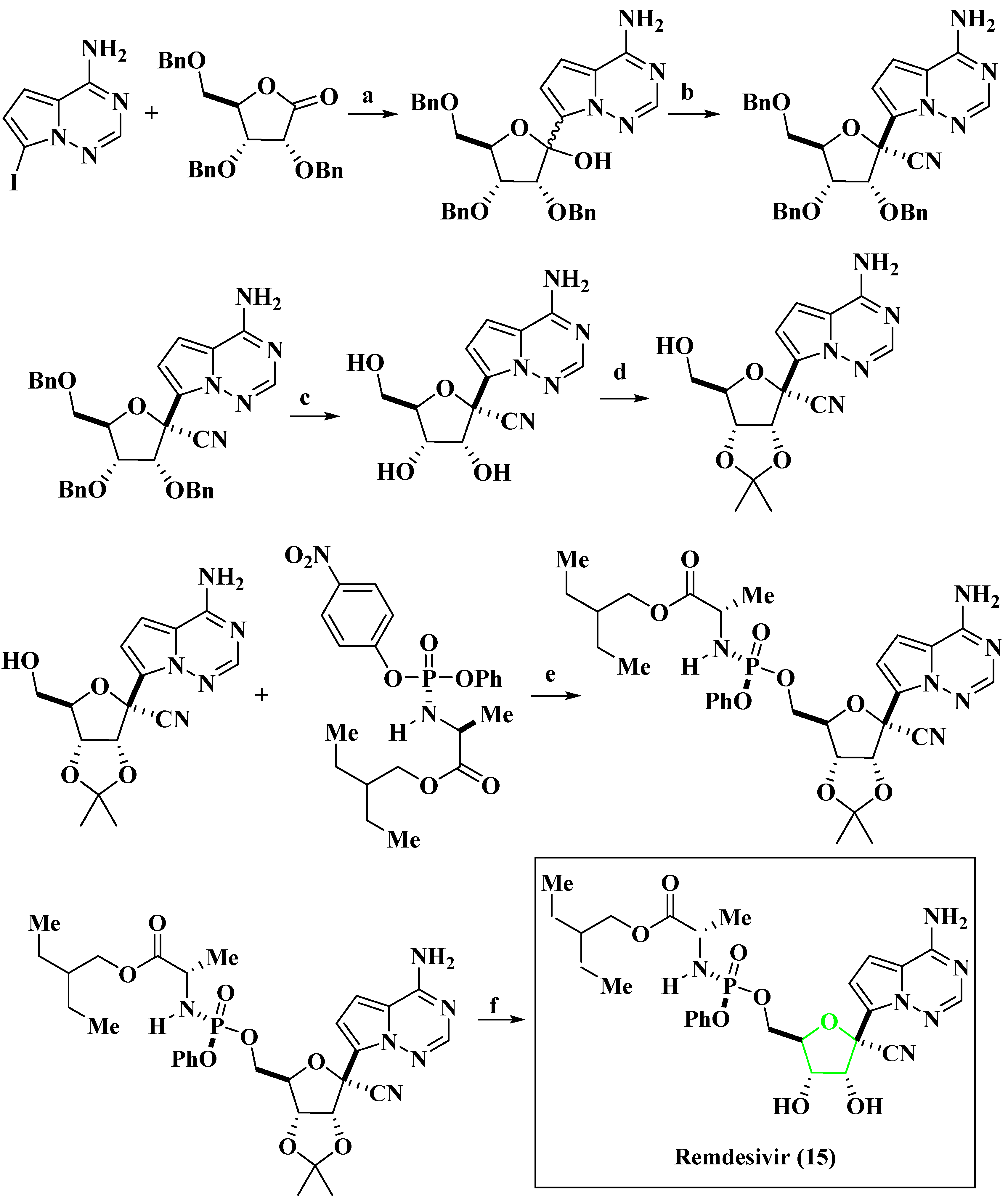
- Pardo, J.; Shukla, A.M.; Chamarthi, G.; Gupte, A. The journey of remdesivir: From Ebola to COVID-19. Drugs Context 2020, 9, 1–9. [Google Scholar] [CrossRef] [PubMed]
- Chun, B.K.; Clarke, M.O.H.; Doerffler, E.; Hui, H.C.; Jordan, R.; Mackman, R.L.; Parrish, J.P.; Ray, A.S.; Siegel, D.; Gilead Sciences, Inc. Methods for Treating Filoviridae Virus Infections. Patent WO 2016069826 A1, 5 July 2016. [Google Scholar]
- Taylor, R.; Kotian, P.; Warren, T.; Panchal, R.; Bavari, S.; Julander, J.; Dobo, S.; Rose, A.; El-Kattan, Y.; Taubenheim, B.; et al. BCX4430 – A broad-spectrum antiviral adenosine nucleoside analog under development for the treatment of Ebola virus disease. Infect. Public Health 2016, 9, 220–226. [Google Scholar] [CrossRef]
- De Clercq, E. Ebola virus (EBOV) infection: Therapeutic strategies. Biochem. Pharmacol. 2015, 93, 1–10. [Google Scholar] [CrossRef]
- Picazo, E.; Giordanetto, F. Small molecule inhibitors of ebola virus infection. Drug Discov. Today 2015, 20, 277–286. [Google Scholar] [CrossRef] [PubMed]
- Shannon, A.; Canard, B. Development of a robust and convenient dual-reporter high-throughput screening assay for SARS-CoV-2 antiviral drug discovery. Antivir. Res. 2023, 210, 105501–105516. [Google Scholar] [CrossRef]
- Scott, J.T.; Sharma, R.; Meredith, L.W.; Dunning, J.; Moore, C.E.; Sahr, F.; Ward, S.; Goodfellow, I.; Horby, P. Pharmacokinetics of TKM-130803 in Sierra Leonean patients with Ebola virus disease: Plasma concentrations exceed target levels, with drug accumulation in the most severe patients. EBioMedicine 2020, 52, 102601–102611. [Google Scholar] [CrossRef] [PubMed]
- Verdonck, S.; Pu, S.Y.; Sorrell, F.J.; Elkins, J.M.; Froeyen, M.; Gao, L.J.; Prugar, L.I.; Dorosky, D.E.; Brannan, J.M.; Barouch-Bentov, R.; et al. Synthesis and Structure−Activity Relationships of 3,5-Disubstitutedpyrrolo[2,3-b]pyridines as Inhibitors of Adaptor-Associated Kinase 1 with Antiviral Activity. J. Med. Chem. 2019, 62, 5810–5831. [Google Scholar] [CrossRef] [PubMed]
- Janeba, Z. Development of Small-Molecule Antivirals for Ebola. Med. Res. Rev. 2015, 35, 1175–1194. [Google Scholar] [CrossRef] [PubMed]
- Zhou, P.; Yang, X.L.; Wang, X.G.; Hu, B.; Zhang, L.; Zhang, W.; Si, H.R.; Zhu, Y.; Li, B.; Huang, C.L.; et al. A pneumonia outbreak associated with a new coronavirus of probable bat origin. Nature 2020, 579, 270–273. [Google Scholar] [CrossRef]
- Mascellino, M.T.; Di Timoteo, F.; De Angelis, M.; Oliva, A. Overview of the Main Anti-SARS-CoV-2 Vaccines: Mechanism of Action, Efficacy and Safety. Infect. Drug Resist. 2021, 14, 3459–3476. [Google Scholar] [CrossRef]
- Quiros-Roldan, E.; Amadasi, S.; Zanella, I.; Degli Antoni, M.; Storti, S.; Tiecco, G.; Castelli, F. Monoclonal Antibodies against SARS-CoV-2: Current Scenario and Future Perspectives. Pharmaceuticals 2021, 14, 1272. [Google Scholar] [CrossRef]
- Wang, X.; Sacramento, C.Q.; Jockusch, S.; Chaves, O.A.; Tao, C.; Fintelman-Rodrigues, N.; Chien, M.; Temerozo, J.R.; Li, X.; Kumar, S.; et al. Combination of antiviral drugs inhibits SARS-CoV-2 polymerase and exonuclease and demonstrates COVID-19 therapeutic potential in viral cell culture. Commun. Biol. 2022, 5, 154–168. [Google Scholar] [CrossRef]
- Dube, T.; Ghosh, A.; Mishra, J.; Kompella, U.B.; Panda, J.J. Repurposed Drugs, Molecular Vaccines, Immune-Modulators, and Nanotherapeutics to Treat and Prevent COVID-19 Associated with SARS-CoV-2, a Deadly Nanovector. Adv. Ther. 2021, 4, 2000172–2000202. [Google Scholar] [CrossRef]
- Marzolini, C.; Kuritzkes, D.R.; Marra, F.; Boyle, A.; Gibbons, S.; Flexner, C.; Pozniak, A.; Boffito, M.; Waters, L.; Burger, D. Recommendations for the Management of Drug–Drug Interactions Between the COVID-19 Antiviral Nirmatrelvir/Ritonavir (Paxlovid) and Comedications. Clin. Pharmacol. Ther. 2022, 112, 1191–1200. [Google Scholar] [CrossRef]
- Rubin, R. Baricitinib Is First Approved COVID-19 Immunomodulatory Treatment. JAMA 2022, 327, 2281. [Google Scholar] [CrossRef]
- Amici, C.; Di Caro, A.; Ciucci, A.; Chiappa, L.; Castilletti, C.; Martella, V.; Decaro, N.; Buonavoglia, C.; Capobianchi, M.R.; Santoro, M.G. Indomethacin has a potent antiviral activity against SARS coronavirus. Antivir. Ther. 2006, 11, 1021–1030. [Google Scholar] [CrossRef] [PubMed]
- Tegeder, I.; Pfeilschifter, J.; Geisslinger, G. Cyclooxygenase-independent actions of cyclooxygenase inhibitors. FASEB J. 2001, 15, 2057–2072. [Google Scholar] [CrossRef] [PubMed]
- Wood, J.M.; Evans, G.B.; Grove, T.L.; Almo, S.C.; Cameron, S.A.; Furneaux, R.H.; Harris, L.D.J. Chemical Synthesis of the Antiviral Nucleotide Analogue ddhCTP. Org. Chem. 2021, 86, 8843–8850. [Google Scholar] [CrossRef] [PubMed]
- Stachulski, A.V.; Taujanskas, J.; Pate, S.L.; Rajoli, R.K.R.; Aljayyoussi, G.; Pennington, S.H.; Ward, S.A.; Hong, W.D.; Biagini, G.A.; Owen, A.; et al. Therapeutic Potential of Nitazoxanide: An Appropriate Choice for Repurposing versus SARS-CoV-2? ACS Infect. Dis. 2021, 7, 1317–1331. [Google Scholar] [CrossRef]
- Gizzi, A.S.; Grove, T.L.; Arnold, J.J.; Jose, J.; Jangra, R.K.; Garforth, S.J.; Du, Q.; Cahill, S.M.; Dulyaninova, N.G.; Love, J.D.; et al. A naturally occurring antiviral ribonucleotide encoded by the human genome. Nature 2018, 558, 610–614. [Google Scholar] [CrossRef]
- Jung, E.; Soto-Acosta, R.; Xie, J.; Wilson, D.J.; Dreis, C.D.; Majima, R.; Edwards, T.C.; Geraghty, R.J.; Chen, L. Bisubstrate Inhibitors of Severe Acute Respiratory Syndrome Coronavirus-2 Nsp14 Methyltransferase. ACS Med. Chem. Lett. 2022, 13, 1477–1484. [Google Scholar] [CrossRef]
- Braaten, K.P.; Laufer, M.R. Human Papillomavirus (HPV), HPV-Related Disease, and the HPV Vaccine. Rev. Obstet. Gynecol. 2008, 1, 2–10. [Google Scholar] [PubMed]
- Rivera, A.; Tyring, S.K. Therapy of cutaneous human Papillomavirus infections. Dermatol. Ther. 2004, 17, 441–448. [Google Scholar] [CrossRef]
- Beadle, J.R.; Valiaeva, N.; Yang, G.; Yu, J.H.; Broker, T.R.; Aldern, K.A.; Harden, E.A.; Keith, K.A.; Prichard, M.N.; Hartman, T.; et al. Synthesis and Antiviral Evaluation of Octadecyloxyethyl Benzyl 9-[(2- Phosphonomethoxy)ethyl]guanine (ODE-Bn-PMEG), a Potent Inhibitor of Transient HPV DNA Amplification. J. Med. Chem. 2016, 59, 10470–10478. [Google Scholar] [CrossRef]
- Briggs, D.J. The role of vaccination in rabies prevention. Curr. Opin. Virol. 2012, 2, 309–314. [Google Scholar] [CrossRef]
- Da Silva, S.; Oliveira Silva Martins, D.; Jardim, A.C. A Review of the Ongoing Research on Zika Virus Treatment. Viruses 2018, 10, 255. [Google Scholar] [CrossRef] [PubMed]
- Barrows, N.J.; Campos, R.K.; Powell, S.T.; Prasanth, K.R.; Schott-Lerner, G.; Soto-Acosta, R.; Galarza-Muñoz, G.; McGrath, E.L.; Urrabaz-Garza, R.; Gao, J. A Screen of FDA-Approved Drugs for Inhibitors of Zika Virus Infection. Cell Host Microbe 2016, 20, 259–270. [Google Scholar] [CrossRef]
- Zhou, K.; Li, C.; Shi, W.; Hu, X.; Nandakumar, K.S.; Jiang, S.; Zhang, N. Current Progress in the Development of Zika Virus Vaccines. Vaccines 2021, 9, 1004. [Google Scholar] [CrossRef] [PubMed]
- Yao, Y.; Huo, T.; Lin, Y.L.; Nie, S.; Wu, F.; Hua, Y.; Wu, J.; Kneubehl, A.R.; Vogt, M.B.; Rico-Hesse, R.; et al. Discovery, X-ray Crystallography and Antiviral Activity of Allosteric Inhibitors of Flavivirus NS2B-NS3 Protease. Am. Chem. Soc. 2019, 141, 6832–6836. [Google Scholar] [CrossRef] [PubMed]
- Tricarico, P.M.; Caracciolo, I.; Crovella, S.; D’Agaro, P. Zika virus induces inflammasome activation in the glial cell line U87-MG. Biochem. Biophys. Res. Commun. 2017, 492, 597–602. [Google Scholar] [CrossRef] [PubMed]
- Lahon, A.; Arya, R.P.; Kneubehl, A.R.; Vogt, M.B.; Dailey Garnes, N.J.M.; Rico-Hesse, R. Characterization of a Zika Virus Isolate from Colombia. PLoS Negl. Trop. Dis. 2016, 10, e0005019. [Google Scholar] [CrossRef]
- Zhu, Y.; Liang, M.; Yu, J.; Zhang, B.; Zhu, G.; Huang, Y.; He, Z.; Yuan, J. Repurposing of Doramectin as a New Anti-Zika Virus Agent. Viruses 2023, 15, 1068. [Google Scholar] [CrossRef]
- Martinez-Lopez, A.; Persaud, M.; Chavez, M.P.; Zhang, H.; Rong, L.; Liu, S.; Wang, T.T.; Sarafianos, S.G.; Diaz-Griffero, F. Glycosylated diphyllin as a broad-spectrum antiviral agent against Zika virus. EBioMedicine 2019, 47, 269–283. [Google Scholar] [CrossRef]
- Li, P.C.; Jang, J.; Hsia, C.Y.; Groomes, P.V.; Lian, W.; de Wispelaere, M.; Pitts, J.D.; Wang, J.; Kwiatkowski, N.; Gray, N.S.; et al. Small Molecules Targeting the Flavivirus E Protein with Broad-Spectrum Activity and Antiviral Efficacy in Vivo. ACS Infect. Dis. 2019, 5, 460–472. [Google Scholar] [CrossRef]
- Lian, W.; Jang, J.; Potisopon, S.; Li, P.C.; Rahmeh, A.; Wang, J.; Kwiatkowski, N.P.; Gray, N.S.; Yang, P.L. Discovery of Immunologically Inspired Small Molecules That Target the Viral Envelope Protein. ACS Infect. Dis. 2018, 4, 1395–1406. [Google Scholar] [CrossRef]
- Murdin, A.D.; Barreto, L.; Plotkin, S. Inactivated poliovirus vaccine: Past and present experience. Vaccine 1996, 14, 735–746. [Google Scholar] [CrossRef]
- Amanna, I.J.; Slifka, M.K. Current trends in West Nile virus vaccine development. Expert Rev. Vaccines 2014, 13, 589–608. [Google Scholar] [CrossRef]
- Harder, T.; Siedler, A. Systematic Review and Meta-analysis of Chickenpox Vaccination and Risk of Herpes Zoster: A Quantitative View on the “Exogenous Boosting Hypothesis”. Clin. Infect. Dis. 2019, 69, 1329–1338. [Google Scholar] [CrossRef] [PubMed]
- Davidson, R.N.; Lynn, W.; Savage, P.; Wansbrough-Jones, M.H. Chickenpox pneumonia: Experience with antiviral treatment. Thorax 1988, 43, 627–630. [Google Scholar] [CrossRef] [PubMed]
- Ward, P.; Small, I.; Smith, J.; Suter, P.; Dutkowski, R.J. Oseltamivir (Tamiflu®) and its potential for use in the event of an influenza pandemic. Antimicrob. Chemother. 2005, 55, 5–21. [Google Scholar] [CrossRef] [PubMed]
- Lin, L.Z.; Fang, J.M. Total Synthesis of Anti-Influenza Agents Zanamivir and Zanaphosphor via Asymmetric Aza-Henry Reaction. Org. Lett. 2016, 18, 4400–4403. [Google Scholar] [CrossRef] [PubMed]
- Shie, J.J.; Fang, J.M.J. Development of effective anti-influenza drugs: Congeners and conjugates – a review. Biomed. Sci. 2019, 26, 84–104. [Google Scholar] [CrossRef]
- Rowse, M.; Qiu, S.; Tsao, J.; Yamauchi, Y.; Wang, G.; Luo, M. Reduction of Influenza Virus Envelope’s Fusogenicity by Viral Fusion Inhibitors. ACS Infect. Dis. 2016, 2, 47–53. [Google Scholar] [CrossRef]
- DeFilippis, V.; Früh, K. Host cell targets for antiviral therapy: An update. Future Virol. 2006, 1, 509–518. [Google Scholar] [CrossRef]
- Cha, H.M.; Kim, U.I.; Ahn, S.B.; Lee, M.K.; Lee, H.; Bang, H.; Jang, Y.; Kim, S.S.; Bae, M.A.; Kim, K.; et al. Evaluation of Antiviral Activity of Gemcitabine Derivatives against Influenza Virus and Severe Acute Respiratory Syndrome Coronavirus. ACS Infect. Dis. 2023, 9, 1033–1045. [Google Scholar] [CrossRef]
- Das, P.; Deng, X.; Zhang, L.; Roth, M.G.; Fontoura, B.M.A.; Phillips, M.A.; De Brabander, J.K. SAR-Based Optimization of a 4-Quinoline Carboxylic Acid Analogue with Potent Antiviral Activity. ACS Med. Chem. Lett. 2013, 4, 517–521. [Google Scholar] [CrossRef] [PubMed]
- Monath, T.P. SAR-Based Optimization of a 4-Quinoline Carboxylic Acid Analogue with Potent Antiviral Activity. Lancet Infect. Dis. 2001, 1, 11–20. [Google Scholar] [CrossRef] [PubMed]


| Compound | Substitution | Activity | ||
|---|---|---|---|---|
| −R1 | −R2 | Inhibition Concentration (nM) | Inhibitor Constant (Ki) | |
 |  |  | IC90 = 4.1 | 16 pM |
 |  | IC90 = 1.4 | 14 pM | |
 |  | IC50 = 0.22 | 14 pM | |
 |  | IC50 = 0.7 | 15 fM | |
 |  | IC50 = 1 | 8 pM | |
| Compound | Substitution | Activity | ||
|---|---|---|---|---|
| −R1 | −R2 | EC50 (nM) | Inhibitor Constant (Ki) (nM) | |
 |  |  | 2.93 | 0.84 |
 |  | 4.29 | 5.8 | |
 |  | 2.32 | 6.4 | |
 |  | 6.89 | 5.8 | |
 |  | 6.04 | 7.4 | |
 |  | 3.63 | 5.2 | |
 |  | 5.53 | 1.8 | |
 |  | 4.30 | 1.9 | |
 |  | 3.46 | 4.3 | |
 |  | 9.2 | 1.6 | |
 |  | 4.48 | 8.1 | |
 |  | 3.08 | 3.5 | |
 |  | 2.68 | 1.6 | |
 |  | 2.25 | 3.8 | |
 |  | 0.10 | 1.95 | |
 |  | 0.10 | 0.33 | |
 |  | 2.93 | 0.086 | |
| Sl. No. | Drug Name | Drug Target | Mechanism of Action | Ways of Use | Side Effect | Brand Name |
|---|---|---|---|---|---|---|
| 1. | Zalcitabine [135] | Reverse transcriptase/RNase H (Human immunodeficiency virus 1) | Inhibiting reverse transcriptase and terminating the viral DNA chain | Oral | Liver failure, inflammation of the pancreas | Hivid |
| 2. | Amprenavir [136] | Human immunodeficiency virus type 1 protease | Blocks the active site of HIV protease to prevent cleavage of the viral precursor proteins | Oral | Diarrhea, kidney failure, weakness | Agenerase |
| 3. | Maraviroc [137] | C-C chemokine receptor type 5 | Blocks GP120-CCR5 interaction to inhibit HIV entry | Oral | Liver problems, nausea, allergic reactions | Selzentry |
| 4. | Tenofovir disoproxil Fumarate [138] | Reverse transcriptase/RNaseH (Human immunodeficiency virus 1) | Competes with dATP and inhibits the activity of HIV RT | Oral | Trouble sleeping, dizziness, diarrhea | Viread |
| 5. | Emtricitabine [139] | Reverse transcriptase/RNase H (Human immunodeficiency virus 1) | Through nucleoside reverse transcriptase inhibition | Oral | Body aches, ough, diarrhea, fever | Truvada |
| 6. | Cabotegravir [140] | Integrase (Human immunodeficiency virus 1) | By HIV-1 integrase inhibition | Oral | Abnormal dreams, dark urine, difficulty in breathing | Vocabria |
| 7. | Lenacapavir [141] | Gag-Pol polyprotein | Lenacapavir works by blocking the HIV-1 virus’ protein shell (the capsid), thereby interfering with multiple essential steps of the viral lifecycle | Oral/subcutaneous injection | Nausea | Sunlenca |
| Sl. No. | Antiviral Agent | Drug Target | Activity |
|---|---|---|---|
| 1. |  | HIV [142] | Methylthiazole derivatives effectively inhibit HIV-1 entry into cells, demonstrating low micromolar potency (IC50 = 1.6 µM) and a favorable selectivity index (SI) in both single-cycle TZM-bl cell assays and multicycle MT-2 cell assays. |
| 2. |  | HIV-1 and HIV-2 [143] | 3″-alkenyl-substituted TSAO derivatives exhibit anti-HIV-1 and anti-HIV-2 activity at subtoxic concentrations against HIV-2 RT involved the RNA-dependent DNA polymerase assay with EC50(HIV-1MT-4) = 0.06 ± 0.03 µM and EC50(HIV-2MT-4) >10 µM. |
| 3. |  | HIV [144] | 4′-ethynyl-2-fluoro-2′-deoxyadenosine demonstrates exceptional potency in inhibiting HIV-1 replication in phytohemagglutinin-activated peripheral blood mononuclear cells with EC50(HIV-1NL4-3) = 50 pM. |
| 4. |  | HIV-1 [145] | Deoxythreosyl phosphonate nucleosides exhibit potent anti-HIV-1 activity (EC50(HIV-1) = 2.53 μM) by binding effectively to the active site pocket of HIV-1 reverse transcriptase. These compounds show no cytotoxicity, even at high concentrations (CC50 > 316 µM). The incorporation kinetics of these compounds into DNA were studied using their diphosphate form as a substrate and HIV-1 reverse transcriptase as the catalyst. |
| 5. |  | HIV-1 [146] | Flavopiridol, a cyclin-dependent kinase (CDK) inhibitor, blocks HIV-1 Tat transactivation and viral replication by inhibiting P-TEFb kinase activity. The cytotoxicity of Flavopiridol and its analogues was evaluated using an MTT-based cell viability assay, demonstrating their potential as anti-HIV-1 therapeutics with EC50 = 7.4 nM. |
| Compound | Substitution | Activity | |||
|---|---|---|---|---|---|
| −R1 | −R2 | −R3 | −R4 | IC50 (nM) | |
 |  |  |  |  | 2 |
 |  |  |  | 247 | |
 |  |  |  | 2 | |
 |  |  |  | 4 | |
 |  |  |  | 4 | |
 |  |  |  | 2 | |
 |  |  |  | 7 | |
 |  |  |  | 1 | |
 |  |  |  | 7 | |
 |  |  |  | 2 | |
| Compound | Ubstitution | Activity | |
|---|---|---|---|
 | −R1 | −R2 | IC50 (μM) |
| 4-Cl-C6H4 | 4-F4-C6H4 | 0.019 | |
| 4-Cl-C6H4 | 2,4-DiF-C6H3 | 0.075 | |
| 4-CF3-C6H4 | C6H5 | 0.024 | |
| 2-F,4-CF3-C6H3 | C6H5 | 0.014 | |
| 2-F,4-CF3-C6H3 | 4-F4-C6H4 | 0.015 | |
| Compound | Substitution | Activity | |||
|---|---|---|---|---|---|
| −R1 | −R2 | −R3 | EC50 (nM) | Inhibitor Constant (Ki) (nM) | |
 |  |  |  | 24 | 19 ± 2.7 |
 |  |  | 6.6 | 7.8 ± 1.1 | |
 |  |  | 6.3 | 6.1 ± 1.1 | |
 |  |  | 10 | 9.2 ± 0.9 | |
 |  |  | 4.5 | 7.1 ± 1.1 | |
 |  |  | 3.1 | 3.9 ± 0.7 | |
| Sl. No. | Drug Name | Drug Target | Mechanism of Action | Ways of Use | Side Effect | Brand Name |
|---|---|---|---|---|---|---|
| 1. | Asunaprevir [147] | Genome polyprotein (Hepatitis C virus genotype 1b) | Inhibits the proteolytic activity of HCV NS3/4A protease | Oral | Flu-like symptoms, skin rash, irritability, headache | Sunvepra |
| 2. | Boceprevir [163] | NS3/4A protein (Hepatitis C Virus) | Inhibits the proteolytic activity of HCV NS3/4A protease | Oral | Vomiting, dry skin, fever, sore throat | Victrelis |
| 3. | Grazoprevir [164] | NS3/4A protein (Hepatitis C Virus) | Inhibits the proteolytic activity of HCV NS3/4A protease | Oral | Headache, nausea, trouble breathing | Zepatier |
| 4. | Sofosbuvir [165] | RNA-dependent RNA-polymerase (Hepatitis C Virus) | Inhibits RNA-dependent RNA polymerases of HCV NS5B (non-structural protein 5B) | Oral | Fatigue, headache, decreased appetite | Vosevi |
| 5. | Daclatasvir [166] | Nonstructural protein 5A (Hepatitis C Virus) | Disrupts the NS5A proteins that have undergone hyperphosphorylation; interferes with the functioning of newly formed HCV replication complexes. | Oral | Headache, feeling tired, nausea | Daklinza |
| Sl. No. | Antiviral Agent | Drug Target | Activity |
|---|---|---|---|
| 1. |  | Hepatitis C Virus (HCV) [167] | The fatty acid synthase inhibitors showed good antiviral activity in a cell-based HCV replicon assay and an acceptable selectivity index. Their observed cell permeability in an MDCK permeability assay supports these findings. The antiviral activities align with the biochemical inhibition (IC50 values > 30 μM) of Hfasn [168]. |
| 2. |  | HCV [169] | Imidazo[2,1-b]thiazole targets HCV NS4B, specifically the second amphipathic α helix (4BAH2). The effectiveness of the compound against Huh-7 cells was assessed, screening an EC50 value of 18 nM [170]. |
| 3. |  | HCV [171] | The efficacy of the compound against Huh-7 cells shows promising activity, which reflects in the EC50 value (16 nM). |
| Compound | Substitution | Activity | |
|---|---|---|---|
| −R1 | −R2 | IC50 (μM) | |
 |  |  | 0.03 |
 |  | 0.128 | |
 |  | >100 | |
| Compound | Substitution | Activity |
|---|---|---|
| −R | IC50 (μM) | |
 |  | 18 |
 | 1970 | |
 | 207 | |
 | 40 | |
 | 2313 | |
 | 19.2 | |
 | 162 | |
 |  | 210 |
 | 1137 | |
 | 121 | |
 | 880 | |
 | 280 | |
 | 565 | |
 | 1317 |
| Sl. No. | Drug Name | Drug Target | Mechanism of Action | Ways of Use | Side Effect | Brand Name |
|---|---|---|---|---|---|---|
| 1. | Entecavir [185] | DNA polymerase | Inhibits the activity of HBV DNA polymerase | Oral | Headache, dizziness, nausea | Baraclude |
| 2. | Telbivudine [186] | DNA polymerase | Inhibits the activity of HBV DNA polymerase | Oral | Diarrhea, cough, headache, dizziness | Tyzeka |
| 3. | Lamivudine [187] | Protein P (HBV-F) | Inhibits reverse transcriptase | Oral | Nausea, diarrhea, headaches | Lamivudine |
| Sl. No. | Drug Name | Drug Target | Mechanism of Action | Ways of Use | Side Effect | Brand Name |
|---|---|---|---|---|---|---|
| 1. | Ribavirin [198] | RNA-directed RNA polymerase L (HPIV-2) | Targets viral RNA polymerase to inhibit mRNA synthesis | Oral or inhaled | Hemolytic anemia, asthenia, rigors, fevers | Rebetol |
| 2. | RSV-IGIV [199] | RSV G protein | Prevents the binding of RSV surface glycoproteins F and G | - | - | RespiGam |
| 3. | Palivizumab [200] | Fusion glycoprotein F0 (Human respiratory syncytial virus B) | Prevents the binding of RSV surface glycoprotein F | Injection | Sore throat, runny nose, vomiting | Synagis |
| Sl. No. | Antiviral Agent | Drug Target | Activity |
|---|---|---|---|
| 1. |  | Respiratory syncytial virus [201] | The anti-RSV property of the compound tested with Hep2 cells with IC50 = 1.2 nM. The cell viability was measured using MTT reagents and cell cytotoxicity was evaluated through parallel assessments with plaque reduction assays. |
| 2. |  | Respiratory syncytial virus [202] | The screening of the compounds against nonattenuated respiratory syncytial virus was done by high-throughput protocol with EC50 = 0.36–0.55 μM. |
| Sl. No. | Drug Name | Drug Target | Mechanism of Action | Ways of Use | Side Effects | Brand Name |
|---|---|---|---|---|---|---|
| 1. | Valganciclovir [212] | DNA polymerase | Inhibits the activity of viral DNA polymerase | Oral | Diarrhea, upset stomach, dizziness | Valcyte |
| 2. | Ganciclovir [213] | DNA polymerase catalytic subunit | DNA polymerase to inhibit viral DNA synthesis | Oral | Diarrhea, loss of appetite, increased sweating | Cytovene |
| Sl. No. | Antiviral Agent | Drug Target | Activity |
|---|---|---|---|
| 1. |  | HCMV [214] | Synthesized trichlorinated indole nucleosides were tested for activity against human cytomegalovirus (HCMV). Cytotoxicity was assessed using two methods: microscopic inspection of uninfected HFF cells [215] and crystal violet staining with spectrophotometric quantitation in KB cells [216]. The IC50 was found to be 0.23 µM. |
| 2. |  | HCMV [217] | The compound was assayed for antiviral activity against HCMV (Davis, VR-807) cells in a cytotoxic assay. The EC50 value of 53.1 µM showed only moderate antiviral activity against HCMV. |
| Sl. No. | Drug Name | Drug Target | Mechanism of Action | Ways of Use | Side Effect | Brand Name |
|---|---|---|---|---|---|---|
| 1. | Idoxuridine [95] | DNA polymerase | HSV DNA polymerase to inhibit viral DNA synthesis | Used as an ointment | Eye irritation or pain, swelling of the eye | Dendrid |
| 2. | Trifluridine [220] | DNA polymerase | Inhibits HSV DNA replication | Eye drop | Eye pain, mild burning of eyes | Viroptic |
| 3. | Brivudine [221] | DNA polymerase | Inhibits HSV DNA replication | Oral | No side effect | Zostex |
| Sl. No. | Drug Name | Drug Target | Mechanism of Action | Ways of Use | Side Effect | Brand Name |
|---|---|---|---|---|---|---|
| 1. | BCX4430 [227] | RNA-directed RNA polymerase L | Acts as a nonobligate RNA chain terminator upon incorporation into viral RNA | Intramuscular or oral | - | BioCryst |
| 2. | Neplanocin A [228] | S-adenosyl-l-homocysteine (SAH) hydrolase inhibitor | - | - | - | - |
| 3. | Lectins [229] | - | - | - | - | - |
| 4. | Remdesivir [230] | RNA-directed RNA polymerase L | Inhibits the viral RNA polymerase enzyme, which is essential for the replication of the virus | Intravenously | Nausea, vomiting, diarrhea, and elevated liver enzymes | Veklury |
| 5. | TKM-130803 [231] | mRNA of EBOV | Degrades the viral RNA of the Ebola virus | Intravenous infusion | Immune reactions, off-target effects, and toxicity due to high doses | - |
| Sl. No. | Antiviral Agent | Drug Target | Activity |
|---|---|---|---|
| 1. |  | Ebola virus [232] | 3,5-Disubstitutedpyrrolo[2,3 b]pyridines demonstrate potent activity against DENV-infected primary dendritic cells and exhibit anti-EBOV activity. A LanthaScreen binding assay was conducted to assess their effectiveness against the Ebola virus (EC50 = 0.59 μM). |
| 2. |  | Ebola virus [233] | Isoxazole analogues were tested as inhibitors of Ebola GP-mediated cell entry, with a promising IC50 value in the range of 30 μM. |
| 3. |  | Ebola virus [200] | - |
| Sl. No. | Antiviral Agent | Drug Target | Activity |
|---|---|---|---|
| 1. |  | SARS-CoV-2 [241] | Indomethacin exhibits potent antiviral activity against SARS coronavirus by selectively inhibiting viral RNA synthesis, independent of its COX inhibition and anti-inflammatory properties [242], with a promising EC50 value of 50 μM. |
| 2. |  | SARS-CoV-2 [243] | 3′-Deoxy-3′,4′-didehydro-cytidine triphosphate (ddhCTP) is a novel antiviral molecule that specifically targets non-native RNA polymerases. Its production does not affect cell viability or growth rate. |
| 3. |  | SARS-CoV-2 [244] | Nitazoxanide (NTZ), originally an antiparasitic agent, shows potent activity against various RNA and DNA viruses, including SARS-CoV-2 (EC50 = 3.16 μM). Further studies are needed to understand its mode of action and specificity [245]. |
| 4. |  | SARS-CoV-2 [246] | This compound targets the SARS-CoV-2 methyltransferases MTase and Nsp14. Which demonstrates superior anti-SARS-CoV-2 inhibition (EC50 = 0.72 μM), as evidenced by the compound’s high antiviral activity and low cytotoxicity. |
| Sl. No. | Antiviral Agent | Drug Target | Activity |
|---|---|---|---|
| 1. |  | Human papillomavirus (HPV-18) [249] | The compound octadecyloxyethyl benzyl 9-[(2-phosphonomethoxy)ethyl]guanine effectively inhibited the amplification of HPV-11 plasmid DNA in transfected cells (EC50 = 0.10 μM). Cell viability was assessed using CellTiter-Glo reagent (Promega) and measured with a luminometer. |
| Sl. No. | Antiviral Agent | Drug Target | Activity |
|---|---|---|---|
| 1. |  | Zika virus [254] | These compounds have demonstrated significant efficacy in both cellular and in vivo studies, particularly in U87 glioma cells infected with the ZIKV FLR strain [255], with IC50 values ranging from 200–790 nM. Their mode of action involves binding to an allosteric pocket of NS3, which is a viable target within the Flavivirus protease. This binding occurs in contrast to the shallow active site, which typically recognizes polar and positively charged residues in the substrate, such as arginine (Arg) or lysine (Lys) [256]. |
| 2. |  | Zika virus [257] | Doramectin has been investigated as a potential broad-spectrum antiviral agent against ZIKV. It has shown strong inhibitory effects against ZIKV infection in SNB19 cells. It shows an EC50 value of less than 3 μM. |
| 3. |  | Zika virus [258] | Another substituted Glycosylated diphyllin has shown strong inhibitory effects against ZIKV infection in various cell lines, including CHME3 cells (human microglia cells), with IC50 values ranging from 10–70 Nm. |
| 4. |  | Zika virus [259] | Cyanohydrazones have also exhibited noteworthy antiviral activity against ZIKV, with an IC90 value of 4.2 ± 0.2. These compounds target the E-mediated membrane fusion process, effectively inhibiting ZIKV infection [260]. |
| Sl. No. | Antiviral Agent | Drug Target | Activity |
|---|---|---|---|
| 1. |  | Influenza virus [267] | Sialosyl sulfonate is a potent inhibitor of influenza virus replication. Sialosyl sulfonates have shown their ability to block influenza A virus H3N2 (Perth/16/2009) infection of MDCK cells in vitro, using an in-situ ELISA method with IC50 > 1000 μM. |
| 2. |  | [267] | The above-mentioned method (Table 25, Sl. No. 1) was used to determine the activity of the compound. The IC50 value was found to be 0.7 μM. |
| 3. |  | [267] | The above-mentioned method (Table 25, Sl. No. 1) was used to determine the activity of the compound. It showed an IC50 > 0.02 μM. |
| 4. |  | [267] | The above-mentioned method (Table 25, Sl. No. 1) was used to determine the activity of the compound. The IC50 value for this compound was found to be 550 μM. |
| 5. |  | Influenza virus [268] | Through intervention in these host–virus interactions, the substituted furan-succinimide derivative can successfully impede viral replication and restrict the advancement of the infection, with the EC50 value determined to be 50 pM [269]. |
| 6. |  | Influenza virus [270] | In human cells (HeLa), the compound was evaluated for its antiviral activity (EC90 = 11.4–15.9 μM), more precisely the polymerase activity of influenza A virus PR8. To assess this, a negative-sense EGFP gene, flanked with 5′ and 3′ UTRs derived from the NS segment, was cloned under the control of the human RNA polymerase I promoter. |
| 7. |  | Influenza virus [271] | The compound inhibits human dihydroorotate dehydrogenase (DHODH) and viral replication of WSN-Influenza, with an EC50 of 41 nM. |
Disclaimer/Publisher’s Note: The statements, opinions and data contained in all publications are solely those of the individual author(s) and contributor(s) and not of MDPI and/or the editor(s). MDPI and/or the editor(s) disclaim responsibility for any injury to people or property resulting from any ideas, methods, instructions or products referred to in the content. |
© 2023 by the authors. Licensee MDPI, Basel, Switzerland. This article is an open access article distributed under the terms and conditions of the Creative Commons Attribution (CC BY) license (https://creativecommons.org/licenses/by/4.0/).
Share and Cite
Manna, S.; Das, K.; Santra, S.; Nosova, E.V.; Zyryanov, G.V.; Halder, S. Structural and Synthetic Aspects of Small Ring Oxa- and Aza-Heterocyclic Ring Systems as Antiviral Activities. Viruses 2023, 15, 1826. https://doi.org/10.3390/v15091826
Manna S, Das K, Santra S, Nosova EV, Zyryanov GV, Halder S. Structural and Synthetic Aspects of Small Ring Oxa- and Aza-Heterocyclic Ring Systems as Antiviral Activities. Viruses. 2023; 15(9):1826. https://doi.org/10.3390/v15091826
Chicago/Turabian StyleManna, Sibasish, Koushik Das, Sougata Santra, Emily V. Nosova, Grigory V. Zyryanov, and Sandipan Halder. 2023. "Structural and Synthetic Aspects of Small Ring Oxa- and Aza-Heterocyclic Ring Systems as Antiviral Activities" Viruses 15, no. 9: 1826. https://doi.org/10.3390/v15091826