Article

Antigenic Characterization of Infectious Bronchitis Virus in the South China during 2021–2022

1
College of Veterinary Medicine, South China Agricultural University, Guangzhou 510642, China
2
National and Regional Joint Engineering Laboratory for Medicament of Zoonosis Prevention and Control, Guangzhou 510642, China
3
Key Laboratory of Animal Vaccine Development, Ministry of Agriculture, Guangzhou 510642, China
4
Key Laboratory of Zoonosis Prevention and Control of Guangdong Province, Guangzhou 510642, China
5
College of Veterinary Medicine, Yunnan Agricultural University, Kunming 650201, China
6
Shanghai Veterinary Research Institute (SHVRI), Chinese Academy of Agricultural Sciences (CAAS), Shanghai 200241, China
*
Authors to whom correspondence should be addressed.
These authors contributed equally to this work.
Viruses 2023, 15(6), 1273; https://doi.org/10.3390/v15061273
Submission received: 3 May 2023 / Revised: 26 May 2023 / Accepted: 28 May 2023 / Published: 29 May 2023
(This article belongs to the Special Issue Enteric and Respiratory Viruses in Animals 2023)

Abstract

:
Avian infectious bronchitis is a serious and highly contagious disease that is caused by the infectious bronchitis virus (IBV). From January 2021 to June 2022, 1008 chicken tissue samples were collected from various regions of southern China, and 15 strains of the IBV were isolated. Phylogenetic analysis revealed that the strains mainly comprised the QX type, belonging to the same genotype as the currently prevalent LX4 type, and identified four recombination events in the S1 gene, among which lineages GI-13 and GI-19 were most frequently involved in recombination. Further study of seven selected isolates revealed that they caused respiratory symptoms, including coughing, sneezing, nasal discharge, and tracheal sounds, accompanied by depression. Inoculation of chicken embryos with the seven isolates resulted in symptoms such as curling, weakness, and bleeding. Immunization of specific pathogen-free (SPF) chickens with inactivated isolates produced high antibody levels that neutralized the corresponding strains; however, antibodies produced by vaccine strains were not effective in neutralizing the isolates. No unambiguous association was found between IBV genotypes and serotypes. In summary, a new trend in IBV prevalence has emerged in southern China, and currently available vaccines do not provide protection against the prevalent IBV strains in this region, facilitating the continued spread of IBV.

1. Introduction

Avian infectious bronchitis (IB) is caused by the avian infectious bronchitis virus (IBV) and is a significant economic threat to poultry farms. Although the IBV has a wide range of hosts, galli and pheasants are generally believed to be its natural hosts. In chickens, IBV infection primarily causes respiratory tract infections, nephritis, and decreased productivity [1]. In layer hens, infection can result not only in clinical symptoms but also in permanent and irreversible damage to the reproductive system, resulting in a loss of maximum egg production and a reduction in the quality of eggs [2]. Some IBV strains can cause intestinal, glandular, and muscular diseases. IB is one of the most significant infectious diseases affecting the poultry industry worldwide. Currently, vaccines remain the most critical measure for prevention and control, with live-attenuated vaccines being the most widely used [3].
The IBV belongs to the Gammacoronavirus genus of the Coronaviridae family in the order Nidovirales [4] The IBV genome encodes four structural proteins: nucleocapsid (N) protein, envelope (E) protein, membrane (M) glycoprotein, and spike (S) glycoprotein [5]. The S protein is the major structural protein of the IBV and is cleaved into S1 and S2 subunits upon translation, facilitating the binding of the virus to its receptor and the subsequent entry into host cells [6]. The S1 protein contains an independent receptor-binding domain that induces neutralizing antibodies, inhibits blood clotting, and generates epitopes for serotype-specific antibodies [7]. Like SARS-CoV-2, IBV replication depends on RNA polymerases and lacks proofreading capabilities, making it prone to mutations or recombination during replication [8]. As the pressure on antibodies induced by IBV vaccines increases, the genetic variation in the IBV is accelerated, leading to the emergence of new genotypes and serotypes [9]. Insertions, deletions, point mutations, and recombinations in the S1 nucleotide sequence can directly lead to the emergence of several serotypes/genotypes. Differences in S1 proteins among the different strains have resulted in weakened cross-protection among the strains [10]; therefore, analysis of the nucleotide sequence encoded by S1 has conventionally been used to determine the genetic type of IBV [11]. Among the numerous classifications based on the S1 gene, the most commonly used classification currently divides the IBV into at least 35 lineages, including seven genotypes (GI-GVII) with many additional inter-lineage recombinants [12].
The IBV has been highly prevalent in China for a long time, with a wide geographical range of infections that has been harmful to the development of the poultry industry. Since its first isolation in 1996, the lineage GI-19 (QX-type) has become the most prevalent IBV strain in China in the last decade [13]. Moreover, lineages GI-7 (TW-type) and GI-13 (4/91-type) have also been reported as major IBV strains in China, with lineages GI-1 (Mass-type), GI-9 (Ark-type), and GI-28 (LDT3-type) sporadically reported [14,15]. The isolation rate of lineage GVI-1 IBV strains has also increased in recent years [16,17], as part of the critical continuous epidemiological surveillance of the complex prevalence of the IBV in China.
In this study, we conducted epidemiological surveillance of the IBV in China from January 2021 to June 2022. We identified the S1 gene in 15 IBV strains isolated from clinical samples and investigated its prevalence, genotype, and recombination. Additionally, we explored the antigenic characteristics of the isolated strains and investigated the relationship between genotype and serotype.

2. Materials and Methods

2.1. Virus Isolation and Identification

Tissue samples of the trachea, lungs, kidneys, spleens, intestines, and joint effusion were collected from deceased chickens suspected of IBV infection across 168 chicken farms located in the Yunnan, Guizhou, Guangxi, and Guangdong provinces of China in 2021–2022. All sick chickens were immunized with H120 or 4/91. The samples were frozen and thawed three times, treated with PBS containing 200 U/mL penicillin and 200 µg/mL streptomycin, then centrifuged at 7000× g for 5 min. After maintaining the samples at 4 °C for 3 h, the viruses were propagated three times by blind passaging. In each blind passage, 9-day-old embryos of specific pathogen-free (SPF) chickens were inoculated with 0.2 mL of the supernatant of each isolate via the allantoic cavity. The allantoic fluids were harvested after incubation at 37 °C for 48 h post-inoculation. RT-PCR was used to identify the presence of the IBV and samples infected with other viruses were excluded to ensure the presence of only the IBV. The primers used for RT-PCR are listed in Supplementary Table S1.

2.2. Cloning and Sequencing of S1

According to the IBV sequence published in GenBank, the primer pair of IBV-S1 was used to amplify S1. The HiScript II One Step RT-PCR Kit (Vazyme, Nanjing, China) instructions were followed, using the total RNA extracted from the virus obtained from the chicken embryo allantoic fluid as the template. RT-PCR involved reverse transcription at 50 °C for 30 min, followed by 94 °C for 3 min, 30 cycles of denaturation at 94 °C for 10 s, annealing at 54 °C for 30 s, and extension at 72 °C for 30 s, with a final extension at 72 °C for 5 min. The RT-PCR products were approximately 1743 bp and were detected by electrophoresis on a 1.0% agarose gel, then observed using an ultraviolet transilluminator. PCR amplicons were purified from the agarose gels using the Gel DNA Extraction Mini Kit (Vazyme, Nanjing, China) according to the manufacturer’s instructions. Purified PCR products were then inserted into the pMD19-T vector (Takara, Beijing, China) and transformed into DH5α Escherichia coli competent cells. This was followed by the sequencing of the plasmid inserts.

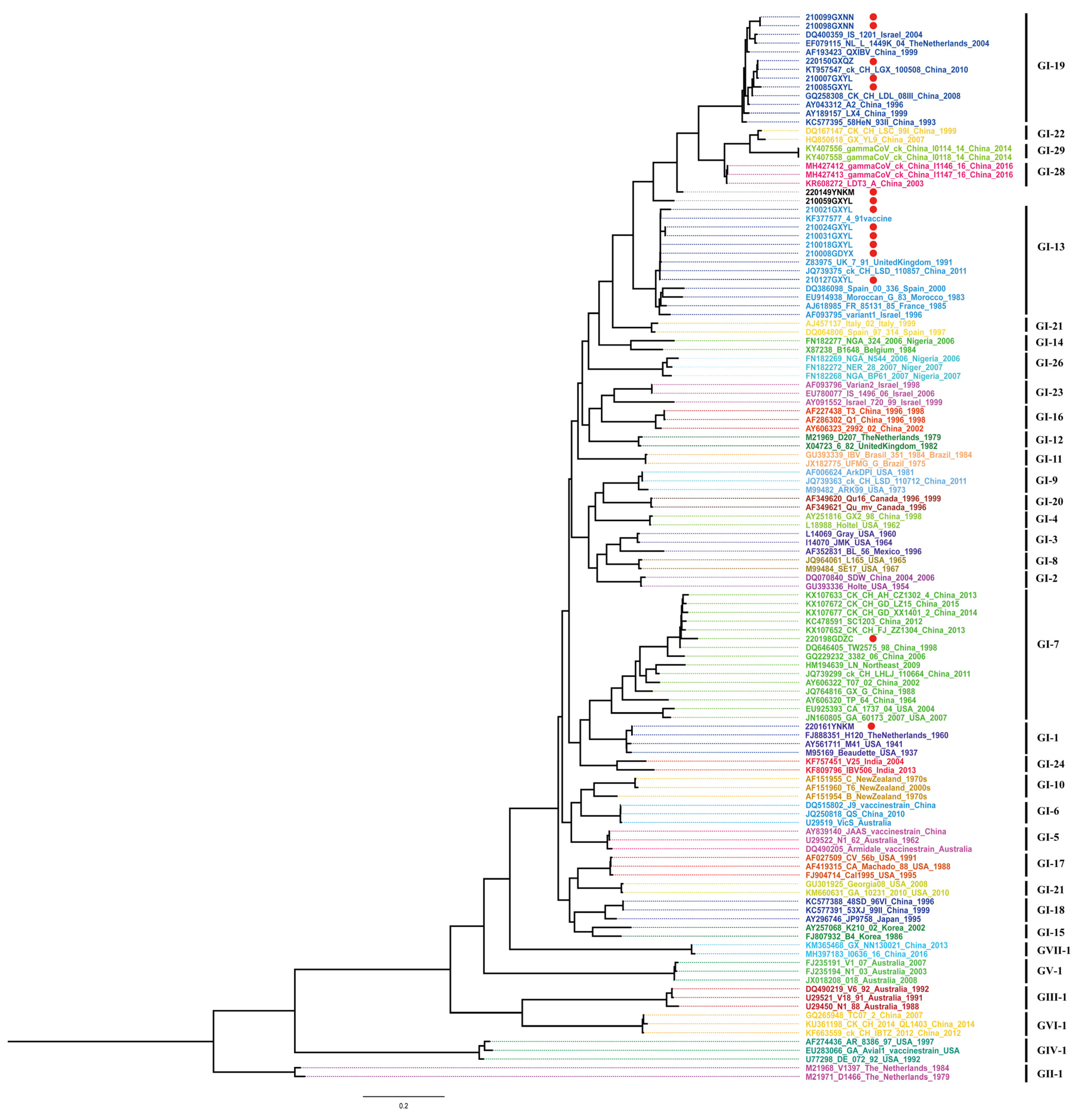
2.3. Phylogenetic Analysis

In addition to the IBVs isolated in this study, 107 S1 gene sequences of 35 lineage IBV strains were obtained from GenBank (NCBI, Bethesda, MD, USA). All sequences were aligned using MAFFT v7.221.3 [18]. A maximum likelihood (ML) tree was constructed based on the Sl gene sequences and GTR+F+R5 model, selected with Modelfinder [19], using IQ-TREE software [20] with 1000 bootstrap replicates. The ML tree was visualized using Figtree version 1.4.4 (http://tree.bio.ed.ac.uk/software/figtree/ (accessed on 2 May 2023)).

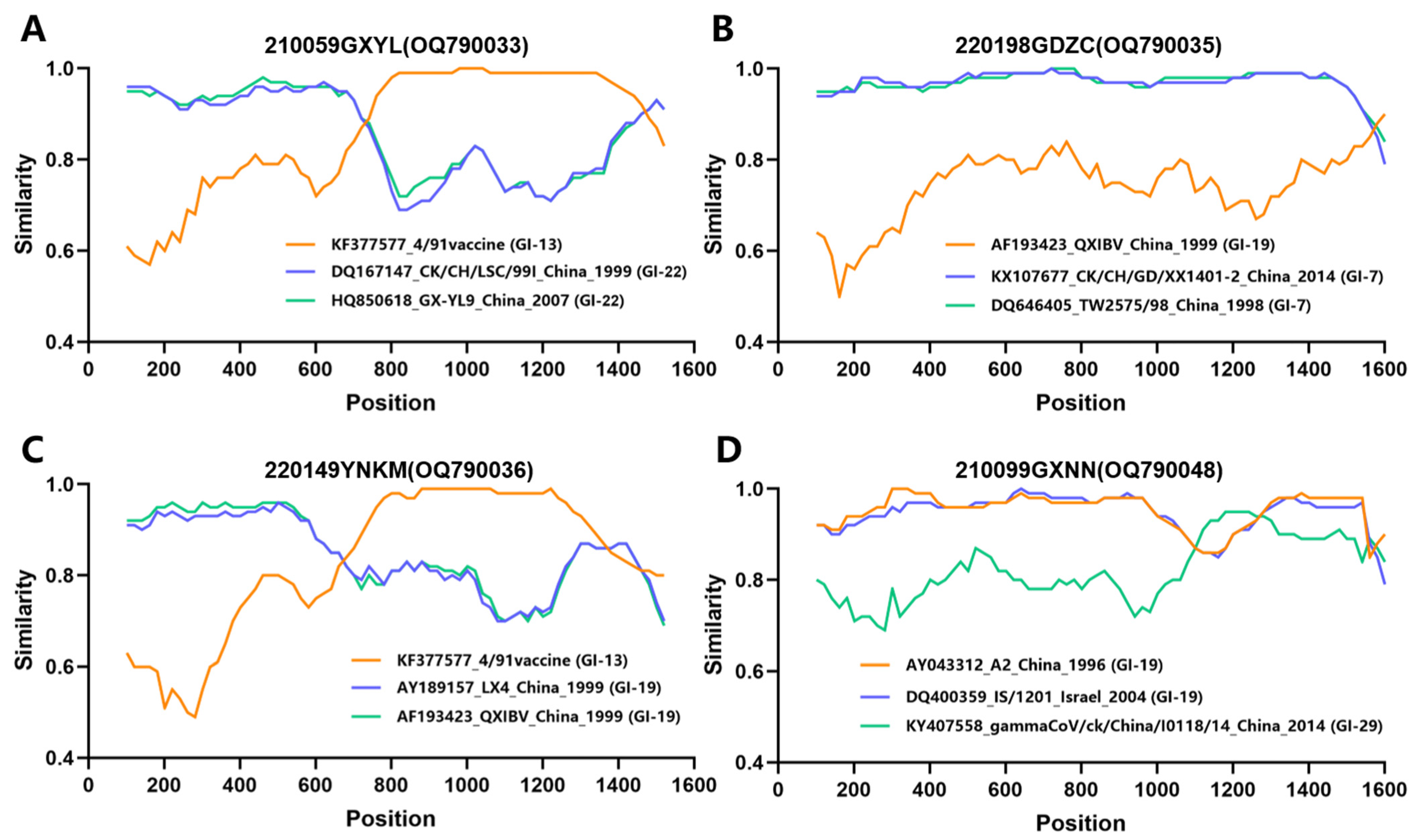
2.4. Recombination Analysis

Putative recombination events and parental strains were identified using the Recombination Detection Program version 4.0 (RDP 4.0; Simmonics, University of Warwick, Coventry, UK). Multiple methods and default program settings were used to analyze the data, including RDP Bootscan, GeneCony, Maxchi, Chimaera, SiSscan, LARD, 3Seq, and PhylPro [21]. The most likely recombinant fragments (p ≤ 10−12) were determined along with the possible parental virus and the beginning and end points. The potential recombination events and breakpoints were determined by similarity plot (SimPlots) analysis in SimPlot version 3.5.1, using a window of 200 bp and a step size of 20 bp.

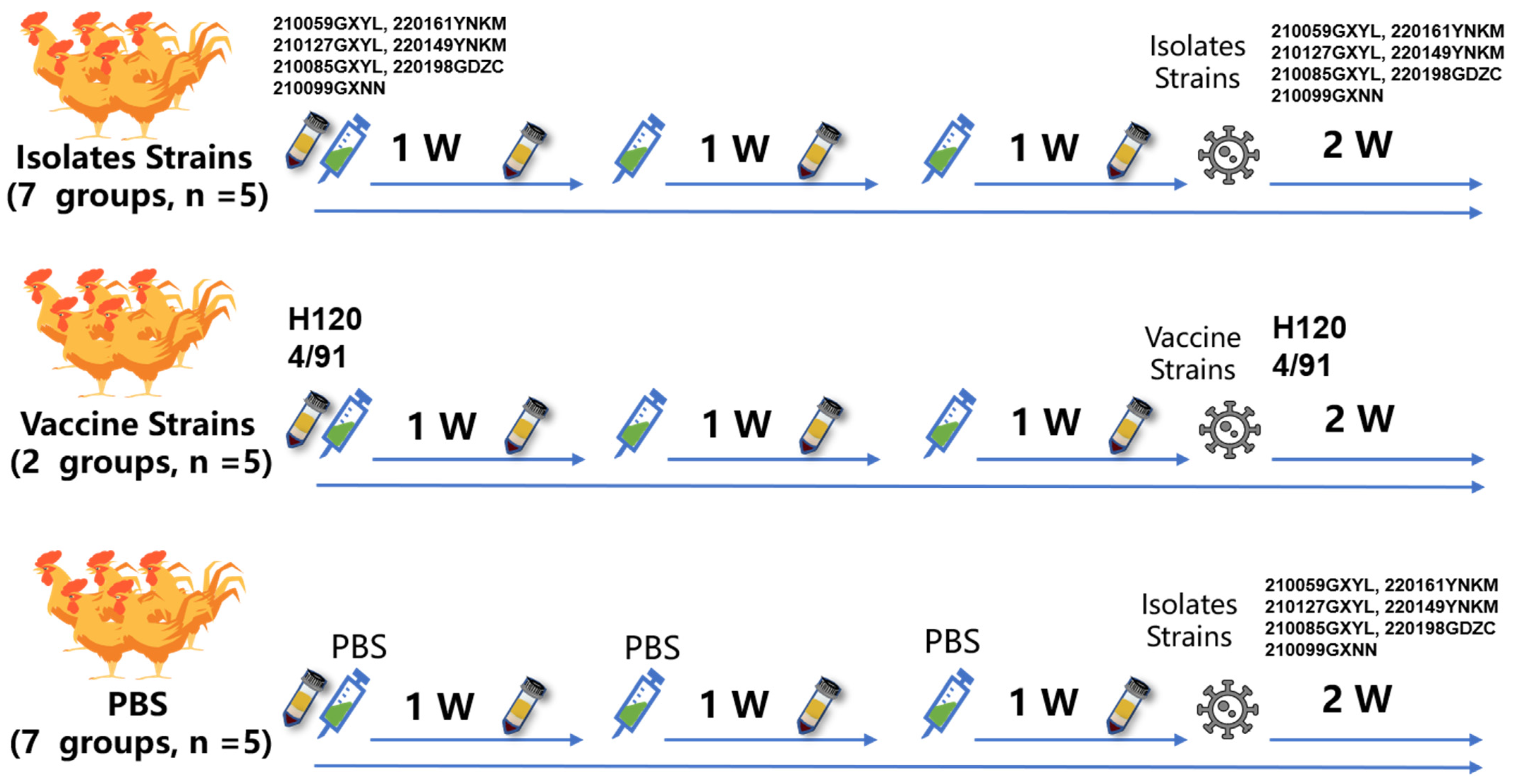
2.5. Inactivated IBV Vaccine and Testing

Twenty-eight-day-old SPF chickens were purchased from Guangdong Wens Dahuanong Biotechnology Co., Ltd. (Yunfu, China) and fed in a negative-pressure biosafety isolation chamber. The chickens had free access to water and commercial feed. The immunization and virus challenge procedures are shown in Figure 1. We used ultracentrifugation to concentrate the virus to achieve an EID50 value higher than 5.5. Viral stocks were inactivated with the addition of formaldehyde (Sigma, Shanghai, China) to 0.2%. The formaldehyde inactivated antigen solution was emulsified with oil adjuvant (ISA78VG, Seppic, Paris, France) at a ratio of 25:75 (w/w). The chickens were immunized with the oil-emulsion vaccine using 105.5 EID50 in 0.2 mL per chicken. Vaccination and challenge studies used the 28-day-old SPF chickens in 9 groups of 5 chickens each. Groups 1 to 7 were vaccinated intramuscularly with 200 µL IBV isolate strains of oil-emulsion vaccine containing 105.5 EID50 antigens per chicken. Groups 8 and 9 were vaccinated intramuscularly with 200 µL IBV H120 or 4/91 strains of oil-emulsion vaccine containing 105.5 EID50 antigens per chicken. Groups 10 to 16 were inoculated with sterile PBS and used as infection control that was unvaccinated but challenged. Booster vaccinations were carried out after 1 and 2 w, and the chickens were challenged with 0.2 mL of IBV isolate or vaccine strains containing 105.5 EID50 virus. Serum samples were obtained before each immunization and IBV challenge, then tested by ELISA (IDEXX 99-09262, Westbrook, ME, USA) and neutralization tests.

2.6. Antigenic Correlation Coefficient

The correlation coefficient (R) = ( r 1 × r 2 ) , where r1 = heterologous serum titer 1/homologous serum titer 1 and r2 = heterologous serum titer 2/homologous serum titer 2. When R > 80%, it was considered the same serotype. When R was between 25% and 80%, it was considered a different subtype of the same serotype. When R ≤ 25%, it was considered a different serotype.

3. Results

3.1. Epidemic Information and S1 Sequences of Clinical Samples

Between January 2021 and June 2022, 1008 samples were collected from broiler farms in southern China. After excluding the samples that were co-infected with the IBV and other viruses, a total of 15 IBV strains were isolated and identified, including 11 isolates from Guangxi, two from Yunnan, and two from Guangdong province. The accession numbers and the clinical information for each strain are summarized in Table 1.
The 15 isolates of the IBV exhibited different clinical symptoms, of which eight strains (53.33%) were from chickens with typical respiratory clinical symptoms, including wheezing, coughing, sneezing, tracheal rales, and nasal discharge, and autopsy showed hemorrhagic points in the lungs and trachea; three strains (20%) were from chickens with typical intestinal symptoms, such as severe dehydration and weight loss, and autopsy showed hemorrhagic points in the intestines; three strains (20%) were from chickens with typical joint inflammation symptoms, with swollen and fluid-filled foot joints visible; and one strain (6.67%) was from chickens with typical renal symptoms, such as kidney enlargement and urate-filled ureters, and autopsy showed hemorrhagic points in the kidneys.
The nucleic acid sequences, amino acid sequence lengths, and cleavage sites of S1 in the 15 IBV isolates were analyzed. Within S1, there were four different nucleotide (1611, 1617, 1620, and 1626) and deduced amino acid (537, 539, 540, and 542) sequence lengths. The most common S1 was 1620 nucleotides long, accounting for 60% of the total IBV isolates. There were five types of cleavage sites, including HRRRR, RRFRR, SRRLR, and RRSRR, all of which were similar to those reported previously (Feng et al., 2014).

3.2. Evolutionary Genetic and Recombination Analysis of S1

A phylogenetic tree of S1, including 107 reference strains and the 15 IBV isolates, was constructed using IQ-TREE. As shown in Figure 2, the 15 IBV isolates comprised four main genotypes: GI-19 (QX), GI-13 (4/91), GI-1 (Mass), and GI-7 (TW-I), with GI-19 (QX) being the predominant genotype. Two isolates, 220149YNKM and 210059GXYL, formed independent evolutionary branches and were considered variant strains.
Based on different genotypes, the 15 IBV isolates were aligned and compared with the H120 and 4/91 vaccine strains. The results showed that the nucleotide and amino acid sequence similarities were 77 to 99.9% and 74.4 to 99.6%, respectively (Table 2). The isolates of each genotype showed the highest similarity to the corresponding vaccine strain. GI-7 (TW-I) did not have any equivalent vaccine strains and the similarity of S1 with the two major vaccine strains was low.
Recombination analysis of the S1 gene sequences of all 15 IBV strains isolated in this study was performed using RDP 4.0. Four isolates, 210059GXYL, 210099GXNN, 220149YNKM, and 220198GDZC, exhibited recombination events (Figure 3). The breakpoint positions and specific P-values for each recombination event detection method are listed in Supplementary Table S2. As shown in Figure 3, recombination of multiple IBV lineages was observed, and lineage GI-13 and GI-19 IBVs were identified as the major and minor parental strains, respectively, in these four recombination events. Together, these results indicated that the prevalent IBV lineages had a higher probability of being involved in recombination events.

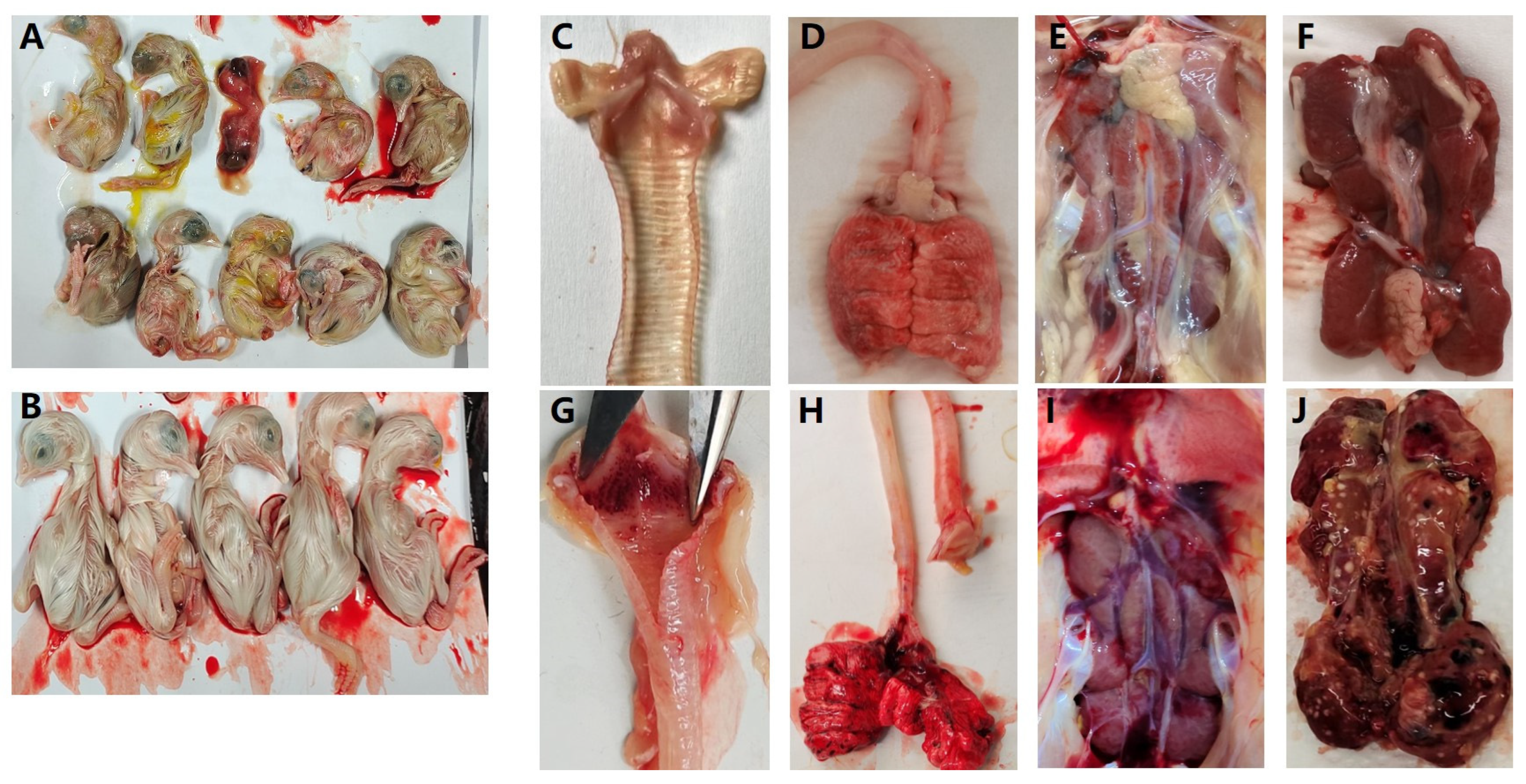
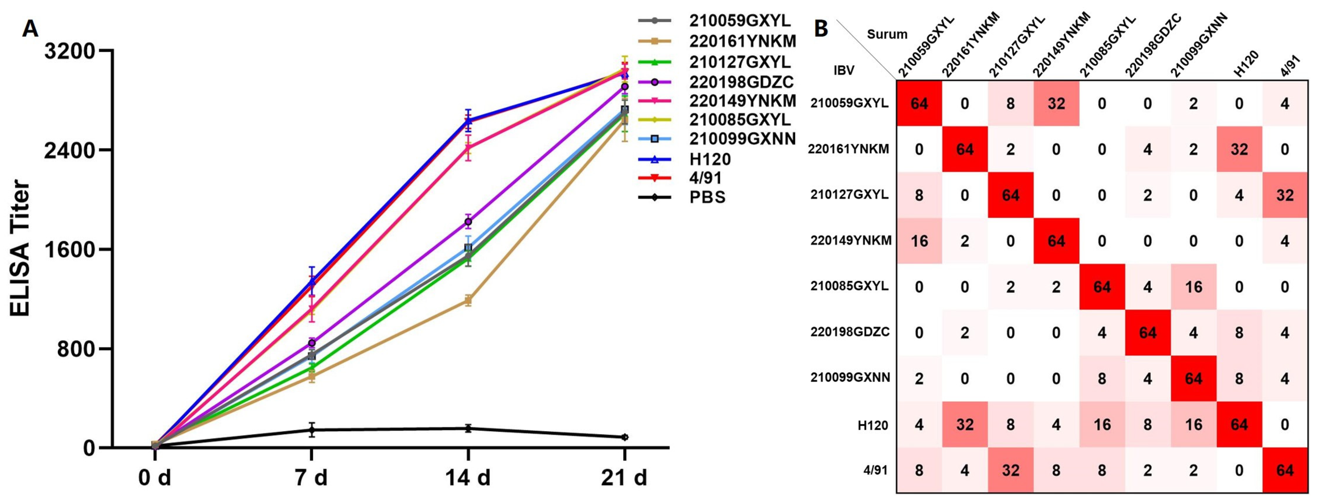
3.3. Antigenic Characterization of Isolate Strains

To investigate whether the recombinant strains had any impact on the virulence of the IBV, typical virulent strains were selected as representative branches for antigenic characterization studies, including four recombinant strains and three typical strains of GI-1, 13, and 19. After inoculating SPF chicken embryos with the isolates, all seven isolates caused embryonic lesions and death, together with various symptoms such as weak, curled, and dissolved embryos (Figure 4A,B).
The isolated IBV strains were inactivated and administered to SPF chickens for immunization. After the third immunization, the chickens were challenged with the corresponding strains and no typical IBV symptoms were observed in the immunized SPF chickens. In contrast, unimmunized chickens infected with the isolated strains exhibited symptoms such as depression, ruffled feathers, and coughing, consistent with the symptoms of the source-diseased chicken (Supplementary Table S3). Autopsy results confirmed this finding, as bleeding points in the trachea and lungs and typical IBV infection symptoms in the kidneys were only observed in the unimmunized group (Figure 4C–J). This further demonstrated the severe damage caused by IBV infection and the efficacy of immunization with the isolated strains in protecting against IBV infection.
ELISA revealed that the immunized chickens produced high levels of IBV antibodies, with the highest level reached after the third immunization, and had high neutralizing titers against the corresponding strains (Figure 5A). To confirm whether the recombinant strain had immune escape ability, cross-serum neutralization experiments were conducted using different groups of serum. The cross-protective antibodies generated by different genotypes of IBV strains had lower protection rates, and the serum antibodies produced by the H120 and 4/91 vaccine strains had poor neutralizing ability against the isolated strains. Only 32 had neutralizing ability against 220161YNKM and 210127GXYL, indicating that differences in serotypes among isolates of the prevalent IBV strains in the southern region of China may have developed an immune escape ability against vaccines (Figure 5B).
Further analysis of the antigenic correlation between the vaccine strains and isolated strains confirmed significant differences in antigenicity, with hemagglutination inhibition antigen correlation coefficients ranging from 2.21 to 74.56 (Table 3). The antigen correlation coefficient between different isolated and vaccine strains was as low as 1.05 and as high as 80.03 between 210059GXYL and 220149YNKM. Although 210085GXYL and 210099GXNN were both GI-19, and 210127GXYL and 4/91 were both GI-13, their R values were all less than 80, indicating that the genotypes and serotypes did not fully match. Therefore, the existing H120 and 4/91 vaccines may not effectively prevent or control IBV infection. It is essential to use appropriate vaccines to achieve ideal preventive and control effects.

4. Discussion

Coronaviruses comprise a large family and are widely found in nature. A growing body of research suggests that SARS-CoV-2, which caused a global pandemic in late 2019, may have originated in nature [22]. Therefore, the study of coronaviruses in different species has received increasing attention from the scientific community. Chicken-derived coronaviruses (IBVs) are widespread worldwide and cause significant losses and threats to the poultry industry. In this study, an epidemiological survey of IBV conducted in South China isolated 15 IBV strains from 1008 infected chicken tissue samples collected between January 2021 and June 2022. Compared with the analyses by Lian et al. (2021) in South China between April 2019 and March 2020 (139/420) and Xu et al. (2018) in China from January 2016 to December 2017 (213/801), the IBV isolation rate reported in the current study was significantly reduced [9,15]. A credible explanation for this result is that the clinical samples we collected included chickens that did not have respiratory diseases due to IBV infection. We also isolated other pathogens, including the avian influenza virus (AIV), the Newcastle disease virus (NDV), the adenovirus, and others, including AIV-mixed infections. As these pathogens may have caused the disappearance of the IBV during chicken embryo passage, we excluded these samples. Due to the varying levels of biosafety in aquaculture farms across different provinces and the significant differences in sample numbers, monitoring bias was also an inevitable challenge. These potential variables in collected samples will be addressed in future studies. Overall, our results showed that the GI-19 (QX-type) IBV lineage still dominated the epidemic. Other IBV lineages were sporadically isolated, similar to the results of recent research in China [9,23].
Recombination is an important contributing factor to the emergence and evolution of the IBV, as well as the emergence of new coronaviruses and novel diseases [12,24]. We identified four recombination events in the S1 gene, among which the IBVs from lineages GI-13 and GI-19 were more frequently involved in recombination, resulting in wider tissue tropism and leading to a more diverse range of clinical symptoms, including respiratory, pulmonary, renal, and even arthritic symptoms. Our findings indicate that these major prevalent lineages may play key roles in the transmission of the IBV in South China.
Ji et al. (2020) found that the homology between IBV variant strains in central China and vaccine strains was only 67.4–89.8% [23]. In this study, the nucleotide and amino acid sequence similarities were 77–99.9% and 74.4–99.6%, respectively, between isolated IBV strains and H120 or 4/91, indicating the limited effectiveness of the vaccine against the variant strains. The S1 protein has conformation-dependent epitopes that induce virus neutralization and serotype-specific antibodies. To investigate whether mutations in the S1 gene affect the protective efficacy of IBV vaccine strains, seven isolate strains were selected from genotype and isolation sites, among which 210059GXYL, 220198GDZC, 220149YNKM, and 210099GXNN were potential strains for S1 gene recombination, 210085GXYL was a GI-19 strain isolated from Guangxi, 210127GXYL was a GI-13 strain isolated from Guangxi, and 220161YNKM was the only GI-1 strain isolated from Yunnan. Then, we examined plasma samples obtained from SPF chickens immunized three times with inactivated isolates. The immunized SPF chickens were found to have high antibody levels and sufficient protective power against the corresponding strains. No clinical symptoms were observed in the immunized SPF chickens after challenge and the results of the serum neutralization experiments confirmed this conclusion. However, the H120 and 4/91 vaccines were unable to provide sufficient protection against the isolate, indicating that the recombinant strain may have developed an immune escape from the current vaccines, limiting their effectiveness in disease control. Li et al. (2022) conducted a survey of IBV prevalence in southwest China and observed the same phenomenon: IBV vaccines were unable to suppress virus release [25]. Therefore, effective vaccines targeting local strains and improved vaccination strategies must be developed.
The IB vaccines currently used in China are mainly targeted against MASS strains, such as H120, which differ in genotype from isolate strains, such as GI-19. Serotype is a reference for representing IBV antigenic variation. Although the nucleotide homology between the isolated and vaccine strains ranged from 77–99.9%, the antigenic correlation coefficient was low and the correlation between strains of the same genotype was less than 80%, indicating that the genotype and serotype did not match. Thus, S1 alone was insufficient to characterize the serotype or protectotype. Chen et al. (2015) analyzed five H120-recombinant strains and found that H120 did not protect against two IBV strains with a genotype- and serotype-matching rate of only 60% [26]. Using reverse genetics, Shan et al. (2018) further demonstrated that recombination of the S1 gene affects the IBV serotype [27]. Yan et al. (2021) also found that the ongoing evolution of IBV field strains through genetic recombination and mutation leads to outbreaks among vaccinated chicken populations, and suggested that evaluating the virulence of the virus strain based solely on the S1 is insufficient [28]. These studies indicated that antigenic variation in IBV cannot be measured solely by genotype; therefore, the selection of serotypes is an important reference index for IBV vaccine application, to conduct continuous epidemiological surveillance against the IBVs, and to screen for the most prevalent genotypes and serotypes to identify vaccine candidates for IB prevention and control in China.
In summary, we isolated 15 IBV strains from South China between 2021 and 2022, with nucleotide homology ranging from 77–99.9% and divided them into four genotypes, with GI-19 serving as the main genotype. Currently, vaccines are no longer effective in protecting against the prevalent IBV strains in southern China, as these strains have developed immune escape. Our study provides valuable information for the control of the IBV in southern China, which may help guide the design of vaccination strategies.

Supplementary Materials

The following supporting information can be downloaded at: https://www.mdpi.com/article/10.3390/v15061273/s1, Table S1: The primers used for RT-PCR; Table S2: Recombinant breakpoints, genes, and major and minor related sequences of genomic recombination events; Table S3: Clinical observations of chickens in the two-week post challenge.

Author Contributions

W.Y. was responsible for experiment design, data analysis, and writing the manuscript. T.L., W.J., Y.H., Q.W. and J.R. were responsible for performing experiments. L.F., Q.L. and C.D. were responsible for suggestions during the experiment’s performance. L.C., B.X. and T.R. were responsible for revising the manuscript. All authors have read and agreed to the published version of the manuscript.

Funding

This research was funded by the Science and Technology Program of Forestry Technology Innovation Project of Guangdong Province, No. 2023KJCX028), the Guangdong Province (No. 2021B1212030015), the Guangdong Provincial Special Fund for Modern Agriculture Industry Technology Innovation Teams (No. 2023KJ119), the National Natural Science Foundation of China (No. 31872492 and 31902251), and the Special Project on Strategy for Rejuvenation of Guangdong Provincial Villages in 2022 (No. 5500-F22039).

Institutional Review Board Statement

The animal study protocol was approved by the South China Agricultural University Experimental Animal Welfare Ethics Committee (permit number: 2022b017). All experiments were performed in animal biosafety level 3 (ABSL-3) facilities.

Informed Consent Statement

Not applicable.

Data Availability Statement

The genomic data presented in this study are available from GenBank (accession numbers: OQ790027-OQ790033, OQ790035-OQ790037, OQ790043-OQ790048).

Conflicts of Interest

The authors declare no conflict of interest.

References

  1. Sjaak de Wit, J.J.; Cook, J.K.; van der Heijden, H.M. Infectious bronchitis virus variants: A review of the history, current situation and control measures. Avian Pathol. 2011, 40, 223–235. [Google Scholar] [CrossRef] [PubMed]
  2. Cook, J.K.A.; Jackwood, M.; Jones, R.C. The long view: 40 years of infectious bronchitis research. Avian Pathol. 2012, 41, 239–250. [Google Scholar] [CrossRef] [PubMed]
  3. Bande, F.; Arshad, S.S.; Bejo, M.H.; Moeini, H.; Omar, A.R. Progress and Challenges toward the Development of Vaccines against Avian Infectious Bronchitis. J. Immunol. Res. 2015, 2015, 424860. [Google Scholar] [CrossRef] [PubMed]
  4. Cavanagh, D. Coronaviruses in poultry and other birds. Avian Pathol. 2005, 34, 439–448. [Google Scholar] [CrossRef]
  5. Cavanagh, D. Coronavirus avian infectious bronchitis virus. Vet. Res. 2007, 38, 281–297. [Google Scholar] [CrossRef]
  6. Shang, J.; Zheng, Y.; Yang, Y.; Liu, C.; Geng, Q.; Luo, C.; Zhang, W.; Li, F. Cryo-EM structure of infectious bronchitis coronavirus spike protein reveals structural and functional evolution of coronavirus spike proteins. PLoS Pathog. 2018, 14, e1007009. [Google Scholar] [CrossRef]
  7. Ali, A.; Ojkic, D.; Elshafiee, E.A.; Shany, S.; El-Safty, M.M.; Shalaby, A.A.; Abdul-Careem, M.F. Genotyping and In Silico Analysis of Delmarva (DMV/1639) Infectious Bronchitis Virus (IBV) Spike 1 (S1) Glycoprotein. Genes 2022, 13, 1617. [Google Scholar] [CrossRef]
  8. Kabinger, F.; Stiller, C.; Schmitzova, J.; Dienemann, C.; Kokic, G.; Hillen, H.S.; Hobartner, C.; Cramer, P. Mechanism of molnupiravir-induced SARS-CoV-2 mutagenesis. Nat. Struct. Mol. Biol. 2021, 28, 740–746. [Google Scholar] [CrossRef]
  9. Xu, L.; Han, Z.; Jiang, L.; Sun, J.; Zhao, Y.; Liu, S. Genetic diversity of avian infectious bronchitis virus in China in recent years. Infect. Genet. Evol. 2018, 66, 82–94. [Google Scholar] [CrossRef]
  10. Xia, X. Domains and Functions of Spike Protein in SARS-CoV-2 in the Context of Vaccine Design. Viruses 2021, 13, 109. [Google Scholar] [CrossRef]
  11. Valastro, V.; Holmes, E.C.; Britton, P.; Fusaro, A.; Jackwood, M.W.; Cattoli, G.; Monne, I. S1 gene-based phylogeny of infectious bronchitis virus: An attempt to harmonize virus classification. Infect. Genet. Evol. 2016, 39, 349–364. [Google Scholar] [CrossRef] [PubMed]
  12. Ma, T.; Xu, L.; Ren, M.; Shen, J.; Han, Z.; Sun, J.; Zhao, Y.; Liu, S. Novel genotype of infectious bronchitis virus isolated in China. Vet. Microbiol. 2019, 230, 178–186. [Google Scholar] [CrossRef] [PubMed]
  13. Feng, K.; Xue, Y.; Wang, F.; Chen, F.; Shu, D.; Xie, Q. Analysis of S1 gene of avian infectious bronchitis virus isolated in southern China during 2011–2012. Virus Genes 2014, 49, 292–303. [Google Scholar] [CrossRef] [PubMed]
  14. Zhao, Y.; Zhang, H.; Zhao, J.; Guo-Zhong, Z.; Jin, J.-H.; Zhang, G.-Z. Evolution of infectious bronchitis virus in China over the past two decades. J. Gen. Virol. 2016, 97, 1566–1574. [Google Scholar] [CrossRef]
  15. Lian, J.; Wang, Z.; Xu, Z.; Chen, T.; Shao, G.; Zhang, X.; Qin, J.; Xie, Q.; Lin, W. Distribution and molecular characterization of avian infectious bronchitis virus in southern China. Poult. Sci. 2021, 100, 101169. [Google Scholar] [CrossRef]
  16. Ren, M.; Sheng, J.; Ma, T.; Xu, L.; Han, Z.; Li, H.; Zhao, Y.; Sun, J.; Liu, S. Molecular and biological characteristics of the infectious bronchitis virus TC07-2/GVI-1 lineage isolated in China. Infect. Genet. Evol. 2019, 75, 103942. [Google Scholar] [CrossRef]
  17. Chen, L.; Xiang, B.; Hong, Y.; Li, Q.; Du, H.; Lin, Q.; Liao, M.; Ren, T.; Xu, C. Phylogenetic analysis of infectious bronchitis virus circulating in southern China in 2016–2017 and evaluation of an attenuated strain as a vaccine candidate. Arch. Virol. 2021, 166, 73–81. [Google Scholar] [CrossRef]
  18. Katoh, K.; Standley, D.M. MAFFT Multiple Sequence Alignment Software Version 7: Improvements in Performance and Usability. Mol. Biol. Evol. 2013, 30, 772–780. [Google Scholar] [CrossRef]
  19. Kalyaanamoorthy, S.; Minh, B.Q.; Wong, T.K.F.; Von Haeseler, A.; Jermiin, L.S. ModelFinder: Fast model selection for accurate phylogenetic estimates. Nat. Methods 2017, 14, 587–589. [Google Scholar] [CrossRef]
  20. Nguyen, L.-T.; Schmidt, H.A.; Von Haeseler, A.; Minh, B.Q. IQ-TREE: A Fast and Effective Stochastic Algorithm for Estimating Maximum-Likelihood Phylogenies. Mol. Biol. Evol. 2015, 32, 268–274. [Google Scholar] [CrossRef]
  21. Martin, D.P. Recombination Detection and Analysis Using RDP3. Bioinform. DNA Seq. Anal. 2009, 537, 185–205. [Google Scholar] [CrossRef]
  22. Temmam, S.; Vongphayloth, K.; Baquero, E.; Munier, S.; Bonomi, M.; Regnault, B.; Douangboubpha, B.; Karami, Y.; Chrétien, D.; Sanamxay, D.; et al. Bat coronaviruses related to SARS-CoV-2 and infectious for human cells. Nature 2022, 604, 330–336. [Google Scholar] [CrossRef]
  23. Ji, J.; Gao, Y.; Chen, Q.; Wu, Q.; Xu, X.; Kan, Y.; Yao, L.; Bi, Y.; Xie, Q. Epidemiological investigation of avian infectious bronchitis and locally determined genotype diversity in central China: A 2016–2018 study. Poult. Sci. 2020, 99, 3001–3008. [Google Scholar] [CrossRef] [PubMed]
  24. Lee, C.-W.; Jackwood, M.W. Evidence of genetic diversity generated by recombination among avian coronavirus IBV. Arch. Virol. 2000, 145, 2135–2148. [Google Scholar] [CrossRef] [PubMed]
  25. Li, S.; Chen, W.; Shen, Y.; Xia, J.; Fan, S.; Li, N.; Luo, Y.; Han, X.; Cui, M.; Zhao, Y.; et al. Molecular characterization of infectious bronchitis virus in Southwestern China for the protective efficacy evaluation of four live vaccine strains. Vaccine 2022, 40, 255–265. [Google Scholar] [CrossRef] [PubMed]
  26. Chen, L.; Zhang, T.; Han, Z.; Liang, S.; Xu, Y.; Xu, Q.; Chen, Y.; Zhao, Y.; Shao, Y.; Li, H.; et al. Molecular and antigenic characteristics of Massachusetts genotype infectious bronchitis coronavirus in China. Vet. Microbiol. 2015, 181, 241–251. [Google Scholar] [CrossRef]
  27. Shan, D.; Fang, S.; Han, Z.; Ai, H.; Zhao, W.; Chen, Y.; Jiang, L.; Liu, S. Effects of hypervariable regions in spike protein on pathogenicity, tropism, and serotypes of infectious bronchitis virus. Virus Res. 2018, 250, 104–113. [Google Scholar] [CrossRef]
  28. Yan, W.; Qiu, R.; Wang, F.; Fu, X.; Li, H.; Cui, P.; Zhai, Y.; Li, C.; Zhang, L.; Gu, K.; et al. Genetic and pathogenic characterization of a novel recombinant avian infectious bronchitis virus derived from GI-1, GI-13, GI-28, and GI-19 strains in Southwestern China. Poult. Sci. 2021, 100, 101210. [Google Scholar] [CrossRef]
Figure 1. Grouping information and timing of immunization, virus challenge, and serum acquisition.
Viruses 15 01273 g001
Figure 2. Maximum likelihood (ML) tree based on the S1 gene from 35 lineages of IBV. The tree was constructed using IQ-TREE software with the GTR+F+R5 model. Red circles indicate the stains isolated in this study.
Viruses 15 01273 g002
Figure 3. Recombination events in the S1 gene from the isolated IBV strains. Simplot analysis was performed to detect recombination within the S1 gene from (A) 210059GXYL, (B) 220198GDZC, (C) 220149YNKM, and (D) 210099GXNN. The y-axis represents the ratio of identity within a 200-bp wide sliding window centered on the position plotted, with a 20 bp step size between plots.
Viruses 15 01273 g003
Figure 4. (A) SPF chicken embryos infected by IBV isolated strains; (B) control; (CF) the trachea, lung, and kidney from immunizated chickens with isolated strains; (GJ) the trachea, lung, and kidney from immunizated chickens with PBS.
Viruses 15 01273 g004
Figure 5. (A) Antibody response of chickens after immunization with isolated strains and vaccine strains; (B) cross-serum neutralization experiments between isolated strains and vaccine strains.
Viruses 15 01273 g005
Table 1. Background and S1 information of domestic isolates of the IBV from 2021 to 2022.
IBV IsolatesAccession NumberProvince 1Date (Day-Month-Year)Vaccination HistoryClinical TypeLength of S1 (nt/aa) 2Cleavage Recognition Motifs 3Genotype
210007GXYLOQ790027Guangxi05-08-2021H120 + H120A1620/540HRRRRGI-19
210008GDYXOQ790028Guangdong11-08-2021H120 + H120A1617/539RRSRRGI-13
210018GXYLOQ790029Guangxi06-09-2021H120 + H120RS1617/539RRSRRGI-13
210021GXYLOQ790030Guangxi06-09-2021H120 + H120I1617/539HRRRRGI-13
210024GXYLOQ790031Guangxi08-09-2021H120 + H120A1620/540RRSRRGI-13
210031GXYLOQ790032Guangxi16-09-2021H120RS1620/540RRSRRGI-13
210059GXYLOQ790033Guangxi29-10-2021H120RS1626/542SRRLR
220198GDZCOQ790035Guangdong07-11-2021H120RS1620/540HRRRRGI-7
220149YNKMOQ790036Yunnan18-11-2021H120 + H120RS1617/539RRFRR
210085GXYLOQ790037Guangxi03-11-2021H120 + H120I1620/540HRRRRGI-19
210127GXYLOQ790043Guangxi12-11-20214/91I1617/539RRSRRGI-13
220150GXQZOQ790044Guangxi19-01-2022H120 + H120NP1620/540HRRRRGI-19
220161YNKMOQ790045Yunnan10-03-2022H120RS1611/537RRFRRGI-1
210098GXNNOQ790047Guangxi07-11-2021H120RS1620/540HRRRRGI-19
210099GXNNOQ790048Guangxi07-11-2021H120RS1620/540HRRRRGI-19
Abbreviations: IBV, infectious bronchitis virus; NP, nephropathogenic; RS, respiratory; A, Arthritis; I, Intestinal. 1: Province where the viruses were isolated; 2: Length of nucleotides and deduced amino acids; 3: Cleavage recognition motifs of S1; R, arginine; F, phenylalanine; H, histidine; T, threonine; K, lysine; L, leucine; S, serine.
Table 2. Similarity in nucleotides and amino acids between isolate strains and vaccine strains.
aa (%)
nt (%) 1234567891011121314151617
1***78.778.980.480.680.68080.486.597.279.199.477.29594.57778.9210007GXYL
278.6***99.496.598.298.285.875.682.178.699.178.774.878.67874.699.4210008GDYX
378.699.8***96.798.398.38675.882.378.799.378.97578.778.274.899.6210018GXYL
48097.597.5***95.495.484.777.380.880.296.380.474.48079.574.397210021GXYL
579.898.898.896.5***10086.27683.780.49880.674.880.379.774.698.3210024GXYL
679.898.898.896.5100***86.27683.780.49880.674.880.379.774.698.3210031GXYL
779.987.987.98788.288.2***77.887.179.885.879.875.18079.474.986210059GXYL
879.8777778.477.277.278.4***77.380.275.880.28080.479.979.875.8220198GDZC
985.585.885.884.686.686.689.377.9***86.182.586.576.586.786.176.382.3220149YNKM
1097.378.678.679.979.879.879.979.485.5***78.997.47794.894.376.878.7210085GXYL
1178.799.799.897.498.798.787.977.18678.7***79.175.478.978.475.299.3210127GXYL
1299.478.778.780.280808079.785.697.478.9***76.894.894.376.678.9220150GXQZ
1377.778.678.677.878.278.278.480.679.377.978.777.7***77.777.299.675220161YNKM
1495.178.478.579.979.679.679.880.385.494.678.695.377.7***99.377.678.7210098GXNN
1595.178.378.479.879.579.579.680.285.294.578.595.177.499.6***7778.2210099GXNN
1677.678.578.577.7787878.480.679.277.778.677.699.977.677.2***74.8FJ888351_H120 vaccine
1778.699.899.997.798.898.887.97785.878.699.878.778.678.578.478.5***KF377577_4/91vaccine
1234567891011121314151617
Table 3. Antigenic correlation between vaccine strains and isolated strains.
H1204/91210059GXYL220161YNKM210127GXYL220149YNKM210085GXYL220198GDZC210099GXNN
H1201006.542.2174.5610.063.414.7127.614.12
4/912.11009.945.3363.4111.126.578.7669.44
210059GXYL3.2121.71001.0510.4980.034.214.255.46
220161YNKM54.363.622.4410014.5211.3512.9621.3115.23
210127GXYL16.1164.4315.465.11006.823.2210.079.08
220149YNKM1.7518.8759.864.876.821005.975.414.12
210085GXYL2.764.434.2112.9612.342.7810016.7235.78
220198GDZC35.117.583.6718.654.625.4116.7210017.46
210099GCYL37.2121.25.467.619.084.1224.7117.46100
Disclaimer/Publisher’s Note: The statements, opinions and data contained in all publications are solely those of the individual author(s) and contributor(s) and not of MDPI and/or the editor(s). MDPI and/or the editor(s) disclaim responsibility for any injury to people or property resulting from any ideas, methods, instructions or products referred to in the content.

