Article

Comparative Evaluation of the Foot-and-Mouth Disease Virus Permissive LF-BK αVβ6 Cell Line for Senecavirus A Research

1
SAIC, Plum Island Animal Disease Center, Greenport, NY 11944, USA
2
U.S. Department of Agriculture, Animal and Plant Health Inspection Service, National Veterinary Services Laboratories, Foreign Animal Disease Diagnostic Laboratory, Plum Island Animal Disease Center, Greenport, NY 11944, USA
3
U.S. Department of Homeland Security Science & Technology Directorate, Plum Island Animal Disease Center, Greenport, NY 11944, USA
*
Author to whom correspondence should be addressed.
Viruses 2022, 14(9), 1875; https://doi.org/10.3390/v14091875
Submission received: 10 August 2022 / Revised: 23 August 2022 / Accepted: 24 August 2022 / Published: 25 August 2022
(This article belongs to the Section Animal Viruses)

Abstract

:
Senecavirus A (SVA) is a member of the family Picornaviridae and enzootic in domestic swine. SVA can induce vesicular lesions that are clinically indistinguishable from Foot-and-mouth disease, a major cause of global trade barriers and agricultural productivity losses worldwide. The LF-BK αVβ6 cell line is a porcine-derived cell line transformed to stably express an αVβ6 bovine integrin and primarily used for enhanced propagation of Foot-and-mouth disease virus (FMDV). Due to the high biosecurity requirements for working with FMDV, SVA has been considered as a surrogate virus to test and evaluate new technologies and countermeasures. Herein we conducted a series of comparative evaluation in vitro studies between SVA and FMDV using the LF-BK αVβ6 cell line. These include utilization of LF-BK αVβ6 cells for field virus isolation, production of high virus titers, and evaluating serological reactivity and virus susceptibility to porcine type I interferons. These four methodologies utilizing LF-BK αVβ6 cells were applicable to research with SVA and results support the current use of SVA as a surrogate for FMDV.

1. Introduction

Senecavirus A (SVA), also identified as Seneca Valley Virus, is the only member of the genus Senecavirus and is considered an emerging pathogen in swine. Similar to other animal picornaviruses, SVA possesses a positive sense single stranded RNA genome encapsulated by four viral-encoded structural proteins. The SVA icosahedral capsid is approximately 27 nm in diameter and comprised of 60 protomers consisting of the individual capsid proteins, VP4, VP2, VP3, and VP1, all derived from the P1 polypeptide [1].
Initially isolated and described as a cell culture contaminant [1], SVA is enzootic in swine populations in the United States and other countries [2,3,4]. In the United States, estimated SVA seroprevalence for grower-finisher pigs and sows was 12.2% and 34.0%, respectively [2]. Clinically, SVA is associated with vesicular disease outbreaks and increased neonate mortality [5,6,7,8,9,10,11]. The vesicular lesions on the snout, oral mucosa, hooves, and coronary bands are clinically indistinguishable from those produced by Foot-and-mouth disease virus (FMDV), a high consequence transboundary animal disease pathogen and the causative agent of Foot-and-mouth disease (FMD) [3,6,9,12].
The LF-BK αVβ6 cell line is a porcine-derived cell line transformed to stably express the bovine αVβ6 integrin, the primary receptor for FMDV [13,14]. The LF-BK αVβ6 cell line is highly permissive to FMDV and to other swine viruses [15,16]. A subclone of the LF-BK αVβ6 cell line, identified as PIPEC, is used to grow African Swine Fever modified live viruses [17]. In this report we evaluated the LF-BK αVβ6 cell line for use in isolation and propagation of SVA from a field sample submitted for diagnostic investigation of swine vesicular disease to the U.S. Department of Agriculture, Animal and Plant Health Inspection Service, National Veterinary Services Laboratories, Foreign Animal Disease Diagnostic Laboratory. Following SVA virus purification, we adopted and evaluated procedures for usage of the LF-BK αVβ6 cells to determine SVA virus titers, serum neutralizing antibodies to SVA, and sensitivity to type I porcine interferon.
We found that four, widely used methodologies for FMDV research were directly applicable to work with SVA, enabling the potential use of SVA as a surrogate virus to FMDV for preliminary screening of new FMDV countermeasures independent of the high-level biocontainment facilities required for FMDV research.

2. Materials and Methods

2.1. Origin of SVA Positive Sample and Initial Propagation on Porcine-Derived Cell Lines

Instituto Biologico-Rim Suino-2 (IB-RS-2) cell culture inoculated with a field sample for a diagnostic investigation of swine vesicular lesions, was confirmed as positive for SVA by PCR and kindly provided by the U.S. Department of Agriculture, Animal and Plant Health Inspection Service, National Veterinary Services Laboratories, Foreign Animal Disease Diagnostic Laboratory (FADDL) at the Plum Island Animal Disease Center. Cell culture media was centrifuged at 500× g for 5 min and clarified supernatant aliquoted into cryovials for storage at −70 °C.
Porcine-derived cell line LF-BK αVβ6 was grown in DMEM media with 10% fetal bovine serum (FBS) (Hyclone™, Marlborough, MA, USA), 1% minimum essential media non-essential amino acids (MEM NEAA) (Gibco™, Waltham, MA, USA), 1% Sodium Pyruvate (Gibco™, Waltham, MA, USA), and 1% Antibiotic-Antimycotic (Anti-Anti) (Gibco™, Waltham, MA, USA). Porcine-derived PK-15 (ATCC CCL-33) and IB-RS-2 cell lines were grown in DMEM media with 10% FBS (Hyclone™, Marlborough, MA, USA) and 1% Anti-Anti (Gibco™, Waltham, MA, USA). Cells were cultured in T-25 flasks to full confluence and inoculated with 10 μL of the SVA-PCR positive clarified supernatant. Inoculated cells were incubated at 37 °C in 5% CO2 until significant cell sloughing was observed, typically, 2–4 days post infection (dpi). Cells were cryofractured at −70 °C, and cellular debris was removed by centrifugation at 500× g for 5 min followed by aliquoting and storage of supernatant at −70 °C until further use.

2.2. Polyethylene Glycol Precipitation

Cleared cell culture media from inoculated cells was transferred to a 500 mL glass bottle with 80 mg/mL of polyethylene glycol (PEG) and 23 mg/mL of NaCl followed by stirring overnight at 4 °C. After overnight incubation, the solution was centrifuged at approximately 7649× g for 25 min using an SS-34 rotor in an RC-6 centrifuge (Thermo Scientific, Waltham, MA, USA). Pellets were resuspended in 3 mL of TNE buffer, pH 7.5, (50 mM Tris-HCl, 100 mM NaCl, 5 mM EDTA), aliquoted, and stored at −70 °C until further use.

2.3. Sucrose Density Gradient Centrifugation

Sucrose solutions at 20%, 35%, 42.5%, 50%, and 65% (w/v) were prepared in 1X NT buffer (0.15 M NaCl, 0.05 M Tris-HCl), pH 7.8. Sucrose solutions were dispensed into 14 × 95 mm polypropylene tubes (Beckman Coulter, Indianapolis, IN, USA) at decreasing concentrations: 0.8 mL of 65%, 2 mL of 50%, 2 mL of 42.5%, 2 mL of 35%, and 1.8 mL of 20%. To prevent mixing, tubes were stored at −70 °C after the addition of each layer until solid, and boundaries marked.
One day prior to use, sucrose gradients were thawed at 4 °C. Up to 4 mL of PEG precipitated sample was applied to each gradient and centrifuged at 100,000× g for 2 h using a SW50ti rotor in an Optima™ L-70 XP ultracentrifuge (Beckman Coulter®, Indianapolis, IN, USA). Individual layers were harvested by pipetting and stored at −70 °C until further use.

2.4. Western Blotting

Samples from each gradient layer were diluted to 1X with 4X NuPage™ loading buffer (Invitrogen™, Waltham, MA, USA), heated at 95 °C for 10 min, applied to NuPAGE™ 4–12% Bis-Tris protein gels (Invitrogen™, Waltham, MA, USA) and run according to manufacturer’s instructions. The iBlot™ 2 Dry Blotting System (Life Technologies™, Waltham, MA, USA) was used according to manufacturer’s instructions for transfer to nitrocellulose membranes utilizing iBlot™ 2 Transfer Stacks (Invitrogen™, Waltham, MA, USA).
Membranes were incubated in 5% w/v nonfat dry milk blocking buffer solution for 1 h with shaking at room temperature followed by two rinses and a 5-min wash in 1X Phosphate Buffered Saline with 0.05% Tween™ 20 pH 7.4 (PBS-T). Primary antibody incubation was performed overnight at 4 °C with a rabbit anti-SVA VP1 polyclonal antibody (Alpha Diagnostic International, San Antonio, TX, USA) at a 1:1000 dilution followed by three 5-min washes in 1X PBS-T. A 1:500 dilution of goat HRP conjugated anti-rabbit secondary antibody (Thermo Scientific, Waltham, MA, USA), diluted in 1X PBS-T, was applied to the membranes, and incubated for 1 h at room temperature. Membranes were washed three times for 5 min each with PBS-T. To visualize bands, a solution of either 3′,3′-diaminobenzidine, made using SIGMAFAST™ (Sigma-Aldrich, St. Louis, MO, USA) 3,3′-diaminobenzidine tablets (Sigma-Aldrich®, St. Louis, MO, USA), or KPL TrueBlue™ (Tacoma, WA, USA) Peroxidase Substrate (Sera care) were prepared as per manufacturer’s instructions and applied to membranes and incubated at room temperature with shaking for 1 h or until bands appeared.

2.5. Electron Microscopy

Samples from both the 20% and 35% sucrose fractions of PEG-precipitated LF-BK αVβ6 propagated SVA were evaluated by negative staining on a Hitachi 7600 transmission electron microscope with a 2 k × 2 k AMT camera at 80 kV. Prior to application of sample, 300-mesh formvar/carbon coated grids were inverted on 1% alcian blue for 5 min followed by a rinse in ddH2O. Samples were adhered at a 1:5 dilution for 5 min; 2% uranyl acetate was added while blotting across Whatman 50 filter paper.

2.6. SVA Plaque Purification and Propagation on LF-BK αVβ6 Cells

The LF-BK αVβ6 cells were cultured to 100% confluence in 6-well plates, rinsed with Dulbecco’s phosphate-buffered saline (DPBS) followed by the application of 2 mL of media containing 2% FBS. The 35% sucrose fractions were diluted from 10−3 to 10−9 and added to individual wells and incubated at 37 °C in 5% CO2 for 1 h with periodic agitation. Media was removed, and cells rinsed with 2 mL of DPBS followed by the addition of 1.5 mL of 10% agar in cell culture media to each well. Plates were incubated at room temperature for 20 min followed by 2 to 3 days at 37 °C with 5% CO2 and observed daily for cytopathic effect (CPE). Viruses harvested from individual plaques were inoculated in 6 well plates of LF-BK αVβ6 cells and cultured at 37 °C in 5% CO2 for 3–4 days. Supernatant was harvested and evaluated for SVA VP1 by Western blotting as described in Section 2.4.
For serial passaging of selected plaques, inoculated T-25 flasks were harvested 72 to 96 h post infection by cryofracture at −70 °C. Cell culture media was centrifugated at 500× g for 5 min and supernatant aliquoted and stored at −70 °C until further use.

2.7. Determination of Plaque Forming Units Per mL (PFU/mL)

Plaque assays were performed in 6-well plates on LF-BK αVβ6 cells seeded at 1.2 × 10⁶ cells/well and incubated overnight at 37 °C with 5% CO2. Cells were washed with 1 mL DMEM media and inoculated with 0.2 mL/well of 10-fold serial dilutions of supernatant from infected flasks. Plates were incubated at 37 °C for 1 h, rocking plates every 10 min to allow for virus absorption. Media was removed by aspiration and 2 mL of overlay media consisting of 45% of 2X Modified Eagle Medium, 50% of Gum Tragacanth, 2% FBS, 1% NEAA, and 2% Anti-Anti was added to each well and incubated at 37 °C in 5% CO2 for 48 h. Following incubation, the overlay media was removed by aspiration; cells were stained with Histochoice Tissue Fixative (Sigma-Aldrich, St. Louis, MO, USA), and plaques were counted for determination of plaque forming units (PFUs) per mL of virus supernatant.

2.8. Determination of 50% Tissue Culture Infectious Dose (TCID50)

Quantification of TCID50 of SVA samples was performed on LF-BK αVβ6 cells in 96-well plates. Supernatant from SVA infected cell culture was diluted from 10−1 to 10−11 and 100 µL/well dispersed into plates and incubated at 37 °C in 5% CO2 for up to 72 h. Wells were scored for CPE under a light microscope, and the titer (TCID50/mL), was calculated using the Spearman-Kärber method [18,19].

2.9. Sequencing of the Isolated SVA Strain

Sequencing of the VP1 region was performed using primer SVA-1C556F and SVA-2A22R as previously described [20]. For full sequencing, primers were designed from the SVA genome with the highest homology to the VP1 region, GenBank Accession: MH634515, and are listed in Table 1. Based on the genetic uniqueness, the SVA isolate was subsequently referred to as SVA-LP8.

2.10. Equilibrium Cesium Chloride Density Gradient Centrifugation

For comparison of particle density between SVA and FMDV by cesium chloride (CsCl) density gradient centrifugation, FMDV A/SAU/26/95 (kindly provided by APHIS FADDL) was cultured in passage 3 BHK-21 cells and inactivated using binary ethylenimine using standard methods. Inactivated virus was PEG precipitated using the same methodology as described in Section 2.2.
SVA-LP8 and FMDV A/SAU/26/95 PEG precipitated viruses was separately layered on 2 mL cesium chloride 2-step discontinuous gradients, 1.38 g/cm3 over 1.42 g/cm3, prepared in TEN buffer (0.05 M Tris, 0.001 M EDTA, 0.15 M NaCl, pH 7.4). Gradients were centrifuged at 217,485× g for 18 h using a SW40Ti rotor in an Optima L-70 XP ultracentrifuge (Beckman Coulter, Indianapolis, IN, USA). Visible bands were collected and dialyzed against PBS at 4 °C using 10K MWCO Slide-A-Lyzer Dialysis Cassettes (Thermo Fisher Scientific, Waltham, MA, USA) prior to Western blotting, as described in Section 2.4.

2.11. SVA-LP8 Propagation and Titration in Four Cell Lines

SVA-LP8 was propagated in PK-15, IB-RS-2, LF-BK αVβ6, and BHK-21 cells and virus titers evaluated. Swine derived cell lines, PK-15, IB-RS-2, and LF-BK αVβ6, were cultured as described in Section 2.1, BHK-21 cells were cultured in DMEM media with 10% FBS (Hyclone™, Marlborough, MA, USA), 1% MEM NEAA (Gibco™, Waltham, MA, USA), 1% Sodium Pyruvate (Gibco™, Waltham, MA, USA), 1% L-Glutamine (Gibco™, Waltham, MA, USA) and 1% Anti-Anti (Gibco™, Waltham, MA, USA). Cell cultures in T-25 flasks were infected with 2 mL of SVA at ≈ 102.5 TCID50/mL and sampled over 72 h. Infection was performed in technical duplicate for PK-15, IB-RS-2, LF-BK αVβ6 cell lines. SVA titers in the supernatants were determined on LF-BK αVβ6 cells at 24, 48, and 72 hpi and the results averaged together.

2.12. Determining Sensitivity of SVA-LP8 and FMDV to Type I Porcine Interferons

Porcine interferon α (IFNα) and interferon β (IFNβ) were produced in transiently transfected cell culture using previously designed plasmids and methodology [21]. LF-BK αVβ6 cells were seeded in 96-well plates at a density of 40,000 cells per well and incubated overnight at 37 °C in 5% CO2. After incubation cell culture media was removed, and cells were washed twice using 100 μL of DPBS (Gibco™, Waltham, MA, USA). Dilutions of porcine IFNα and IFNβ aliquots, ranging from 1:100 to 1:409,600, were applied to wells in a total volume of 100 μL, with eight wells utilized per dilution. Plates were incubated at 37 °C in 5% CO2 overnight.
After incubation, media was removed and 100 μL per well of infection media (DMEM, 10% FBS, 1% NEAA, 1% L-Glutamine, and 1% Anti-anti) applied containing either FMDV O1 Manisa/69 (3.3 log10 TCID50/mL) [22], or SVA-LP8 (2.9 log10 TCID50/mL). Plates were incubated at 37 °C in 5% CO2 for 48 h, scoring for CPE at 24 and 48 hpi.

2.13. Determination of Virus Neutralizing Antibody Titers in Swine Serum Samples

Virus neutralizing antibody titers were measured in 96-well plates. Four-fold dilutions of heat inactivated serum samples were mixed with approximately 100 TCID50 of either SVA or FMDV per well in duplicate and incubated at 37 °C with 5% CO2 for 1 h before adding 2 × 10⁴ LF-BK αVβ6 cells per well and incubating for 72 h. Wells were scored for the presence of CPE under a light microscope and titers calculated by using the Spearman-Kärber method. Titers were expressed as the log10 of the dilution that neutralized 50% of the wells (assay lower and upper limits of detection were 0.6 log10 and 3.0 log10, respectively.

3. Results and Discussion

3.1. Cultivation of Submitted Field Sample in Porcine Derived Cell Lines

Supernatant from a FADDL vesicular disease investigation that tested negative for FMDV and positive for SVA was inoculated into LF-BK αVβ6, PK-15, and IB-RS-2 cell cultures. At 4 dpi supernatant was applied to discontinuous sucrose gradients. All three gradients demonstrated multiple diffuse bands at different densities suggestive of a potentially diverse viral population within the sample, Figure 1A. Western blotting with a polyclonal antibody against the SVA VP1 protein demonstrated strong reactivity in both the 20% and 35% fractions of LF-BK αVβ6 cultures. PK-15 and IB-RS-2 cell supernatants showed weak reactivity in the 20% fraction only, Figure 1B.
The 20 and 35% sucrose fractions from infected LF-BK αVβ6 supernatants were examined by negative stain transmission electron microscopy. Particles of the expected size for SVA capsids were identified in both fractions, Figure 1C, and the 20% fraction also contained putative empty procapsids, Figure 1D, supporting previous reports of empty procapsids during SVA replication [23].

3.2. SVA Purification Using LF-BK αVβ6 Cells

To reduce the carryover risk of lower molecular weight viruses, either porcine or environmental, the 35% sucrose fraction was utilized for plaque purification on LF-BK αVβ6 cells. Cells were evaluated daily for up to four days, and 20 plaques selected. Multiple plaques demonstrated reactivity by Western blot to SVA VP1 polyclonal antibody and supernatant used to continue multiple passages. Subsequently, a single plaque, designated SVA-LP8, was selected for further characterization by genome sequencing.
VP1 sequence results from SVA-LP8 passages 2 and 5 were compared to the original field sample, and no nucleotide changes were detected. A nucleotide BLAST alignment identified a deposited SVA sequence in GenBank, Accession: MH634515, with the highest level of VP1 nucleotide identity to SVA-LP8. Using this sequence primers were designed to allow for sequencing of the SVA-LP8 genome, Table 1. SVA-LP8 genome sequencing identified 6803 nucleotides, including the polypeptide coding region of 6546nt. The consensus nucleotide sequence has been submitted to Genbank, Accession: OP184809. A BLASTN search of NCBI identified strong matches with several other SVA sequences from North America but none with 100% identity. The highest homology, 98.16%, represented SVA USA/IN_Purdue_1581/2016, GenBank: KY618836.1. No sequences representing 100% homology were found from a BLASTp search of the 2181 amino acid polyprotein produced from the SVA-LP8 coding region. The closest homology was to SVA/US/CA/17-60-2D/2017 GenBank: MH634509.1 and contained a 14 amino acid difference.

3.3. SVA-LP8 Characterization

SVA-LP8 propagated on LF-BK αVβ6 cells, produced circular plaques with diameters ranging from 3 to 7 mm after 48 h of incubation, Figure 2A, and a titer of 1.9 × 10¹⁰ PFU/mL. This titer is higher than 106 to 108 PFU/mL titers typically observed with various strains of FMDV [13]. The infected cell culture supernatant was positive for SVA, illustrated by a Western blot of VP1, Figure 2B.
Cultured SVA-LP8 was concentrated by PEG precipitation prior to application on a discontinuous CsCl gradients utilizing the same densities as for FMDV, 1.38 g/cm3 and 1.42 g/cm3. Application of FMDV produced bands corresponding to infectious virus in the 1.42 g/cm3 fraction and empty particles in the 1.38 g/cm3 fraction, Figure 3A. SVA-LP8 produced a discrete band containing high titer infectious virus within the 1.38 g/cm3 layer, similar to FMDV empty particles, Figure 3A, agreeing with a previously established density of 1.33 g/cm3 [24]. The isolated CsCl band was characterized by both silver staining for total proteins and Western blotting for SVA VP1, Figure 3B, confirming the presence of antigen of appropriate size for SVA capsid proteins and specifically the presence of SVA VP1.

3.4. SVA-LP8 Propagation in Four Cell Lines

Purification and concentration by CsCl gradient produced a stock of SVA-LP8 used to screen four cell lines for viral propagation. Cells were cultured to confluence in T-25 flasks and infected with CsCl-purified SVA. In addition to the porcine PK-15, IB-RS-2, and LF-BK αVβ6 cell lines, we also evaluated the BHK-21 cell line, previously reported to grow both FMDV and SVA [13,25,26]. While SVA-LP8 grew in all four cell lines, it reached the highest titers within 24 h post infection in LF-BK αVβ6, 9.58 log10 to 9.71 log10 TCID50/mL, Figure 4. The PK-15 cell line had lower titers, 8.26 log10 and 9.15 log10 determined at 48 h dpi. IB-RS-2 cells produced the least amount of virus, 6.87 log10, over 72-h, Figure 4. These results correlate with observed Western blot results from cell cultures infected with the source field sample, Figure 1B, and confirm that SVA grows rapidly and to a higher titer in LF-BK αVβ6 cells than in other cell lines previously reported for SVA research. Previously published titers obtained from LF-BK αVβ6, BHK-21, and IB-RS-2 cells following FMDV infection correlated with these results [13].

3.5. Sensitivity to Type I Porcine Interferons

FMDV is sensitive to both IFNα and IFNβ, [27], and molecular FMD vaccine constructs have incorporated methods to express IFNα to enhance protection [28]. SVA sensitivity to porcine IFNα and IFNβ was evaluated using the LF-BK αVβ6 cell line and compared to that of FMDV O1 Manisa. SVA-LP8 demonstrated a similar sensitivity to both IFNα and IFNβ, Table 2. Interferon antiviral activity against FMDV and SVA was most similar at 48 h post infection. These results raise the possibility that SVA could be used as a FMDV surrogate for screening of cytokines with anti-FMDV potential.

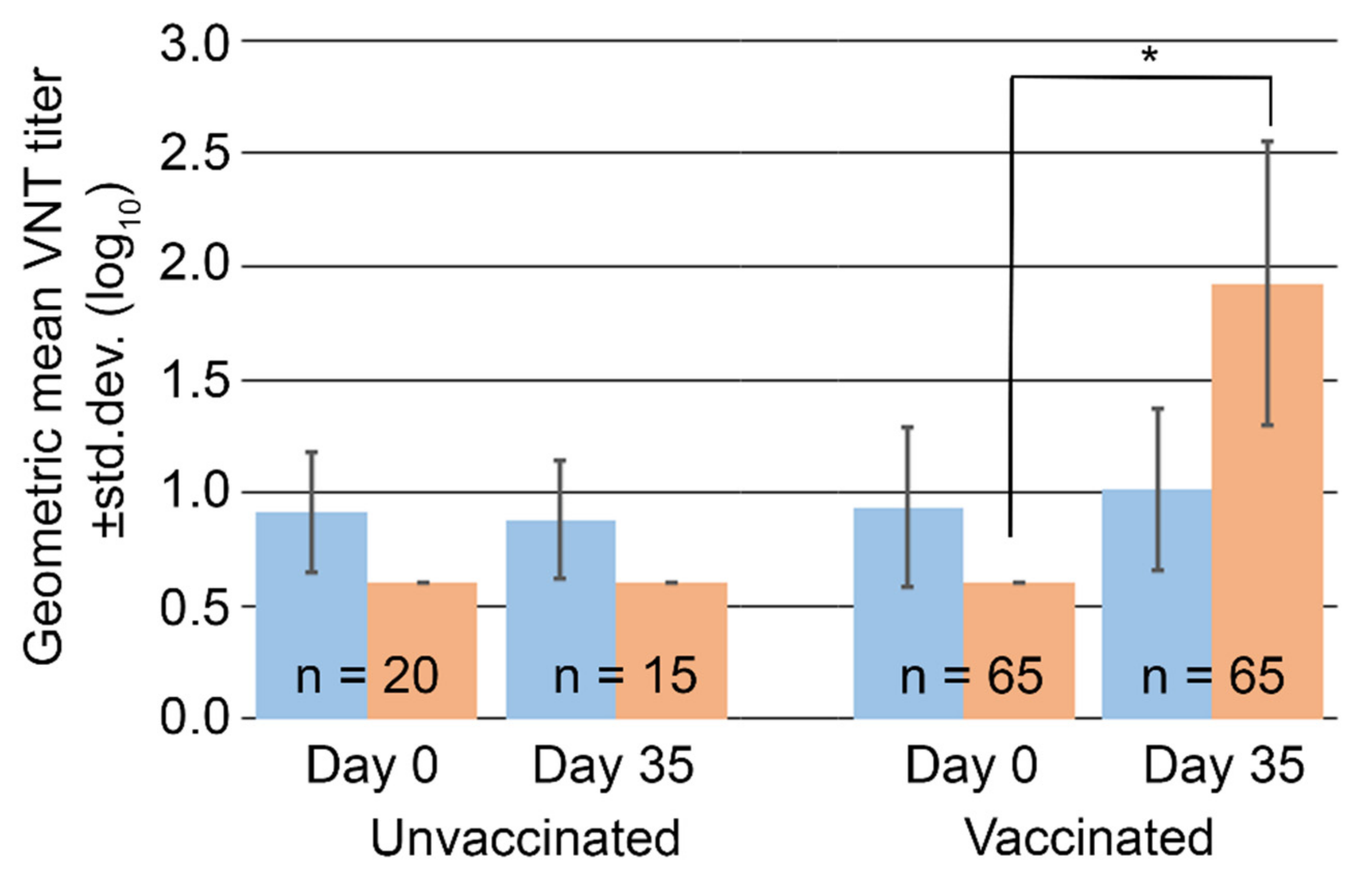
3.6. SVA Neutralizing Test Antibody Titers in Serum from Swine Used in FMDV Vaccination Studies

The presence of neutralizing antibodies can be indicative of past infection or vaccination. LF-BK αVβ6 cells can be utilized to determine virus neutralizing antibody titers against FMDV in vaccine studies to evaluate stimulation of an adaptive immune response prior to challenge. Swine serum samples from FMD vaccinated and unvaccinated pigs in previous FMD vaccination studies [29] were screened for antibody titers to SVA both before and after vaccination but prior to challenge. All pigs had been adapted to the facility at least seven days prior to vaccination on Day 0. Serum was screened on study days 0 and 35, Supplemental Table S1.
On Day 0, 62% of the population, (53/85), had SVA antibody titers above the 0.6 log10 limit of detection, with a mean of 0.93 ± 0.33 log10 and a high of 2.4 log10, Table 3. This is suggestive of previous exposure to SVA and somewhat expected since SVA is enzootic among swine populations in the U.S. This contrasts with the absence of FMDV antibody titers at Day 0 in all the pigs, Table 3. After 35 days on site, SVA antibody titers increased, remained unchanged or decreased in 42% (36/85), 36% (31/85), and 21% (18/85) of study animals, respectively. Despite these changes, the mean titers of the population, 0.99 ± 0.35 log10, remained within the standard deviation for both time points. During this time there was no presentation of clinical signs associated with SVA infection.
When animals were sorted by FMD vaccination status, a small increase in average SVA antibody titers among vaccinated animals was observed at Day 35, Figure 5. However, the difference was not statistically significant when compared to either unvaccinated animals at Day 35 (p = 0.54, Student’s paired t-test) or to the same animals at Day 0 prior to vaccination (p = 0.08, Student’s paired t-test). As expected, antibody titers against FMDV were significantly higher in vaccinated animals compared to unvaccinated controls (p < 0.001, Student’s paired t-test), Figure 5.
While SVA VNTs were determined in this study as an indicator of prior exposure it is unknown if they would indicate protection from virus challenge. In FMD, the presentation of neutralizing antibodies after vaccination is not necessarily indicative of an individuals clinical outcome in swine [29]. These results do validate the need to pre-screen swine for neutralizing antibody titers in SVA experimental studies, as pre-existing antibody levels may continue for a lengthy time after delivery to laboratory facilities.

4. Conclusions

In this report, we evaluated the ability of the porcine-derived LF-BK αVβ6 cell line to be used for isolation, culturing, and characterization of a SVA field isolate. We found that SVA grew quickly and to high titers in LF-BK αVβ6 cells. LF-BK αVβ6 cells were useful for SVA isolation and purification, measurement of SVA titers (in both PFU and TCID50), the evaluation of the sensitivity to porcine Type I interferon, and the quantification of SVA serum neutralizing antibody titers in swine. The presence of pre-existing antibody titers in swine utilized for FMD and other swine vesicular disease vaccine studies may complicate the interpretation of clinical observations and highlights a need for preliminary screening of pig sera prior to challenge studies.
Due to its high infectivity and devastating economic consequences, research on FMDV is limited to a small number of high-biocontainment facilities. In contrast, SVA can be handled in a BSL2 facility with fewer restrictions. Our work demonstrated that many methodologies used for work with FMDV are directly translatable to SVA. This translatability increases the likelihood that SVA may be useful as a surrogate virus to test and select countermeasures that may also be effective against FMDV.

Supplementary Materials

The following supporting information can be downloaded at: https://www.mdpi.com/article/10.3390/v14091875/s1; Table S1: SVA and FMDV geometric mean neutralizing antibody titers (log10) collected on days 0 and 35 for FMD vaccinated and unvaccinated swine.

Author Contributions

Conceptualization: J.B. and M.P.; Methodology: L.M., J.B. and M.P.; Validation: J.M. and J.S.; Investigation: V.P., J.M., L.M., J.B., B.C., W.H. and M.P.; Writing—Original Draft Preparation: M.P.; Writing—Review and Editing: M.P. and J.G.N.; Visualization: J.M., B.C. and M.P.; Supervision: M.P. and J.G.N. All authors have read and agreed to the published version of the manuscript.

Funding

This research was funded by the U.S. DHS Science and Technology Directorate (S&T), and SAIC was funded through an agreement under contract 70RSAT19D00000004.

Institutional Review Board Statement

Animal study protocols were approved by the Plum Island Animal Disease Center Institutional Review Board and the Institutional Animal Care and Use Committee of Plum Island Animal Disease Center (protocol code 240.01-19-D approved 18 June 2019).

Informed Consent Statement

Not applicable.

Data Availability Statement

Individual animal SVA and FMDV VNTs at Day 0 and Day 35 along with FMD vaccination status can be found in Table S1. Any additional data presented in this study is available upon request from the corresponding author.

Acknowledgments

The authors thank Barbara Kamicker and David Brake for their support with the manuscript and Vivian O’Donnell of USDA APHIS for her discussions and support on this project.

Conflicts of Interest

The authors have no conflict of interest to declare.

References

  1. Hales, L.M.; Knowles, N.J.; Reddy, P.S.; Xu, L.; Hay, C.; Hallenbeck, P.L. Complete genome sequence analysis of Seneca Valley virus-001, a novel oncolytic picornavirus. J. Gen. Virol. 2008, 89, 1265–1275. [Google Scholar] [CrossRef] [PubMed]
  2. Houston, E.; Gimenez-Lirola, L.G.; Magtoto, R.; Mora-Diaz, J.C.; Baum, D.; Pineyro, P.E. Seroprevalence of Senecavirus A in sows and grower-finisher pigs in major swine producing-states in the United States. Prev. Vet. Med. 2019, 165, 1–7. [Google Scholar] [CrossRef] [PubMed]
  3. Leme, R.A.; Zotti, E.; Alcântara, B.K.; Oliveira, M.V.; Freitas, L.A.; Alfieri, A.F. Senecavirus A: An Emerging Vesicular Infection in Brazilian Pig Herds. Transbound. Emerg. Dis. 2015, 62, 603–611. [Google Scholar] [CrossRef]
  4. Zhu, Z.; Yang, F.; Chen, P.; Liu, H.; Cao, W.; Zhang, K.; Liu, X.; Zheng, H. Emergence of novel Seneca Valley virus strains in China, 2017. Transbound. Emerg. Dis. 2017, 64, 1024–1029. [Google Scholar] [CrossRef]
  5. Baker, K.L.; Mowrer, C.; Canon, A.; Linhares, D.C.L.; Rademacher, C.; Karriker, L.A.; Holtkamp, D.J. Systematic Epidemiological Investigations of Cases of Senecavirus A in US Swine Breeding Herds. Transbound. Emerg. Dis. 2017, 64, 11–18. [Google Scholar] [CrossRef] [PubMed]
  6. Vannucci, F.A.; Linhares, D.C.; Barcellos, D.E.; Lam, H.C.; Collins, J.; Marthaler, D. Identification and Complete Genome of Seneca Valley Virus in Vesicular Fluid and Sera of Pigs Affected with Idiopathic Vesicular Disease, Brazil. Transbound. Emerg. Dis. 2015, 62, 589–593. [Google Scholar] [CrossRef] [PubMed]
  7. Wu, Q.; Zhao, X.; Bai, Y.; Sun, B.; Xie, Q.; Ma, J. The First Identification and Complete Genome of Senecavirus A Affecting Pig with Idiopathic Vesicular Disease in China. Transbound. Emerg. Dis. 2017, 64, 1633–1640. [Google Scholar] [CrossRef]
  8. Zhang, J.; Pineyro, P.; Chen, Q.; Zheng, Y.; Li, G.; Rademacher, C.; Derscheid, R.; Guo, B.; Yoon, K.-J.; Madson, D.; et al. Full-Length Genome Sequences of Senecavirus A from Recent Idiopathic Vesicular Disease Outbreaks in, U.S. Swine. Genome Announc. 2015, 3, e01270-15. [Google Scholar] [CrossRef] [PubMed]
  9. Joshi, L.; Mohr, K.A.; Clement, T.; Hain, K.S.; Myers, B.; Yaros, J.; Nelson, E.A.; Christopher-Hennings, J.; Gava, D.; Schaefer, R.; et al. Detection of the Emerging Picornavirus Senecavirus A in Pigs, Mice, and Houseflies. J. Clin. Microbiol. 2016, 54, 1536–1545. [Google Scholar] [CrossRef]
  10. Joshi, L.; Fernandes, M.H.V.; Clement, T.; Lawson, S.; Pillatzki, A.; Resende, T.P.; Vannucci, F.A.; Kutish, G.F.; Nelson, E.A.; Diel, D.G. Pathogenesis of Senecavirus A infection in finishing pigs. J. Gen. Virol. 2016, 97, 3267–3279. [Google Scholar] [CrossRef]
  11. Buckley, A.C.; Michael, D.D.; Faaberg, K.S.; Guo, B.; Yoon, K.J.; Lager, K.M. Comparison of historical and contemporary isolates of Senecavirus A. Vet. Microbiol. 2021, 253, 108946. [Google Scholar] [CrossRef] [PubMed]
  12. Pasma, T.; Davidson, S.; Shaw, S.L. Idiopathic vesicular disease in swine in Manitoba. Can. Vet. J. 2008, 49, 84–85. [Google Scholar] [PubMed]
  13. LaRocco, M.; Krug, P.W.; Kramer, E.; Ahmed, Z.; Pacheco, J.M.; Duque, H.; Baxt, B.; Rodriguez, L. A continuous bovine kidney cell line constitutively expressing bovine alphavbeta6 integrin has increased susceptibility to foot-and-mouth disease virus. J. Clin. Microbiol. 2013, 51, 1714–1720, Correction in J. Clin. Microbiol. 2015, 53, 755. [Google Scholar] [CrossRef] [PubMed]
  14. Jackson, T.; Sheppard, D.; Denyer, M.; Blakemore, W.; King, A.M. The epithelial integrin alphavbeta6 is a receptor for foot-and-mouth disease virus. J. Virol. 2000, 74, 4949–4956. [Google Scholar] [CrossRef] [PubMed]
  15. Fukai, K.; Morioka, K.; Yamada, M.; Nishi, T.; Yoshida, K.; Kitano, R.; Yamazoe, R.; Kanno, T. Comparative performance of fetal goat tongue cell line ZZ-R 127 and fetal porcine kidney cell line LFBK-alphavbeta6 for Foot-and-mouth disease virus isolation. J. Vet. Diagn. Investig. 2015, 27, 516–521. [Google Scholar] [CrossRef]
  16. Gray, A.R.; Wood, B.A.; Henry, E.; Azhar, M.; King, D.P.; Mioulet, V. Evaluation of Cell Lines for the Isolation of Foot-and-Mouth Disease Virus and Other Viruses Causing Vesicular Disease. Front. Vet. Sci. 2020, 7, 426. [Google Scholar] [CrossRef]
  17. Borca, M.V.; Rai, A.; Ramirez-Medina, E.; Silva, E.; Velazquez-Salinas, L.; Vuono, E.; Pruitt, S.; Espinoza, N.; Gladue, D.P. A Cell Culture-Adapted Vaccine Virus against the Current African Swine Fever Virus Pandemic Strain. J. Virol. 2021, 95, e0012321. [Google Scholar] [CrossRef]
  18. Kärber, G. Beitrag zur kollektiven Behandlung pharmakologischer Reihenversuche. Naunyn-Schmiedebergs Arch. Exp. Pathol. Pharmakol. 1931, 162, 480–483. [Google Scholar] [CrossRef]
  19. Spearman, C. The Method of ‘Right and Wrong Cases’ (‘Constant Stimuli’) without Gauss’s Formulae. Br. J. Psychol. 1908, 2, 227–242. [Google Scholar] [CrossRef]
  20. Bracht, A.J.; O’Hearn, E.S.; Fabian, A.W.; Barrette, R.W.; Sayed, A. Real-Time Reverse Transcription PCR Assay for Detection of Senecavirus A in Swine Vesicular Diagnostic Specimens. PLoS ONE 2016, 11, e0146211. [Google Scholar] [CrossRef] [PubMed]
  21. Puckette, M.; Barrera, J.; Schwarz, M.; Rasmussen, M. Method for quantification of porcine type I interferon activity using luminescence, by direct and indirect means. BMC Biotechnol. 2022, 22, 13. [Google Scholar] [CrossRef] [PubMed]
  22. Carrillo, C.; Tulman, E.R.; Delhon, G.; Lu, Z.; Carreno, A.; Vagnozzi, A.; Kutish, G.F.; Rock, D.L. Comparative genomics of foot-and-mouth disease virus. J. Virol. 2005, 79, 6487–6504. [Google Scholar] [CrossRef] [PubMed]
  23. Strauss, M.; Jayawardena, N.; Sun, E.; Easingwood, R.A.; Burga, L.N.; Bostina, M. Cryo-Electron Microscopy Structure of Seneca Valley Virus Procapsid. J. Virol. 2018, 92, e01927-17. [Google Scholar] [CrossRef] [PubMed]
  24. Reddy, P.S.; Burroughs, K.D.; Hales, L.M.; Ganesh, S.; Jones, B.; Idamakanti, N.; Hay, C.; Li, S.S.; Skele, K.L.; Vasko, A.-J.; et al. Seneca Valley virus, a systemically deliverable oncolytic picornavirus, and the treatment of neuroendocrine cancers. J. Natl. Cancer Inst. 2007, 99, 1623–1633. [Google Scholar] [CrossRef]
  25. Yang, F.; Zhu, Z.; Cao, W.; Liu, H.; Zhang, K.; Tian, H.; Liu, X.; Zheng, H. Immunogenicity and protective efficacy of an inactivated cell culture-derived Seneca Valley virus vaccine in pigs. Vaccine 2018, 36, 841–846. [Google Scholar] [CrossRef]
  26. Qian, S.; Fan, W.; Liu, T.; Wu, M.; Zhang, H.; Cui, X.; Zhou, Y.; Hu, J.; Wei, S.; Chen, H.; et al. Seneca Valley Virus Suppresses Host Type I Interferon Production by Targeting Adaptor Proteins MAVS, TRIF, and TANK for Cleavage. J. Virol. 2017, 91, e00823-17. [Google Scholar] [CrossRef]
  27. Chinsangaram, J.; Koster, M.; Grubman, M.J. Inhibition of L-deleted foot-and-mouth disease virus replication by alpha/beta interferon involves double-stranded RNA-dependent protein kinase. J. Virol. 2001, 75, 5498–5503. [Google Scholar] [CrossRef]
  28. Chinsangaram, J.; Moraes, M.P.; Koster, M.; Grubman, M.J. Novel viral disease control strategy: Adenovirus expressing alpha interferon rapidly protects swine from foot-and-mouth disease. J. Virol. 2003, 77, 1621–1625. [Google Scholar] [CrossRef]
  29. Puckette, M.; Primavera, V.; Martel, E.; Barrera, J.; Hurtle, W.; Clark, B.; Kamicker, B.; Zurita, M.; Brake, D.; Neilan, J. Transiently Transfected Mammalian Cell Cultures: An Adaptable and Effective Platform for Virus-like Particle-Based Vaccines against Foot-and-Mouth Disease Virus. Viruses 2022, 14, 989. [Google Scholar] [CrossRef]
Figure 1. Supernatant from an IB-RS-2 flask inoculated with a submitted field sample cultured in three porcine-derived cell lines, LF-BK αVβ6, PK-15, or IB-RS-2. (A) PEG precipitated supernatant was applied to discontinuous sucrose gradients ranging in density from 20% to 65% and demonstrated diffuse materials at all densities. (B) Western blotting of harvested gradient layers demonstrated strong SVA antigen in 20% and 35% fractions from LF-BK αVβ6 cultured samples and weak antigen in the 20% fraction for PK-15 and IB-RS-2 cultured samples. (C) The 20% and 35% fractions from the LF-BK αVβ6 gradient were examined by transmission electron microscopy using negative staining, and viral particles were observed. Both samples contained particles of the expected size for SVA. (D) The 20% fraction also contained structures resembling empty SVA procapsids.
Viruses 14 01875 g001
Figure 2. (A) Plaque-purified SVA produced 3 to 7 mm plaques on LF-BK αVβ6 cells after 48 h of incubation and (B) demonstrated strong reactivity to polyclonal anti-SVA VP1 antibody.
Viruses 14 01875 g002
Figure 3. (A) Cesium chloride gradient of PEG precipitated supernatant from SVA-LP8 inoculated LF-BK αVβ6 cells produced a discrete white band just above the 1.42 g/cm3 density. (B) The band harvested from the gradient demonstrated bands of the expected size for individual viral proteins and capsid subunits when run on a denaturing gel and stained. Polyclonal anti-SVA VP1 antibody was reactive with individual VP1 as well as VP1 containing capsid subunits.
Viruses 14 01875 g003
Figure 4. SVA titers, in log10 TCID50/mL, obtained from LF-BK αVβ6 (blue), PK-15 (green), and BHK-21 (orange), and IB-RS-2 (dark blue) cell lines following infection with ≈2.5 log10 TCID50/mL of SVA. Titer was determined by the presentation of CPE at 24, 48, and 72 h post inoculation on LF-BK αVβ6 cells and averaged together, the standard deviation is represented by error bars.
Viruses 14 01875 g004
Figure 5. Mean virus neutralizing antibody titers (log10) against SVA, blue, and FMDV, orange. No statistically significant differences in SVA titers between unvaccinated and FMD vaccinated swine are observed unlike neutralizing titers against FMD which increased in response to FMD vaccination. * represents p < 0.001 using a Student’s paired t-test.
Viruses 14 01875 g005
Table 1. Primers utilized for amplification and sequencing of isolated SVA.
Forward PrimerReverse Primer
Primer IDSequencePrimer IDSequence
AmplificationSVF1GAAGGACTGGGCATGAGGGSVR550AGTTCCCGCTGTAGCTCGC
SVF450GTGATTGCTACCACCATGAGTACASVR1650CGATAGTATGTACCAAGAGACTGC
SVF1550CGAAACCACCCTTGATGTCAAACSVR2750TTAGAGCCAGGAGCCGCCA
SVF2650GATTACACCCTCCGTCTCCCSVR3850TCCAGTCTTTGACTGTATCCATGG
SVF3050GTCACGGTGGTCTCACTGGASVR4250GGCGGAGCTCTGCTTGGC
SVF4100ACGACCAGATTGAATACCTCCAGASVR5300TCACCACGGATTGTGAAGCT
SVF5200GAGCGAGAATGCTTATGACGGSVR6400CGATAGCGGAGCCAAGGAGAA
SVF6300GTCTGACCCTGATGTCTTCTGGSVR7300TTCTGTTCCGACTGAGTTCTCCCA
SequencingSVF1GAAGGACTGGGCATGAGGGSVR-SEQ1ATCCAAGGCACGCTAAGGC
SVF-SEQ1CCACCATGAGTACATGGTTCTCCSVR-SEQ2TCCGGTAGTCGTCAGACATTTCC
SVF-SEQ2CAGTCTCTTGGTACATACTATCGGCSVR-SEQ3AGTCTCGGCGTTGTCGGTG
SVF-SEQ3ATTGAGGCAGGTAACACTGACACSVR-SEQ4CTGGTGGAGGAGGCGGTTCTA
SVF-SEQ4GGCAGTGAGTACCAGGCTTCTSVR-SEQ5CTCTGAGGACCACCACAACGG
SVF-SEQ5CCAGATTGAATACCTCCAGAACCTCSVR5300TCACCACGGATTGTGAAGCT
SVF-SEQ6GCGAGAATGCTTATGACGGSVR-SEQ6CTATGACGGTCCAGAAGACATC
SVF-SEQ7GCCGCCAAGTTTCAATCCSVR7300TTCTGTTCCGACTGAGTTCTCCCA
Table 2. Sensitivity of FMDV and SVA to type I interferons on LF-BK αVβ6 cells. Interferon activity is presented as units of Interferon Antiviral Activity capable of inhibit 50% of viral growth (IFNAA50) per 1 mL after 24 and 48 h of incubation as calculated by the Spearman-Kärber Method.
CytokineVirusTiter (log10) TCID50/mLTime (h)IFNAA50 (log10) Units/mL
IFNαFMDV3.3245.14
484.88
SVA2.9245.63
484.84
IFNβFMDV3.3245.48
485.4
SVA2.9245.96
485.21
Table 3. SVA and FMDV neutralizing antibody titers in swine serum.
Neutralizing Antibody Titers (log10)SVAFMDV
Day 0Day 35Day 0Day 35
0.632258517
0.9211904
1.2292807
1.50408
1.823011
2.101010
2.410012
2.70006
3.00004
Publisher’s Note: MDPI stays neutral with regard to jurisdictional claims in published maps and institutional affiliations.

