Review

The Role of Latency-Associated Transcripts in the Latent Infection of Pseudorabies Virus

Key Laboratory of Zoonosis Prevention and Control of Guangdong Province, College of Veterinary Medicine, South China Agricultural University, Guangzhou 510642, China
*
Author to whom correspondence should be addressed.
These authors contributed equally to this work.
Viruses 2022, 14(7), 1379; https://doi.org/10.3390/v14071379
Submission received: 30 May 2022 / Revised: 22 June 2022 / Accepted: 22 June 2022 / Published: 24 June 2022
(This article belongs to the Special Issue Pseudorabies Virus)

Abstract

:
Pseudorabies virus (PRV) can cause neurological, respiratory, and reproductive diseases in pigs and establish lifelong latent infection in the peripheral nervous system (PNS). Latent infection is a typical feature of PRV, which brings great difficulties to the prevention, control, and eradication of pseudorabies. The integral mechanism of latent infection is still unclear. Latency-associated transcripts (LAT) gene is the only transcriptional region during latent infection of PRV which plays the key role in regulating viral latent infection and inhibiting apoptosis. Here, we review the characteristics of PRV latent infection and the transcriptional characteristics of the LAT gene. We also analyzed the function of non-coding RNA (ncRNA) produced by the LAT gene and its importance in latent infection. Furthermore, we provided possible strategies to solve the problem of latent infection of virulent PRV strains in the host. In short, the detailed mechanism of PRV latent infection needs to be further studied and elucidated.

1. Introduction

Pseudorabies virus (PRV) belongs to the family Herpesviridae, subfamily Alphaherpesvirinae, and the Varicellovirus genus [1]. The PRV genome is 142 kb of linear double-stranded DNA with 70 different coding genes and one latency-associated transcript (LAT) site. It consists of a unique long region (UL), a unique short region (US), internal repetitive sequences (IRS), and terminal repetitive sequences (TRS) [2]. The natural host of PRV is pigs, but it can infect most mammals, including cattle, sheep, cats, dogs, mink, and rodents [3,4,5,6,7,8,9]. There are significant differences in PRV infection between natural and non-natural hosts [10,11]. In natural hosts, PRV can cause neurological, respiratory, and reproductive diseases and establish latent infection in the peripheral nervous system (PNS) of surviving pigs, but death in adult pigs is uncommon [11,12,13,14]. In non-natural hosts, PRV infection is characterized by severe pruritus, a short duration of disease, and rapid death [10,11]. The mortality rate of non-natural hosts is up to 100%, and consequently latent infection rarely occurs [10,11,15]. However, under laboratory conditions, PRV can establish activatable latent infection in non-natural hosts, which is of great significance in the study of herpesvirus latent infection [16,17,18,19].
PRV has been eradicated or controlled through the use of gene-deficient vaccines and differentiating infected from vaccinated animals (DIVA) strategy in many countries. However, since 2011, the emergence of mutant strains of PRV has made pseudorabies come back in China, one of the world’s largest pig breeding countries [20,21,22,23,24]. Tong et al. found that PRV mutant strain JS-2012 caused earlier clinical symptoms and higher mortality compared to PRV classic strain SC in 15, 30, and 60-day-old pigs [25]. In protection assays, the Bartha-K61 vaccine provided 100% protection against classic strain, but only partial protection against JS-2012 strain or HeN1 strain [25,26,27].The mutant strains have been prevalent in pig farms immunized with PRV vaccine, which shows that the existing PRV vaccine cannot prevent infection caused by the new PRV mutant strains [20,22,25,26,27]. In recent years, human infections of PRV have been increasingly reported. From 2018 to 2022, more than 20 cases of human PRV infection have been found [28,29,30,31,32,33,34,35,36,37,38,39]. Liu et al. isolated and identified a human PRV strain hSD-1/2019 which had high pathogenicity to mice and pigs [28]. Most of the patients infected with PRV are workers related to the pig industry, and they are directly or indirectly infected with PRV through conjunctiva, skin wounds, and syringe stab wounds. PRV is not only costly to the pig industry but also a serious threat to humans. Therefore, the eradication of PRV should be accelerated all over the world.
Latent infection is the major impediment to eradication of PRV. PRV can establish latent infection in the PNS of pigs. During the latent infection of PRV, no clinical symptoms and infectious virions exist in pigs. When stimulated by stressors, the latent virus can be reactivated, and then productive infection occurs. According to the investigation of pig farms, PRV was in a state of latent infection most of the time and latent virions were prone to reactivation in winter and spring [40]. Interestingly, although reactivated virions were detected in pigs, no clinical symptoms were observed. These virions were excreted into the environment, resulting in the spread of PRV and the infection of other susceptible animals. Therefore, controlling latent infection plays an important role in the eradication of PRV.
The integral mechanism of latent infection is still unclear. The LAT gene is the only active gene during the latent infection. It can transcribe a variety of non-coding RNAs (ncRNAs) which are involved in the establishment, maintenance, and reactivation of viral latent infection, as well as the inhibition of productive infection and anti-apoptosis [41]. Here, we mainly summarized the characteristics of PRV latent infection, the transcription and function of the LAT gene, so as to provide new perspectives for future research on PRV latent infection.

2. The Characteristics of PRV Latent Infection

During the natural infection of pigs, PRV replicates in the epithelial cells of the nasal mucosa and invades sensory nerve endings by membrane fusion. The virus particles entering the axon terminals are retrogradely transported to the neuron nucleus [42,43]. Then, the capsid docks near the nuclear foramen, PRV genome is released, and the tegument protein VP16 activates the immediate early gene IE180 of PRV to form a productive infection [1]. During latent infection, the expression of immediate early genes (IE gene) is affected by many factors, such as VP16, Oct-1 (a member of the Oct protein family), HCF (a cellular protein), and LAT [44]. In sensory neurons, Oct proteins (except Oct-1) can prevent the formation of VP16/Oct-1/HCF complexes, thus inhibiting the transcription of IE genes [44]. Moreover, in the nucleus, the viral genome binds to the nucleosome and further inhibits the expression of IE180 during latent infection [41]. IE180 is a potent transcriptional activator which is required for efficient transcription of early (E) gene and late (L) gene of PRV, so it is essential for viral replication [1]. The expression product of IE180 can bind to the promoter of LAT and inhibit the transcription of LAT genes [45]. Therefore, when the IE180 gene is silenced, IE180-mediated transcription of E gene and L gene is restricted, while LAT gene transcriptional activity is enhanced. Thus, LAT gene is the only transcriptional region during latent infection [46].
The PRV genome mainly exists in the nerve tissue, especially in the trigeminal ganglion, which is the most reliable tissue for detecting latent PRV during latent infection [47]. The olfactory bulb and medulla oblongata can also contain the latent genome. The PRV latent genome is mainly confined to the nucleus in the form of linear and unintegrated, and a small number of latent genomes exist in the form of ring [47]. The positive cells of latent infection are distributed in different regions of the neural tissue in the form of aggregation [47]. In latently infected neurons, the number of PRV genome is stable and is not related to the length of the latency period [47]. Latent infections are generally stable but can be reactivated under stress, such as restraint, exposure to cold, or transport [47,48].
Latent infection requires the co-regulation of viruses, neurons, and the host immune system [49,50]. When the three are balanced, the virus can establish a latent infection in the host. The conditions for the establishment of latent infection are as follows: firstly, the viral genome enters the nucleus of neurons, and the vast majority of genes are restricted for transcription and translation. Secondly, in order to avoid latent genome loss, the virus takes certain measures to promote the survival of infected cells and evade host immunity [51]. For example, LAT has an anti-apoptotic effect and can prolong the survival time of neurons [41]. Finally, latent viruses can monitor and manipulate the environment of host cells for reactivation.

3. Transcriptional Characteristics of LAT Gene

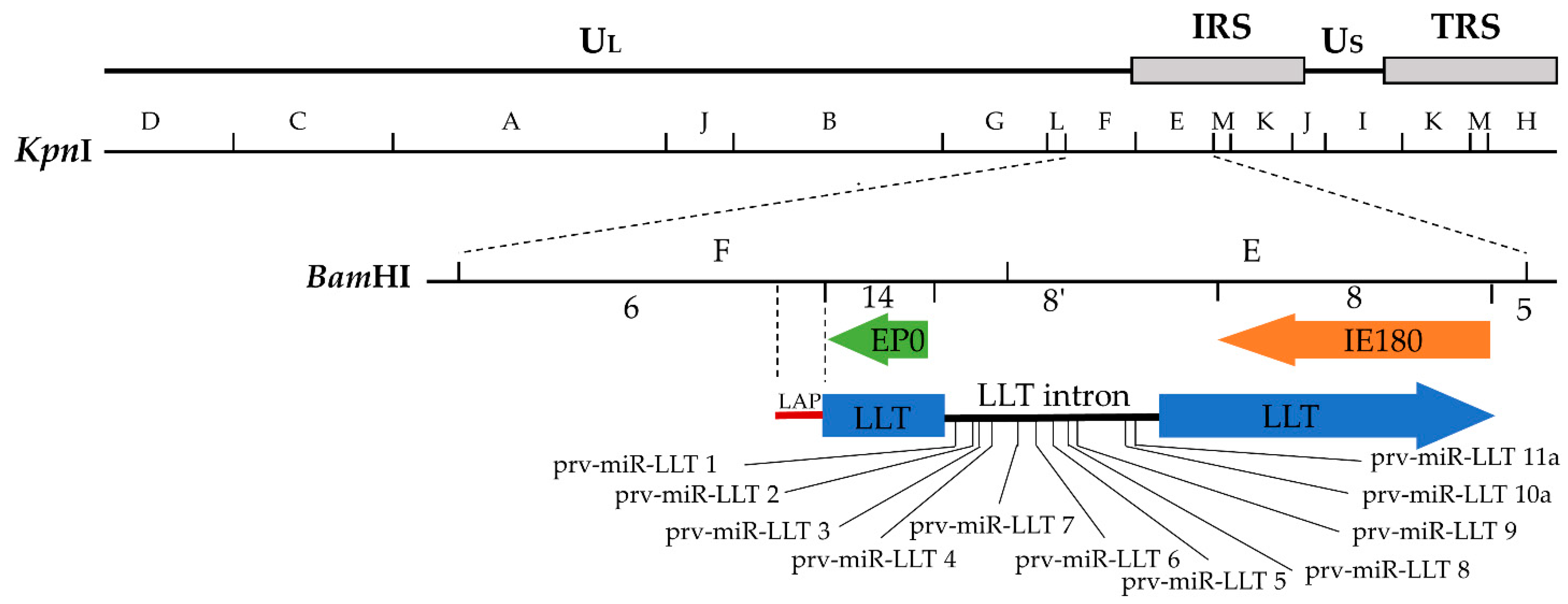
3.1. Transcriptional Region and Sizes of the LAT Gene

The study of latent viral gene expression is restricted because less than 1% of ganglion neurons contain latent viral genomes [47]. Among the latently infected neurons, Rock et al. detected DNA and mRNA of the virus by in situ hybridization, which proved that the PRV genome has transcriptional activity during the latent infection [46]. Researchers soon discovered that mRNAs produced during latent infection were transcribed from a region between 0.69 and 0.77 map units of the PRV genome [52]. This region is about 11 kb, which covers the early protein 0 (EP0) gene and IE180 gene, and the transcriptional direction is opposite to that of the IE180 and EP0 genes [53,54,55]. These mRNAs are collectively referred to as LATs, including various sizes of mRNA, in which 0.95 kb, 1.0 kb, 2.0 kb, 8.0 kb, and 8.4 kb mRNA are generally detected [52,56,57]. The mRNA of 8.4 kb (some refer to 8.5 kb) is called large latency transcript (LLT) [52,55,57,58]. Through in situ hybridization analysis of LATs, it was found that LATs were mainly confined to the nucleus of neurons and a small part of them existed in the cytoplasm [44,46].

3.2. The Structure and Function of LAT Promoter

The structure of the LAT promoter (LAP) overlaps with the promoter of UL1-3.5 gene cluster in the opposite direction [57]. LAP contains 2 TATA boxes, 3 CAAT boxes, and 2 GC boxes, which is a dual regulatory promoter. The first latent activation promoter (LAP1) contains the first TATA box and three CAAT boxes, and the second latent activation promoter (LAP2) contains the second TATA box and two GC boxes. LAP1 is the basic promoter of LAT gene expression during PRV latent infection. It initiates transcription of LAT in nerve tissue and produces LLT with 4.6 kb intron [58]. LLT starts at 34 nucleotides downstream of the first TATA box in LAP1 and can be spliced into different sizes of RNAs. The whole nucleotide sequence of 2.0 kb mRNA is contained in the LLT, and it lacks the intron of 4.6 kb. However, the LAT of 2.0 kb is regulated by LAP2, which starts at about 243 bp downstream of the LLT transcription initiation site and ends at the junction of BamHI fragments 8′ and 8 [52]. Whether in latent or lytic infection, nerve or non-nerve cells, in vivo or in vitro, LAP2 has no specific activity, and it is responsible for regulating the transcription of 1.0 kb, 2.0 kb, and 8.0 kb LATs [52,58]. When LAP2 and LAP1 coexist, LAP2 can enhance the activity of LAP1 [57].
LAP1 mediates the transcription of LLT [58]. The nerve cells were infected by the recombinant PRV strain with a deletion of the LAP1 region in vitro. The mRNA of 2.0 kb and 8.0 kb could be obtained from the infected nerve cells, but the LLT of 8.4 kb could not be detected. The recombinant strain could establish latent infection in the pigs, but the LLT of 8.4 kb could not be detected in trigeminal nerve. Therefore, LAP1 is the key promoter of LLT, and LLT is not required for the establishment of PRV latent infection [58]. The role of LLT needs to be further understood in PRV latent infection.
LAP is neuron-specific in vivo [59,60]. Taharaguchi et al. established a transgenic mouse line containing LAP linked with the chloramphenicol acetyltransferase (CAT) gene [60]. The expression level of the CAT gene in different tissues of transgenic mice was evaluated by enzyme-linked immunosorbent assay (ELISA). It was found that CAT was almost exclusively expressed in nerve tissue, and the expression level was the highest in the trigeminal nerve [60]. The expression of CAT in trigeminal ganglion neurons was further verified by in situ hybridization. In the absence of viral proteins, LAP is not only active but also neuron-specific, indicating that LAP may be regulated by neuronal transcription factors, and is independent of viral proteins. However, in the studies by Ou and Taharaguchi et al., it has been shown that the inhibition of LAP by IE180 is caused by the formation of a stable complex of IE180, cellular protein(s), and the IE180 binding site located on LAP, suggesting that LAP can be regulated by viral and host protein(s) [45,60,61]. In order to further understand the molecular regulatory mechanism of LAP, the host protein that regulates the neuron-specificity of LAP needs to be discovered. In the study of Taharaguchi et al., there were significant differences in the expression level of the CAT gene in different neural tissues, suggesting that LAP activity varies in different neuronal environments, which may be related to the differences of neuronal transcription factors and neuronal morphology in various nerve tissues [60,62]. It has been proved that dexamethasone can activate latent infection of PRV [48,63]. When transgenic mice were treated with dexamethasone, it was found that dexamethasone did not affect LAP-mediated CAT transcription and translation. We speculate that dexamethasone induced viral reactivation may be irrelevant to LAP [60]. Therefore, it is important to analyze the interaction between LAP, viral protein, and host protein for the study of PRV latent infection.

4. The Role of LAT Gene in Latent Infection

Non-coding RNA (ncRNA) molecules are small and have various regulatory functions in virus replication, virus persistence, immune escape, and cellular transformation [64]. Compared with proteins, the regulation of latent infection by ncRNAs is more desirable: first, the LAT gene is the only gene with transcriptional activity during latent infection, with can transcribe a variety of ncRNAs, but does not produce bioactive proteins [65,66,67,68,69,70]; second, ncRNAs lack antigenicity and are more likely to evade host cellular immunity [64]; third, the rich functions of ncRNAs are suitable for the regulation of latent infection [64]; finally, compared with proteins, the regulatory function of ncRNAs is mild, and the regulatory mode is ideal in the setting of latent infection [71]. Therefore, the ncRNA is of great significance for the research of PRV latent infection. The LAT gene can transcribe several different types of ncRNAs, such as microRNA (miRNA), small RNA (sRNA), long non-coding RNA (lncRNA), and short non-coding RNA (sncRNA), etc. [65,66,67,68,69,70]. At present, studies on PRV latent infection are mainly focused on miRNA, and few reports on other ncRNAs. PRV LAT can transcribe many kinds of ncRNAs just like that of Herpes simplex virus type 1 (HSV-1). PRV EP0 and IE180 are homologues of HSV-1 ICP0 (infected cell polypeptide 0) and ICP4 (infected cell polypeptide 4), respectively [72]. The ncRNAs produced by HSV-1 LAT, such as miR-H2 and miR-H6, can inhibit the expression of ICP0 and ICP4 [73,74]. The ncRNAs transcribed by PRV LAT can target EP0 and IE180 mRNAs. Based on the structural similarity of homologous proteins, we speculate that some ncRNAs transcribed by PRV may have functions analogous to those of HSV. HSV’s research on LAT ncRNA is more abundant than PRV’s. Therefore, we will analyze the function of PRV LAT ncRNAs combined with HSV to comprehensively elucidate their role in latent infection.

4.1. MicroRNAs Transcribed by the LAT Gene and Host Cell

MicroRNA (miRNA) is about 20–24 nucleotides (nt) in size and has a variety of important regulatory functions [75]. It can regulate target mRNA by altering its stability or inhibiting its transcription. With the development of sequencing technology, the miRNAs of most herpesviruses have been identified, including PRV. However, the specific function of PRV miRNAs in the process of PRV infection is still unclear.
Alphaherpesvirinae has been shown to transcribe a variety of miRNAs which are usually clustered in the viral genome. The viral miRNAs are limited to LAT sites or adjacent regions and can be transcribed by each strand of the genome [72]. Anselmo et al. identified five viral miRNAs (prv-miR-LLT 1 to prv-miR-LLT 5) in PRV-infected porcine dendritic cells (DCs) by deep sequencing [76]. These miRNAs are all transcribed by the intron of LLT [76]. The sizes of prv-miR-LLT 1, prv-miR-LLT 2, prv-miR-LLT 3, and prv-miR-LLT 5 are between 21 and 23nt. Prv-miR-LLT 4 is a mature miRNA with a size of 18nt. Using gene target analysis of miRNAs (prv-miR-1,2,3,4,5), it was found that the possible targets of Prv-miR-LLT 1-5 located in LLT, EP0, and IE180. Based on Anselmo’s study, Wu et al. identified 11 viral miRNAs (prv-miR-LLT 1 to prv-miR-LLT 11) in porcine epithelial cell line (PK-15) infected with PRV by the same method [77] (Figure 1). Gene target analysis showed that prv-miR-LLT 1 and prv-miR-LLT 9 could target IE180 and LLT, and prv-miR-LLT 2 could target EP0 and LLT. It was also found that 11 viral miRNAs could target 235 host genes. GO enrichment analysis showed that these 235 host genes are involved in apoptosis, host immune response, cell metabolism, and virus replication [77]. These results suggest that viral miRNAs can play an important role in regulating the interaction between virus and host.
Although these viral miRNAs were detected during productive infection of PRV, they were produced by LAT and could regulate the latent infection of the virus. As a member of the Alphaherpesvirinae subfamily, the viral miRNA of PRV may have similar functions to that of HSV. MiRNAs of HSV can regulate viral latent infection by down-regulating the expression of IE genes or E genes [73,78]. In HSV-1 infected cells, LAT, as the primary miRNA precursor, can transcribe various miRNAs. Among them, HSV-1 miR-H2 can inhibit the expression of ICP0 through targeting its mRNA [73]. ICP0 is an effective activator of virus reactivation, so miR-H2 inhibits the expression of ICP0, thereby hindering virus reactivation [73,79]. When interfering with the transcription of HSV-1 miR-H2, reverse consequences will occur, including the increase of ICP0 expression, viral reactivation, and the neurovirulence of HSV [74]. Therefore, miR-H2 can regulate the conversion between latency and reactivation of the virus. In addition, HSV-1 miR-H6 can inhibit the expression of ICP4. The LAT gene of herpes simplex virus type 2 (HSV-2) can also produce many different miRNAs, among which miR-I and miR-II can reduce the expression of the neurovirulence factor ICP34.5, and miR-III can block the expression of the ICP0 gene [78,80]. MiRNAs not only regulate the latent infection, but also the productive infection. Timoneda et al. detected 8 miRNAs transcribed by LLT intron in PRV infected pigs. The expression of these viral miRNAs was obviously changed at different times of acute infection and significantly increased only in the early stage of virus infection. Thus, the authors speculated that the 8 miRNAs could be involved in the establishment of productive infection of the virus [65]. Another interesting phenomenon was also observed. The 20 viral miRNAs detected from PK15 cell line by using Illumina deep sequencing were derived from the open reading frame (ORF), IRS, and TRS regions of the PRV genome [81].
Host miRNAs, like viral miRNAs, can participate in the regulation of virus infection. Many studies have shown that PRV infection can affect the expression of miRNAs in host cells [76,77,81,82]. Members of the miR-146 family can regulate host inflammatory and immune responses [83,84]. In PRV infected mouse neuroblastoma cells, miR-146b-5p was significantly up-regulated after PRV infection, which could promote PRV replication and negatively regulate type I interferon response [82]. Furthermore, other host miRNAs can also target multiple viral genes such as miR-1249-3p, miR-6538, miR-466k, and miR-714 to regulate the PRV infection [82].

4.2. Other Non-Coding RNAs Transcribed by LAT Gene

Long non-coding RNA (lncRNA) has the function of regulating gene expression [67]. In latently infected ganglia, the LAT gene of HSV can express different sizes of lncRNAs. They can accumulate in the nucleus of latently infected neurons to induce the formation of facultative heterochromatin, promote lytic gene silencing, and evade the host immune response [66,70]. PRV can up-regulate the expression of host lncRNAs in infected cells, thus promoting the replication of itself [85].
Peng et al. identified two ncRNAs in the first 1.5 kb LAT region of HSV which were different from the typical miRNA structure [86]. They possessed the feature of sRNA, so the two ncRNAs were called LAT sRNA1 and sRNA2.The first 1.5 kb LAT region of HSV plays an important role in suppressing productive infection, resisting apoptosis, maintaining latent infection, and ensuring a high rate of viral reactivation [87,88]. According to the complementary pairing of the two sRNAs with ICP4 mRNA and the position of their corresponding DNA sequence, it is speculated that sRNA1 and sRNA2 could inhibit the translation of ICP4 mRNA and apoptosis. Shen et al. confirmed this speculation [69]. ICP4 is essential for productive infection and viral reactivation. The LAT sRNA2 can inhibit the translation of ICP4 mRNA, so the functions of the latter can be affected in regulating the virus infection. LAT sRNA1 has a stronger ability to suppress productive infection than sRNA2, but it has no significant effect on the expression of ICP0 or ICP4 protein. It is predicted that LAT sRNA1 may target mRNA of VP16 and UL8 to inhibit the production of regulatory proteins necessary for viral replication. Single point mutations in LAT sRNA1 and sRNA2 can reduce the ability of LAT to inhibit apoptosis [69]. Therefore, sRNA1 and sRNA2 take an important role in the anti-apoptotic effect of LAT.
In some studies, LAT sRNA1 and sRNA2 are named LAT sncRNA1 and LAT sncRNA2, respectively. LAT sncRNA1 and sncRNA2 can regulate the signal pathway of retinoic acid-induced gene I (RIG-I) to improve cell survival [68]. Herpesvirus entry medium (HVEM) is a cell surface protein that mediates the attachment and entry of HSV into cells. HVEM can regulate the cellular immune response and inhibit apoptosis. In latently infected neurons, LAT can up-regulate the expression of HVEM through the interaction of LAT sncRNA1 and sncRNA2 with the HVEM promoters, thereby promoting cell survival and helping the virus escape host immunity [89].

4.3. LAT Encoding Protein in Latent Infection

Open reading frames (ORFs) are DNA sequences that are capable of encoding proteins. Eight potential ORFs exist in the first 1.5 kb LAT of HSV-1. Introducing point mutations into the ATG of ORFs can reduce the activity of LAT in inhibiting apoptosis [90]. Although no bioactive LAT encoding protein was detected in latently infected neurons, its biological function was confirmed in vitro [91]. HSV-1 LAT encoding protein can restore the replication level of the ICP0 gene-deficient strain in vitro and improve the growth level of the virus in vivo. Therefore, it was implied that the LAT encoding protein might have functions similar to ICP0 in the reactivation of virus latent infection [91,92].

5. Effect of Pseudorabies Vaccine on Virus Latent Infection

The PRV vaccine could not prevent the establishment of latent infection of wild-type strains. As early as 1981, it was reported that latent infection of PRV still existed in pig farms vaccinated with live attenuated PRV vaccine [93]. Since then, a large number of studies have confirmed that the clinical symptoms and mortality of infected pigs can be reduced by active immunization of the inactivated vaccine, live attenuated vaccine, and subunit vaccine or passive immunization of maternal antibody, but latent infection of the virulent PRV strain cannot be prevented in the PNS of the host [18,26,94,95,96,97,98]. Inoculation with the PRV vaccine cannot prevent the virulent strain from establishing latent infection in host pigs, but it can reduce the amount and time of virus excretion after the virulent strain reactivated [99].
Live attenuated PRV vaccines can also establish latent infections in PNS of pigs like virulent strains [100]. The latent infection of the PRV gene deletion vaccine can affect that of wild-type strains, and there is a significant negative correlation between them [101]. By increasing the level of latent infection of the gene deletion vaccine, we can reduce or eliminate that of wild-type PRV in the host. However, one problem that needs to be considered is genetic recombination. Virus recombination is affected by the dose of the inoculated virus, the time interval between the two viruses, the distance between marker mutations, genetic homology, virulence, and latency [102,103,104]. Homologous recombination often occurs among the same Alphaherpesvirinae [104]. The PRV gene deletion vaccine may recombine with the wild-type virus strain to produce a highly virulent variant strain, or a variant strain that cannot be differentiated by serological method [105]. To avoid this, the biosafety of live attenuated vaccines must be carefully evaluated. The LAT gene is a nonessential factor for the establishment of viral latent infection, but it plays a pivotal role in the reactivation of the virus [58]. Mahjoub et al. obtained a PRV mutant with 9 LAT miRNAs deletions. This mutant could establish latent infection, but it could not be reactivated [106]. In HSV, the deletion of LAT fragments can affect the reactivation rate, the virulence, and the ability of anti-apoptosis [74,107,108]. The specific molecular mechanism of LAT still needs to be explored. LAT plays a key role in regulating the conversion between virus latency and reactivation, inhibiting apoptosis to prolong the survival time of neurons. Perhaps we could take some measures to modify the LAT gene to develop a non-reactivated PRV vaccine.

6. Conclusions and Perspectives

In this review, we summarize the characteristics of PRV latent infection, the transcriptional characteristics and functions of LAT gene, and the effects of PRV vaccine on the establishment of latent infection of virulent PRV strains. Studying LAT ncRNA can provide a new perspective for elucidating the molecular mechanism of PRV latent infection. We can also take some effective measures to control the latent infection of wild-type PRV in pigs, such as developing effective vaccines or drugs to inhibit the establishment or reactivation of latent infection of wild type PRV. During latent infection of PRV, no infectious virions are produced, but the genome of the virus can be detected in the host. However, latent infection of the virus can be detected by serology, in situ hybridization, tissue co-culture, polymerase chain reaction (PCR), fluorescence quantitative PCR, and real-time recombinant enzyme-assisted amplification [46,109,110,111,112,113]. Similar to the PRV virulent strain, the PRV gene deletion vaccine can also establish latent infection in the PNS of pigs. When pigs are inoculated with two vaccines of different gene deletions, genetic recombination may occur between them, and then new strains may be produced. Therefore, only one gene deletion vaccine is recommended in the same pig farm or the same animal individual to avoid genetic recombination between vaccine strains [102,103].
The LAT gene is closely related to latent infection. The latent infection of the PRV gene deletion vaccine was negatively correlated with that of wild-type PRV strains, and the LAT gene could affect the reactivation rate of the virus. Therefore, we can modify the LAT gene to develop a genetic engineering vaccine to restrict the latent infection of PRV wild-type strain in pigs. The ncRNAs transcribed by the LAT gene play an important role in the process of viral latent infection. Therefore, RNA interference and RNA silencing are applied to these ncRNAs to regulate the latency and reactivation of PRV. At present, there is little research on the ncRNA of PRV LAT. Therefore, in order to further understand the molecular mechanism of PRV latent infection, we can emphasize the study of ncRNAs produced by PRV LAT.
PRV can be transmitted from pigs to humans, threatening human health [28,29,30,31,32,33,34,35,36,37,38,39]. PRV infection can cause fever, headache, endophthalmitis, and acute encephalitis [28,29,30,31,32,33,34,35,36,37,38,39]. Antiherpes drugs such as valaciclovir, penciclovir, and phosphonoformate can control the symptoms of patients, but the visual and nerve damage caused by PRV is irreversible. PRV can establish reactivatable latent infection in mice [16,114,115]. Humans are non-natural hosts like mice, so it is possible for PRV to establish reactivatable latent infection in the body, resulting in irregular recurrence of the disease. Therefore, the subsequent progress of PRV-infected patients should be tracked in the long-term to evaluate the probability. If the molecular mechanism of latent infection is clear, some specific targets may be found, and then relevant drugs can be developed to block the latency and reactivation of PRV.

Author Contributions

Conceptualization of the manuscript, J.D., C.J., Z.W., J.L. and Q.J.; writing—original draft preparation, J.D. and C.J.; writing—review and editing, J.D., Z.W., J.L., Q.J. and C.J. All authors have read and agreed to the published version of the manuscript.

Funding

This research was supported by the grants for Da Hua Nong Award Fund for training young teachers of Veterinary College (No. 5500-A17003) and Natural Science Foundation of Guangdong Province (No. 2018A030313163).

Institutional Review Board Statement

Not applicable.

Informed Consent Statement

Not applicable.

Data Availability Statement

Not applicable.

Conflicts of Interest

The authors declare no conflict of interest.

References

  1. Hengartner, L.E.; Reynolds, A.E.; Hengartner, C.J. Molecular Biology of Pseudorabies Virus: Impact on Neurovirology and Veterinary Medicine. Microbiol. Mol. Biol. 2005, 69, 462–500. [Google Scholar] [CrossRef] [Green Version]
  2. Klupp, B.G.; Hengartner, C.J.; Mettenleiter, T.C.; Enquist, L.W. Complete, Annotated Sequence of the Pseudorabies Virus Genome. J. Virol. 2004, 78, 17. [Google Scholar] [CrossRef] [Green Version]
  3. Hugoson, G.; Rockborn, G. On the Occurrence of Pseudorabies in Sweden II. An Outbreak in Dogs Caused by Feeding Abattoir Offal. Zentralbl Vet. B 2010, 19, 641–645. [Google Scholar] [CrossRef] [PubMed]
  4. Capua, I.; Fico, R.; Banks, M.; Tamba, M.; Calzetta, G. Isolation and Characterisation of an Aujeszky’s Disease Virus Naturally Infecting a Wild Boar (Sus Scrofa). Vet. Microbiol. 1997, 55, 141–146. [Google Scholar] [CrossRef]
  5. Cheng, Z.; Kong, Z.; Liu, P.; Fu, Z.; Zhang, J.; Liu, M.; Shang, Y. Natural Infection of a Variant Pseudorabies Virus Leads to Bovine Death in China. Transbound. Emerg. Dis. 2020, 67, 518–522. [Google Scholar] [CrossRef]
  6. Kaneko, C.; Kaneko, Y.; Sudaryatma, P.E.; Mekata, H.; Kirino, Y.; Yamaguchi, R.; Okabayashi, T. Pseudorabies Virus Infection in Hunting Dogs in Oita, Japan: Report from a Prefecture Free from Aujeszky’s Disease in Domestic Pigs. J. Vet. Med. Sci. 2021, 83, 680–684. [Google Scholar] [CrossRef]
  7. Marcaccini, A.; López Peña, M.; Quiroga, M.I.; Bermúdez, R.; Nieto, J.M.; Alemañ, N. Pseudorabies Virus Infection in Mink: A Host-Specific Pathogenesis. Vet. Immunol. Immunopathol. 2008, 124, 264–273. [Google Scholar] [CrossRef]
  8. Laval, K.; Vernejoul, J.B.; Van Cleemput, J.; Koyuncu, O.O.; Enquist, L.W. Virulent Pseudorabies Virus Infection Induces a Specific and Lethal Systemic Inflammatory Response in Mice. J. Virol. 2018, 92, e01614-18. [Google Scholar] [CrossRef] [Green Version]
  9. Di Marco Lo Presti, V.; Moreno, A.; Castelli, A.; Ippolito, D.; Aliberti, A.; Amato, B.; Vitale, M.; Fiasconaro, M.; Pruiti Ciarello, F. Retrieving Historical Cases of Aujeszky’s Disease in Sicily (Italy): Report of a Natural Outbreak Affecting Sheep, Goats, Dogs, Cats and Foxes and Considerations on Critical Issues and Perspectives in Light of the Recent EU Regulation 429/2016. Pathogens 2021, 10, 1301. [Google Scholar] [CrossRef]
  10. Laval, K.; Enquist, L.W. The Neuropathic Itch Caused by Pseudorabies Virus. Pathogens 2020, 9, 254. [Google Scholar] [CrossRef] [Green Version]
  11. Sehl, J.; Teifke, J.P. Comparative Pathology of Pseudorabies in Different Naturally and Experimentally Infected Species—A Review. Pathogens 2020, 9, 633. [Google Scholar] [CrossRef] [PubMed]
  12. Gutekunst, D.C.; Pirtle, E.C.; Miller, L.D.; Stewart, W.C. Isolation of Pseudorabies Virus from Trigeminal Ganglia of a Latently Infected Sow. Am. J. Vet. Res. 1980, 41, 1315–1316. [Google Scholar] [PubMed]
  13. Brown, T.M.; Osorio, F.A.; Rock, D.L. Detection of Latent Pseudorabies Virus in Swine Using in Situ Hybridization. Vet. Microbiol. 1990, 24, 273–280. [Google Scholar] [CrossRef]
  14. Romero, C.H.; Meade, P.N.; Homer, B.L.; Shultz, J.E.; Lollis, G. Potential Sites of Virus Latency Associated with Indigenous Pseudorabies Viruses in Feral Swine. J. Wildl. Dis. 2003, 39, 567–575. [Google Scholar] [CrossRef] [Green Version]
  15. Brittle, E.E.; Reynolds, A.E.; Enquist, L.W. Two Modes of Pseudorabies Virus Neuroinvasion and Lethality in Mice. J. Virol. 2004, 78, 12951–12963. [Google Scholar] [CrossRef] [Green Version]
  16. Seiichi, T.; Takashi, I.; Masashi, S.; Kazuaki, M. Acetylcholine Reactivates Latent Pseudorabies Virus in Mice. J. Virol. Methods 1998, 70, 103–106. [Google Scholar] [CrossRef]
  17. Seiichi, T.; Kazuaki, M. Activation of Latent Pseudorabies Virus Infection in Mice Treated with Acetylcholine. Exp. Anim. 2002, 51, 407–709. [Google Scholar] [CrossRef] [Green Version]
  18. Osorio, F.A.; Rock, D.L. A Murine Model of Pseudorabies Virus Latency. Microb. Pathog. 1992, 12, 39–46. [Google Scholar] [CrossRef]
  19. Seiichi, T.; Kazuaki, M. Analysis of the Mechanism of Reactivation of Latently Infecting Pseudorabies Virus by Acetylcholine. J. Vet. Med. Sci. 2014, 76, 719–722. [Google Scholar] [CrossRef] [Green Version]
  20. Sun, Y.; Liang, W.; Liu, Q.; Zhao, T.; Zhu, H.; Hua, L.; Peng, Z.; Tang, X.; Stratton, C.; Zhou, D.; et al. Epidemiological and Genetic Characteristics of Swine Pseudorabies Virus in Mainland China between 2012 and 2017. PeerJ 2018, 6, e5785. [Google Scholar] [CrossRef] [Green Version]
  21. Zheng, H.; Jin, Y.; Hou, C.; Li, X.; Zhao, L.; Wang, Z.; Chen, H. Seroprevalence Investigation and Genetic Analysis of Pseudorabies Virus within Pig Populations in Henan Province of China during 2018–2019. Infect. Genet. Evol. 2021, 92, 104835. [Google Scholar] [CrossRef] [PubMed]
  22. Ren, Q.; Ren, H.; Gu, J.; Wang, J.; Jiang, L.; Gao, S. The Epidemiological Analysis of Pseudorabies Virus and Pathogenicity of the Variant Strain in Shandong Province. Front. Vet. Sci. 2022, 9, 806824. [Google Scholar] [CrossRef] [PubMed]
  23. Lin, Y.; Tan, L.; Wang, C.; He, S.; Fang, L.; Wang, Z.; Zhong, Y.; Zhang, K.; Liu, D.; Yang, Q.; et al. Serological Investigation and Genetic Characteristics of Pseudorabies Virus in Hunan Province of China from 2016 to 2020. Front. Vet. Sci. 2021, 8, 762326. [Google Scholar] [CrossRef] [PubMed]
  24. Sun, Y.; Luo, Y.; Wang, C.H.; Yuan, J.; Li, N.; Song, K.; Qiu, H.J. Control of Swine Pseudorabies in China: Opportunities and Limitations. Vet. Microbiol. 2016, 183, 119–124. [Google Scholar] [CrossRef]
  25. Tong, W.; Liu, F.; Zheng, H.; Liang, C.; Zhou, Y.; Jiang, Y.; Shan, T.; Gao, F.; Li, G.; Tong, G. Emergence of a Pseudorabies Virus Variant with Increased Virulence to Piglets. Vet. Microbiol. 2015, 181, 236–240. [Google Scholar] [CrossRef] [PubMed]
  26. An, T.; Peng, J.; Tian, Z.; Zhao, H.; Li, N.; Liu, Y.; Chen, J.; Leng, C.; Sun, Y.; Chang, D.; et al. Pseudorabies Virus Variant in Bartha-K61-Vaccinated Pigs, China, 2012. Emerg. Infect. Dis. 2013, 19, 1749–1755. [Google Scholar] [CrossRef]
  27. Yu, Z.; Tong, W.; Zheng, H.; Li, L.; Li, G.; Gao, F.; Wang, T.; Liang, C.; Ye, C.; Wu, J.; et al. Variations in Glycoprotein B Contribute to Immunogenic Difference between PRV Variant JS-2012 and Bartha-K61. Vet. Microbiol. 2017, 208, 97–105. [Google Scholar] [CrossRef]
  28. Liu, Q.; Wang, X.; Xie, C.; Ding, S.; Yang, H.; Guo, S.; Li, J.; Qin, L.; Ban, F.; Wang, D.; et al. A Novel Human Acute Encephalitis Caused by Pseudorabies Virus Variant Strain. Clin. Infect. Dis. 2021, 73, e3690–e3700. [Google Scholar] [CrossRef]
  29. Ai, J.; Weng, S.; Cheng, Q.; Cui, P.; Li, Y.; Wu, H.; Zhu, Y.; Xu, B.; Zhang, W. Human Endophthalmitis Caused By Pseudorabies Virus Infection, China, 2017. Emerg. Infect. Dis. 2018, 24, 1087–1090. [Google Scholar] [CrossRef] [Green Version]
  30. Fan, S.; Yuan, H.; Liu, L.; Li, H.; Wang, S.; Zhao, W.; Wu, Y.; Wang, P.; Hu, Y.; Han, J.; et al. Pseudorabies Virus Encephalitis in Humans: A Case Series Study. J. Neurovirol. 2020, 26, 556–564. [Google Scholar] [CrossRef]
  31. Wang, Y.; Nian, H.; Li, Z.; Wang, W.; Wang, X.; Cui, Y. Human Encephalitis Complicated with Bilateral Acute Retinal Necrosis Associated with Pseudorabies Virus Infection: A Case Report. Int. J. Infect. Dis. 2019, 89, 51–54. [Google Scholar] [CrossRef] [PubMed] [Green Version]
  32. Yang, X.; Guan, H.; Li, C.; Li, Y.; Wang, S.; Zhao, X.; Zhao, Y.; Liu, Y. Characteristics of Human Encephalitis Caused by Pseudorabies Virus: A Case Series Study. Int. J. Infect. Dis. 2019, 87, 92–99. [Google Scholar] [CrossRef] [Green Version]
  33. Yang, H.; Han, H.; Wang, H.; Cui, Y.; Liu, H.; Ding, S. A Case of Human Viral Encephalitis Caused by Pseudorabies Virus Infection in China. Front. Neurol. 2019, 10, 534. [Google Scholar] [CrossRef] [PubMed] [Green Version]
  34. Zheng, L.; Liu, X.; Yuan, D.; Li, R.; Lu, J.; Li, X.; Tian, K.; Dai, E. Dynamic Cerebrospinal Fluid Analyses of Severe Pseudorabies Encephalitis. Transbound. Emerg. Dis. 2019, 66, 2562–2565. [Google Scholar] [CrossRef] [PubMed]
  35. Hu, F.; Wang, J.; Peng, X.Y. Bilateral Necrotizing Retinitis Following Encephalitis Caused by the Pseudorabies Virus Confirmed by Next-Generation Sequencing. Ocul. Immunol. Inflamm. 2021, 29, 922–925. [Google Scholar] [CrossRef]
  36. Yan, W.; Hu, Z.; Zhang, Y.; Wu, X.; Zhang, H. Case Report: Metagenomic Next-Generation Sequencing for Diagnosis of Human Encephalitis and Endophthalmitis Caused by Pseudorabies Virus. Front. Med. 2022, 8, 753988. [Google Scholar] [CrossRef] [PubMed]
  37. Wang, D.; Tao, X.; Fei, M.; Chen, J.; Guo, W.; Li, P.; Wang, J. Human Encephalitis Caused by Pseudorabies Virus Infection: A Case Report. J. Neurovirol. 2020, 26, 442–448. [Google Scholar] [CrossRef]
  38. Liu, Y.; Li, Y.; Tong, F.; Tian, M.; Li, M.; Wang, L.; Zou, Y.; Duan, J.; Bu, H.; He, J. Human Encephalitis Complicated With Ocular Symptoms Associated With Pseudorabies Virus Infection: A Case Report. Front. Neurol. 2022, 13, 878007. [Google Scholar]
  39. Zhou, Y.; Nie, C.; Wen, H.; Long, Y.; Zhou, M.; Xie, Z.; Hong, D. Human Viral Encephalitis Associated with Suid Herpesvirus 1. Neurol. Sci. 2022, 43, 2681–2692. [Google Scholar] [CrossRef]
  40. Motovski, A.; Kunev, Z.; Stoichev, P. Epizootic Process on a Farm Chronically Infected with Aujeszky’s Disease. Vet.-Meditsinski Nauki 1977, 14, 16–21. [Google Scholar]
  41. Zhang, Y.; Zeng, L.-S.; Wang, J.; Cai, W.-Q.; Cui, W.; Song, T.-J.; Peng, X.-C.; Ma, Z.; Xiang, Y.; Cui, S.-Z.; et al. Multifunctional Non-Coding RNAs Mediate Latent Infection and Recurrence of Herpes Simplex Viruses. Infect. Drug Resist. 2021, 14, 5335–5349. [Google Scholar] [CrossRef] [PubMed]
  42. Smith, G. Herpesvirus Transport to the Nervous System and Back Again. Annu. Rev. Microbiol. 2012, 66, 153–176. [Google Scholar] [CrossRef] [PubMed] [Green Version]
  43. Koyuncu, O.O.; Hogue, L.B.; Enquist, L.W. Virus Infections in the Nervous System. Cell Host Microbe 2013, 13, 379–393. [Google Scholar] [CrossRef] [PubMed] [Green Version]
  44. Preston, C.M. Repression of Viral Transcription during Herpes Simplex Virus Latency. Microbiology 2000, 81, 1–19. [Google Scholar] [CrossRef] [PubMed]
  45. Ou, C.J.; Wong, M.; Huang, C.; Chang, T.J. Suppression of Promoter Activity of the LAT Gene by IE180 of Pseudorabies Virus. Virus Genes 2002, 13, 227–239. [Google Scholar] [CrossRef] [PubMed]
  46. Rock, D.L.; Hagemoser, W.A.; Osorio, F.A.; McAllister, H.A. Transcription from the Pseudorabies Virus Genome during Latent Infection. Brief Report. Arch. Virol. 1988, 98, 99–106. [Google Scholar] [CrossRef]
  47. Rziha, H.J.; Mettenleiter, T.C.; Ohlinger, V.; Wittmann, G. Herpesvirus (Pseudorabies Virus) Latency in Swine: Occurrence and Physical State of Viral DNA in Neural Tissues. Virology 1986, 155, 600–613. [Google Scholar] [CrossRef]
  48. Tanaka, S.; Mannen, K. Effect of Mild Stress in Mice Latently Infected Pseudorabies Virus. Exp. Anim. 2003, 52, 383–386. [Google Scholar] [CrossRef] [Green Version]
  49. Szpara, M.L.; Kobiler, O.; Enquist, L.W. A Common Neuronal Response to Alphaherpesvirus Infection. J. Neuroimmune Pharmacol. 2010, 5, 418–427. [Google Scholar] [CrossRef] [Green Version]
  50. Shu, M.; Du, T.; Zhou, G.; Roizman, B. Role of Activating Transcription Factor 3 in the Synthesis of Latency-Associated Transcript and Maintenance of Herpes Simplex Virus 1 in Latent State in Ganglia. Proc. Natl. Acad. Sci. USA 2015, 112, E5420–E5426. [Google Scholar] [CrossRef] [Green Version]
  51. Zhang, R.; Tang, J. Evasion of I Interferon-Mediated Innate Immunity by Pseudorabies Virus. Front. Microbiol. 2021, 12, 801257. [Google Scholar] [CrossRef] [PubMed]
  52. Jin, L.; Scherba, G. Expression of the Pseudorabies Virus Latency-Associated Transcript Gene during Productive Infection of Cultured Cells. J. Virol. 1999, 73, 9781–9788. [Google Scholar] [CrossRef] [PubMed] [Green Version]
  53. Priola, S.A.; Gustafson, D.P.; Wagner, E.K.; Stevens, J.G. A Major Portion of the Latent Pseudorabies Virus Genome Is Transcribed in Trigeminal Ganglia of Pigs. J. Virol. 1990, 64, 4755–4760. [Google Scholar] [CrossRef] [PubMed] [Green Version]
  54. Priola, S.A.; Stevens, J.G. The 5′ and 3′ Limits of Transcription in the Pseudorabies Virus Latency Associated Transcription Unit. Virology 1991, 182, 852–856. [Google Scholar] [CrossRef]
  55. Cheung, A.K. Cloning of the Latency Gene and the Early Protein 0 Gene of Pseudorabies Virus. J. Virol. 1991, 65, 5260–5271. [Google Scholar] [CrossRef] [Green Version]
  56. Cheung, A.K. Detection of Pseudorabies Virus Transcripts in Trigeminal Ganglia of Latently Infected Swine. J. Virol. 1989, 63, 2908–2913. [Google Scholar] [CrossRef] [Green Version]
  57. Cheung, A.K.; Smith, T.A. Analysis of the Latency-Associated Transcript/UL1-3.5 Gene Cluster Promoter Complex of Pseudorabies Virus. Arch. Virol. 1999, 144, 381–391. [Google Scholar] [CrossRef]
  58. Jin, L.; Schnitzlein, W.M.; Scherba, G. Identification of the Pseudorabies Virus Promoter Required for Latency-Associated Transcript Gene Expression in the Natural Host. J. Virol. 2000, 74, 6333–6338. [Google Scholar] [CrossRef] [Green Version]
  59. Ou, C.J.; Chen, Y.; Huang, C. Cloning and Characterization of the Pseudorabies Virus Latency-Associated Transcript Promoter. Taiwan Vet. J. 2002, 28, 252–259. [Google Scholar]
  60. Taharaguchi, S.; Yoshino, S.; Amagai, K.; Ono, E. The Latency-Associated Transcript Promoter of Pseudorabies Virus Directs Neuron-Specific Expression in Trigeminal Ganglia of Transgenic Mice. J. Gen. Virol. 2003, 84, 2015–2022. [Google Scholar] [CrossRef]
  61. Taharaguchi, S.; Kobayashi, T.; Yoshino, S.; Ono, E. Analysis of Regulatory Functions for the Region Located Upstream from the Latency-Associated Transcript (LAT) Promoter of Pseudorabies Virus in Cultured Cells. Vet. Microbiol. 2002, 85, 197–208. [Google Scholar] [CrossRef]
  62. Papageorgiou, K.; Grivas, I.; Chiotelli, M.; Theodoridis, A.; Panteris, E.; Papadopoulos, D.; Petridou, E.; Papaioannou, N.; Nauwynck, H.; Kritas, S.K. Age-Dependent Invasion of Pseudorabies Virus into Porcine Central Nervous System via Maxillary Nerve. Pathogens 2022, 11, 157. [Google Scholar] [CrossRef] [PubMed]
  63. Tham, K.M.; Motha, M.X.J.; Horner, G.W.; Ralston, J.C. Polymerase Chain Reaction Amplification of Latent Aujeszky’s Disease Virus in Dexamethasone Treated Pigs. Arch. Virol. 1994, 136, 197–205. [Google Scholar] [CrossRef]
  64. Tycowski, K.T.; Guo, Y.E.; Lee, N.; Moss, W.N.; Vallery, T.K.; Xie, M.; Steitz, J.A. Viral Noncoding RNAs: More Surprises. Genes Dev. 2015, 29, 567–584. [Google Scholar] [CrossRef] [PubMed] [Green Version]
  65. Timoneda, O.; Núñez-Hernández, F.; Balcells, I.; Muñoz, M.; Castelló, A.; Vera, G.; Pérez, L.J.; Egea, R.; Mir, G.; Córdoba, S.; et al. The Role of Viral and Host MicroRNAs in the Aujeszky’s Disease Virus during the Infection Process. PLoS ONE 2014, 9, e86965. [Google Scholar] [CrossRef] [PubMed] [Green Version]
  66. Ahmed, W.; Liu, Z.F. Long Non-Coding RNAs: Novel Players in Regulation of Immune Response Upon Herpesvirus Infection. Front. Immunol. 2018, 9, 761. [Google Scholar] [CrossRef] [PubMed]
  67. Cheng, J.T.; Wang, L.; Wang, H.; Tang, F.R.; Cai, W.Q.; Sethi, G.; Xin, H.W.; Ma, Z. Insights into Biological Role of LncRNAs in Epithelial-Mesenchymal Transition. Cells 2019, 8, 1178. [Google Scholar] [CrossRef] [PubMed] [Green Version]
  68. Silva, L.F.D.; Jones, C. Small Non-Coding RNAs Encoded within the Herpes Simplex Virus Type 1 Latency Associated Transcript (LAT) Cooperate with the Retinoic Acid Inducible Gene I (RIG-I) to Induce Beta-Interferon Promoter Activity and Promote Cell Survival. Virus Res. 2013, 175, 101–109. [Google Scholar] [CrossRef] [Green Version]
  69. Shen, W.; Silva, M.S.E.; Jaber, T.; Vitvitskaia, O.; Li, S.; Henderson, G.; Jones, C. Two Small RNAs Encoded within the First 1.5 Kilobases of the Herpes Simplex Virus Type 1 Latency-Associated Transcript Can Inhibit Productive Infection and Cooperate To Inhibit Apoptosis. J. Virol. 2009, 83, 9131–9139. [Google Scholar] [CrossRef] [Green Version]
  70. Cliffe, A.R.; Garber, D.A.; Knipe, D.M. Transcription of the Herpes Simplex Virus Latency-Associated Transcript Promotes the Formation of Facultative Heterochromatin on Lytic Promoters. J. Virol. 2009, 83, 8182–8190. [Google Scholar] [CrossRef] [Green Version]
  71. Grey, F. Role of MicroRNAs in Herpesvirus Latency and Persistence. J. Gen. Virol. 2015, 96, 739–751. [Google Scholar] [CrossRef] [PubMed]
  72. Jurak, L.; Griffiths, A.; Coen, D.M. Mammalian Alphaherpesvirus MiRNAs. Biochim. Biophys. Acta BBA Gene Regul. Mech. 2011, 1809, 641–653. [Google Scholar] [CrossRef] [PubMed] [Green Version]
  73. Umbach, J.L.; Kramer, M.F.; Jurak, L.; Karnowski, H.W.; Coen, D.M.; Cullen, B.R. MicroRNAs Expressed by Herpes Simplex Virus 1 during Latent Infection Regulate Viral MRNAs. Nature 2008, 454, 780–783. [Google Scholar] [CrossRef] [PubMed] [Green Version]
  74. Jiang, X.; Brown, D.; Osorio, N.; Hsiang, C.; BenMohamed, L.; Wechsler, S.L. Increased Neurovirulence and Reactivation of the Herpes Simplex Virus Type 1 Latency-Associated Transcript (LAT)-Negative Mutant DLAT2903 with a Disrupted LAT MiR-H2. J. Neurovirol. 2016, 22, 38–49. [Google Scholar] [CrossRef] [Green Version]
  75. Kincaid, R.P.; Sullivan, C.S. Virus-Encoded MicroRNAs: An Overview and a Look to the Future. PLoS Pathog. 2012, 8, e1003018. [Google Scholar] [CrossRef] [Green Version]
  76. Anselmo, A.; Flori, L.; Jaffrezic, F.; Rutigliano, T.; Cecere, M.; Cortes-Perez, N.; Lefevre, F.; Rogel-Gaillard, C.; Giuffra, E. Co-Expression of Host and Viral MicroRNAs in Porcine Dendritic Cells Infected by the Pseudorabies Virus. PLoS ONE 2011, 6, e17374. [Google Scholar] [CrossRef] [Green Version]
  77. Wu, Y.Q.; Chen, Q.J.; He, H.B.; Chen, D.S.; Chen, L.L.; Chen, H.C.; Liu, Z.F. Pseudorabies Virus Infected Porcine Epithelial Cell Line Generates a Diverse Set of Host MicroRNAs and a Special Cluster of Viral MicroRNAs. PLoS ONE 2012, 7, e30988. [Google Scholar] [CrossRef] [Green Version]
  78. Tang, S.; Patel, A.; Krause, P.R. Novel Less-Abundant Viral MicroRNAs Encoded by Herpes Simplex Virus 2 Latency-Associated Transcript and Their Roles in Regulating ICP34.5 and ICP0 MRNAs. J. Virol. 2009, 83, 1433–1442. [Google Scholar] [CrossRef] [Green Version]
  79. Hobbs, W.E.; Brough, D.E.; Kovesdi, I.; DeLuca, N.A. Efficient Activation of Viral Genomes by Levels of Herpes Simplex Virus ICP0 Insufficient To Affect Cellular Gene Expression or Cell Survival. J. Virol. 2001, 75, 3391–3403. [Google Scholar] [CrossRef] [Green Version]
  80. Tang, S.; Bertke, A.S.; Patel, A.; Wang, K.; Cohen, J.I.; Krause, P.R. An Acutely and Latently Expressed Herpes Simplex Virus 2 Viral MicroRNA Inhibits Expression of ICP34.5, a Viral Neurovirulence Factor. Proc. Natl. Acad. Sci. USA 2008, 105, 10931–10936. [Google Scholar] [CrossRef] [Green Version]
  81. Liu, F.; Zheng, H.; Tong, W.; Li, G.X.; Tian, Q.; Liang, C.; Li, L.W.; Zheng, X.C.; Tong, G.Z. Identification and Analysis of Novel Viral and Host Dysregulated MicroRNAs in Variant Pseudorabies Virus-Infected PK15 Cells. PLoS ONE 2016, 11, e0151546. [Google Scholar] [CrossRef] [PubMed]
  82. Li, Y.; Zheng, G.; Zhang, Y.; Yang, X.; Liu, H.; Chang, H.; Wang, X.; Zhao, J.; Wang, C.; Chen, L. MicroRNA Analysis in Mouse Neuro-2a Cells after Pseudorabies Virus Infection. J. Neurovirol. 2017, 23, 430–440. [Google Scholar] [CrossRef] [PubMed]
  83. Saba, R.; Sorensen, D.L.; Booth, S.A. MicroRNA-146a: A Dominant, Negative Regulator of the Innate Immune Response. Front. Immunol. 2014, 5, 578. [Google Scholar] [CrossRef]
  84. Taganov, K.D.; Boldin, M.P.; Chang, K.J.; Baltimore, D. NF-ΚB-Dependent Induction of MicroRNA MiR-146, an Inhibitor Targeted to Signaling Proteins of Innate Immune Responses. Proc. Natl. Acad. Sci. USA 2006, 103, 12481–12486. [Google Scholar] [CrossRef] [PubMed] [Green Version]
  85. Fang, L.; Gao, Y.; Liu, X.; Bai, J.; Jiang, P.; Wang, X. Long Non-Coding RNA LNC_000641 Regulates Pseudorabies Virus Replication. Vet. Res. 2021, 52, 1–13. [Google Scholar] [CrossRef]
  86. Peng, W.; Vitvitskaia, O.; Carpenter, D.; Wechsler, S.L.; Jones, C. Identification of Two Small RNAs within the First 1.5-Kb of the Herpes Simplex Virus Type 1–Encoded Latency-Associated Transcript. J. Neurovirol. 2008, 14, 41–52. [Google Scholar] [CrossRef]
  87. Ahmed, M.; Lock, M.; Miller, C.G.; Fraser, N.W. Regions of the Herpes Simplex Virus Type 1 Latency-Associated Transcript That Protect Cells from Apoptosis In Vitro and Protect Neuronal Cells In Vivo. J. Virol. 2002, 76, 717–729. [Google Scholar] [CrossRef] [Green Version]
  88. Nicoll, M.P.; Proenc, J.T.; Efstathiou, S. The Molecular Basis of Herpes Simplex Virus Latency. FEMS Microbiol. Rev. 2012, 36, 684–705. [Google Scholar] [CrossRef]
  89. Allen, S.J.; Rhode-Kurnow, A.; Mott, K.R.; Jiang, X.; Carpenter, D.; Rodriguez-Barbosa, J.I.; Jones, C.; Wechsler, S.L.; Ware, C.F.; Ghiasi, H. Interactions between Herpesvirus Entry Mediator (TNFRSF14) and Latency-Associated Transcript during Herpes Simplex Virus 1 Latency. J. Virol. 2014, 88, 1961–1971. [Google Scholar] [CrossRef] [Green Version]
  90. Carpenter, D.; Henderson, G.; Hsiang, C.; Osorio, N.; BenMohamed, L.; Jones, C.; Wechsler, S.L. Introducing Point Mutations into the ATGs of the Putative Open Reading Frames of the HSV-1 Gene Encoding the Latency Associated Transcript (LAT) Reduces Its Anti-Apoptosis Activity. Microb. Pathog. 2008, 44, 98–102. [Google Scholar] [CrossRef] [Green Version]
  91. Thomas, S.K.; Lilley, C.E.; Latchman, D.S.; Coffin, R.S. A Protein Encoded by the Herpes Simplex Virus (HSV) Type 1 2-Kilobase Latency-Associated Transcript Is Phosphorylated, Localized to the Nucleus, and Overcomes the Repression of Expression from Exogenous Promoters When Inserted into the Quiescent HSV Genome. J. Virol. 2002, 76, 4056–4067. [Google Scholar] [CrossRef] [PubMed] [Green Version]
  92. Thomas, S.K.; Gough, G.; Latchman, D.S.; Coffin, R.S. Herpes Simplex Virus Latency-Associated Transcript Encodes a Protein Which Greatly Enhances Virus Growth, Can Compensate for Deficiencies in Immediate-Early Gene Expression, and Is Likely To Function during Reactivation from Virus Latency. J. Virol. 1999, 73, 6618–6625. [Google Scholar] [CrossRef] [Green Version]
  93. Mock, R.E.; Crandell, R.A.; Mesfin, G.M. Induced Latency in Pseudorabies Vaccinated Pigs. Can. J. Comp. Med. Rev. Can. Med. Comp. 1981, 45, 56–59. [Google Scholar]
  94. Mengeling, W.L. Virus Reactivation in Pigs Latently Infected with a Thymidine Kinase Negative Vaccine Strain of Pseudorabies Virus. Arch. Virol. 1991, 120, 57–70. [Google Scholar] [CrossRef] [PubMed]
  95. Volz, D.M.; Lager, K.M.; Mengeling, W.L. Latency of a Thymidine Kinase-Negative Pseudorabies Vaccine Virus Detected by the Polymerase Chain Reaction. Arch. Virol. 1992, 122, 341–348. [Google Scholar] [CrossRef] [PubMed]
  96. van Oirschot, J.T.; Gielkens, A.L. In Vivo and in Vitro Reactivation of Latent Pseudorabies Virus in Pigs Born to Vaccinated Sows. Am. J. Vet. Res. 1984, 45, 567–571. [Google Scholar]
  97. McCaw, M.B.; Osorio, F.A.; Wheeler, J.; Xu, J.; Erickson, G.A. Effect of Maternally Acquired Aujeszky’s Disease (Pseudorabies) Virus-Specific Antibody in Pigs on Establishment of Latency and Seroconversion to Differential Glycoproteins after Low Dose Challenge. Vet. Microbiol. 1997, 55, 91–98. [Google Scholar] [CrossRef]
  98. Wittmann, G.; Ohlinger, V.; Rziha, J.H. Occurrence and Reactivation of Latent Aujeszky’s Disease Virus Following Challenge in Previously Vaccinated Pigs. Arch. Virol. 1983, 75, 29–41. [Google Scholar] [CrossRef]
  99. Schoenbaum, M.A.; Beran, G.W.; Murphy, D.P. Pseudorabies Virus Latency and Reactivation in Vaccinated Swine. Am. J. Vet. Res. 1990, 51, 334–338. [Google Scholar]
  100. Lu, J.J.; Yuan, W.Z.; Zhu, Y.P.; Hou, S.H.; Wang, X.J. Latent Pseudorabies Virus Infection in Medulla Oblongata from Quarantined Pigs. Transbound. Emerg. Dis. 2021, 68, 543–551. [Google Scholar] [CrossRef]
  101. Schang, L.M.; Kutish, G.F.; Osorio, F.A. Correlation between Precolonization of Trigeminal Ganglia by Attenuated Strains of Pseudorabies Virus and Resistance to Wild-Type Virus Latency. J. Virol. 1994, 68, 8470–8476. [Google Scholar] [CrossRef] [PubMed] [Green Version]
  102. Thiry, E.; Meurens, F.; Muylkens, B.; McVoy, M.; Gogev, S.; Thiry, J.; Vanderplasschen, A.; Epstein, A.; Keil, G.; Schynts, F. Recombination in Alphaherpesviruses. Rev. Med. Virol. 2005, 15, 89–103. [Google Scholar] [CrossRef] [PubMed]
  103. Thiry, E.; Muylkens, B.; Meurens, F.; Gogev, S.; Thiry, J.; Vanderplasschen, A.; Schynts, F. Recombination in the Alphaherpesvirus Bovine Herpesvirus 1. Vet. Microbiol. 2006, 113, 171–177. [Google Scholar] [CrossRef]
  104. Meurens, F.; Schynts, F.; Keil, G.M.; Muylkens, B.; Vanderplasschen, A.; Gallego, P.; Thiry, E. Superinfection Prevents Recombination of the Alphaherpesvirus Bovine Herpesvirus 1. J. Virol. 2004, 78, 3872–3879. [Google Scholar] [CrossRef] [PubMed] [Green Version]
  105. Maes, R.K.; Sussman, M.D.; Vilnis, A.; Thacker, B.J. Recent Developments in Latency and Recombination of Aujeszky’s Disease (Pseudorabies) Virus. Vet. Microbiol. 1997, 55, 13–27. [Google Scholar] [CrossRef]
  106. Mahjoub, N.; Dhorne-Pollet, S.; Fuchs, W.; Ahanda, E.; Lange, E.; Klupp, B.; Arya, A.; Loveland, J.E.; Lefevre, F.; Mettenleiter, T.C.; et al. A 2.5-Kilobase Deletion Containing a Cluster of Nine MicroRNAs in the Latency-Associated-Transcript Locus of the Pseudorabies Virus Affects the Host Response of Porcine Trigeminal Ganglia during Established Latency. J. Virol. 2015, 89, 428–442. [Google Scholar] [CrossRef] [Green Version]
  107. Mott, K.R. The Bovine Herpesvirus-1 LR ORF2 Is Critical for This Gene’s Ability to Restore the High Wild-Type Reactivation Phenotype to a Herpes Simplex Virus-1 LAT Null Mutant. J. Gen. Virol. 2003, 84, 2975–2985. [Google Scholar] [CrossRef]
  108. Harrison, K.S.; Zhu, L.; Thunuguntla, P.; Jones, C. Herpes Simplex Virus 1 Regulates β-Catenin Expression in TG Neurons during the Latency-Reactivation Cycle. PLoS ONE 2020, 15, e0230870. [Google Scholar] [CrossRef]
  109. Thiery, R.; Boutin, P.; Arnauld, C.; Jestin, A. Pseudorabies Virus Latency: A Quantitative Approach by Polymerase Chain Reaction. Acta Vet. Hung. 1994, 42, 277–287. [Google Scholar]
  110. White, A.K.; Ciacci-Zanella, J.; Galeota, J.; Ele, S.; Osorio Fernando, A. Comparison of the Abilities of Serologic Tests to Detect Pseudorabies-Infected Pigs during the Latent Phase of Infection. Am. J. Vet. Res. 1996, 57, 608–611. [Google Scholar]
  111. Tu, F.; Zhang, Y.; Xu, S.; Yang, X.; Zhou, L.; Ge, X.; Han, J.; Guo, X.; Yang, H. Detection of Pseudorabies Virus with a Real-time Recombinase-aided Amplification Assay. Transbound. Emerg. Dis. 2021, 1–9. [Google Scholar] [CrossRef] [PubMed]
  112. Yoon, H.A.; Eo, S.K.; Aleyas, A.G.; Park, S.O.; Lee, J.H.; Chae, J.S.; Cho, J.G.; Song, H.J. Molecular Survey of Latent Pseudorabies Virus Infection in Nervous Tissues of Slaughtered Pigs by Nested and Real-Time PCR. J. Microbiol. 2005, 43, 430–436. [Google Scholar] [PubMed]
  113. Cheng, T.Y.; Henao-Diaz, A.; Poonsuk, K.; Buckley, A.; van Geelen, A.; Lager, K.; Harmon, K.; Gauger, P.; Wang, C.; Ambagala, A.; et al. Pseudorabies (Aujeszky’s Disease) Virus DNA Detection in Swine Nasal Swab and Oral Fluid Specimens Using a GB-Based Real-Time Quantitative PCR. Prev. Vet. Med. 2021, 189, 105308. [Google Scholar] [CrossRef]
  114. Flatschart, R.B.; Maurício, R. Acute and Latent Infection in Mice with a Virulent Strain of Aujeszky’s Disease Virus. Braz. J. Microbiol. 2000, 31, 308–311. [Google Scholar] [CrossRef]
  115. Ren, C.Z.; Hu, W.Y.; Zhang, J.W.; Wei, Y.Y.; Yu, M.L.; Hu, T.J. Establishment of Inflammatory Model Induced by Pseudorabies Virus Infection in Mice. J. Vet. Sci. 2021, 22, e20. [Google Scholar] [CrossRef] [PubMed]
Figure 1. Schematic of the PRV genome and location of the LAT gene. The images from top to bottom show the structure of the PRV genome, the KpnI restriction enzyme map, the BamHI restriction enzyme map, the transcription location and direction of LAT, IE180, and EP0 genes, the LAP location, and the location of prv-miR-LLT 1-11 in the LLT intron.
Viruses 14 01379 g001
Publisher’s Note: MDPI stays neutral with regard to jurisdictional claims in published maps and institutional affiliations.

