Review

Transport of Prions in the Peripheral Nervous System: Pathways, Cell Types, and Mechanisms

1
Department of Medical Microbiology and Immunology, School of Medicine, Creighton University, Omaha, NE 68178, USA
2
Department of Pharmacy Science, School of Pharmacy and Health Professions, Creighton University, Omaha, NE 68178, USA
*
Author to whom correspondence should be addressed.
Viruses 2022, 14(3), 630; https://doi.org/10.3390/v14030630
Submission received: 28 February 2022 / Revised: 16 March 2022 / Accepted: 17 March 2022 / Published: 18 March 2022
(This article belongs to the Special Issue Prion Neuroinvasion 2.0)

Abstract

:
Prion diseases are transmissible protein misfolding disorders that occur in animals and humans where the endogenous prion protein, PrPC, undergoes a conformational change into self-templating aggregates termed PrPSc. Formation of PrPSc in the central nervous system (CNS) leads to gliosis, spongiosis, and cellular dysfunction that ultimately results in the death of the host. The spread of prions from peripheral inoculation sites to CNS structures occurs through neuroanatomical networks. While it has been established that endogenous PrPC is necessary for prion formation, and that the rate of prion spread is consistent with slow axonal transport, the mechanistic details of PrPSc transport remain elusive. Current research endeavors are primarily focused on the cellular mechanisms of prion transport associated with axons. This includes elucidating specific cell types involved, subcellular machinery, and potential cofactors present during this process.

1. Introduction

Prion diseases are protein misfolding disorders that can have an infectious, sporadic, or genetic etiology, and lead to the slow, progressive, and inevitable demise of the host through neuronal degeneration of the CNS [1,2,3]. Although prion diseases were initially identified early in the 20th century, the exact nature of the infectious agent responsible for such disorders has only recently been determined. Initially described as slow viruses, prion diseases were thought to be caused by a slowly progressive viral infection [4,5,6,7]. It is now established that prion diseases are caused by a proteinaceous infectious particle, or prion [1,8,9]. Importantly, prions do not contain a nucleic acid genome like viral particles, but have templating abilities that allow propagation in the host [10,11].
The prion protein (PrPC) is an endogenous protein coded by the PRNP gene that is present within most mammalian species with paralogues in certain reptiles such as turtles [12]. Although the exact functions of PrPC are unknown, there is evidence that it participates in a variety of roles including neurogenesis, neuronal development, and synaptic function [13,14,15,16]. In the infectious prion disease etiology, PrPC undergoes an aberrant conformational change into misfolded prion protein (PrPSc) that is induced by the interaction of exogenous PrPSc with endogenous PrPC. In sporadic prion diseases PrPC is thought to spontaneously adopt the self-propagating infectious PrPSc conformation. In familial forms of prion diseases, mutations in the PRNP gene result in an increased propensity of PrPC adopting the PrPSc conformation compared to wild type PrPC [17,18]. The tertiary and/or quaternary structure of PrPSc results in an increased resistance to degradation by proteases, heat, pH, and various other environmental factors compared to PrPC [19,20,21,22,23,24,25]. In addition, whereas PrPC is considered a monomer, PrPSc can aggregate within cells leading to cellular dysfunction, neurodegeneration, gliosis, and spongiosis [2,3,17,26,27]. This CNS pathology is what ultimately leads to the clinical signs of disease (ataxia, tremor, behavioral changes, lethargy, etc.) and the death of the host.
The structural conformation of PrPSc is hypothesized to encode prion strain diversity [28]. Prion strains are operationally defined as heritable strain-specific phenotypes of disease that are characterized by incubation period, clinical signs of disease, CNS pathology, and distribution of prions in the host [29,30]. Prion strain-specific biochemical and biological properties of PrPSc can include electrophoretic mobility, stability of PrPSc in the presence of protein denaturants, PrPSc aggregate size distribution, and rate of PrPSc formation [30,31,32,33]. It is unclear how these differences in strain-specific properties of PrPSc result in differences in the phenotype of disease. Recent work, however, suggests that PrPSc particle size can influence prion formation efficiency and PrPSc clearance and that strain-specific ratios of these PrPSc particle sizes may provide a mechanistic basis for strain-specific rates of prion formation, incubation period of disease, and tissue tropism [34,35,36].
Prion diseases affect a wide variety of mammalian species. Notable examples of prion diseases with an infectious etiology include kuru, variant Creutzfeldt Jacob disease (vCJD), bovine spongiform encephalopathy (BSE), transmissible mink encephalopathy (TME), chronic wasting disease (CWD), and scrapie. Kuru occurs in the Fore people of Papa New Guinea and is thought to be caused by the consumption of nervous tissues from individuals afflicted with CJD via a practice of mortuary feasts of deceased family members [27,37,38,39,40,41,42,43,44,45]. BSE was first described in the United Kingdom in 1987 [46] and was propagated in cattle by the feeding of BSE-tainted meat and bone meal back to cattle [47,48]. The BSE epidemic caused a public health crisis in Europe, resulting in the transmission of BSE to humans through the emergence of vCJD [49,50,51]. TME is a prion disease of ranch-raised mink that was initially thought to be caused by feeding mink scrapie-infected sheep tissue [52,53,54]. Epidemiological and experimental evidence from the Stetsonville outbreak of TME, however, suggested that TME is caused by feeding mink downer cattle infected with an unrecognized BSE-like disease [55,56]. Subsequent studies supporting this hypothesis showed that atypical sporadic L-type BSE is the source of TME [57]. CWD is an emerging prion disease of cervids that was first described in a captive deer facility in Colorado in 1969 and was officially characterized as a prion disease in 1980 [58,59]. CWD has continued to spread throughout the United States and North America and is currently found in 27 states and 3 Canadian provinces [60,61]. Recently, CWD cases have been identified in Norway, Sweden, and Finland. Compared to North American CWD, the Scandinavian CWD cases have PrPSc and transmission properties suggesting that it is a unique strain of CWD [24,62,63,64,65,66,67]. Notably, CWD prions persist in the environment and are highly contagious resulting in horizontal transmission in both captive and free-ranging cervid populations [24,25,68]. Scrapie in sheep and goats was described as early as the 18th century and is still present in sheep and goat populations today [7,37,69,70,71,72,73,74,75,76]. Regarded as the prototypical prion disease, scrapie has played a large role in understanding prion disease pathogenesis. Like CWD, it can persist in the environment and spread through horizontal transmission [77,78,79]. Notably, placentas of scrapie positive ewes contain prion infectivity and may be a source of infection in utero (vertical transmission) or through environmental contamination early in the lambing stages [80].

2. Prions Spread along Defined Neuroanatomical Pathways

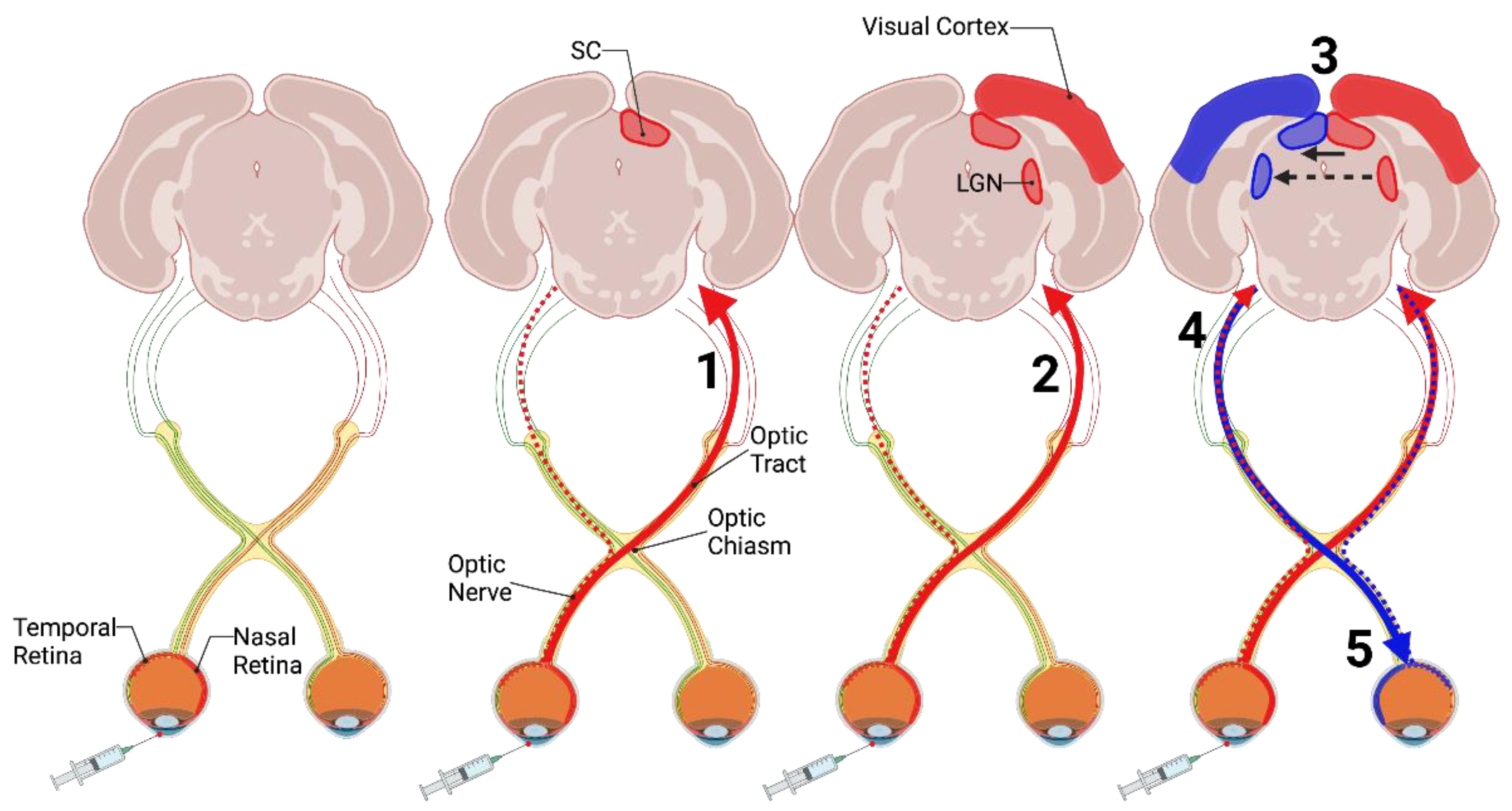
2.1. Intraocular Inoculation of Prions

Prions spread along defined neuroanatomical pathways following the intraocular route of infection [81,82]. Evidence that the prion agent is transported in CNS tissues via known neural pathways was established by determining the temporal and spatial spread of prion replication and pathology following intraocular injection of mice with the scrapie agent. Inoculation of the retina resulted in detection of prion infectivity in the contralateral superior colliculus (SC) via the optic nerve (Figure 1, [81,83]). In addition to the contralateral SC, scrapie infectivity was also identified in the contralateral lateral geniculate nucleus (LGN) and the contralateral visual cortex at later time points post-infection (Figure 1, [82,84]). Importantly, infectivity and vacuolation pathology was greater in the contralateral SC and LGN compared to the ipsilateral SC and LGN [82,85]. Since the rodent visual pathways almost completely decussate [86,87,88], this suggested the transport of prions from the injection site in the eye to the CNS occurs along defined neuroanatomical tracks. To support this hypothesis, enucleation of the retina just prior to or immediately after scrapie inoculation delayed the onset of clinical signs of disease, the increase in infectivity, and the development of vacuolation pathology [85]. This observation indicates that when the retina was removed, scrapie prions were not able to effectively access CNS structures as the neuronal pathway was eliminated. Following this centripetal (periphery to CNS) prion spread, a centrifugal (CNS to periphery) prion spread was also observed.
At later timepoints post infection, the uninoculated contralateral optic nerve and retina developed scrapie infectivity [81] indicating prions spread from the peripheral structures to central structures, and then spread back into unaffected peripheral structures (Figure 1). Since this phenomenon is observed later in the disease process it may be less efficient compared to the initial spread of infectivity. As the major neuronal connections of the inoculated retina are to the contralateral SC and LGN [82,86], scrapie prions will more easily spread along these pathways and lead to earlier and more significant pathology. In contrast, the projection to the ipsilateral SC and LGN is more modest from the inoculated retina, and there may also be minor connections between the bilateral SC and LGN structures allowing for attenuated prion spread between the structures [86,89,90,91]. This results in prion invasion and the spread of pathology that takes longer (via more minor neuronal connections) and is less prominent at the end stage of disease. Once infected, the ipsilateral SC and LGN can transport prion infectivity to the uninoculated retina and its respective optic nerve via retrograde transport or the ipsilateral visual cortex via anterograde transport (Figure 1). Overall, these data indicate the anterograde transport of scrapie prions through established neuroanatomical pathways to central structures that can then spread contiguously or through other neuroanatomical connections within the CNS.

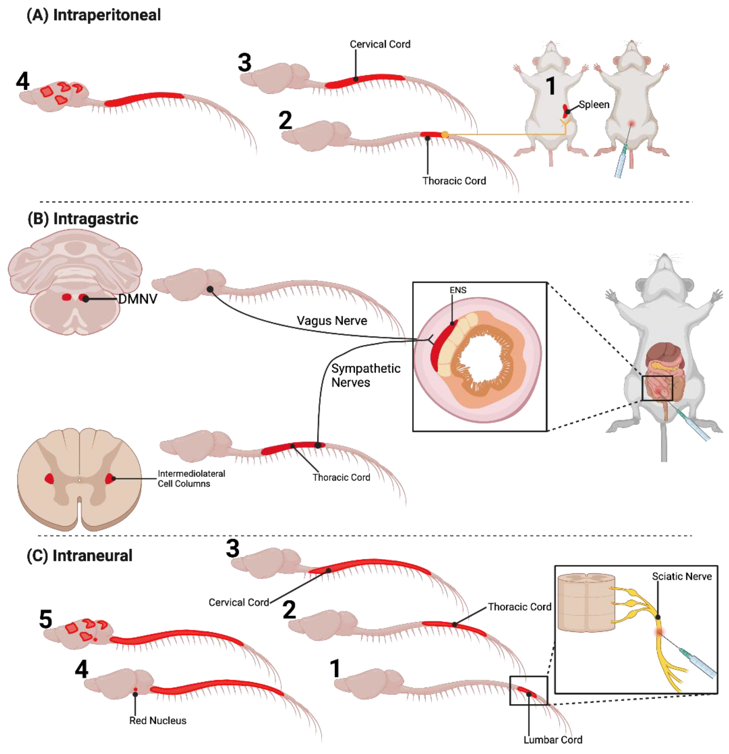
2.2. Extraneural Inoculation of Prions

Prions spread along defined neuroanatomical pathways following extraneural routes of infection. Intraperitoneal (i.p.) injection of scrapie resulted in scrapie infectivity that was first detected in the spleen [92,93,94]. Within the spleen and other LRS tissues, PrPSc has been shown to accumulate on follicular dendritic cells (FDCs) and within tingible body macrophages (TBM, [95,96]). Elimination of FDCs results in a failure of prions to establish infection in secondary LRS tissues and suggests that FDCs actively replicate PrPSc while TBMs scavenge PrPSc [96,97,98]. Accumulation of PrPSc on FDCs has been shown to occur for the duration of prion infection and does not result in cellular dysfunction [99,100,101,102]. Evidence suggests that PrPSc produced by FDCs can infect sympathetic nerves that innervate LRS tissue, as denervation or increasing the physical distance between FDCs and nerve fibers can extend the incubation period of disease or prevent disease transmission [103]. Following i.p. inoculation, prions were found to spread from the spleen to the thoracic spinal cord via the splenic sympathetic axons, with subsequent spread caudally and rostrally to the lumbar and cervical spinal cord, respectively, prior to detection in the brain (Figure 2A, [83,94,104]). In the brain, scrapie infectivity was first detected in caudal brain structures such as the medulla and the midbrain and eventually spread to the cerebellum and the cerebral cortex.
Per os infection of prion agent results in neuroinvasion along defined anatomical pathways that involve the sympathetic and parasympathetic pathways innervating the gut. Intragastric prion inoculation of mice resulted in detection of infectivity in the Peyer’s patches of the gut, followed by spread of infectivity via the myenteric plexus and the enteric nervous system (ENS) to the thoracic spinal cord and finally the brain (Figure 2B, [105]). Consistent with this pathway of prion transport, splenectomy did not prolong the incubation period following intragastric inoculation. In sheep naturally infected with scrapie, PrPSc immunoreactivity was first detected in the Peyer’s patches. Later in the disease course, PrPSc immunoreactivity was detected in the myenteric plexus and ENS, the celiac sympathetic ganglia near the thoracic spinal cord, subsequently in the intermediolateral cell column of the thoracic spinal cord, and finally in the brain [106]. Simultaneously, PrPSc immunoreactivity was also found in the parasympathetic nodose ganglia and subsequently in the dorsal motor nucleus of the vagal nerve (DMNV). Oral BSE challenge of cattle also identified PrPSc immunoreactivity and infectivity that spread from the ENS to the brain via sympathetic pathways involving the thoracic spinal cord and parasympathetic pathways involving the vagus nerve [107,108,109]. Oral inoculation of hamsters with 263K scrapie prions recapitulated many of the findings from the sheep studies where 263K PrPSc was detected in the ENS, then in the sympathetic ganglia, thoracic spinal cord, and later in the medulla [110,111,112]. Importantly, scrapie was also observed to invade the CNS through the DMNV independent of the thoracic spinal cord pathway [113,114]. In this case, scrapie prions were hypothesized to invade the ENS, then travel through the vagal nerve parasympathetic pathway, directly invade the DMNV, and subsequently transneuronally spread throughout the CNS (Figure 2B). Neuroinvasion via the DMNV is also observed in deer per os inoculated with CWD [115,116]. Overall, these data indicate the retrograde spread of PrPSc through the autonomic nervous system via sympathetic and parasympathetic pathways after extraneural inoculation.

2.3. Intraneural Inoculation of Prions

Prions spread along defined neuroanatomical pathways following inoculation into peripheral nerves [117,118]. Unilateral inoculation of scrapie prions into the sciatic nerve of hamsters resulted in a significantly shortened incubation time compared to all other inoculation routes with the exception of intravenous and intracerebral routes [117]. Importantly, scrapie infectivity was detected in spinal cord segments at earlier timepoints post-infection compared to other extraneural infection (intraperitoneal, oral, subcutaneous, etc.) routes suggesting that scrapie prions were able to bypass the spleen and other LRS tissues, and directly invade and spread via axonal networks (Figure 2C). Consistent with these studies, a detailed analysis of the temporal and spatial spread of hyper (HY) PrPSc following inoculation of the sciatic nerve revealed that prions initially spread to ipsilateral ventral motor neurons in the lumbar spinal cord followed by transport along the rubrospinal, corticospinal, vestibulospinal, and reticulospinal descending motor pathways [118]. Axons of these four motor pathways synapse either directly or indirectly on VMNs [119]. HY PrPSc was detected in progressively more rostral spinal cord segments starting with the T10-T13 segment corresponding to where the sciatic nerve enters the lumbar spinal cord (Figure 2C). Within the brain, PrPSc was detected in the ventral portion of the contralateral RN (origin of the rubrospinal tract) which decussates and has direct projections to ventral motor neurons found in the lumbar spinal cord associated with the inoculated sciatic nerve. Other regions that are associated with the sciatic nerve motor pathways were also affected including the contralateral hindlimb motor cortex (origin of the corticospinal tract) and the ipsilateral lateral vestibular nucleus (origin of the vestibulospinal tract). Since HY PrPSc was not detected in the spleen following sciatic nerve inoculation, neuroinvasion and subsequent spread was via the sciatic nerve and not from sympathetic innervation from the spleen [118,120]. In addition to HY, sciatic nerve inoculation experiments with drowsy (DY) or 139H strain of prions, resulted in transport of PrPSc along the same four descending motor pathways [119,121]. Intralingual inoculation of HY has demonstrated PrPSc deposition in the hypoglossal nucleus, consistent with PrPSc spread via the hypoglossal nerve [122]. Overall, PrPSc is transneuronally transported along defined neuroanatomical pathways in the anterograde (intraocular inoculation) or retrograde (intraperitoneal, intragastric, or intraneural) direction. This is also independent of prion strain, suggesting this is a common feature of prions. A summary of the discussed transport pathways can be found in Table 1.

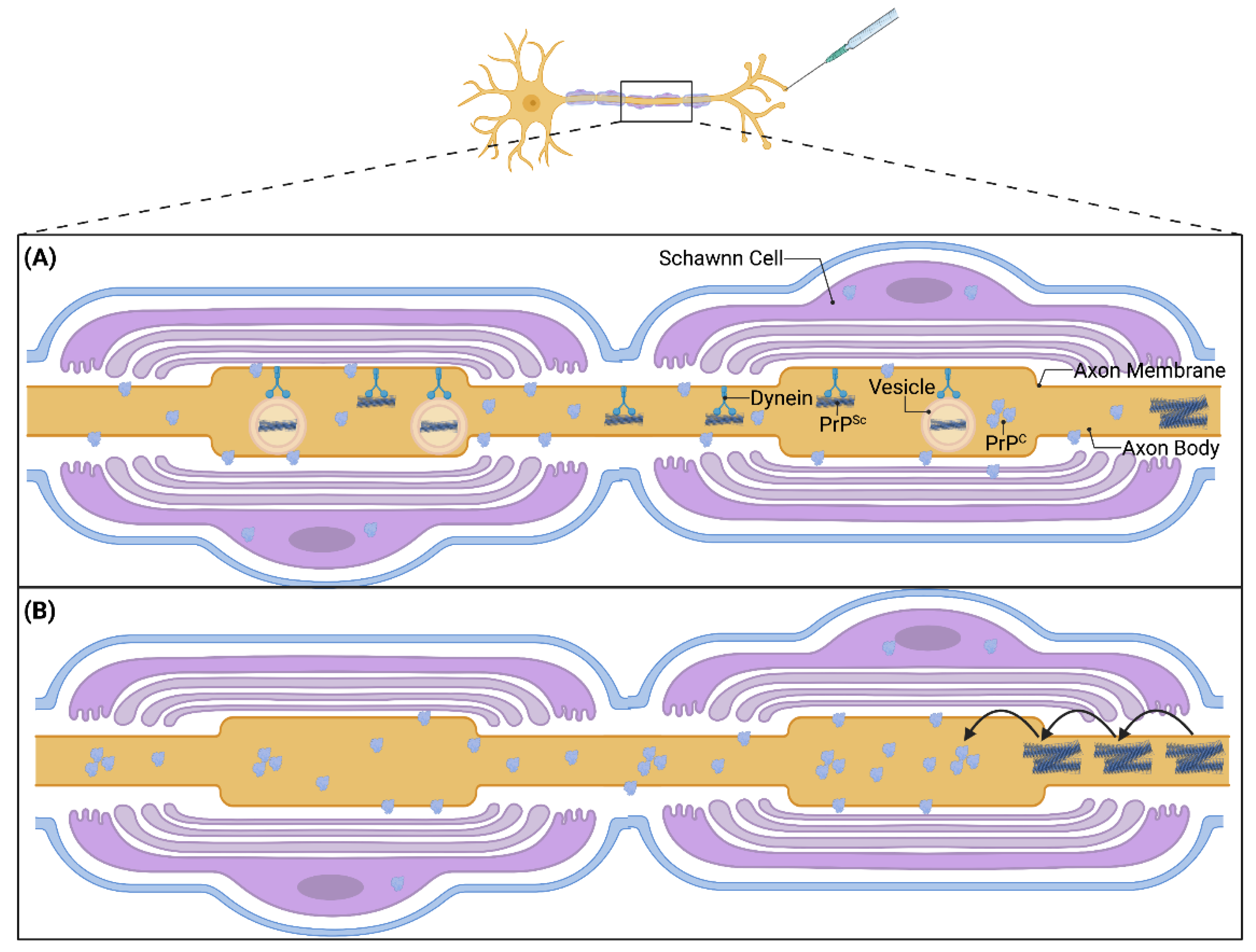
3. Cell Types Involved in Prion Transport

While it is well-established that prions invade and spread through the peripheral nerves to access the CNS, the mechanism by which this phenomenon occurs has remained elusive. Neurotropic viruses such as rabies and pseudorabies travel from the periphery to the CNS via axons. For example, the rabies virus utilizes microtubule polymerization as it undergoes retrograde transport along motor neuron axons [123]. Herpes viruses also utilize microtubules to spread along both sensory and motor neurons [124]. Since neurotropic viruses and prions spread along established neuroanatomical pathways after infection, the processes utilized by these viral agents serve as a starting point to elucidate the mechanisms involved in prion transport.
The exact cellular location of PrPSc within neuronal pathways is unknown. In the nervous system, PrPC is expressed not only in neurons and axons, but it has also been found in Schwann cells that myelinate axons in the PNS, oligodendrocytes that myelinate axons in the CNS, as well as astrocytes and microglia that support CNS neurons [16,125]. PrPSc was localized to the adaxonal membrane in peripheral nerves of scrapie-infected sheep [126] and was also described in adaxonal location in peripheral nerves of CJD patients after immunohistochemical analyses [127], implicating neuronal axons as a pathway of PrPSc spread. In addition, PrPSc aggregates traveled along neuritic projections in in vitro neuronal cultures [128]. To complement this finding, Schwann cells with ablated PrPC expression using two different transgenetic mouse models did not slow the progression and incubation period of scrapie after intraperitoneal inoculation in mice [129,130]. This further strengthens the overarching finding that prions invade the CNS through peripheral neuronal pathways and utilize axons to propagate along these pathways towards central structures. These studies also suggest that the contribution of Schwann cells and oligodendrocytes are not required for PrPSc transport. There is still speculation on the exact location of PrPSc within an axonal segment. PrPC has been noted as a GPI anchored membrane protein [13], but its conversion into PrPSc with altered properties may translocate it to another area on the axon, and confocal laser scanning imaging has detected PrPSc on the axonal periphery and within the axonal cytoplasm [131]. PrPSc populations have also been localized to endosomal and lysosomal compartments (Figure 3A), suggesting that PrPSc may also utilize intracellular membranous vesicles for movement within cells [128,132]. Interestingly, it was found that only a small fraction of PrPSc was associated with membrane-bound vesicles, indicating free PrPSc is also present within the cell. These findings suggest different roles of PrPSc based on axonal location and cell compartment where one population of PrPSc is primarily transported to other structures while another population aggregates and causes disease pathology. Alternatively, this may indicate multiple modalities of PrPSc transport that utilize different subcellular mechanisms.

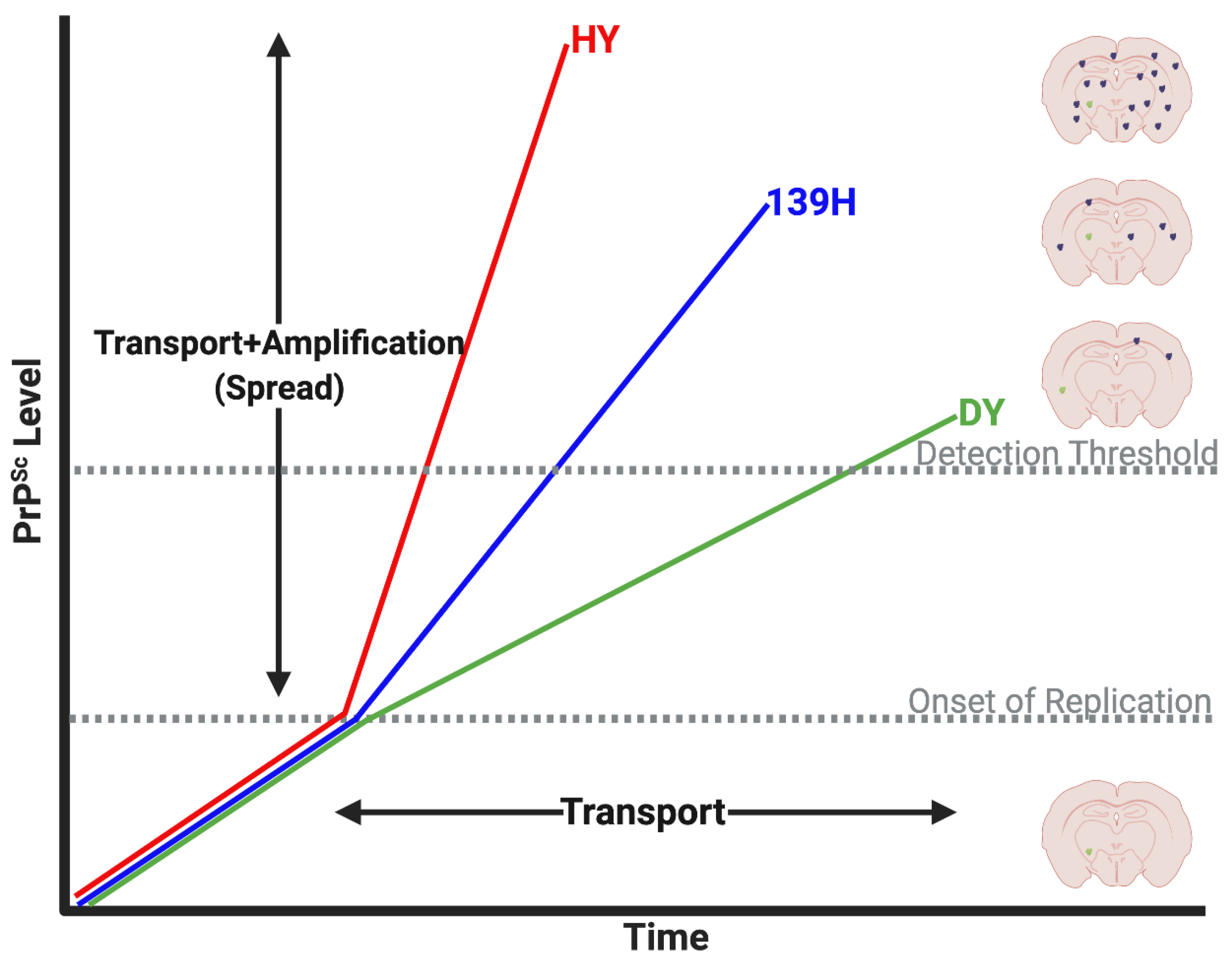
4. Prion Transport Rate and Machinery

The velocity of PrPSc transport and whether prions utilize a fast or slow mode of axonal transport may provide insight into the mechanisms of prion transport. Axonal transport includes two molecular motors, kinesin and dynein. Kinesin is the main component involved in anterograde transport (from soma to synapse) while dynein takes part in retrograde transport (from synapse to soma). Axonal transport can also be divided into two main types, fast axonal transport and slow axonal transport which includes two components, a and b. Both transport mechanisms utilize kinesin and dynein motors and are differentiated based on their speeds, cargoes, subtypes of kinesin and dynein, and cofactors [133]. Fast axonal transport moves organelles and vesicles containing neurotransmitters at upwards of 400 mm/day. Slow axonal transport primarily moves cytoskeletal elements such as microtubules and neurofilaments (slow component a) or cytosolic proteins (slow component b) at less than 8 mm/day [134].
Estimates of the rate of prion spread is consistent with slow axonal transport mechanism (Figure 3, [104,117,118,119]). Importantly, prion spread was determined by assaying for prion infectivity via animal bioassay or immunodetection of PrPSc. These assays do not discriminate between the original inoculum and newly converted prions, therefore, the time to detection of infectivity or PrPSc may be a combination of both transport and replication, hence the use of the term prion “spread” (Figure 4). Initial estimates of prion spread calculated from the detection of scrapie infectivity in different tissues at various times post infection determined that mouse adapted scrapie prions spread approximately 1–2 mm/day [104,117]. Subsequent studies corroborated that the spread of prions in the nervous system is consistent with slow axonal transport [85,112]. In a series of studies that utilized the sciatic nerve inoculation paradigm and three hamster-adapted rodent prion strains, strain-specific differences in the rate of PrPSc spread were identified. In these studies, the rate of spread of HY, 139H and DY PrPSc in the nervous system, as determined by immunohistochemistry, was 1.10 ± 0.11, 1.80 ± 0.27 and 4.14 ± 0.35 mm/day, respectively [119,121]. While these values were noted to be statistically different, they are still within the range of slow axonal transport. The reason for the observed differences is unknown, however these strains were shown to have differing rates of prion formation with the HY strain having a faster rate of prion formation compared to 139H and DY [34]. It is possible, that all three strains move along axons at the same velocity but since HY PrPSc amplifies faster than PrPSc from the other two strains, it may reach a level of abundance that is detectable by IHC faster than either DY or 139H (Figure 4). Alternatively, these findings could indicate that different strains utilize different host mechanisms of PrPSc transport. Overall, the actual velocity of PrPSc transport is unknown, as PrPSc may traffic to tissues much earlier in the disease process that can only be elucidated through experiments that directly detect inoculum PrPSc. A summary of prion transport rate studies can be found in Table 1.
The role of endogenous PrPC in the transport of PrPSc from the periphery to the CNS is poorly understood. Landmark studies confirmed the requirement of PrPC for successful prion infection [135,136] since PrPC expression is a necessity for prion formation. Studies utilizing PrPC expressing neural grafts found that not only did the target CNS tissue require PrPC for successful infection and pathology, but the peripheral pathways leading to the tissue also required PrPC expression to propagate pathology from the periphery to the CNS [137,138]. This may indicate a need for a continuous supply of PrPC from the site of inoculation to the target site in the CNS for successful infection and clinical pathology of CNS structures. It is possible that PrPC is required for axonal transport or that PrPC must be present along the whole tract to serve as a substrate for continual conversion of PrPC to PrPSc all the way to the CNS in a “domino-like” fashion (Figure 3B). Alternatively, the failure to establish infection in the PrPC containing grafts following peripheral inoculation may be due to the grafts not being synaptically connected to the peripheral neuronal pathways and therefore not allowing PrPSc to invade the graft. Interestingly, multiple cell culture studies have concluded that although endogenous PrPC may play a role in initial PrPSc uptake into the cell, it is not necessary for infection of the cell or PrPSc transport between cells [139,140]. Evidence from imaging studies conducted on in vitro primary neuronal cultures found that PrPC knockout neuronal cells could still take up and transport PrPSc within neurites [128] so PrPSc may not require endogenous PrPc for transport.

5. Conclusions

Studies of the peripheral to central transport of prions have yielded many results of prion spread into the CNS. As evidenced by the studies above, prions spread along defined neuronal projections that innervate the inoculation site. Peripheral extraneural inoculation allows many prion strains to replicate in the LRS and travel along sympathetic projections toward the spinal cord where the agent spreads rostrally toward the brain. In the case of per os inoculation, prions spread along sympathetic and parasympathetic projections of the ENS and invade the spinal cord and DMNV, respectively. Peripheral intraneural injections demonstrate prion spread along defined neuroanatomical pathways that connect peripheral axonal pathways with specific brainstem nuclei and/or cortical areas. Currently, efforts are being undertaken to understand mechanisms behind prion spread. So far, PrPC has been demonstrated to be a necessary part of successful prion infection, and evidence has indicated prions utilize axonal networks for peripheral to central transport. The approximate spread of prions is consistent with a slow axonal transport mechanism, yet the exact speed, subcellular mechanisms, and specific cofactors behind this transport phenomenon are still unknown. Overall, future research should focus on elucidating these mechanisms to further our understanding of peripheral prion disease pathogenesis.

Author Contributions

Writing—original draft preparation, J.C.B. and S.M.K.; Writing—review and editing, J.C.B., S.M.K. and A.E.K. All authors have read and agreed to the published version of the manuscript.

Funding

NIH NINDS 1R01NS107246.

Conflicts of Interest

The authors declare no conflict of interest.

References

  1. Prusiner, S.B. Novel proteinaceous infectious particles cause scrapie. Science 1982, 216, 136–144. [Google Scholar] [CrossRef] [PubMed] [Green Version]
  2. Geschwind, M.D. Prion Diseases. Contin. (Minneap. Minn.) 2015, 21, 1612–1638. [Google Scholar] [CrossRef] [PubMed]
  3. Sigurdson, C.J.; Bartz, J.C.; Glatzel, M. Cellular and Molecular Mechanisms of Prion Disease. Annu. Rev. Pathol. Mech. Dis. 2019, 14, 497–516. [Google Scholar] [CrossRef]
  4. Cole, M. Slow virus diseases. Rocky Mt. Intern. Med. 1975, 72, 294–296. [Google Scholar]
  5. Fuccillo, D.A.; Kurent, J.E.; Sever, J.L. Slow virus diseases. Annu. Rev. Microbiol. 1974, 28, 231–234. [Google Scholar] [CrossRef] [PubMed] [Green Version]
  6. Gajdusek, D.C. Slow virus diseases of the central nervous system. Am. J. Clin. Pathol. 1971, 56, 320–332. [Google Scholar] [CrossRef] [Green Version]
  7. Hadlow, W.J.; Eklund, C.M. Scrapie—A virus-induced chronic encephalopathy of sheep. Res. Publ. Assoc. Res. Nerv. Ment. Dis. 1968, 44, 281–306. [Google Scholar]
  8. Ma, J.; Wang, F. Prion disease and the ‘protein-only hypothesis’. Essays Biochem. 2014, 56, 181–191. [Google Scholar] [CrossRef]
  9. Griffith, J.S. Self-replication and scrapie. Nature 1967, 215, 1043–1044. [Google Scholar] [CrossRef]
  10. Deleault, N.R.; Harris, B.T.; Rees, J.R.; Supattapone, S. Formation of native prions from minimal components in vitro. Proc. Natl. Acad. Sci. USA 2007, 104, 9741–9746. [Google Scholar] [CrossRef] [Green Version]
  11. Alper, T.; Haig, D.A.; Clarke, M.C. The scrapie agent: Evidence against its dependence for replication on intrinsic nucleic acid. J. Gen. Virol. 1978, 41, 503–516. [Google Scholar] [CrossRef] [PubMed]
  12. Calzolai, L.; Lysek, D.A.; Pérez, D.R.; Güntert, P.; Wüthrich, K. Prion protein NMR structures of chickens, turtles, and frogs. Proc. Natl. Acad. Sci. USA 2005, 102, 651–655. [Google Scholar] [CrossRef] [PubMed] [Green Version]
  13. Legname, G. Elucidating the function of the prion protein. PLoS Pathog. 2017, 13, e1006458. [Google Scholar] [CrossRef] [PubMed] [Green Version]
  14. Fernandez-Funez, P.; Sanchez-Garcia, J.; Rincon-Limas, D.E. Drosophila models of prionopathies: Insight into prion protein function, transmission, and neurotoxicity. Curr. Opin. Genet. Dev. 2017, 44, 141–148. [Google Scholar] [CrossRef]
  15. Vassallo, N.; Herms, J. Cellular prion protein function in copper homeostasis and redox signalling at the synapse. J. Neurochem. 2003, 86, 538–544. [Google Scholar] [CrossRef]
  16. Westergard, L.; Christensen, H.M.; Harris, D.A. The cellular prion protein (PrP(C)): Its physiological function and role in disease. Biochim. Biophys. Acta 2007, 1772, 629–644. [Google Scholar] [CrossRef] [Green Version]
  17. Ironside, J.W.; Ritchie, D.L.; Head, M.W. Prion diseases. Handb. Clin. Neurol. 2017, 145, 393–403. [Google Scholar] [CrossRef]
  18. Baldwin, K.J.; Correll, C.M. Prion Disease. Semin. Neurol. 2019, 39, 428–439. [Google Scholar] [CrossRef]
  19. Pattinson, I.H. Resistance of Scrapie Agent to Formalin. J. Comp. Pathol. 1965, 75, 159–164. [Google Scholar] [CrossRef]
  20. Dickinson, A.G.; Taylor, D.M. Resistance of scrapie agent to decontamination. N. Engl. J. Med. 1978, 299, 1413–1414. [Google Scholar] [CrossRef]
  21. Gibbs, C.J.; Gajdusek, D.C.; Latarjet, R. Unusual resistance to ionizing radiation of the viruses of kuru, Creutzfeldt-Jakob disease, and scrapie. Proc. Natl. Acad. Sci. USA 1978, 75, 6268–6270. [Google Scholar] [CrossRef] [Green Version]
  22. Brown, P.; Liberski, P.P.; Wolff, A.; Gajdusek, D.C. Resistance of scrapie infectivity to steam autoclaving after formaldehyde fixation and limited survival after ashing at 360 degrees C: Practical and theoretical implications. J. Infect. Dis. 1990, 161, 467–472. [Google Scholar] [CrossRef] [PubMed]
  23. Caughey, B.; Raymond, G.J.; Kocisko, D.A.; Lansbury, P.T. Scrapie infectivity correlates with converting activity, protease resistance, and aggregation of scrapie-associated prion protein in guanidine denaturation studies. J. Virol. 1997, 71, 4107–4110. [Google Scholar] [CrossRef] [PubMed] [Green Version]
  24. Yuan, Q.; Telling, G.; Bartelt-Hunt, S.L.; Bartz, J.C. Dehydration of Prions on Environmentally Relevant Surfaces Protects Them from Inactivation by Freezing and Thawing. J. Virol. 2018, 92, e02191-17. [Google Scholar] [CrossRef] [PubMed] [Green Version]
  25. Saunders, S.E.; Yuan, Q.; Bartz, J.C.; Bartelt-Hunt, S. Effects of solution chemistry and aging time on prion protein adsorption and replication of soil-bound prions. PLoS ONE 2011, 6, e18752. [Google Scholar] [CrossRef] [Green Version]
  26. Jeffrey, M.; Goodbrand, I.A.; Goodsir, C.M. Pathology of the transmissible spongiform encephalopathies with special emphasis on ultrastructure. Micron 1995, 26, 277–298. [Google Scholar] [CrossRef]
  27. Gajdusek, D.C.; Zigas, V. Studies on the pathogenesis of kuru; a clinical, pathological and epidemiological study of a chronic, progressive, degenerative disease of the central nervous system achieving epidemic proportions among the natives of the Eastern Highlands of New Guinea. Klin. Wochenschr. 1958, 36, 445–459. [Google Scholar] [CrossRef]
  28. Morales, R. Prion strains in mammals: Different conformations leading to disease. PLoS Pathog. 2017, 13, e1006323. [Google Scholar] [CrossRef] [Green Version]
  29. Bartz, J.C. Prion Strain Diversity. Cold Spring Harb. Perspect. Med. 2016, 6, a024349. [Google Scholar] [CrossRef]
  30. Hill, A.F.; Collinge, J. Prion strains and species barriers. Contrib. Microbiol. 2004, 11, 33–49. [Google Scholar] [CrossRef]
  31. Aguzzi, A.; Heikenwalder, M.; Polymenidou, M. Insights into prion strains and neurotoxicity. Nat. Rev. Mol. Cell Biol. 2007, 8, 552–561. [Google Scholar] [CrossRef]
  32. Collinge, J.; Clarke, A.R. A general model of prion strains and their pathogenicity. Science 2007, 318, 930–936. [Google Scholar] [CrossRef] [PubMed] [Green Version]
  33. Weissmann, C. Thoughts on mammalian prion strains. Folia Neuropathol. 2009, 47, 104–113. [Google Scholar] [PubMed]
  34. Shikiya, R.A.; Langenfeld, K.A.; Eckland, T.E.; Trinh, J.; Holec, S.A.M.; Mathiason, C.K.; Kincaid, A.E.; Bartz, J.C. PrPSc formation and clearance as determinants of prion tropism. PLoS Pathog. 2017, 13, e1006298. [Google Scholar] [CrossRef] [PubMed]
  35. Cortez, L.M.; Nemani, S.K.; Duque Velásquez, C.; Sriraman, A.; Wang, Y.; Wille, H.; McKenzie, D.; Sim, V.L. Asymmetric-flow field-flow fractionation of prions reveals a strain-specific continuum of quaternary structures with protease resistance developing at a hydrodynamic radius of 15 nm. PLoS Pathog. 2021, 17, e1009703. [Google Scholar] [CrossRef] [PubMed]
  36. Bett, C.; Lawrence, J.; Kurt, T.D.; Orru, C.; Aguilar-Calvo, P.; Kincaid, A.E.; Surewicz, W.K.; Caughey, B.; Wu, C.; Sigurdson, C.J. Enhanced neuroinvasion by smaller, soluble prions. Acta Neuropathol. Commun. 2017, 5, 32. [Google Scholar] [CrossRef] [PubMed] [Green Version]
  37. Hulland, T.J. Scrapie and Kuru. Can. Vet. J. 1960, 1, 496. [Google Scholar] [PubMed]
  38. Gajdusek, D.C.; Zigas, V. Degenerative disease of the central nervous system in New Guinea; the endemic occurrence of kuru in the native population. N. Engl. J. Med. 1957, 257, 974–978. [Google Scholar] [CrossRef] [PubMed]
  39. Gajdusek, D.C.; Zigas, V. Kuru; clinical, pathological and epidemiological study of an acute progressive degenerative disease of the central nervous system among natives of the Eastern Highlands of New Guinea. Am. J. Med. 1959, 26, 442–469. [Google Scholar] [CrossRef]
  40. Gajdusek, D.C.; Reid, L.H. Studies on kuru. IV. The kuru pattern in Moke, a representative Fore village. Am. J. Trop. Med. Hyg. 1961, 10, 628–638. [Google Scholar] [CrossRef]
  41. Gajdusek, D.C.; Zigas, V. Studies on kuru. I. The ethnologic setting of kuru. Am. J. Trop. Med. Hyg. 1961, 10, 80–91. [Google Scholar] [CrossRef] [PubMed]
  42. Gajdusek, D.C.; Zigas, V.; Baker, J. Studies on kuru. III. Patterns of kuru incidence: Demographic and geographic epidemiological analysis. Am. J. Trop. Med. Hyg. 1961, 10, 599–627. [Google Scholar] [CrossRef]
  43. Gajdusek, D.C. Kuru: An appraisal of five years of investigation. Eugen. Q. 1962, 9, 69–74. [Google Scholar] [CrossRef]
  44. Gajdusek, D.C. Kuru. Trans. R. Soc. Trop. Med. Hyg. 1963, 57, 151–169. [Google Scholar] [CrossRef]
  45. Liberski, P.P.; Gajos, A.; Sikorska, B.; Lindenbaum, S. Kuru, the First Human Prion Disease. Viruses 2019, 11, 232. [Google Scholar] [CrossRef] [PubMed] [Green Version]
  46. Wells, G.A.; Scott, A.C.; Johnson, C.T.; Gunning, R.F.; Hancock, R.D.; Jeffrey, M.; Dawson, M.; Bradley, R. A novel progressive spongiform encephalopathy in cattle. Vet. Rec. 1987, 121, 419–420. [Google Scholar] [CrossRef] [PubMed]
  47. Wilesmith, J.W.; Wells, G.A.; Cranwell, M.P.; Ryan, J.B. Bovine spongiform encephalopathy: Epidemiological studies. Vet. Rec. 1988, 123, 638–644. [Google Scholar] [CrossRef] [PubMed]
  48. Wilesmith, J.W.; Ryan, J.B.; Atkinson, M.J. Bovine spongiform encephalopathy: Epidemiological studies on the origin. Vet. Rec. 1991, 128, 199–203. [Google Scholar] [CrossRef]
  49. Bruce, M.E.; Will, R.G.; Ironside, J.W.; McConnell, I.; Drummond, D.; Suttie, A.; McCardle, L.; Chree, A.; Hope, J.; Birkett, C.; et al. Transmissions to mice indicate that ‘new variant’ CJD is caused by the BSE agent. Nature 1997, 389, 498–501. [Google Scholar] [CrossRef]
  50. Hill, A.F.; Desbruslais, M.; Joiner, S.; Sidle, K.C.; Gowland, I.; Collinge, J.; Doey, L.J.; Lantos, P. The same prion strain causes vCJD and BSE. Nature 1997, 389, 448–450. [Google Scholar] [CrossRef]
  51. Houston, F.; Andréoletti, O. Animal prion diseases: The risks to human health. Brain Pathol. 2019, 29, 248–262. [Google Scholar] [CrossRef] [PubMed] [Green Version]
  52. Hartsough, G.R.; Burger, D. Encephalopathy of mink. I. Epizootiologic and clinical observations. J. Infect. Dis. 1965, 115, 387–392. [Google Scholar] [CrossRef] [PubMed]
  53. Hanson, R.P.; Eckroade, R.J.; Marsh, R.F.; Zu Rhein, G.M.; Kanitz, C.L.; Gustafson, D.P. Susceptibility of mink to sheep scrapie. Science 1971, 172, 859–861. [Google Scholar] [CrossRef] [PubMed]
  54. Marsh, R.F.; Hanson, R.P. On the origin of transmissible mink encephalopathy. In Slow Transmissible Diseases of the Nervous System; Prusiner, S.B., Hadlow, W.J., Eds.; Academic Press: New York, NY, USA, 1979; Volume 1, pp. 451–460. [Google Scholar]
  55. Marsh, R.F.; Bessen, R.A.; Lehmann, S.; Hartsough, G.R. Epidemiological and experimental studies on a new incident of transmissible mink encephalopathy. J. Gen. Virol. 1991, 72 Pt 3, 589–594. [Google Scholar] [CrossRef] [PubMed]
  56. Marsh, R.F.; Bessen, R.A. Epidemiologic and experimental studies on transmissible mink encephalopathy. Dev. Biol. Stand. 1993, 80, 111–118. [Google Scholar] [PubMed]
  57. Baron, T.; Bencsik, A.; Biacabe, A.G.; Morignat, E.; Bessen, R.A. Phenotypic similarity of transmissible mink encephalopathy in cattle and L-type bovine spongiform encephalopathy in a mouse model. Emerg. Infect. Dis. 2007, 13, 1887–1894. [Google Scholar] [CrossRef]
  58. Adcock, J.L.; Keiss, R.E. Locoism in elk. A disease resembling cerebral pseudolipidosis. Bull. Wildl. Dis. Assoc. 1969, 5, 121–124. [Google Scholar] [CrossRef]
  59. Williams, E.S.; Young, S. Chronic wasting disease of captive mule deer: A spongiform encephalopathy. J. Wildl. Dis. 1980, 16, 89–98. [Google Scholar] [CrossRef] [Green Version]
  60. Sigurdson, C.J.; Aguzzi, A. Chronic wasting disease. Biochim. Biophys. Acta 2007, 1772, 610–618. [Google Scholar] [CrossRef]
  61. CDC. Chronic Wasting Disease (CWD), Occurrence, January 2022. Available online: https://www.cdc.gov/prions/cwd/occurrence.html (accessed on 23 February 2022).
  62. Benestad, S.L.; Mitchell, G.; Simmons, M.; Ytrehus, B.; Vikøren, T. First case of chronic wasting disease in Europe in a Norwegian free-ranging reindeer. Vet. Res. 2016, 47, 88. [Google Scholar] [CrossRef]
  63. Pirisinu, L.; Tran, L.; Chiappini, B.; Vanni, I.; Di Bari, M.A.; Vaccari, G.; Vikøren, T.; Madslien, K.I.; Våge, J.; Spraker, T.; et al. Novel Type of Chronic Wasting Disease Detected in Moose (Alces alces), Norway. Emerg. Infect. Dis. 2018, 24, 2210–2218. [Google Scholar] [CrossRef] [PubMed] [Green Version]
  64. Nonno, R.; Di Bari, M.A.; Pirisinu, L.; D’Agostino, C.; Vanni, I.; Chiappini, B.; Marcon, S.; Riccardi, G.; Tran, L.; Vikøren, T.; et al. Studies in bank voles reveal strain differences between chronic wasting disease prions from Norway and North America. Proc. Natl. Acad. Sci. USA 2020, 117, 31417–31426. [Google Scholar] [CrossRef] [PubMed]
  65. Ågren, E.O.; Sörén, K.; Gavier-Widén, D.; Benestad, S.L.; Tran, L.; Wall, K.; Averhed, G.; Doose, N.; Våge, J.; Nöremark, M. First Detection of Chronic Wasting Disease in Moose (Alces alces) in Sweden. J. Wildl. Dis. 2021, 57, 461–463. [Google Scholar] [CrossRef]
  66. Güere, M.E.; Våge, J.; Tharaldsen, H.; Kvie, K.S.; Bårdsen, B.J.; Benestad, S.L.; Vikøren, T.; Madslien, K.; Rolandsen, C.M.; Tranulis, M.A.; et al. Chronic wasting disease in Norway-A survey of prion protein gene variation among cervids. Transbound. Emerg. Dis. 2021, 1–12. [Google Scholar] [CrossRef]
  67. Tranulis, M.A.; Gavier-Widén, D.; Våge, J.; Nöremark, M.; Korpenfelt, S.L.; Hautaniemi, M.; Pirisinu, L.; Nonno, R.; Benestad, S.L. Chronic wasting disease in Europe: New strains on the horizon. Acta Vet. Scand. 2021, 63, 48. [Google Scholar] [CrossRef]
  68. Williams, E.S. Chronic wasting disease. Vet. Pathol. 2005, 42, 530–549. [Google Scholar] [CrossRef]
  69. Detwiler, L.A. Scrapie. Rev. Sci. Tech. 1992, 11, 491–537. [Google Scholar] [CrossRef] [Green Version]
  70. Plummer, P.J. Scrapie-A Disease of Sheep: A Review of the literature. Can. J. Comp. Med. Vet Sci. 1946, 10, 49–54. [Google Scholar]
  71. Dickinson, A.G.; Mackay, J.M. Genetical Control of the Incubation Period in Mice of the Neurological Disease, Scrapie. Heredity 1964, 19, 279–288. [Google Scholar] [CrossRef] [PubMed] [Green Version]
  72. Dickinson, A.G.; Mackay, J.M.; Zlotnik, I. Transmission by Contact of Scrapie in Mice. J. Comp. Pathol. 1964, 74, 250–254. [Google Scholar] [CrossRef]
  73. Dickinson, A.G.; Young, G.B.; Stamp, J.T.; Renwick, C.C. An analysis of natural scrapie in Suffolk sheep. Heredity 1965, 20, 485–503. [Google Scholar] [CrossRef] [PubMed] [Green Version]
  74. Dickinson, A.G.; Meikle, V.M.; Fraser, H. Identification of a gene which controls the incubation period of some strains of scrapie agent in mice. J. Comp. Pathol. 1968, 78, 293–299. [Google Scholar] [CrossRef]
  75. Dickinson, A.G.; Stamp, J.T.; Renwick, C.C.; Rennie, J.C. Some factors controlling the incidence of scrapie in Cheviot sheep injected with a Cheviot-passaged scrapie agent. J. Comp. Pathol. 1968, 78, 313–321. [Google Scholar] [CrossRef]
  76. Kimberlin, R.H. Biochemical changes in scrapie affeced brain. Biochem. J. 1969, 114, 20P–22P. [Google Scholar] [CrossRef] [PubMed] [Green Version]
  77. Georgsson, G.; Sigurdarson, S.; Brown, P. Infectious agent of sheep scrapie may persist in the environment for at least 16 years. J. Gen. Virol. 2006, 87, 3737–3740. [Google Scholar] [CrossRef] [PubMed]
  78. Saunders, S.E.; Bartelt-Hunt, S.L.; Bartz, J.C. Prions in the environment: Occurrence, fate and mitigation. Prion 2008, 2, 162–169. [Google Scholar] [CrossRef] [Green Version]
  79. Hawkins, S.A.; Simmons, H.A.; Gough, K.C.; Maddison, B.C. Persistence of ovine scrapie infectivity in a farm environment following cleaning and decontamination. Vet. Rec. 2015, 176, 99. [Google Scholar] [CrossRef]
  80. Andréoletti, O.; Lacroux, C.; Chabert, A.; Monnereau, L.; Tabouret, G.; Lantier, F.; Berthon, P.; Eychenne, F.; Lafond-Benestad, S.; Elsen, J.M.; et al. PrP(Sc) accumulation in placentas of ewes exposed to natural scrapie: Influence of foetal PrP genotype and effect on ewe-to-lamb transmission. J. Gen. Virol. 2002, 83, 2607–2616. [Google Scholar] [CrossRef]
  81. Buyukmihci, N.; Goehring-Harmon, F.; Marsh, R.F. Neural pathogenesis of experimental scrapie after intraocular inoculation of hamsters. Exp. Neurol. 1983, 81, 396–406. [Google Scholar] [CrossRef]
  82. Fraser, H.; Dickinson, A.G. Targeting of scrapie lesions and spread of agent via the retino-tectal projection. Brain Res. 1985, 346, 32–41. [Google Scholar] [CrossRef]
  83. Kimberlin, R.H.; Walker, C.A. Pathogenesis of scrapie (strain 263K) in hamsters infected intracerebrally, intraperitoneally or intraocularly. J. Gen. Virol. 1986, 67 Pt 2, 255–263. [Google Scholar] [CrossRef] [PubMed]
  84. Scott, J.R.; Davies, D.; Fraser, H. Scrapie in the central nervous system: Neuroanatomical spread of infection and Sinc control of pathogenesis. J. Gen. Virol. 1992, 73 Pt 7, 1637–1644. [Google Scholar] [CrossRef]
  85. Scott, J.R.; Fraser, H. Transport and targeting of scrapie infectivity and pathology in the optic nerve projections following intraocular infection. Prog. Clin. Biol. Res. 1989, 317, 645–652. [Google Scholar] [CrossRef] [PubMed]
  86. Sefton, A.J.; Dreher, B. Visual System. In The Rat Nervous System, Forebrain and Midbrain; Paxinos, G., Ed.; Academic Press Inc.: Orlando, FL, USA, 1985; Volume 1, pp. 169–210. [Google Scholar]
  87. Jeffery, G. Retinal ganglion cell death and terminal field retraction in the developing rodent visual system. Brain Res. 1984, 315, 81–96. [Google Scholar] [CrossRef]
  88. Dreher, B.; Sefton, A.J.; Ni, S.Y.; Nisbett, G. The morphology, number, distribution and central projections of Class I retinal ganglion cells in albino and hooded rats. Brain Behav. Evol. 1985, 26, 10–48. [Google Scholar] [CrossRef] [PubMed]
  89. Fish, S.E.; Goodman, D.K.; Kuo, D.C.; Polcer, J.D.; Rhoades, R.W. The intercollicular pathway in the golden hamster: An anatomical study. J. Comp. Neurol. 1982, 204, 6–20. [Google Scholar] [CrossRef] [PubMed]
  90. Mackay-Sim, A.; Sefton, A.J.; Martin, P.R. Subcortical projections to lateral geniculate and thalamic reticular nuclei in the hooded rat. J. Comp. Neurol. 1983, 213, 24–35. [Google Scholar] [CrossRef]
  91. Hubel, D.H.; Wiesel, T.N. Cortical and callosal connections concerned with the vertical meridian of visual fields in the cat. J. Neurophysiol. 1967, 30, 1561–1573. [Google Scholar] [CrossRef]
  92. Kimberlin, R.H.; Walker, C.A. Pathogenesis of mouse scrapie: Dynamics of agent replication in spleen, spinal cord and brain after infection by different routes. J. Comp. Pathol. 1979, 89, 551–562. [Google Scholar] [CrossRef]
  93. Kimberlin, R.H.; Walker, C.A. Pathogenesis of mouse scrapie: Evidence for neural spread of infection to the CNS. J. Gen. Virol. 1980, 51, 183–187. [Google Scholar] [CrossRef]
  94. Kimberlin, R.H.; Walker, C.A. The role of the spleen in the neuroinvasion of scrapie in mice. Virus Res. 1989, 12, 201–211. [Google Scholar] [CrossRef]
  95. Muramoto, T.; Kitamoto, T.; Tateishi, J.; Goto, I. The sequential development of abnormal prion protein accumulation in mice with Creutzfeldt-Jakob disease. Am. J. Pathol. 1992, 140, 1411–1420. [Google Scholar]
  96. Brown, K.L.; Stewart, K.; Ritchie, D.L.; Mabbott, N.A.; Williams, A.; Fraser, H.; Morrison, W.I.; Bruce, M.E. Scrapie replication in lymphoid tissues depends on prion protein-expressing follicular dendritic cells. Nat. Med. 1999, 5, 1308–1312. [Google Scholar] [CrossRef] [PubMed]
  97. Brown, K.L.; Stewart, K.; Ritchie, D.; Fraser, H.; Morrison, W.I.; Bruce, M.E. Follicular dendritic cells in scrapie pathogenesis. Arch. Virol. Suppl. 2000, 16, 13–21. [Google Scholar] [CrossRef]
  98. Bruce, M.E.; Brown, K.L.; Mabbott, N.A.; Farquhar, C.F.; Jeffrey, M. Follicular dendritic cells in TSE pathogenesis. Immunol. Today 2000, 21, 442–446. [Google Scholar] [CrossRef]
  99. Fraser, H.; Dickinson, A.G. Studies of the lymphoreticular system in the pathogenesis of scrapie: The role of spleen and thymus. J. Comp. Pathol. 1978, 88, 563–573. [Google Scholar] [CrossRef]
  100. Aguzzi, A.; Nuvolone, M.; Zhu, C. The immunobiology of prion diseases. Nat. Rev. Immunol. 2013, 13, 888–902. [Google Scholar] [CrossRef]
  101. McCulloch, L.; Brown, K.L.; Bradford, B.M.; Hopkins, J.; Bailey, M.; Rajewsky, K.; Manson, J.C.; Mabbott, N.A. Follicular dendritic cell-specific prion protein (PrP) expression alone is sufficient to sustain prion infection in the spleen. PLoS Pathog. 2011, 7, e1002402. [Google Scholar] [CrossRef] [Green Version]
  102. Castro-Seoane, R.; Hummerich, H.; Sweeting, T.; Tattum, M.H.; Linehan, J.M.; Fernandez de Marco, M.; Brandner, S.; Collinge, J.; Klöhn, P.C. Plasmacytoid dendritic cells sequester high prion titres at early stages of prion infection. PLoS Pathog. 2012, 8, e1002538. [Google Scholar] [CrossRef] [Green Version]
  103. Prinz, M.; Heikenwalder, M.; Junt, T.; Schwarz, P.; Glatzel, M.; Heppner, F.L.; Fu, Y.X.; Lipp, M.; Aguzzi, A. Positioning of follicular dendritic cells within the spleen controls prion neuroinvasion. Nature 2003, 425, 957–962. [Google Scholar] [CrossRef]
  104. Kimberlin, R.H.; Walker, C.A. Pathogenesis of mouse scrapie: Patterns of agent replication in different parts of the CNS following intraperitoneal infection. J. R. Soc. Med. 1982, 75, 618–624. [Google Scholar] [PubMed]
  105. Kimberlin, R.H.; Walker, C.A. Pathogenesis of scrapie in mice after intragastric infection. Virus Res. 1989, 12, 213–220. [Google Scholar] [CrossRef]
  106. van Keulen, L.J.; Schreuder, B.E.; Vromans, M.E.; Langeveld, J.P.; Smits, M.A. Pathogenesis of natural scrapie in sheep. Arch. Virol. Suppl. 2000, 16, 57–71. [Google Scholar] [CrossRef]
  107. Kaatz, M.; Fast, C.; Ziegler, U.; Balkema-Buschmann, A.; Hammerschmidt, B.; Keller, M.; Oelschlegel, A.; McIntyre, L.; Groschup, M.H. Spread of classic BSE prions from the gut via the peripheral nervous system to the brain. Am. J. Pathol. 2012, 181, 515–524. [Google Scholar] [CrossRef]
  108. Ackermann, I.; Ulrich, R.; Tauscher, K.; Fatola, O.I.; Keller, M.; Shawulu, J.C.; Arnold, M.; Czub, S.; Groschup, M.H.; Balkema-Buschmann, A. Prion Infectivity and PrP. Int. J. Mol. Sci. 2021, 22, 1310. [Google Scholar] [CrossRef]
  109. Hoffmann, C.; Ziegler, U.; Buschmann, A.; Weber, A.; Kupfer, L.; Oelschlegel, A.; Hammerschmidt, B.; Groschup, M.H. Prions spread via the autonomic nervous system from the gut to the central nervous system in cattle incubating bovine spongiform encephalopathy. J. Gen. Virol. 2007, 88, 1048–1055. [Google Scholar] [CrossRef]
  110. Beekes, M.; McBride, P.A. Early accumulation of pathological PrP in the enteric nervous system and gut-associated lymphoid tissue of hamsters orally infected with scrapie. Neurosci. Lett. 2000, 278, 181–184. [Google Scholar] [CrossRef]
  111. McBride, P.A.; Beekes, M. Pathological PrP is abundant in sympathetic and sensory ganglia of hamsters fed with scrapie. Neurosci. Lett. 1999, 265, 135–138. [Google Scholar] [CrossRef]
  112. Beekes, M.; Baldauf, E.; Diringer, H. Sequential appearance and accumulation of pathognomonic markers in the central nervous system of hamsters orally infected with scrapie. J. Gen. Virol. 1996, 77 Pt 8, 1925–1934. [Google Scholar] [CrossRef]
  113. Beekes, M.; McBride, P.A.; Baldauf, E. Cerebral targeting indicates vagal spread of infection in hamsters fed with scrapie. J. Gen. Virol. 1998, 79 Pt 3, 601–607. [Google Scholar] [CrossRef]
  114. McBride, P.A.; Schulz-Schaeffer, W.J.; Donaldson, M.; Bruce, M.; Diringer, H.; Kretzschmar, H.A.; Beekes, M. Early spread of scrapie from the gastrointestinal tract to the central nervous system involves autonomic fibers of the splanchnic and vagus nerves. J. Virol. 2001, 75, 9320–9327. [Google Scholar] [CrossRef] [PubMed] [Green Version]
  115. Fox, K.A.; Jewell, J.E.; Williams, E.S.; Miller, M.W. Patterns of PrPCWD accumulation during the course of chronic wasting disease infection in orally inoculated mule deer (Odocoileus hemionus). J. Gen. Virol. 2006, 87, 3451–3461. [Google Scholar] [CrossRef] [PubMed]
  116. Sigurdson, C.J.; Spraker, T.R.; Miller, M.W.; Oesch, B.; Hoover, E.A. PrP(CWD) in the myenteric plexus, vagosympathetic trunk and endocrine glands of deer with chronic wasting disease. J. Gen. Virol. 2001, 82, 2327–2334. [Google Scholar] [CrossRef] [PubMed]
  117. Kimberlin, R.H.; Hall, S.M.; Walker, C.A. Pathogenesis of mouse scrapie. Evidence for direct neural spread of infection to the CNS after injection of sciatic nerve. J. Neurol. Sci. 1983, 61, 315–325. [Google Scholar] [CrossRef]
  118. Bartz, J.C.; Kincaid, A.E.; Bessen, R.A. Retrograde transport of transmissible mink encephalopathy within descending motor tracts. J. Virol. 2002, 76, 5759–5768. [Google Scholar] [CrossRef] [Green Version]
  119. Ayers, J.I.; Kincaid, A.E.; Bartz, J.C. Prion strain targeting independent of strain-specific neuronal tropism. J. Virol. 2009, 83, 81–87. [Google Scholar] [CrossRef] [Green Version]
  120. Kratzel, C.; Mai, J.; Madela, K.; Beekes, M.; Krüger, D. Propagation of scrapie in peripheral nerves after footpad infection in normal and neurotoxin exposed hamsters. Vet. Res. 2007, 38, 127–139. [Google Scholar] [CrossRef] [PubMed] [Green Version]
  121. Langenfeld, K.A.; Shikiya, R.A.; Kincaid, A.E.; Bartz, J.C. Incongruity between Prion Conversion and Incubation Period following Coinfection. J. Virol. 2016, 90, 5715–5723. [Google Scholar] [CrossRef] [Green Version]
  122. Bartz, J.C.; Kincaid, A.E.; Bessen, R.A. Rapid prion neuroinvasion following tongue infection. J. Virol. 2003, 77, 583–591. [Google Scholar] [CrossRef] [Green Version]
  123. Davis, B.M.; Rall, G.F.; Schnell, M.J. Everything You Always Wanted to Know About Rabies Virus (But Were Afraid to Ask). Annu. Rev. Virol. 2015, 2, 451–471. [Google Scholar] [CrossRef]
  124. Diefenbach, R.J.; Miranda-Saksena, M.; Douglas, M.W.; Cunningham, A.L. Transport and egress of herpes simplex virus in neurons. Rev. Med. Virol. 2008, 18, 35–51. [Google Scholar] [CrossRef]
  125. Follet, J.; Lemaire-Vieille, C.; Blanquet-Grossard, F.; Podevin-Dimster, V.; Lehmann, S.; Chauvin, J.P.; Decavel, J.P.; Varea, R.; Grassi, J.; Fontès, M.; et al. PrP expression and replication by Schwann cells: Implications in prion spreading. J. Virol. 2002, 76, 2434–2439. [Google Scholar] [CrossRef] [PubMed] [Green Version]
  126. Groschup, M.H.; Beekes, M.; McBride, P.A.; Hardt, M.; Hainfellner, J.A.; Budka, H. Deposition of disease-associated prion protein involves the peripheral nervous system in experimental scrapie. Acta Neuropathol. 1999, 98, 453–457. [Google Scholar] [CrossRef]
  127. Hainfellner, J.A.; Budka, H. Disease associated prion protein may deposit in the peripheral nervous system in human transmissible spongiform encephalopathies. Acta Neuropathol. 1999, 98, 458–460. [Google Scholar] [CrossRef] [PubMed]
  128. Magalhães, A.C.; Baron, G.S.; Lee, K.S.; Steele-Mortimer, O.; Dorward, D.; Prado, M.A.; Caughey, B. Uptake and neuritic transport of scrapie prion protein coincident with infection of neuronal cells. J. Neurosci. 2005, 25, 5207–5216. [Google Scholar] [CrossRef] [PubMed] [Green Version]
  129. Bradford, B.M.; Tuzi, N.L.; Feltri, M.L.; McCorquodale, C.; Cancellotti, E.; Manson, J.C. Dramatic reduction of PrP C level and glycosylation in peripheral nerves following PrP knock-out from Schwann cells does not prevent transmissible spongiform encephalopathy neuroinvasion. J. Neurosci. 2009, 29, 15445–15454. [Google Scholar] [CrossRef] [Green Version]
  130. Halliez, S.; Chesnais, N.; Mallucci, G.; Vilotte, M.; Langevin, C.; Jaumain, E.; Laude, H.; Vilotte, J.L.; Béringue, V. Targeted knock-down of cellular prion protein expression in myelinating Schwann cells does not alter mouse prion pathogenesis. J. Gen. Virol. 2013, 94, 1435–1440. [Google Scholar] [CrossRef] [Green Version]
  131. Kovács, G.G.; Preusser, M.; Strohschneider, M.; Budka, H. Subcellular localization of disease-associated prion protein in the human brain. Am. J. Pathol. 2005, 166, 287–294. [Google Scholar] [CrossRef] [Green Version]
  132. Shearin, H.; Bessen, R.A. Axonal and transynaptic spread of prions. J. Virol. 2014, 88, 8640–8655. [Google Scholar] [CrossRef] [Green Version]
  133. Maday, S.; Twelvetrees, A.E.; Moughamian, A.J.; Holzbaur, E.L. Axonal transport: Cargo-specific mechanisms of motility and regulation. Neuron 2014, 84, 292–309. [Google Scholar] [CrossRef] [Green Version]
  134. Goldstein, L.S.; Yang, Z. Microtubule-based transport systems in neurons: The roles of kinesins and dyneins. Annu. Rev. Neurosci. 2000, 23, 39–71. [Google Scholar] [CrossRef]
  135. Büeler, H.; Aguzzi, A.; Sailer, A.; Greiner, R.A.; Autenried, P.; Aguet, M.; Weissmann, C. Mice devoid of PrP are resistant to scrapie. Cell 1993, 73, 1339–1347. [Google Scholar] [CrossRef]
  136. Prusiner, S.B.; Groth, D.; Serban, A.; Koehler, R.; Foster, D.; Torchia, M.; Burton, D.; Yang, S.L.; DeArmond, S.J. Ablation of the prion protein (PrP) gene in mice prevents scrapie and facilitates production of anti-PrP antibodies. Proc. Natl. Acad. Sci. USA 1993, 90, 10608–10612. [Google Scholar] [CrossRef] [PubMed] [Green Version]
  137. Brandner, S.; Raeber, A.; Sailer, A.; Blättler, T.; Fischer, M.; Weissmann, C.; Aguzzi, A. Normal host prion protein (PrPC) is required for scrapie spread within the central nervous system. Proc. Natl. Acad. Sci. USA 1996, 93, 13148–13151. [Google Scholar] [CrossRef] [Green Version]
  138. Brandner, S.; Isenmann, S.; Raeber, A.; Fischer, M.; Sailer, A.; Kobayashi, Y.; Marino, S.; Weissmann, C.; Aguzzi, A. Normal host prion protein necessary for scrapie-induced neurotoxicity. Nature 1996, 379, 339–343. [Google Scholar] [CrossRef] [PubMed]
  139. Priola, S.A. Cell biology of prion infection. Handb. Clin. Neurol. 2018, 153, 45–68. [Google Scholar] [CrossRef]
  140. Greil, C.S.; Vorberg, I.M.; Ward, A.E.; Meade-White, K.D.; Harris, D.A.; Priola, S.A. Acute cellular uptake of abnormal prion protein is cell type and scrapie-strain independent. Virology 2008, 379, 284–293. [Google Scholar] [CrossRef] [Green Version]
Figure 1. Prion spread following intraocular inoculation. Inoculation of prions into the retina of rodents results in anterograde spread of prion agent from the inoculated retina to the contralateral superior colliculus (SC; 1) and lateral geniculate nucleus (LGN), and visual cortex (solid red line and red structures; 2). As the animal approaches clinical endpoint, the ipsilateral SC, LGN, and visual cortex are also affected to a lesser extent (blue structures). This is thought to be due to reciprocal connections between the contralateral SC (solid black arrow; 3) and LGN (dashed black arrow; 3) and ipsilateral anterograde spread from the inoculated retina (dashed red line; 4). The uninoculated optic nerve and retina are also affected through retrograde prion spread (solid blue line; 5). Image created with BioRender.com, accessed on 28 February 2022.
Viruses 14 00630 g001
Figure 2. Prions spread along defined neuroanatomical pathways. (A) Prions inoculated through the intraperitoneal route (i.p.) initially accumulate in the spleen (1) before spreading along autonomic nerves into the thoracic spinal cord (2). Subsequent spread is rostrally within the spinal cord (3) to the brainstem and eventually into the brain (4). (B) Intragastric prion inoculation results in PrPSc accumulation in the myenteric plexus of the enteric nervous system (ENS) then subsequent spread along sympathetic efferent nerves to the thoracic spinal cord or directly to the dorsal motor nucleus of the vagus (DMNV) via parasympathetic vagal efferents. As with i.p. routes, spread occurs rostrally within the spinal cord to the brain. (C) Intraneural infection of prions results in spread along defined neuroanatomical pathways. In the case of the sciatic nerve, prion spread occurs along nerve roots to the VMNs of the lumbar spinal cord (1). Subsequent spread of prions in the spinal cord occurs in the rostral direction (2 and 3) toward brainstem nuclei that include the red nucleus (RN), vestibular nucleus, and cortex (4 and 5). Image created with BioRender.com, accessed on 28 February 2022.
Viruses 14 00630 g002
Figure 3. Prion Transport Mechanisms. Current theories of PrPSc axonal transport after neuroinvasion include retrograde PrPSc transport on dynein motors (A) in a traditional axonal transport paradigm. Of note, PrPSc may be free aggregates or contained within membranous vesicles such as endosomes or lysosomes. A “domino-like” mechanism has also been proposed (B), where PrPSc initiates the conversion of neighboring PrPC, and this conversion chain continues down the length of the axonal projection. Image created with BioRender.com, accessed on 28 February 2022.
Viruses 14 00630 g003
Figure 4. Prion Transport and Spread. PrPSc can traffic to its target directly (transport) or can convert endogenous PrPC anywhere along the pathway and amplify (spread). Currently, detection methods such as immunohistochemistry (IHC) or Western blot (WB) can only measure the spread of PrPSc because threshold levels of PrPSc are required for successful detection. Moreover, this spread has been demonstrated to vary between prion strains and can influence calculation of transport/spread. More sensitive measurement techniques can approximate transport more accurately (requires lower PrPSc threshold for detection or even directly measure PrPSc transport). Note: Light green dots in coronal brain sections represents the original inoculum PrPSc, while the black dots represent newly replicated PrPSc. Image created with BioRender.com, accessed on 15 March 2022.
Viruses 14 00630 g004
Table 1. Summary of prion pathogenesis experiments.
Inoculation SiteStudiesRoute of InvasionNeural PathwaysTargetsTransportRate
IntraocularBuyukmihci et al., 1983
Fraser & Dickinson, 1985
Kimberlin & Walker, 1986
Scott & Fraser, 1989
Scott et al., 1992
Retinal Ganglion CellsOptic Nerve
Optic tract
SC
LGN
Visual Cortex
AnterogradeSlow
1.0 mm/day
IntraperitonealKimberlin & Walker, 1979
Kimberlin & Walker, 1980
Kimberlin & Walker, 1982
Kimberlin & Walker, 1986
Kimberlin & Walker, 1989b
SpleenANS: Splenic Sympathetic NervesBrainstemRetrogradeSlow
0.5–1.0 mm/day
Oral/IntragastricKimberlin & Walker, 1989a
Beekes et al., 1996
Beekes et al., 1998
McBride & Beekes, 1999
Beekes & McBride, 2000
McBride et al., 2001
van Keulen et al., 2000
Sigurdson et al., 2000
Fox et al., 2006
Peyer’s PatchesENS: Sympathetic and Parasympathetic (Vagus) NervesDMNV
Brainstem
RetrogradeSlow
0.8–2.0 mm/day
IntraneuralKimberlin et al., 1983
Bartz et al., 2002
Kratzel et al., 2007
Ayers et al., 2009
Langenfeld et al., 2016
Sciatic NerveSciatic Nerve
Lumbar Spinal Nerves
Lumbar VMNs
RN
Motor Cortex
LVN
RF
RetrogradeSlow
1.0–4.0 mm/day
IntralingualBartz et al., 2003Hypoglossal NerveHypoglossal NerveHypoglossal NucleusRetrogradeND
Abbreviations: ANS, autonomic nervous system; DMNV, dorsal motor nucleus of the vagus; ENS, enteric nervous system; LGN, lateral geniculate nucleus; LVN, lateral vestibular nucleus; ND, not determined; RF, reticular formation; RN, red nucleus; SC, superior colliculus; VMNs, ventral motor neurons.
Publisher’s Note: MDPI stays neutral with regard to jurisdictional claims in published maps and institutional affiliations.

