Article

Genomic Characterisation of UFJF_PfDIW6: A Novel Lytic Pseudomonas fluorescens-Phage with Potential for Biocontrol in the Dairy Industry

by
Humberto Moreira Hungaro
1,*,†,
Pedro Marcus Pereira Vidigal
2,†,
Edilane Cristina do Nascimento
1,
Felipe Gomes da Costa Oliveira
1,
Marco Túlio Pardini Gontijo
3 and
Maryoris Elisa Soto Lopez
4,*
1
Departamento de Ciências Farmacêuticas, Faculdade de Farmácia, Universidade Federal de Juiz de Fora (UFJF), Juiz de Fora 36036-900, MG, Brazil
2
Núcleo de Análise de Biomoléculas (NuBioMol), Campus da UFV, Universidade Federal de Viçosa (UFV), Viçosa 36570-900, MG, Brazil
3
Departamento de Genética, Evolução, Microbiologia e Imunologia, Instituto de Biologia, Universidade Estadual de Campinas (UNICAMP), Campinas 13083-872, SP, Brazil
4
Departamento de Engenharia de Alimentos, Universidade de Córdoba (UNICORDOBA), Córdoba 230002, Colombia
*
Authors to whom correspondence should be addressed.
These authors contributed equally to this work.
Viruses 2022, 14(3), 629; https://doi.org/10.3390/v14030629
Submission received: 30 January 2022 / Revised: 12 March 2022 / Accepted: 15 March 2022 / Published: 17 March 2022
(This article belongs to the Section Bacterial Viruses)

Abstract

:
In this study, we have presented the genomic characterisation of UFJF_PfDIW6, a novel lytic Pseudomonas fluorescens-phage with potential for biocontrol in the dairy industry. This phage showed a short linear double-stranded DNA genome (~42 kb) with a GC content of 58.3% and more than 50% of the genes encoding proteins with unknown functions. Nevertheless, UFJF_PfDIW6’s genome was organised into five functional modules: DNA packaging, structural proteins, DNA metabolism, lysogenic, and host lysis. Comparative genome analysis revealed that the UFJF_PfDIW6’s genome is distinct from other viral genomes available at NCBI databases, displaying maximum coverages of 5% among all alignments. Curiously, this phage showed higher sequence coverages (38–49%) when aligned with uncharacterised prophages integrated into Pseudomonas genomes. Phages compared in this study share conserved locally collinear blocks comprising genes of the modules’ DNA packing and structural proteins but were primarily differentiated by the composition of the DNA metabolism and lysogeny modules. Strategies for taxonomy assignment showed that UFJF_PfDIW6 was clustered into an unclassified genus in the Podoviridae clade. Therefore, our findings indicate that this phage could represent a novel genus belonging to the Podoviridae family.

1. Introduction

Pseudomonas is an important group of bacteria widely distributed in several ecological niches, including plants, soil, water, animals and humans [1]. This genus includes species saprophytes, commensals, and pathogens to plants, insects, animals, and humans, with which some have the potential for biotechnological applications [2]. For example, in the dairy industry, Pseudomonas spp., mainly P. fluorescens, are common contaminants associated with the spoilage of milk and dairy products [3,4]. The growth of Pseudomonas in milk has been long associated with the production of peptidases and lipases, leading to rancid, bitter flavour, off-flavour, milk heat-stability loss, gelation of UHT milk, and reduction of yield in cheese manufacturing [5,6,7].
Bacteriophages have emerged as a promising alternative to bacterial control in the food chain [8]. Although bacteriophages have long traditionally been considered enemies for the dairy industry, especially in fermented dairy products, they have been recognized as a potential solution for controlling pathogenic and spoilage bacteria in this environment [9,10,11]. Examples of biocontrol in the dairy industry have included the use of phages against Staphylococcus aureus in different types of milk and cheese [12,13,14,15]; Listeria monocytogenes in chocolate milk and cheese [16,17,18,19,20], pathogenic Escherichia coli in raw milk and UHT milk [21,22] Salmonella in milk and cheese [23,24], and Pseudomonas in raw milk and UHT milk [25,26,27].
Ideally, bacteriophages exhibit species specificity when used to prevent the growth of undesired bacteria in milk that act as spoilage agents. Bacteriophages that infect lactic acid bacteria represent a great concern in the dairy industry, which might result in production delays, waste of ingredients and raw materials, lower quality of the fermented product, growth of spoilage and/or pathogenic microbes, or even total loss in the processing plant [28]. Therefore, the host range of bacteriophages must be assessed to ensure application safety.
P. fluorescens-infecting phages had been isolated, characterised, and evaluated to the ability to control bacterial contamination in different environments, including milk and dairy products and biofilms on abiotic surfaces [29,30,31,32,33,34,35]. In our previous study, phage UFJF_PfDIW6 isolated from dairy industry wastewater could control P. fluorescens growth and reduce casein hydrolysis in raw milk. UFJF_PfDIW6 was also shown as highly specific to P. fluorescens UFV 041. Using this host, we obtained a titre of 9.7 ± 0.18 log PFU/mL, a latent period of 115 min, a burst size of 145 PFU/infected cell, a thermal resistance at 63 °C or 72 °C for 30 min, and stability at pH 5 to 11 [27].
UFJF_PfDIW6 was assigned as a putative member of the Podoviridae family due to its morphological features. In comparison to phages infecting the other species of the genus Pseudomonas (host taxid: 286; n = 731), only a small number of phage species infecting the P. fluorescens group (host taxid: 136843; n = 37) have been sequenced and are available at the NCBI Virus database (https://www.ncbi.nlm.nih.gov/labs/virus; accessed on 26 October 2021). These P. fluorescens phages were assigned to the Autographiviridae family (n = 17), Myoviridae (n = 5), Podoviridae (n = 5), Siphoviridae (n = 4), Schitoviridae (n = 1), and one is still unclassified. All these families are classified into the Caudovirales order, which was proposed in 1998 by Hans Wolfgang Ackermann to comprise all tailed phages [36]. The advent of the genomic era revealed a much higher genomic diversity for this order, and the paraphyly observed in phylogenetic studies has prompted the International Committee on the Taxonomy of Viruses (ICTV) to disentangle Caudovirales’ taxonomy by defining new genome-based families [36].
Given these findings, the sequencing of the UFJF_PfDIW6 genome and its comparison to other Pseudomonas phages are essential to solve its taxonomy and provide information for its future commercial use as biocontrol agents in the dairy industry.
Here, we report the genome sequence of Pseudomonas fluorescens-phage UFJF_PfDIW6 and a taxonomy proposal to comprise its genomic features, based on the functional prediction of proteins, host spectrum, and comparative analysis to other Pseudomonas phages.

2. Materials and Methods

2.1. Phage Isolation and Propagation

The UFJF_PfDIW6 phage, previously isolated from dairy industry wastewater, was propagated as described by Sambrook and Russell [37] at 30 °C in TSB medium using P. fluorescens UFV 041 (host bacterium), kindly provided by the Culture Collection of the Laboratory of Food Microbiology, Department of Microbiology, Federal University of Viçosa, Brazil.

2.2. Phage DNA Extraction and Sequencing

The UFJF_PfDIW6 phage was propagated in TSB medium containing P. fluorescens UFV 041 in exponential phase at 30 °C for 24 h, centrifuged (10 min, 2202× g, 4 °C), and filtered (0.22 μm pore size). Phage suspension was incubated with nuclease mix (0.25 mg/mL RNAse A, 0.25 mg/mL DNAse I, 150 mM NaCl, 50% v/v glycerol) at 37 °C for 30 min followed by 25 °C for 1 h to remove bacterial DNA and RNA. Then, the phage particles were precipitated using precipitant suspension (20% w/v PEG 8000, 1.76 M NaCl), centrifuged (20 min, 12,000× g, 4 °C) and resuspended in SM buffer (50 mM Tris–HCl (pH 7.5), 0.1 M NaCl, 8 mM MgSO4 × 7H2O). After that, phage DNA was extracted using the Wizard DNA Clean-Up kit with modifications as proposed by Summer [38]. The degradation, purity, and concentration of DNA extracted were evaluated by 1% agarose gel electrophoresis, spectrophotometry (OD260/OD280) using a NanodropTM (Thermo Fisher Scientific, Waltham, MA, USA), and fluorimetry using a Qubit 2.0 fluorometer (Life Technologies (Carlsbad, CA, USA), Invitrogen (Waltham, MA, USA), respectively.
UFJF_PfDIW6 genome library construction was performed using the NEBNext Ultra II DNA Library Prep Kit for Illumina (New England Biolabs, Ipswich, MA, USA) according to the manufacturer’s instructions and sequenced using the Illumina NovaSeq 6000 platform with a read length of 2 × 150 bp by GenOne Biotech (Rio de Janeiro, RJ, Brazil).

2.3. Quality Control, Trimming and Filtering

The quality of sequencing data was assessed using FASTQC version 0.1.16 (https://github.com/s-andrews/FastQC). Adapter sequences were detected and removed from sequencing data using the “auto-detection” setting of TrimGalore version 0.6.7 [39]. Then, paired reads were trimmed for quality and filtered for length using Trimmomatic version 0.39 [40] by selecting the following parameters: HEADCROP:10, CROP:135, SLIDINGWINDOW:4:20, and MINLEN:50.

2.4. Genome Assembly, Gene Prediction and Protein Annotation

The ab initio assembly of the phage genome was performed using the “careful” option of SPAdes version 3.14.1 [41] and all odd k-mers between 21 and 127. Then, paired reads were mapped to the assembly using the BWA-MEM algorithm of BWA version 0.7.17 [42] and insert size metrics were collected using Picard toolkit version 2.26.2 (https://github.com/broadinstitute/picard) to perform an additional step of scaffolding using SSPACE version 3.0 [43]. The final assembly was evaluated using assembly-stats version 1.0.1 (https://github.com/sanger-pathogens/assembly-stats), and the coverage of scaffolds was calculated using BBMap version 38.76 (https://sourceforge.net/projects/bbmap).
The UFJF_PfDIW6 genome was scanned for terminal repeats using PhageTerm version 4.0.0 [44] with default parameters. Genes were predicted using Prokka version 1.14.6 [45] by selecting the following parameters: compliant, kingdom: viruses, gcode: 11, cdsrnaolap, and E-value: 1e−10. Putative promoters were identified using the PhagePromoter (https://galaxy.bio.di.uminho.pt/) [46], by selecting the following parameters: threshold: 0.5, phage family: Podoviridae, host bacteria genus: Pseudomonas, and phage type: virulent. Rho-independent terminators were identified using ARNold [47] (http://rssf.i2bc.paris-saclay.fr/toolbox/arnold/) with default parameters. The host spectrum of UFJF_PfDIW6 was predicted using HostPhinder server version 1.1 (http://cge.cbs.dtu.dk/services/HostPhinder) [48], and its lifestyle was predicted using BACPHLIP version 0.9.3-alpha [49].
UFJF_PfDIW6 genome and predicted proteins were submitted to sequence similarity searches using BLAST [50], considering an E-value threshold ≤1e−10 for selecting significant alignments. The alignments were further evaluated by query coverage, identity and or similarity.
The genome sequence was aligned to viruses genomes (taxonomy ID 10239) available at the NCBI Nucleotide collection (nt) database using the BLASTn tool of the web BLAST server (https://blast.ncbi.nlm.nih.gov/). A search for related prophages was also performed by aligning the phage genome to bacteria genomes (taxonomy ID 2). PHASTER (PHAge Search Tool Enhanced Release) webserver (https://phaster.ca/) [51,52] was used to screen the prophages in the bacteria genomes that showed significant alignments with the UFJF_PfDIW6 genome.
The protein sequences were aligned to viral proteins available at the NCBI Virus database (26 October 2021; Taxonomy ID 10239) using the BLASTp tool of BLAST version 2.12.0. Only significant alignments with overall similarity (similarity × coverage) ≥ 40% were considered for annotation of UFJF_PfDIW6 proteins.
In addition, protein sequences were aligned to profiles with hidden Markov models (HMMs) available at the Virus Orthologous Groups (VOG) database (https://vogdb.csb.univie.ac.at/, release vog207) using HMMER version 3.3.2 [53] by selecting the following parameters for significant alignments: E-value ≤ 1e−10 and bias score = 0.

2.5. Endolysin Screening

Endolysins were screened using HmmerWeb version 2.41.2 (https://www.ebi.ac.uk/Tools/hmmer/) [54] and Protein family database (Pfam) (http://pfam.xfam.org/) [55] applying default parameters. Signal peptides and signal-arrest–release domains were characterized as proposed by [56] and Oliveira et al. [57]. Signal peptides were predicted using SignalP version 5.0 [58] and PrediSi version 1.0 [59] against a Gram-negative database. Transmembrane regions were predicted using SOSUI version 1.1 [60], TMHMM version 2.0 [61], Phobius version 1.01 [61], and Topcons version 1.0 [62].

2.6. Comparative Genomics

The UFJF_PfDIW6’s genome was compared to other genomes of phages and prophages that showed significant alignments in the similarity searches. The selected genomes were aligned using the progressive Mauve algorithm [63] of Mauve version 2.4.0 (http://darlinglab.org/mauve/mauve.htm) to identify conserved locally collinear blocks (LCBs). The synteny and genes’ modular organisation of phages and prophages genomes were evaluated using Clinker version 0.0.23 [64].

2.7. Taxonomy Assignment and Phylogenetic Analysis

UFJF_PfDIW6’s taxonomy assignment was evaluated using the VIRIDIC web server (http://rhea.icbm.uni-oldenburg.de/VIRIDIC/) [65] and were compared to its genome sequence with the genomes of 130 reference species of the Podoviridae family which are recognised by the International Committee on Taxonomy of Viruses (ICTV) (https://talk.ictvonline.org/taxonomy/, release 2020). UFJF_PfDIW6’s genome was also compared to the genomes that showed significant results in similarity searches. We considered a genomic similarity threshold of 95% for species assignment and 70% for the genus.
Phylogenetic analysis was performed by selecting terminase large subunit (TerL) amino acid sequences from UFJF_PfDIW6 and other TerL sequences of Pseudomonas phages available at the NCBI RefSeq database (https://www.ncbi.nlm.nih.gov/refseq/, accessed on 26 October 2021). TerL sequences were aligned by MAFFT version 7.487 [66] using a local pairwise alignment with 100,000 iterations of parameter refinements to improve the alignment. The alignment was trimmed using the “automated1” heuristic method of Trimal version 1.4.1 [67], and the best-fit amino-acid substitution model was selected according to Bayesian Information Criterion (BIC) using Modeltest-NG version 0.1.6 [68]. The Phylogenetic tree was inferred by Maximum Likelihood (ML) using RAxML-NG version 1.0.3 [69]. The Majority Rule (MR) consensus tree was calculated with 1000 bootstrap replicates and then visualised using Figtree version 1.4.4 (https://github.com/rambaut/figtree).

3. Results and Discussion

The UFJF_PfDIW6 phage was previously isolated from dairy industry wastewater and described as showing great potential to be applied for Pseudomonas biocontrol and prevent spoilage of raw milk stored under refrigeration [27]. The plaque morphology of UFJF_PfDIW6 on its propagating host P. fluorescens UFV 041, virion morphology, and a summary of lifecycle characteristics are shown in Figure S1.

3.1. UFJF_PfDIW6’s Genome Features

The genome of the UFJF_PfDIW6 phage was assembled as a circular sequence without terminal repeats, which represents its replicative intermediate. This genome was sequenced with 35,802 fold coverage and is available at the GenBank database (https://www.ncbi.nlm.nih.gov/genbank/) under accession OM418631.
The UFJF_PfDIW6 genome comprises 42,322 bp of linear double-stranded DNA with a GC content of 58.3%. Its sequence has a bidirectional organisation with a gene density of 1.38 gene/Kb, including 58 coding DNA sequences (CDSs), and was opened to the small terminase subunit gene (TerS; g01) (Figure 1 and Table S4). Thirty-eight CDSs encode proteins similar to other phage proteins, while nineteen CDSs encode proteins similar to bacteria’s proteins that possibly represent prophages. Only one CDS encodes a protein that did not significantly align to any sequence. Thirty-one proteins had a predicted function, while twenty-seven were annotated as hypothetical proteins. Twenty-five Rho-independent terminator sequences (Table S6), four promoter sequences recognised by host’s RNA polymerase (RNAP) and one by phage’s RNAP (Table S7) were also found in the UFJF_PfDIW6 genome. In addition, no evidence of RNAP was found among predicted proteins, even among profiles of the Virus Orthologous Groups database (Table S3), and no tRNA genes were found, which suggest that UFJF_PfDIW6 phage could be highly dependent on host translation machinery.
Despite that more than 50% of proteins had no predicted functions, UFJF_PfDIW6’s genome can be organised into five functional modules: (A) DNA packaging; (B) structural proteins; (C) DNA metabolism (replication, processing and modification); (D) lysogenic; and (E) host lysis (Figure 1).
Three major genes encode small and large subunits of terminase, and portal proteins are generally identified in the DNA packaging module of phages’ genomes. Terminase proteins are responsible for recognition and cleavage of viral DNA and its translocation into a preformed empty phage capsid by ATP hydrolysis [70,71]. Portal proteins also play a crucial role in virion assembly, DNA packaging, and DNA delivery in phages of the Caudovirales order. They serve as a portal for phage DNA passage during DNA packaging and ejection [72]. UFJF_PfDIW6’s genome is opened by two genes that encode the terminase small subunit (TerS; g01) and large terminal subunit (TerL; g02). The third gene (g03) encodes a 117-aa protein that did not significantly align to any sequence available in the selected databases, but we speculate that it encodes a portal protein.
The structural module contains nine genes that encode virion’s structural proteins such as head-to-tail-connector (g04), stabilisation protein (g06), capsid protein (g07), tail tubular proteins A (g10), tail tubular proteins B (g11 and g12), internal virion proteins (IVPs) A and B (g13 and g14) and tail spike protein (g17). This module also has two proteins, a structural protein (g08), and a structural lysozyme (g15). Three genes (g05, g09 and g16) that encode proteins with unknown functions are located among the structural genes and are possibly virion structural proteins.
Capsid proteins are essential structural components of the virion and protect the viral genome from the harsh outer environment. The tail structures form a complex macromolecular machine responsible for recognition and penetration of the host cell and channel formation for phage genome injection [73]. Two-tail tubular proteins (A and B) are frequently identified in the genome of Podoviridae phages. Tail tubular A forms the attachment for the virion’s side fibres and mediates the initiation of the infection process working similar to a conformational switch. Side fibre engagement causes the release of capsid contents. Tail tubular B associates directly below the protein A in a nozzle-like structure, extending the tube through which the DNA will pass [74].
Other structural proteins vital in the viral infection process are the Internal Virion Protein (IVPs), which form a transient tube crossing the cell wall to the cellular membrane and protect the viral genome from periplasmic nucleases [75]. IVPs are sometimes associated with lysozymes [74], which have lytic transglycosylase activity and break the peptidoglycan layer of the host cell during the phage’s infection process.
Seven genes were included in the DNA metabolism module for encoding proteins displaying functions related to replication, regulation and modification; they are topoisomerase (g18), endonuclease (g29), DnaC DNA replication protein (g45), recombination protein NimB-like (g48), recombination protein NinG (g50), antitermination Q protein (g51), and HNH homing endonuclease II (g54).
DNA topoisomerases are enzymes that play a crucial role in DNA metabolism, as they can solve DNA topological problems and enable processes such as DNA replication, transcription, and recombination [76]. DnaC proteins are known to recruit the helicase protein to the initiator complexes into replication origins in both phages and bacteria [77]. HNH endonucleases are large group proteins that have been identified from various organisms, including bacteriophages and bacteria. They have been related to DNA packaging and described as a cofactor associated with terminases for many phages [78,79].
Our previous study assessed eight strains of five Pseudomonas species, and the UFJF_PfDIW6 phage was host specific [27]. UFJF_PfDIW6 had a typical lytic phage behaviour during all experimental assays by showing clear lysis plaques on the lawn of P. fluorescens UFV 041 host since its purification (Figure S1), high titres after successive rounds of propagation, and infectivity after storage under both refrigeration and freezing [27]. However, UFJF_PfDIW6 was predicted as having a temperate lifestyle (p = 0.90) by the BACPHLIP algorithm. A detailed analysis of its genome revealed a typical lysogeny module containing genes that encode two Arc family DNA-binding proteins (g19 and g20), antirepressor (g21), integrase (g22), TolA protein (g31), Cro/CI family transcriptional regulator (g41) and CII regulatory (g43) proteins (Figure 1). There is no evidence of gene encoding excisionase in UFJF_PfDIW6’s genome, an essential enzyme in the excision of prophage from the bacterial chromosome to establish the phage’s lytic cycle. However, some authors reported that, eventually, integrase could catalyse both integration and excision reactions, regardless of the presence of an excisionase [80,81].
During lysogeny, the phage genome is integrated into the bacterial chromosome as a prophage, which will be replicated through new host generations [82,83]. The switch between the lytic and lysogenic cycle depends on the presence and regulation of some genes of the phages’ genome, such as genes that encode CII regulatory protein and Cro/CI-like repressor. CII regulatory protein activates the transcription of both CI and integrase leading to lysogeny [84]. Conversely, a Cro-like repressor is a repressor of CI, which leads the phage to the lytic cycle [85].
Many genes encoding hypothetical proteins were annotated among genes of the lysogeny module, and some critical genes for the complete functioning of this module were not found, which suggests that the UFJF_PfDIW6 genome has an incomplete module, or an environmental factor could explain its lytic behaviour. In addition to crucial genes of the lysogeny module, both host incubation temperature and multiplicity of infection (MOI) can affect the lysis–lysogeny decision in the phage infection. For example, in the Enterobacteria Phage Lambda, the lysis–lysogeny decision occurs by cII protein accumulation, which activates transcription of the cI gene and the integrase gene, culminating in lysogen formation [86]. The accumulation of the CII protein depends on no activation of a protease encoded by the host hfl gene, which is inhibited by poor host growth conditions. Similarly, high MOI increases CIII protein, a proteolysis inhibitor of cII protein. Changes in the size and appearance of lysis plaques were observed when host infections by UFJF_PfDIW6 occurred at 30 or 37 °C, following the same conditions previously described [27].
Two genes that encode phage holin (g55) and endolysin (g56) were classified as being components of the host lysis module (Figure 1). UFJF_PfDIW6 was highly specific regarding the host range in previous tests, infecting only the host bacterium among 23 bacterial strains belonging to nine genera [27]. However, this phage reduced the bacterial count of naturally contaminated raw milk, indicating the infection ability of other bacterial species.
The host range of UFJF_PfDIW6 was predicted using its genome to complement previously reported experimental results. Computational tools based on whole phage genome sequences analysis have been frequently used to predict host range [87]. HostPhinder compares k-mers between the query phage genome and the genomes of reference phages with known hosts [48]. No bacterial species from the HostPhinder’s database were predicted as a target to be infected by UFJF_PfDIW6 (Table S5), contrasting to the experimental evidence that confirmed its effectiveness in reducing raw milk microbiota. This divergence could be explained by limitations of HostPhinder’s database regarding its completeness, with a limited number of phage reference genomes and bacterial hosts [48].
Beyond host spectrum analysis, the analysis of the host lysis module revealed some aspects about the interaction of UFJF_PfDIW6 with its bacterial hosts. Genes encoding holin (g55) and endolysin (g56) were annotated in this module. The holin belongs to the bacteriophage holin family, superfamily II-like (Pfam accession PF16082; E-value: 2.1e−15). The PF16082 Pfam is commonly found in the genomes of Pseudomonas bacteriophages, as well as other holin-family proteins: PF05106, PF05449, PF10746, PF13272, PF16080, PF16082, PF16083, PF16085, and PF16931 [88].
The endolysin belongs to the phage lysozyme family (Glycoside hydrolase family 24; PF00959; E-value: 3.7e−4). Among the muramidase superfamily of endolysins, PF00959 is the most common Pfam domain in Pseudomonas phages endolysins [88]. In addition, the endolysin contains a signal peptide with a cleavage site at residue 26 (likelihood: 0.9005), and no transmembrane helices were found on its sequence. These results suggest that predicted endolysin contains a canonical signal peptide compatible with the Sec-mediated transport and no SAR (signal-arrest-release) domain.
The canonical lysis module of bacteriophages includes endolysins and holins [89]. Endolysins are responsible for peptidoglycan (PG) disruption after holins form pores in the cytoplasmatic membrane. These pores provide PG access to endolysins [90]. In the case of SP-endolysins, holins also control the depolarization of the cell membrane and trigger the activation of secreted endolysins [90,91]. This is the case for the UFJF_PfDIW6 phage.
Some bacteriophages also encode pinholins and, in the case of bacteriophages that infect Gram-negative bacteria, spanins [89]. Spanins form junctions between the inner and the outer membrane of Gram-negative bacteria to promote the release of the new viral particles after viral infection [92]. No spanin homolog was found in the genome of the UFJF_PfDIW6 phage.

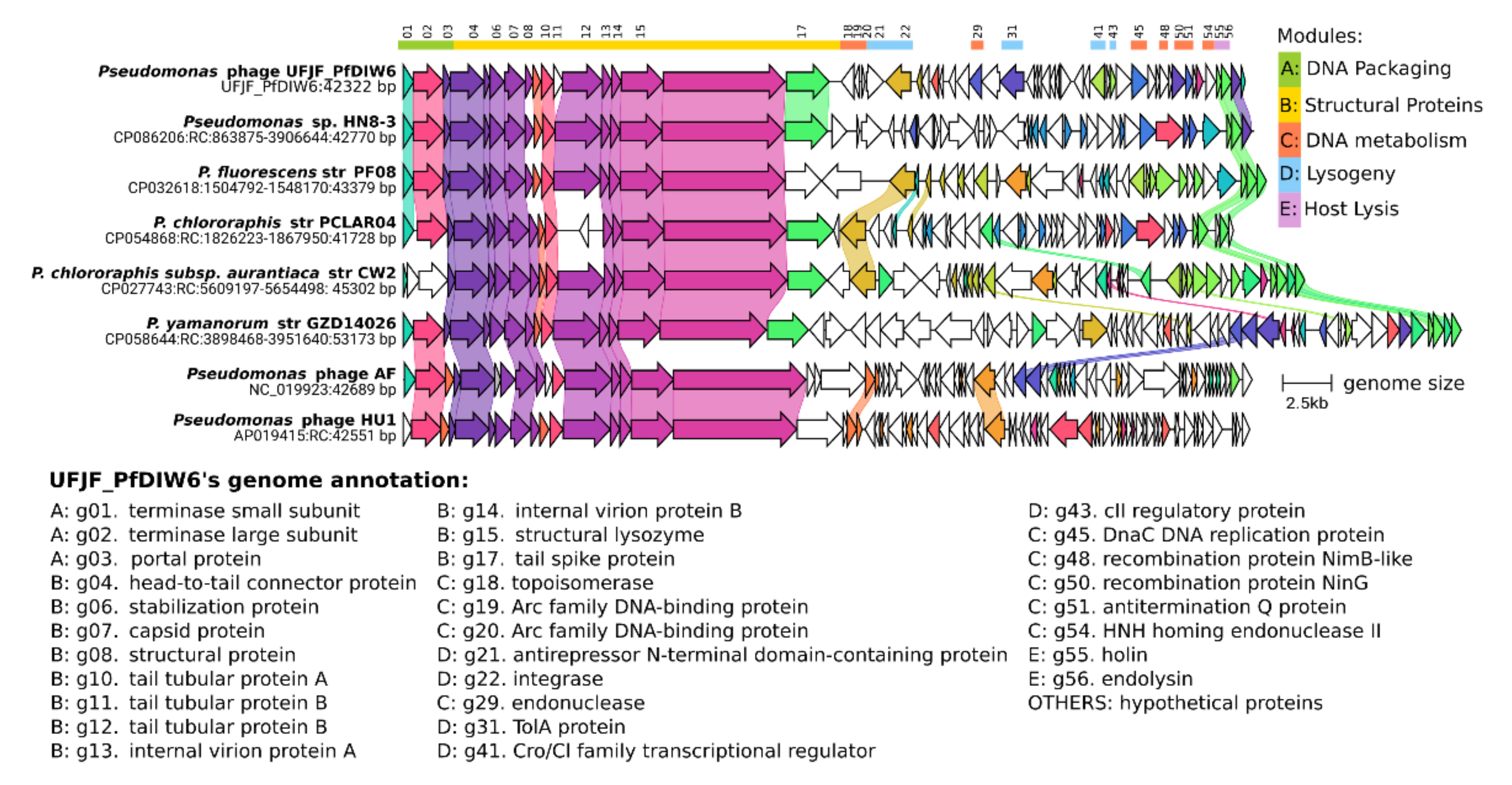
3.2. Comparative Genomics

Similarity searches revealed that the UFJF_PfDIW6’s genome is significantly different from other viral genomes available at the NCBI GenBank database, displaying a maximum coverage of 5% among all alignments (Table S1). This intriguing result led to an investigation into whether its genome could be similar to putative prophages integrated into bacterial genomes once bacteria can harbour several phages that are still uncharacterised [93]. UFJF_PfDIW6’s genome showed higher sequence coverages when aligned with genomes from Pseudomonas species, ranging from 38% to 49% (Table S1). In addition, all bacteria’s genomic regions that were targets of the alignments contain putative prophages classified as intact by the PHASTER.
Five prophage genomes and two genomes of Pseudomonas phages (AF and HU1) were selected and compared to UFJF_PfDIW6’s genome (Figure 1 and Figure S2). The selected genomes were re-opened to the gene that encodes terminase small subunit (TerS). Comparative genomic analysis revealed that they share conserved locally collinear blocks (LCBs) comprising genes of DNA packing and structural proteins modules. UFJF_PfDIW6’s genome shares Pseudomonas prophage’s one LCB, which includes two genes of the host lysis module. The genes included in these LCBs are syntenic and encode proteins related to virion assembly and its interaction with host bacteria. Based on these genomic features, some insights about the host spectrum of UFJF_PfDIW6 phage can be considered, such as its ability to infect other Pseudomonas species.
Among structural proteins, tail spike protein was suggested as being the only virion component of Pseudomonas phage AF that determines its host specificity [94]. The protein encoded by gene g17 of UFJF_PfDIW6’s genome only showed significant similarity with a tail spike protein from a metagenome-assembled genome (MAG) sequence classified as Siphoviridae sp. (Table S2). However, UFJF_PfDIW6’s tail spike protein also showed a high similarity with hypothetical proteins of putative prophages of Pseudomonas sp. HN8-3 (NCBI accession WP_228308670.1; 95% of similarity), P. fragi (WP_169907497.1; 81%), P. lundensis (WP_070413385.1; 81%) and P. lactis (WP_057976804.1; 82). These findings suggest a broader host range spectrum for UFJF_PfDIW6 phage and corroborate the experimental evidence about its ability in reducing raw milk microbiota [27].
In contrast to the structural and host lysis modules, the genomic region, including the modules of DNA metabolism and lysogeny, is the primary point of differentiation among all analysed genomes. This region corresponds to 44% of UFJF_PfDIW6’s genome and is delimited by genes g18 (position 22,019) and g54 (position 40,821), comprising most of the genes that encode proteins with unknown function (Table S4).

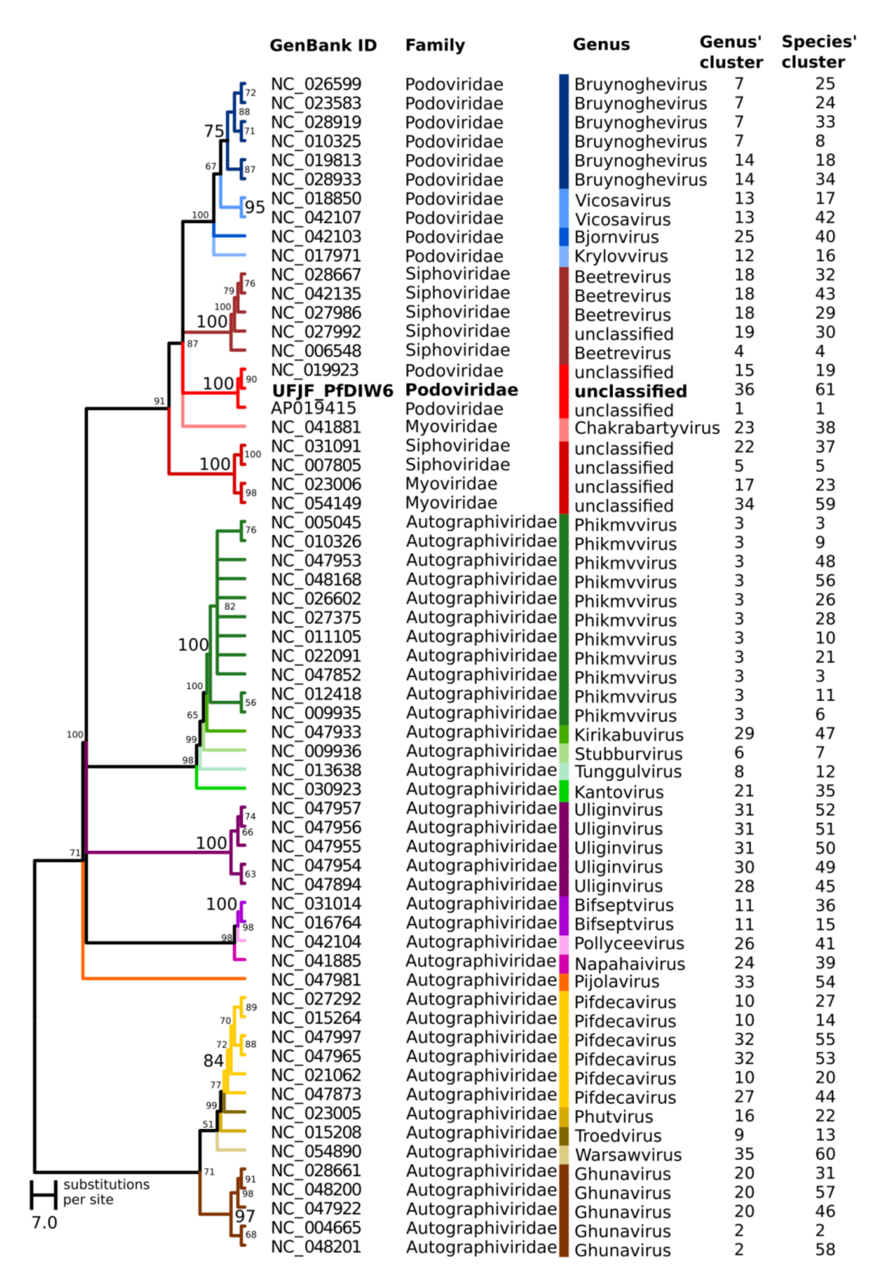
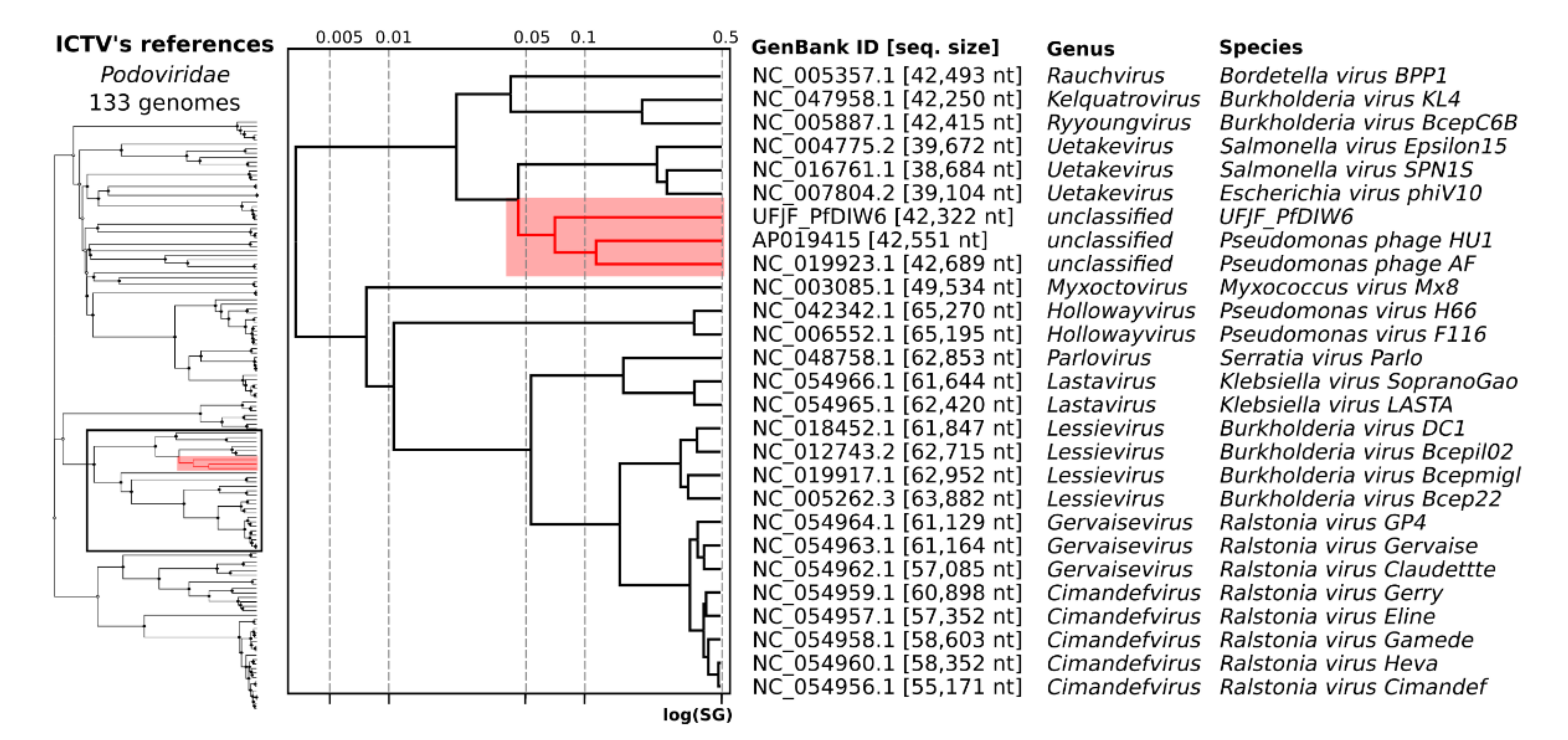
3.3. Taxonomic Assignment

Because comparative genomics revealed conserved regions among UFJF_PfDIW6’s genome and other phages’ genomes, the question is its taxonomic classification.
The genomic similarity matrix calculated by VIRIDIC showed that UFJF_PfDIW6’s genome is different from all reference genomes of species classified in the Podoviridae family listed in the current release of the International Committee on Taxonomy of Viruses (ICTV). UFJF_PfDIW6’s genome was placed in different species and genus clusters from these genomes (Table S8). Among RefSeq genomes, the genome of Salmonella virus Epsilon 15, from the Uetakevirus genus, showed the highest similarity with 5.54%, far from the genomic similarity threshold of 95% for species assignment and 70% for the genus. The genome of prophage identified in Pseudomonas sp. HN8-3 with 47.81% was the most similar among the genomes presented in Figure 1. In addition, the genomes of Pseudomonas phage AF and Pseudomonas phage HU1 showed 6.83% and 10.22% of genomic similarity, respectively (Table S9).
A phylogenetic hypothesis was inferred using the terminase large subunits (TerL) protein sequences from UFJF_PfDIW6 and other Pseudomonas phages as evolutionary markers to solve its taxonomic assignment (Figure 2). All genera were included in monophyletic clades in the phylogenetic tree, supported by high bootstrap values and reinforcing the TerL as an excellent phylogenetic marker for phages’ genus delimitation.
The UFJF_PfDIW6 phage was clustered with Pseudomonas phage AF and Pseudomonas phage HU1, which displayed conserved regions in comparative genomic analysis representing a monophyletic cluster of the unclassified genus in the Podoviridae clade. A genomic similarity matrix was also calculated for these Pseudomonas phages and revealed that even phages previously classified in the same genus could be placed in different genus clusters by VIRIDIC (Figure 2). In addition, UFJF_PfDIW6 phage, Pseudomonas phage AF and Pseudomonas phage HU1 were also placed in different genus and species clusters.
The viral proteomic tree (ViPTree), including the reference genomes of the Podoviridae family, also clustered the genomes of UFJF_PfDIW6 phage, Pseudomonas phage AF and Pseudomonas phage HU1 in a distinct monophyletic cluster of unclassified viruses (Figure 3), such as that observed in the phylogenetic tree based on TerL proteins of Pseudomonas phages. Taken together, all evidence presented here reinforces that the UFJF_PfDIW6 phage is representative of a novel genus of the Podoviridae family, such that Pseudomonas phage AF and Pseudomonas phage HU1 are also single species of novel genera. These findings are in line with the need for taxonomic revision of the order Caudovirales through genome-based approaches [36].

4. Conclusions

In this study, we report a taxonomic proposal for Pseudomonas phage UFJF_PfDIW6 to represent a novel genus of the Podoviridae family. The UFJF_PfDIW6’s genome comprises 42,322 bp of linear double-stranded DNA containing 58 genes organised into five functional modules. Despite UFJF_PfDIW6 showing a lytic behaviour in the in vitro assays on the host lawn, a set of genes related to lysogeny was annotated on its genome. Comparative genomic analysis showed that UFJF_PfDIW6 shares, with other Pseudomonas phages, conserved locally collinear blocks (LCBs) comprising genes of DNA packing and structural protein modules related to virion assembly and host recognition. However, UFJF_PfDIW6’s sequence is distinct from other genomes of virulent phages and prophages, having genomic similarities below the recommended thresholds for genus and species assignments. Phylogenetic analysis, using the terminase large subunit (TerL) protein sequences and viral proteomic tree, clustered UFJF_PfDIW6 into a monophyletic cluster of unclassified viruses, being a single representative of a novel genus in the Podoviridae clade.

Supplementary Materials

The following supporting information can be downloaded at: https://www.mdpi.com/article/10.3390/v14030629/s1, Figure S1. Phenotypic features of Pseudomonas phage UFJF_PfDIW6. (A) Lysis plaques formed by UFJF_PfDIW6 on the lawn of P. fluorescens UFV 041. Incubation conditions: BHI agar at 30 °C for 24 h. (B) Virion morphology. Transmission electron micrograph (TEM) negatively stained with 2% uranyl acetate. Bar magnification: 140,000X. * Tests performed in TSB medium at 30 °C; ** Test performed against 23 bacterial strains belonging to nine genera. The methodology used for phenotypic characterization of UFJF_PfDIW6 was described in detail by Nascimento et. [27]. Figure S2: Conserved locally collinear blocks (LCBs) shared among Pseudomonas UFJF_PfDIW6 phage, Pseudomonas sp. HN8-3 prophage, P. fluorescens str PF08 prophage, P. chlororaphis str PCLAR04 prophage, P. chlororaphis subsp. aurantiaca str CW2 prophage, P. yamanorum str GZD14026 pro-phage, Pseudomonas phage AF, and Pseudomonas phage HU1 genomes. The alignment was performed using progressive Mauve with default parameters. The coloured blocks correspond to the LCBs predicted by Mauve. Horizontal white bars correspond to the Coding DNA sequences (CDSs) annotated in each genome. Table S1: Similarity searches between UFJF_PfDIW6 genome sequence and genomes available at NCBI database. Sequences were aligned using the BLASTn tool BLAST. An E-value threshold ≤ 1e−10 was considered for selecting significant alignments. Table S2: Similarity searches among the proteins annotated in UFJF_PfDIW6 genome sequence and sequences available at NCBI Protein Database (nr). Sequences were aligned using the BLASTp tool of BLAST. An E-value threshold ≤ 1e−10 was considered for selecting significant alignments. Table S3: Functional classification of UFJF_PfDIW6 proteins in Virus Orthologous Groups (VOG). Table S4: Genome annotation of Phage UFJF_PfDIW6. Table S5: Host prediction of phage UFJF_PfDIW6. Table S6: Rho-independent terminators of Phage UFJF_PfDIW6 genome. Table S7: Promoters of Phage UFJF_PfDIW6 genome. Table S8: Intergenomic similarities between UFJF_PfDIW6 genome and genomes of Podoviridae species recognised by the International Committee on Taxonomy of Viruses (ICTV). Genome sequences were compared by VIRIDIC and clustered, considering a genomic similarity threshold of 95% for species assignment and 70% for the genus. Table S9: Intergenomic similarities between UFJF_PfDIW6 genome and genomes presented in Figure 1. Genome sequences were compared by VIRIDIC and clustered, considering a genomic similarity threshold of 95% for species assignment and 70% for the genus.

Author Contributions

Conceptualisation, P.M.P.V., H.M.H., M.E.S.L. and M.T.P.G.; formal analysis, P.M.P.V. and M.T.P.G.; investigation, E.C.d.N. and F.G.d.C.O.; resources, H.M.H. and M.E.S.L.; data curation, P.M.P.V.; writing—original draft preparation, P.M.P.V., H.M.H., M.E.S.L. and M.T.P.G.; writing—review and editing, P.M.P.V., H.M.H., M.E.S.L. and M.T.P.G.; visualisation, P.M.P.V., H.M.H., M.E.S.L. and M.T.P.G.; supervision, P.M.P.V. and H.M.H.; project administration, H.M.H., P.M.P.V. and M.E.S.L.; funding acquisition, H.M.H. and M.E.S.L. All authors have read and agreed to the published version of the manuscript.

Funding

This research was funded by Fundação de Amparo à Pesquisa do Estado de Minas Gerais (FAPEMIG), grant number CAG00390-15, and Conselho Nacional de Desenvolvimento Científico e Tecnológico (CNPq), grant number 427667/2016-0. The APC was funded by Vicerrectoria de Investigación y Extensión–Universidad de Córdoba, Montería–Colombia.

Institutional Review Board Statement

Not applicable.

Informed Consent Statement

Not applicable.

Data Availability Statement

The data presented in this study are openly available at the NCBI (https://www.ncbi.nlm.nih.gov/genbank/). The complete genome sequence of phage UFJF_PfDIW6 is available at GenBank database, accession number OM418631.

Acknowledgments

We thank Cristina Dantas Vanetti of the Universidade Federal de Viçosa (UFV) for providing the host bacterial strain used in this work and the Núcleo de Microscopia e Microanálise of UFV, Brazil, for transmission electron microscopy (TEM) analysis of the phage UFJF_PfDIW6. We are also grateful to the Núcleo de Análise de Biomoléculas (NuBioMol) of the Universidade Federal de Viçosa (UFV) for providing the facilities for data analysis. NuBioMol is financially supported by the following Brazilian agencies: Fundação de Amparo à Pesquisa do Estado de Minas Gerais (FAPEMIG), Coordenação de Aperfeiçoamento de Pessoal de Nível Superior (CAPES), Conselho Nacional de Desenvolvimento Científico e Tecnológico (CNPq), Financiadora de Estudos e Projetos (Finep) and Sistema Nacional de Laboratórios em Nanotecnologias (SisNANO)/Ministério da Ciência, Tecnologia e Informação (MCTI).

Conflicts of Interest

The authors declare no conflict of interest. The funders had no role in the study’s design, in the collection, analyses, or interpretation of data, in the writing of the manuscript, or in the decision to publish the results.

References

  1. De Smet, J.; Hendrix, H.; Blasdel, B.G.; Danis-Wlodarczyk, K.; Lavigne, R. Pseudomonas predators: Understanding and exploiting phage–host interactions. Nat. Rev. Microbiol. 2017, 15, 517–530. [Google Scholar] [CrossRef]
  2. Silby, M.W.; Winstanley, C.; Godfrey, S.A.C.; Levy, S.B.; Jackson, R.W. Pseudomonas Genomes: Diverse and adaptable. FEMS Microbiol. Rev. 2011, 35, 652–680. [Google Scholar] [CrossRef] [PubMed] [Green Version]
  3. Rodrigues, R.d.S.; Machado, S.G.; de Carvalho, A.F.; Nero, L.A. Pseudomonas sp. as the causative agent of anomalous blue discoloration in Brazilian fresh soft cheese (Minas Frescal). Int. Dairy J. 2021, 117, 105020. [Google Scholar] [CrossRef]
  4. Machado, S.G.; Baglinière, F.; Marchand, S.; Van Coillie, E.; Vanetti, M.C.D.; De Block, J.; Heyndrickx, M. The Biodiversity of the Microbiota Producing Heat-Resistant Enzymes Responsible for Spoilage in Processed Bovine Milk and Dairy Products. Front. Microbiol. 2017, 8, 302. [Google Scholar] [CrossRef] [PubMed] [Green Version]
  5. Meng, L.; Zhang, Y.; Liu, H.; Zhao, S.; Wang, J.; Zheng, N. Characterization of Pseudomonas spp. and Associated Proteolytic Properties in Raw Milk Stored at Low Temperatures. Front. Microbiol. 2017, 8, 2158. [Google Scholar] [CrossRef] [PubMed] [Green Version]
  6. Ribeiro Júnior, J.C.; Teider Junior, P.I.; Oliveira, A.L.M.; Rios, E.A.; Tamanini, R.; Beloti, V. Proteolytic and Lipolytic Potential of Pseudomonas spp. from Goat and Bovine Raw Milk. Pesq. Vet. Bras. 2018, 38, 1577–1583. [Google Scholar] [CrossRef]
  7. Matselis, E.; Roussis, I.G. Proteinase and lipase production by Pseudomonas fluorescens. Proteolysis and lipolysis in thermized ewe’s milk. Food Control 1998, 9, 251–259. [Google Scholar] [CrossRef]
  8. Endersen, L.; Coffey, A. The use of bacteriophages for food safety. Curr. Opin. Food Sci. 2020, 36, 1–8. [Google Scholar] [CrossRef]
  9. O’Sullivan, L.; Bolton, D.; Mcauliffe, O.; Coffey, A. Bacteriophages in Food Applications: From Foe to Friend. Annu. Rev. Food Sci. Technol. 2018, 10, 151–172. [Google Scholar] [CrossRef]
  10. O’Sullivan, L.; Bolton, D.; McAuliffe, O.; Coffey, A. The use of bacteriophages to control and detect pathogens in the dairy industry. Int. J. Dairy Technol. 2020, 73, 1–11. [Google Scholar] [CrossRef]
  11. García-Anaya, M.C.; Sepulveda, D.R.; Sáenz-Mendoza, A.I.; Rios-Velasco, C.; Zamudio-Flores, P.B.; Acosta-Muñiz, C.H. Phages as biocontrol agents in dairy products. Trends Food Sci. Technol. 2020, 95, 10–20. [Google Scholar] [CrossRef]
  12. Garcia, P.; Madera, C.; Martínez, B.; Rodriguez, A.; Suárez, J.E. Prevalence of bacteriophages infecting Staphylococcus aureus in dairy samples and their potential as biocontrol agents. J. Dairy Sci. 2009, 92, 3019–3026. [Google Scholar] [CrossRef] [PubMed]
  13. O’Flaherty, S.; Coffey, A.; Meaney, W.J.; Fitzgerald, G.F.; Ross, R.P. Inhibition of bacteriophage K proliferation on Staphylococcus aureus in raw bovine milk. Lett. Appl. Microbiol. 2005, 41, 274–279. [Google Scholar] [CrossRef] [PubMed]
  14. García, P.; Madera, C.; Martínez, B.; Rodríguez, A. Biocontrol of Staphylococcus aureus in curd manufacturing processes using bacteriophages. Int. Dairy J. 2007, 17, 1232–1239. [Google Scholar] [CrossRef]
  15. El Haddad, L.; Roy, J.-P.; Khalil, G.E.; St-Gelais, D.; Champagne, C.P.; Labrie, S.; Moineau, S. Efficacy of two Staphylococcus aureus phage cocktails in cheese production. Int. J. Food Microbiol. 2016, 217, 7–13. [Google Scholar] [CrossRef]
  16. Silva, E.N.G.; Figueiredo, A.C.L.; Miranda, F.A.; Almeida, R.C.d.C. Control of Listeria monocytogenes growth in soft cheeses by bacteriophage P100. Braz. J. Microbiol. 2014, 45, 11–16. [Google Scholar] [CrossRef] [Green Version]
  17. Perera, M.N.; Abuladze, T.; Li, M.; Woolston, J.; Sulakvelidze, A. Bacteriophage cocktail significantly reduces or eliminates Listeria monocytogenes contamination on lettuce, apples, cheese, smoked salmon and frozen foods. Food Microbiol. 2015, 52, 42–48. [Google Scholar] [CrossRef]
  18. Carlton, R.M.; Noordman, W.H.; Biswas, B.; de Meester, E.D.; Loessner, M.J. Bacteriophage P100 for control of Listeria monocytogenes in foods: Genome sequence, bioinformatic analyses, oral toxicity study, and application. Regul. Toxicol. Pharmacol. 2005, 43, 301–312. [Google Scholar] [CrossRef]
  19. Guenther, S.; Loessner, M.J. Bacteriophage biocontrol of Listeria monocytogenes on soft ripened white mold and red-smear cheeses. Bacteriophage 2011, 1, 94–100. [Google Scholar] [CrossRef] [Green Version]
  20. Guenther, S.; Huwyler, D.; Richard, S.; Loessner, M.J. Virulent Bacteriophage for Efficient Biocontrol of Listeria monocytogenes in Ready-To-Eat Foods. Appl. Environ. Microbiol. 2009, 75, 93–100. [Google Scholar] [CrossRef] [Green Version]
  21. McLean, S.K.; Dunn, L.A.; Palombo, E.A. Phage Inhibition of Escherichia coli in Ultrahigh-Temperature-Treated and Raw Milk. Foodborne Pathog. Dis. 2013, 10, 956–962. [Google Scholar] [CrossRef] [PubMed]
  22. Grygorcewicz, B.; Chajęcka-Wierzchowska, W.; Augustyniak, A.; Wasak, A.; Stachurska, X.; Nawrotek, P.; Dołęgowska, B. In-milk inactivation of Escherichia coli O157:H7 by the environmental lytic bacteriophage ECPS-6. J. Food Saf. 2020, 40. [Google Scholar] [CrossRef]
  23. Modi, R.; Hirvi, Y.; Hill, A.; Griffiths, M.W. Effect of Phage on Survival of Salmonella Enteritidis during Manufacture and Storage of Cheddar Cheese Made from Raw and Pasteurized Milk. J. Food Prot. 2001, 64, 927–933. [Google Scholar] [CrossRef] [PubMed]
  24. Zinno, P.; Devirgiliis, C.; Ercolini, D.; Ongeng, D.; Mauriello, G. Bacteriophage P22 to challenge Salmonella in foods. Int. J. Food Microbiol. 2014, 191, 69–74. [Google Scholar] [CrossRef]
  25. Tanaka, C.; Yamada, K.; Takeuchi, H.; Inokuchi, Y.; Kashiwagi, A.; Toba, T. A Lytic Bacteriophage for Controlling Pseudomonas lactis in Raw Cow’s Milk. Appl. Environ. Microbiol. 2018, 84, e00111-18. [Google Scholar] [CrossRef] [Green Version]
  26. Hu, Z.; Meng, X.-C.; Liu, F. Isolation and characterisation of lytic bacteriophages against Pseudomonas spp., a novel biological intervention for preventing spoilage of raw milk. Int. Dairy J. 2016, 55, 72–78. [Google Scholar] [CrossRef]
  27. do Nascimento, E.C.; Sabino, M.C.; Corguinha, L.d.R.; Targino, B.N.; Lange, C.C.; Pinto, C.L.d.O.; Pinto, P.d.F.; Vidigal, P.M.P.; Sant’Ana, A.S.; Hungaro, H.M. Lytic bacteriophages UFJF_PfDIW6 and UFJF_PfSW6 prevent Pseudomonas fluorescens growth in vitro and the proteolytic-caused spoilage of raw milk during chilled storage. Food Microbiol. 2022, 101, 103892. [Google Scholar] [CrossRef]
  28. Fernández, L.; Escobedo, S.; Gutiérrez, D.; Portilla, S.; Martínez, B.; García, P.; Rodríguez, A. Bacteriophages in the Dairy Environment: From Enemies to Allies. Antibiotics 2017, 6, 27. [Google Scholar] [CrossRef] [Green Version]
  29. Eller, M.R.; Salgado, R.L.; Vidigal, P.M.P.; Alves, M.P.; Dias, R.S.; de Oliveira, L.L.; da Silva, C.C.; de Carvalho, A.F.; de Paula, S.O. Complete Genome Sequence of the Pseudomonas fluorescens Bacteriophage UFV-P2. Genome Announc. 2013, 1, e00006-12. [Google Scholar] [CrossRef] [Green Version]
  30. Koberg, S.; Gieschler, S.; Brinks, E.; Wenning, M.; Neve, H.; Franz, C.M.A.P. Genome sequence of the novel virulent bacteriophage PMBT14 with lytic activity against Pseudomonas fluorescens DSM 50090R. Arch. Virol. 2018, 163, 2575–2577. [Google Scholar] [CrossRef]
  31. Sillankorva, S.; Neubauer, P.; Azeredo, J. Isolation and Characterization of a T7-like Lytic Phage for Pseudomonas fluorescens. BMC Biotechnol. 2008, 8, 80. [Google Scholar] [CrossRef] [PubMed] [Green Version]
  32. Sun, W.-J.; Liu, C.-F.; Yu, L.; Cui, F.-J.; Zhou, Q.; Yu, S.-L.; Sun, L. A novel bacteriophage KSL-1 of 2-Keto-gluconic acid producer Pseudomonas fluorescens K1005: Isolation, characterization and its remedial action. BMC Microbiol. 2012, 12, 127. [Google Scholar] [CrossRef] [PubMed] [Green Version]
  33. Wojtus, J.K.; Fitch, J.L.; Christian, E.; Dalefield, T.; Lawes, J.K.; Kumar, K.; Peebles, C.L.; Altermann, E.; Hendrickson, H.L. Complete Genome Sequences of Three Novel Pseudomonas fluorescens SBW25 Bacteriophages, Noxifer, Phabio, and Skulduggery. Genome Announc. 2017, 5, e00725-17. [Google Scholar] [CrossRef] [PubMed] [Green Version]
  34. Xiang, Y.; Wang, S.; Li, J.; Wei, Y.; Zhang, Q.; Lin, L.; Ji, X. Isolation and characterization of two lytic cold-active bacteriophages infecting Pseudomonas fluorescens from the Napahai plateau wetland. Can. J. Microbiol. 2018, 64, 183–190. [Google Scholar] [CrossRef]
  35. Zhang, C.; Zhang, Z.; Li, J.; Qin, K.; Wei, Y.; Zhang, Q.; Lin, L.; Xiuling, J. Complete genome sequence of the lytic cold-active Pseudomonas fluorescens bacteriophage VSW-3 from Napahai plateau wetland. Virus Genes 2016, 53, 146–150. [Google Scholar] [CrossRef]
  36. Turner, D.; Kropinski, A.M.; Adriaenssens, E.M. A Roadmap for Genome-Based Phage Taxonomy. Viruses 2021, 13, 506. [Google Scholar] [CrossRef]
  37. Sambrook, J.; Russell, D.W. Molecular Cloning: A Laboratory Manual; Cold Spring Harbor Laboratory Press: Cold Spring Harbor, NY, USA, 2001; p. 999. [Google Scholar]
  38. Summer, E.J. Preparation of a Phage DNA Fragment Library for Whole Genome Shotgun Sequencing. Methods Mol. Biol. 2009, 502, 27–46. [Google Scholar] [CrossRef]
  39. Krueger, F.; James, F.; Ewels, P.; Afyounian, E.; Schuster-Boeckler, B. FelixKrueger/TrimGalore: v0.6.7-DOI via Zenodo. 2021. Available online: https://zenodo.org/record/5127899#.YjMyx3rMJPZ (accessed on 26 October 2021).
  40. Bolger, A.M.; Lohse, M.; Usadel, B. Trimmomatic: A flexible trimmer for Illumina sequence data. Bioinformatics 2014, 30, 2114–2120. [Google Scholar] [CrossRef] [Green Version]
  41. Bankevich, A.; Nurk, S.; Antipov, D.; Gurevich, A.A.; Dvorkin, M.; Kulikov, A.S.; Lesin, V.M.; Nikolenko, S.I.; Pham, S.; Prjibelski, A.D.; et al. SPAdes: A new genome assembly algorithm and its applications to single-cell sequencing. J. Comput. Biol. 2012, 19, 455–477. [Google Scholar] [CrossRef] [Green Version]
  42. Li, H.; Durbin, R. Fast and accurate short read alignment with Burrows-Wheeler transform. Bioinformatics 2009, 25, 1754–1760. [Google Scholar] [CrossRef] [Green Version]
  43. Boetzer, M.; Henkel, C.V.; Jansen, H.J.; Butler, D.; Pirovano, W. Scaffolding pre-assembled contigs using SSPACE. Bioinformatics 2011, 27, 578–579. [Google Scholar] [CrossRef] [PubMed] [Green Version]
  44. Garneau, J.R.; Depardieu, F.; Fortier, L.-C.; Bikard, D.; Monot, M. PhageTerm: A tool for fast and accurate determination of phage termini and packaging mechanism using next-generation sequencing data. Sci. Rep. 2017, 7, 8292. [Google Scholar] [CrossRef] [PubMed]
  45. Seemann, T. Prokka: Rapid Prokaryotic Genome Annotation. Bioinformatics 2014, 30, 2068–2069. [Google Scholar] [CrossRef] [PubMed]
  46. Sampaio, M.; Rocha, M.; Oliveira, H.; Dias, O. Predicting promoters in phage genomes using PhagePromoter. Bioinformatics 2019, 35, 5301–5302. [Google Scholar] [CrossRef] [PubMed]
  47. Lesnik, E.A.; Sampath, R.; Levene, H.B.; Henderson, T.J.; McNeil, J.A.; Ecker, D.J. Prediction of rho-independent transcriptional terminators in Escherichia coli. Nucleic Acids Res. 2001, 29, 3583–3594. [Google Scholar] [CrossRef] [Green Version]
  48. Villarroel, J.; Kleinheinz, K.A.; Jurtz, V.I.; Zschach, H.; Lund, O.; Nielsen, M.; Larsen, M.V. HostPhinder: A Phage Host Prediction Tool. Viruses 2016, 8, 116. [Google Scholar] [CrossRef]
  49. Hockenberry, A.J.; Wilke, C.O. BACPHLIP: Predicting Bacteriophage Lifestyle from Conserved Protein Domains. PeerJ 2021, 9, e11396. [Google Scholar] [CrossRef]
  50. Altschul, S.F.; Gish, W.; Miller, W.; Myers, E.W.; Lipman, D.J. Basic local alignment search tool. J. Mol. Biol. 1990, 215, 403–410. [Google Scholar] [CrossRef]
  51. Arndt, D.; Grant, J.R.; Marcu, A.; Sajed, T.; Pon, A.; Liang, Y.; Wishart, D.S. PHASTER: A better, faster version of the PHAST phage search tool. Nucleic Acids Res. 2016, 44, W16–W21. [Google Scholar] [CrossRef] [Green Version]
  52. Zhou, Y.; Liang, Y.; Lynch, K.H.; Dennis, J.J.; Wishart, D.S. PHAST: A Fast Phage Search Tool. Nucleic Acids Res. 2011, 39, W347–W352. [Google Scholar] [CrossRef]
  53. Eddy, S.R. Accelerated Profile HMM Searches. PLOS Comput. Biol. 2011, 7, e1002195. [Google Scholar] [CrossRef] [PubMed] [Green Version]
  54. Potter, S.C.; Luciani, A.; Eddy, S.R.; Park, Y.; López, R.; Finn, R.D. HMMER web server: 2018 update. Nucleic Acids Res. 2018, 46, W200–W204. [Google Scholar] [CrossRef] [Green Version]
  55. Finn, R.D.; Bateman, A.; Clements, J.; Coggill, P.; Eberhardt, R.Y.; Eddy, S.R.; Heger, A.; Hetherington, K.; Holm, L.; Mistry, J.; et al. Pfam: The protein families database. Nucleic Acids Res. 2014, 42, D222–D230. [Google Scholar] [CrossRef] [PubMed] [Green Version]
  56. Gontijo, M.T.P.; Vidigal, P.M.P.; Lopez, M.E.S.; Brocchi, M. Bacteriophages that infect Gram-negative bacteria as source of signal-arrest-release motif lysins. Res. Microbiol. 2021, 172, 103794. [Google Scholar] [CrossRef] [PubMed]
  57. Oliveira, H.; Melo, L.D.R.; Santos, S.B.; Nóbrega, F.L.; Ferreira, E.C.; Cerca, N.; Azeredo, J.; Kluskens, L.D. Molecular Aspects and Comparative Genomics of Bacteriophage Endolysins. J. Virol. 2013, 87, 4558–4570. [Google Scholar] [CrossRef] [PubMed] [Green Version]
  58. Almagro Armenteros, J.J.; Tsirigos, K.D.; Sønderby, C.K.; Petersen, T.N.; Winther, O.; Brunak, S.; Von Heijne, G.; Nielsen, H. SignalP 5.0 improves signal peptide predictions using deep neural networks. Nat. Biotechnol. 2019, 37, 420–423. [Google Scholar] [CrossRef] [PubMed]
  59. Hiller, K.; Grote, A.; Scheer, M.; Münch, R.; Jahn, D. PrediSi: Prediction of signal peptides and their cleavage positions. Nucleic Acids Res. 2004, 32, W375–W379. [Google Scholar] [CrossRef]
  60. Hirokawa, T.; Boon-Chieng, S.; Mitaku, S. SOSUI: Classification and secondary structure prediction system for membrane proteins. Bioinformatics 1998, 14, 378–379. [Google Scholar] [CrossRef] [Green Version]
  61. Käll, L.; Krogh, A.; Sonnhammer, E.L. A Combined Transmembrane Topology and Signal Peptide Prediction Method. J. Mol. Biol. 2004, 338, 1027–1036. [Google Scholar] [CrossRef]
  62. Tsirigos, K.D.; Peters, C.; Shu, N.; Käll, L.; Elofsson, A. The TOPCONS web server for consensus prediction of membrane protein topology and signal peptides. Nucleic Acids Res. 2015, 43, W401–W407. [Google Scholar] [CrossRef]
  63. Darling, A.E.; Mau, B.; Perna, N.T.; Batzoglou, S.; Zhong, Y. progressiveMauve: Multiple Genome Alignment with Gene Gain, Loss and Rearrangement. PLoS ONE 2010, 5, e11147. [Google Scholar] [CrossRef] [PubMed] [Green Version]
  64. Gilchrist, C.L.M.; Chooi, Y.-H. clinker & clustermap.js: Automatic generation of gene cluster comparison figures. Bioinformatics 2021, 37, 2473–2475. [Google Scholar] [CrossRef]
  65. Moraru, C.; Varsani, A.; Kropinski, A.M. VIRIDIC—A Novel Tool to Calculate the Intergenomic Similarities of Prokaryote-Infecting Viruses. Viruses 2020, 12, 1268. [Google Scholar] [CrossRef] [PubMed]
  66. Katoh, K.; Standley, D.M. MAFFT Multiple Sequence Alignment Software Version 7: Improvements in Performance and Usability. Mol. Biol. Evol. 2013, 30, 772–780. [Google Scholar] [CrossRef] [PubMed] [Green Version]
  67. Capella-Gutiérrez, S.; Silla-Martínez, J.M.; Gabaldón, T. trimAl: A tool for automated alignment trimming in large-scale phylogenetic analyses. Bioinformatics 2009, 25, 1972–1973. [Google Scholar] [CrossRef] [PubMed]
  68. Darriba, D.; Posada, D.; Kozlov, A.M.; Stamatakis, A.; Morel, B.; Flouri, T. ModelTest-NG: A New and Scalable Tool for the Selection of DNA and Protein Evolutionary Models. Mol. Biol. Evol. 2020, 37, 291–294. [Google Scholar] [CrossRef] [PubMed] [Green Version]
  69. Kozlov, A.M.; Darriba, D.; Flouri, T.; Morel, B.; Stamatakis, A. RAxML-NG: A fast, scalable and user-friendly tool for maximum likelihood phylogenetic inference. Bioinformatics 2019, 35, 4453–4455. [Google Scholar] [CrossRef] [Green Version]
  70. Shen, X.; Li, M.; Zeng, Y.; Hu, X.; Tan, Y.; Rao, X.; Jin, X.; Li, S.; Zhu, J.; Zhang, K.; et al. Functional Identifica-tion of the DNA Packaging Terminase from Pseudomonas aeruginosa Phage PaP3. Arch. Virol. 2012, 157, 2133–2141. [Google Scholar] [CrossRef] [Green Version]
  71. Fujisawa, H.; Morita, M. Phage DNA packaging. Genes Cells 1997, 2, 537–545. [Google Scholar] [CrossRef]
  72. Prevelige, P.E.; Cortines, J.R. Phage assembly and the special role of the portal protein. Curr. Opin. Virol. 2018, 31, 66–73. [Google Scholar] [CrossRef]
  73. Leiman, P.G.; Shneider, M.M. Contractile Tail Machines of Bacteriophages. Adv. Exp. Med. Biol. 2012, 726, 93–114. [Google Scholar] [CrossRef] [PubMed]
  74. Hardies, S.C.; Thomas, J.A.; Black, L.; Weintraub, S.T.; Hwang, C.Y.; Cho, B.C. Identification of structural and morphogenesis genes of Pseudoalteromonas phage φRIO-1 and placement within the evolutionary history of Podoviridae. Virology 2016, 489, 116–127. [Google Scholar] [CrossRef] [PubMed] [Green Version]
  75. Hu, B.; Margolin, W.; Molineux, I.J.; Liu, J. The Bacteriophage T7 Virion Undergoes Extensive Structural Re-modeling during Infection. Science 2013, 339, 576–579. [Google Scholar] [CrossRef] [PubMed] [Green Version]
  76. Champoux, J.J. DNA Topoisomerases: Structure, Function, and Mechanism. Annu. Rev. Biochem. 2003, 70, 369–413. [Google Scholar] [CrossRef] [Green Version]
  77. Learn, B.A.; Um, S.-J.; Huang, L.; McMacken, R. Cryptic single-stranded-DNA binding activities of the phage λ P and Escherichia coli DnaC replication initiation proteins facilitate the transfer of E. coli DnaB helicase onto DNA. Proc. Natl. Acad. Sci. USA 1997, 94, 1154–1159. [Google Scholar] [CrossRef] [Green Version]
  78. Quiles-Puchalt, N.; Carpena, N.; Alonso, J.C.; Novick, R.P.; Marina, A.; Penadés, J.R. Staphylococcal Pathogenicity Island DNA Packaging System Involving Cos-Site Packaging and Phage-Encoded HNH Endonucleases. Proc. Natl. Acad. Sci. USA 2014, 111, 6016–6021. [Google Scholar] [CrossRef] [Green Version]
  79. Kala, S.; Cumby, N.; Sadowski, P.D.; Hyder, B.Z.; Kanelis, V.; Davidson, A.R.; Maxwell, K.L. HNH Proteins Are a Widespread Component of Phage DNA Packaging Machines. Proc. Natl. Acad. Sci. USA 2014, 111, 6022–6027. [Google Scholar] [CrossRef] [Green Version]
  80. Hochhut, B.; Wilde, C.; Balling, G.; Middendorf, B.; Dobrindt, U.; Brzuszkiewicz, E.; Gottschalk, G.; Carniel, E.; Hacker, J. Role of pathogenicity island-associated integrases in the genome plasticity of uropathogenic Escherichia coli strain 536. Mol. Microbiol. 2006, 61, 584–595. [Google Scholar] [CrossRef]
  81. Wilde, C.; Mazel, D.; Hochhut, B.; Middendorf, B.; Le Roux, F.; Carniel, E.; Dobrindt, U.; Hacker, J. Delineation of the recombination sites necessary for integration of pathogenicity islands II and III into the Escherichia coli 536 chromosome. Mol. Microbiol. 2008, 68, 139–151. [Google Scholar] [CrossRef]
  82. Feiner, R.; Argov, T.; Rabinovich, L.; Sigal, N.; Borovok, I.; Herskovits, A.A. A new perspective on lysogeny: Prophages as active regulatory switches of bacteria. Nat. Rev. Microbiol. 2015, 13, 641–650. [Google Scholar] [CrossRef]
  83. Howard-Varona, C.; Hargreaves, K.R.; Abedon, S.T.; Sullivan, M.B. Lysogeny in nature: Mechanisms, impact and ecology of temperate phages. ISME J. 2017, 11, 1511–1520. [Google Scholar] [CrossRef] [PubMed] [Green Version]
  84. Broussard, G.W.; Oldfield, L.M.; Villanueva, V.M.; Lunt, B.L.; Shine, E.E.; Hatfull, G.F. Integration-Dependent Bacteriophage Immunity Provides Insights into the Evolution of Genetic Switches. Mol. Cell 2013, 49, 237–248. [Google Scholar] [CrossRef] [PubMed] [Green Version]
  85. Chang, Y.; Shin, H.; Lee, J.-H.; Park, C.J.; Paik, S.-Y.; Ryu, S. Isolation and Genome Characterization of the Virulent Staphylococcus aureus Bacteriophage SA97. Viruses 2015, 7, 5225–5242. [Google Scholar] [CrossRef] [PubMed] [Green Version]
  86. Shao, Q.; Trinh, J.T.; Zeng, L. High-resolution studies of lysis–lysogeny decision-making in bacteriophage lambda. J. Biol. Chem. 2019, 294, 3343–3349. [Google Scholar] [CrossRef] [PubMed] [Green Version]
  87. Boeckaerts, D.; Stock, M.; Criel, B.; Gerstmans, H.; De Baets, B.; Briers, Y. Predicting bacteriophage hosts based on sequences of annotated receptor-binding proteins. Sci. Rep. 2021, 11, 1467. [Google Scholar] [CrossRef]
  88. Valero-Rello, A. Diversity, specificity and molecular evolution of the lytic arsenal of Pseudomonas phages: In silico perspective. Environ. Microbiol. 2019, 21, 4136–4150. [Google Scholar] [CrossRef]
  89. Young, R. Phage lysis: Three steps, three choices, one outcome. J. Microbiol. 2014, 52, 243–258. [Google Scholar] [CrossRef]
  90. Wang, I.-N.; Smith, D.L.; Young, R. Holins: The Protein Clocks of Bacteriophage Infections. Annu. Rev. Microbiol. 2000, 54, 799–825. [Google Scholar] [CrossRef]
  91. Catalao, M.J.; Gil, F.; Moniz-Pereira, J.; São-José, C.; Pimentel, M. Diversity in bacterial lysis systems: Bacteriophages show the way. FEMS Microbiol. Rev. 2013, 37, 554–571. [Google Scholar] [CrossRef] [Green Version]
  92. Rajaure, M.; Berry, J.; Kongari, R.; Cahill, J.; Young, R. Membrane Fusion during Phage Lysis. Proc. Natl. Acad. Sci. USA 2015, 112, 5497–5502. [Google Scholar] [CrossRef] [Green Version]
  93. Crispim, J.S.; Dias, R.S.; Vidigal, P.M.P.; De Sousa, M.P.; Da Silva, C.C.; Santana, M.F.; De Paula, S.O. Screening and characterization of prophages in Desulfovibrio genomes. Sci. Rep. 2018, 8, 9273. [Google Scholar] [CrossRef] [PubMed]
  94. Cornelissen, A.; Ceyssens, P.-J.; Krylov, V.N.; Noben, J.-P.; Volckaert, G.; Lavigne, R. Identification of EPS-degrading activity within the tail spikes of the novel Pseudomonas putida phage AF. Virology 2012, 434, 251–256. [Google Scholar] [CrossRef] [PubMed] [Green Version]
Figure 1. Genomic features of Pseudomonas phage UFJF_PfDIW6. UFJF_PfDIW6’s genome is shown above all others. Its sequence has a bidirectional organisation with 58 genes (represented by arrows) organised into five functional modules, represented by the coloured bars and indicated in the genes’ annotation list. The numbers above the arrows indicate the genes with predicted functions, shown in the annotation list. The arrow’s colours represent the gene clusters identified by Clinker, which encode similar proteins. The lines connecting the arrows represent gene-encoding proteins that share sequence identity more significant than 40%.
Viruses 14 00629 g001
Figure 2. Maximum likelihood tree of the large terminase subunit of phages infecting the genus Pseudomonas. The midpoint rooted maximum likelihood (ML) tree was obtained by analysing terminase large subunit (TerL) amino acid sequences from UFJF_PfDIW6 and other Pseudomonas phages available at NCBI RefSeq database. The bootstrap values (expressed as percentages) calculated from 1000 replicates are shown beside each node. The monophyletic clades corresponding to each genus are shown in different colours. The genomes of Pseudomonas phages were clustered by VIRIDIC using the genomic similarity threshold of 70% for genus assignment (Genus’ cluster) and 95% for species assignment (Species’ cluster).
Viruses 14 00629 g002
Figure 3. Viral proteomic tree of reference genomes for Podoviridae family. The proteomic tree was obtained from UFJF_PfDIW6 and 130 reference genomes for the genus of Podoviridae phages. The genomes were aligned all-against-all using ViPTree, and the genomic similarity scores (SG) were computed. The cluster, including UFJF_PfDIW6, is highlighted in red. The genomes of Podoviridae phages were also clustered by VIRIDIC using the genomic similarity threshold of 70% for genus assignment (Genus’ cluster) and 95% for species assignment (Species’ cluster).
Viruses 14 00629 g003
Publisher’s Note: MDPI stays neutral with regard to jurisdictional claims in published maps and institutional affiliations.

