Article

Effect of Sun Exposure of the Horse Chestnut (Aesculus hippocastanum L.) on the Occurrence and Number of Cameraria ohridella (Lepidoptera: Gracillariidae)

by
Liliya R. Bogoutdinova
1,
Ekaterina V. Tkacheva
2,3,*,
Ludmila N. Konovalova
4,
Oleg B. Tkachenko
4,
Ludmila S. Olekhnovich
5,
Alexander A. Gulevich
6,
Ekaterina N. Baranova
1,4,* and
Olga V. Shelepova
5
1
Cell Biology Laboratory, All-Russia Research Institute of Agricultural Biotechnology, Timiryazevskaya 42, Moscow 127550, Russia
2
Faculty of Biology, Shenzhen MSU-BIT University, International University Park Road 1, Dayun New Town, Longgang District, Shenzhen 517182, China
3
Department of General Ecology and Hydrobiology, Faculty of Biology, Lomonosov Moscow State University, Moscow 119991, Russia
4
Plant Protection Laboratory, N.V. Tsitsin Main Botanical Garden of Russian Academy of Sciences, Botanicheskaya 4, Moscow 127276, Russia
5
Plant Physiology and Immunity Laboratory, N.V. Tsitsin Main Botanical Garden of Russian Academy of Sciences, Botanicheskaya 4, Moscow 127276, Russia
6
Plant Cell Engineering Laboratory, All-Russia Research Institute of Agricultural Biotechnology, Timiryazevskaya 42, Moscow 127550, Russia
*
Authors to whom correspondence should be addressed.
Forests 2023, 14(6), 1079; https://doi.org/10.3390/f14061079
Submission received: 29 March 2023 / Revised: 7 May 2023 / Accepted: 22 May 2023 / Published: 24 May 2023
(This article belongs to the Special Issue Research, Assessment, and Management of Invasive Plant Species)

Abstract

:
The study of the leafmining moth of the chestnut miner (Cameraria ohridella Deschka & Dymić, 1986) was carried out through the planting of the common horse chestnut (Aesculus hippocastanum L.) in the Main Botanical Garden of the Russian Academy of Sciences. The effect of various degrees of insolation of horse chestnut plants on leaf morphology and the composition of secondary metabolites, as well as the relationship of these parameters with the number and density of C. ohridella populations during the growing season, was studied. The solar influence, it was noted, had a significant impact. Thus, the largest number of the pests was recorded on the leaves of the sunlit side of the tree crown, and the smallest on the leaves of the shady part of the crown. The low content of polyphenols in the pool of secondary metabolites in the tissues of the A. hippocastanum leaves did not deter C. ohridella and poorly protected the plants from this miner, while the significant content of carbohydrates in the leaves reduced the resistance of chestnut plants to damage by the Ohrid leaf miner.

1. Introduction

Horse chestnut Aesculus hippocastanum L. is one of the woody species that retains its decorative appearance throughout the growing season. Beautiful foliage, the spectacular shape of inflorescences, their abundance, long flowering period, and dense spherical crown shape create a unique aesthetic appearance in this plant. In addition, horse chestnut is well adapted to the anthropogenic conditions of the urban environment, which is an important component in the selection of woody plants for landscaping [1].
The application of chemical protection agents in the form of spraying is limited in cities, and stem injections do not lead to significant stable results and injure the trunks, causing the likelihood of stem rot infection with regular use. Therefore, the efforts of researchers are aimed at studying the features of the pathogenesis and development of the pest, at searching for the possibility of regulating its abundance with the use of natural enemies, and at finding ways to influence the physiology of the plant, leading to a decrease in the abundance of the pest. The information received is still insufficient to identify an acceptable solution. Lighting, both natural and artificial, is an important physiological factor that makes it possible to regulate both the metabolism of the affected plant and the attractiveness of this plant for parasitic insects and birds, which are natural enemies of phytophages.
However, this species is resistant to such pests as the chestnut leafmining moth or the Ohrid miner Cameraria ohridella Deschka et Dimic (Lepidoptera, Gracillariidae, Lithocol letinae) [2]. Cameraria ohridella Deschka et Dimic was first discovered and described in Macedonia [3,4], and eventually spread almost everywhere in Europe [5,6,7]. On the territory of Russia, this phytophage was first officially registered in 2003 in the Kaliningrad region [8], and in 2005 phytophage mines were found on A. hippocastanum trees in the arboretum of the Main Botanical Garden in Moscow, where, most likely, the phytophage came with planting material imported from Europe [9].
A feature of C. ohridella’s bioecology is the development of larvae in extensive yellow-brown mines on leaf blades, clearly visible against the background of intact foliage [10]. As a result, the trees quickly lose their decorative appearance, shed their leaves ahead of schedule, reduce growth, and switch to wintering in a weakened state [11]. The high harmfulness of the chestnut leafminer calls into question the feasibility of not only laying new, but also the existence of earlier plantings of horse chestnut in urban green spaces. So far, it has not been fully clarified whether the environmental conditions of growth affect the developmental biology of the orchid miner [12]. One of the factors affecting the number of caterpillars and pupae of C. ohridella is the insolation of the crowns of adult chestnut trees. Thus, in a study performed by Tarwacki et al. (2012), it was demonstrated that leaves located on the sunny side of the chestnut crown are more susceptible to orchid leafminer damage [13], which may have been due to morphological and biochemical differences in the leaves of different parts of the tree crown. With a high degree of insolation and, consequently, high daily sunny radiation per unit area, leaves are formed with a larger mass and with an increased content of secondary metabolites, which play a significant role in the formation of plant protection against biotic stress [14,15]. Multifunctional phenolic compounds are known to be one of the main factors of plant protection against pathogens and pests [16]. In phenolic compounds, hydrolysable tannins are believed to act as inhibitors and thus reduce the digestion of plant tissues, especially hardwoods [17,18].
In addition, the anatomical features of the A. hippocastanum leaf, such as the thickness of the outer wall and the presence of a cuticle and hairs, play an important role [19]. Due to the location of stomata in the abaxial epidermis, a single layer of palisade parenchyma, the larger cell size of the adaxial epidermis compared to the abaxial one may be a physiological and mechanical barrier to feeding C. ohridella larvae [20]. The fight against the orchid miner in chestnut plantings is based on mechanical, chemical, and biological methods. The most frequently recommended type of phytosanitary control against orchid miners is the collection and disposal of fallen leaves—their burning in autumn [21,22]. Various adhesive tapes, liquid adhesives, and chemical treatments are also often used to control C. ohridella [22,23]. However, the most promising, highly effective, selective, and easy-to-apply biological control method is the use of pheromone traps [23].
The development of a monitoring system for the spread of the orchid miner and the protection of horse chestnut trees from this pest should be based on the results of studying the biological characteristics and development of these insects in specific environmental conditions, in particular under insolation of trees, which was the goal of our research. The main objectives of this study were to determine the morphological and biochemical characteristics of chestnut leaves under different levels of daily sunny radiation per unit area of the leaf and to account for the abundance of C. ohridella on the sunny and shady sides of the tree crown during the growing season.

2. Materials and Methods

2.1. Place of Research and Plant Material

The study was conducted on the territory of the Main Botanical Garden of the Russian Academy of Sciences (GBS RAS), Moscow (55.838° N, 37.588° E). Adult trees of horse chestnut Aesculus hippocastanum L. grew in a forest belt oriented from southeast to northwest. These trees, up to 30 m high, vegetate from the third 10 days of April to the second 10 days of October in 2022. Their flowering occurs from the age of 9 and is carried out from the third 10 days of May to the second 10 days of June; fruits ripen in the second half of September [24]. Trees infected with the Ohrid miner were selected for the study.

2.2. Estimation of Orchid Miner Abundance Using Pheromone Traps

Delta sticky pheromone traps with dispensers impregnated with the synthesized sex pheromone of female moths (Pheromon, Russia) were hung on horse chestnut trees in parts of the crown differing in solar illumination after the end of flowering (in mid-June) and used during the entire growing season. Traps in triple biological replication on 5 trees were attached to horizontal branches of the outer part of the tree crown at a height of 1.5–2 m from the ground. The first count was carried out three weeks after hanging—in mid-July (after the emergence of the first adults in mid-June). Then, the sticky insert with pheromone was replaced, and the next count was carried out at the end of August after the flights of the second generation.

2.3. Sampling of Chestnut Leaves Damaged by C. ohridella

Ten leaves of A. hippocastanum (10 pcs.) were selected in triplicate from each of the twelve trees, and a total of thirty leaves from the shaded and illuminated sides of the crown of each tree were collected at the end of the growing season in August 2022. All examined trees were approximately the same age and were located within the same production area of the MBG RAS and were not subjected to any insecticide treatment. Plant material was randomly selected from the lower tier from the illuminated and shaded sides of the crown. The leaves were collected on the same day in the morning hours (9–11 am), under the same dry weather conditions. The stages of phytophages were identified according to the descriptions of C. ohridella’s life cycle [25,26,27].

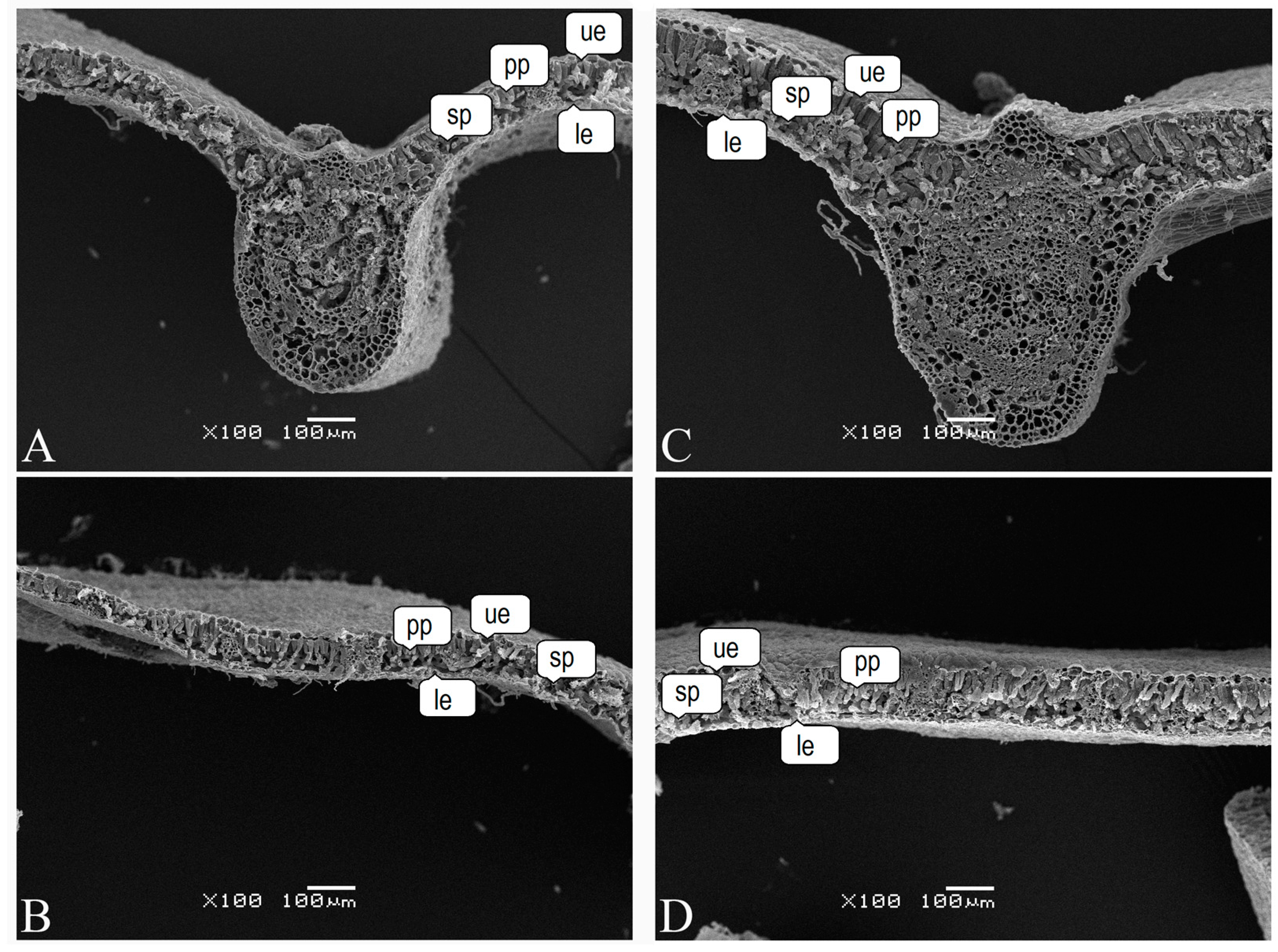
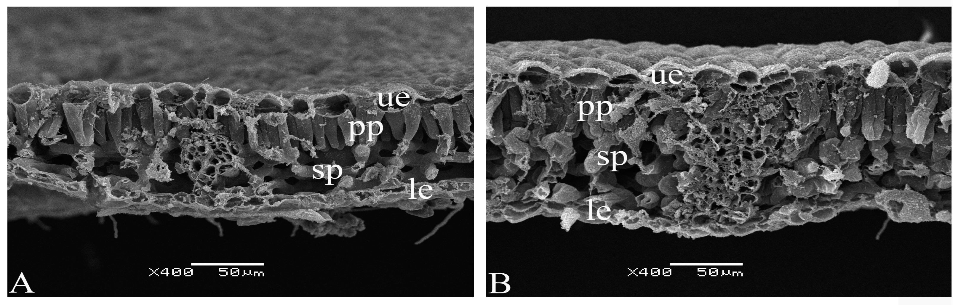
2.4. Scanning Electron Microscopy

Fragments of the leaf middle part with a size of 2–3 mm were fixed during the day at a +4 °C in a 2.5% solution of glutaraldehyde (Merck, Germany) prepared in 0.1 M phosphate buffer (pH is 7.2) with the addition of 1.5% sucrose. Then, the samples were dehydrated at +4 °C for 30 min in each alcohol solution with successively increasing concentration: 30%, 50%, 70%, 96%, and in three changes of absolute ethanol. After that, the samples were transferred to liquid CO2 under pressure in the device for drying at the “critical point” and slowly heated under pressure. When the pressure and temperature together passed the so-called “critical point” (31 °C and 74 bar), the pressure was reduced, thus drying the samples without any damage, bypassing the liquid–gas phase transition. Next, a thin (from 1 nm and more) metal layer was deposited onto the samples to enhance conductivity and add mechanical strength to the sample, as well as to localize the signal on the sample surface (sputtering unit SPI supplies, SPI, Santa-Clara, CA, USA). Samples were stored in closed Petri dishes with granulated silica gel. The photographs were obtained using a JSM-6380LA scanning electron microscope (JEOL Ltd., Tokyo, Japan) at various magnifications. The thickness of the leaf blade was calculated using the Image J program with an accuracy of 0.1 µm. At least 300 cells of the above tissues from three independent leaves were analyzed for each experimental treatment.

2.5. Determination of the Composition of Phenolic Compounds in Chestnut Leaves

From the chestnut leaves selected at contrasting levels of daily solar radiation (from the sunny and shady sides of the tree crown), on which damage from C. ohridella was recorded, averaged samples were formed. Leaf powder samples (1 g) were extracted with methanol (20 mL). Extraction was carried out for 20 min with sonication and occasional shaking. Then, the suspension was centrifuged at 15,000× g for 15 min, and the supernatant was filtered through a 0.25 μm membrane and evaporated to dryness in a helium stream. The identification and content of secondary metabolites in individual extracts were determined by chromato-mass spectrometry (GC-MS) on a JMS-Q1050GC GC-MS chromatograph (JEOL Ltd., Tokyo, Japan). Capillary column DB-5HT (Agilent, Santa-Clara, CA, USA) (length 30 m, inner diameter—0.25 mm, the film thickness—0.52 μm, and gas carrier—helium) was used. The temperature gradients during the analysis were within the range of 40 °C to 280 °C, the injector and interface temperature was 250 °C, while the ionic source was 200 °C. Gas flow in the column was equal to 2.0 mL min–1, split-flow injection mode, with the sample injected into a volume of 1–2 μL of the evaporated extract. The analysis was held for 45 min. The derivation was held using silylation reagent N,O—Bis-trimethylsilyl) trifluoracetamide—BSTFA following the method described in past studies [28,29,30]. Identification of the substances was carried out according to NIST-5: National Institute of Standards and Technology (USA) retention behavior and mass spectra. The scanning range was 33–900 m Z–1, while the identification of substances’ credibility was within 75%–98%. All determinations were carried out in triplicate.

2.6. Statistical Analysis

To test for the normality of the distribution, the Kolmogorov–Smirnov test was performed. Further, to compare the arithmetic means, ANOVA was used, with the Bonferroni correction. Pairwise differences between individual extracts of secondary metabolites were assessed using Tukey’s test. The measurements were performed in the Statistica v. 12.0 PL (StatSoft, Tulsa, OK, USA) program.

3. Results

The first imagoes appeared in mid-June, the number of individuals of the first generation recorded in traps during a three-week collection period (from mid-June to early July) averaged 150 moths, both on the sunny and shady sides of the crown (Figure 1). However, already by the second generation, noted in the period from the last ten days of July to the end of August, the number of individuals in traps in the same period of fishing exceeded the number of imagoes of the first generation by 2.1 and 1.6 times on the sunny and shady sides of the crown, respectively. At the same time, the number of second-generation individuals caught on the sunny part of the crown was statistically significantly higher (1.25 times) than the number of individuals on the shady side.
No significant differences were found between the variants within each month; however, the indicators in July and August differ significantly from each other.
After taking into account the flight activity of the number of second-generation individuals on the leaves of horse chestnut, the number of caterpillars of two age groups (L1–L4; L5 and L6), as well as the number of diapausing/non-diapausing pupae of the orchid miner (Figure 2 and Figure 3) were counted.
Significant differences were found between the variants within each month (except for non diapaused pupae); indicators in July and August are significantly different from each other (Figure 2).
Early age caterpillars (L1–L4) had a flattened body, while late-age caterpillars (L5 and L6) were typical caterpillars with an almost cylindrical body. The structure of the mouthparts was the same for all feeding larval stages (L1–L4). Late-instar larvae (L5 and L6) did not feed and morphologically differed significantly from younger larval stages. The average number of miner caterpillars feeding per one leaf of common horse chestnut was 1.7 times more on the sunny side of the crown than on the shady side. The number of caterpillars of late ages on both parts of the crown did not statistically significantly differ from each other and amounted to 1.3 and 1.5 individuals per leaf on average. At the same time, the number of caterpillars of late instars was significantly less (by four times in the sunny and two times in the shady side of the crown) than the larvae of L1–L4 stages.
Larvae of the fifth stage are known to braid the lower part of the cocoon in the form of a lenticular convexity of the abaxial part of the leaf and behave in one of two ways: they form a shallow deformation of the leaf, and in this case, L6 larvae weave a very loose cocoon and turn into a pupa that does not enter into diapause; or L5 larvae produce a much deeper deformation of the lower surface of the leaf, and the sixth instar larvae that arise from them weave a very dense cocoon (and then the pupae enter diapause) [25]. An analysis of pupae, both falling into diapause and not going into it, showed that the average number of pupae per leaf that did not go into diapause was almost the same, both on the sunlit (16.5 individuals) and on the shady (17.5 individuals) sides of the tree crown. The number of such pupae was 2.2 and 4.4 times greater than the number of pupae falling into diapause on the sunny and shady sides of the crown, respectively. At the same time, the number of pupae falling into diapause on the sunlit side of the chestnut crown was 1.9 times more than on the shady side of the tree crown.
A micromorphological assessment of horse chestnut leaves damaged by larvae (Figure 3) allowed us to describe the characteristic features of the leaf transverse section on the sunny and shady sides of the horse chestnut crown (Figure 4). The leaf blades had a structure typical of mesomorphic leaves with relatively thin outer cell walls of the adaxial and abaxial epidermis, a thin cuticle layer, and a single layer of palisade and spongy parenchyma. Small cuticular grooves are visible on the leaf surface of A. hippocastanum, and a few trichomes are also found. A dense mass of organic matter from the remains of damaged cells and excrement of larvae was found in the mine cavity. In some cells of the palisade and spongy parenchyma, as well as in cells located near the phloem of large vascular bundles, calcium oxalate crystals (Supplementary Materials Figure S2) were formed (Figure 3B).
The area of the palisade parenchyma cells was significantly larger in leaf tissue from the sunny side of the crown than in leaves from the shady side of the crown (Figure 5). This trend was also characteristic of the cells of the lower epidermis, while the area of the cells of the upper epidermis was larger in the sunny part of the crown. Only spongy parenchyma cells did not differ in this parameter (to be added to the text) (Figure 6A). When studying the palisade parenchyma, differences were found in the length of the cells of horse chestnut leaves (Figure 5B). A significant increase in the length of the cells of the palisade parenchyma of the leaves of the shady part of the crown was found to cause a greater thickness of their leaf blade compared to the leaves of the sunny side of the crown—3068.78 µm and 2026.63 µm, respectively.
In addition, a fungal infection was revealed on the surface of the leaf, both on the sunny and shady sides of the crown (Supplementary Materials Figure S3).

Metabolic Analysis

The composition of the main substances contained in the methanol extract prepared from the leaves of A. hippocastanum was determined by GC-MS. A total of 100 metabolites were identified (Supplementary Materials, Table S1). The height of their peaks was at least 0.1% of the instrument scale. Since the content of most compounds did not exceed 0.1 mass. % of the extract, the results consider data only for 45 compounds, the contents of which are above 0.1 mass. % of the extract (Table 1).
The metabolites were divided into the following groups: polyphenolic compounds (10 compounds); cyclic esters (2 compounds); organic acids (11 compounds); and sugars and their derivatives (22 compounds). The share of carbohydrates in the composition in the tissue of leaves collected from the sunny and shady sides of the chestnut trees crown was 49.39% and 38.83%, respectively. Among carbohydrates, D-psicofuranose and methyl galactoside accounted for the largest share. The content of these compounds in the tissue of leaves collected from the sunny and shady sides of the crown was 30.9 and 38.2% of the total amount of carbohydrates, respectively. In the leaves collected from the sunny side of the crown, six types of carbohydrates dominated, and two compounds (methyl-a-N-acetyl-D-galactoside and D-(-)-xylophyranose) accumulated only in this part of the crown. Although 16 compounds dominated in the leaves of the shady side of the crown, the excess of their number was not significant—1.3 times compared with the content of these compounds in the leaves of the sunny side of the crown. Among the 11 organic acids, two compounds dominated—quininic acid and gluonic acid, and both acids accumulated to a greater extent in the leaves of the shady side of the crown (by 1.6 and 1.2 times, respectively). The proportion of polyphenolic compounds was 26.26 and 32.73% of the total composition of metabolites in the leaves of the sunny and shady sides of the horse chestnut crown, respectively. Among the polyphenolic compounds, two sugar alcohols dominated—1,5-angidroglucitol and galactinol—and their content in the leaves of the shady side of the crown was 1.3 times higher than in the leaves of the sunny side of the crown. It should be noted that an ester, gluconolactone, which has strong antioxidant and chelating properties, accumulated in the leaves of the sunny side of the crown.

4. Discussion

Significant damage to plantations of A. hippocastanum trees by the horse chestnut moth (C. ohridella) has been recorded in Moscow since 2006 [9], wherein the density of the phytophage and the dynamics of its abundance during the season change depending on the economic maintenance of plantations and the climatic conditions of the growing season [31]. In the absence of anthropogenic interference in the annual cycle of development of the pest, which is typical for areas with a special conservation status in particular, such as the arboretum of the GBS RAS, a high density of miners was recorded in plantations of horse chestnut [32].
One of the characteristics of the mass distribution of the horse chestnut leafminer is the determination of the abundance and density of populations of C. ohridella during the growing season. In recent years, Delta sticky traps with sex pheromones from C. ohridella females have been used for monitoring purposes [33,34]. During the capture of horse chestnut leafminer imagoes with pheromone traps, two generations of moths on horse chestnut trees were recorded, while up to three generations of C. ohridella were recorded in more southern European cities [35]. Often, in the conditions of a wet growing season, the development of the first generation of the miner is more extended and less productive, and the mass appearance of the second generation is timed to mid-August, i.e., to the period of the beginning of leaf aging. Thus, the number of Ohrid leafminers in the first generation averaged 170 individuals per sticky trap. Additionally, in the second generation it was more numerous—the maximum number of moths reached 350 individuals per trap, which is significantly lower than the number (up to 600–700 moths) recorded in European cities [36]. The trend of a smaller number of imagoes in the first generation recorded in our studies, not reaching the level of the second generation, was also observed in European populations of C. ohridella [36].
It is known that C. ohridella females of the first generation show very low selectivity when choosing A. hippocastanum leaves [37,38], therefore fluctuations in the number of miners on the sunny and shady sides of the crown did not exceed 10% and were statistically insignificant (p ≤ 0.05). However, already in the second generation, the choice of leaves by C. ohridella females could be influenced by intraspecific competition. The leaves of the sunny side of the horse chestnut crown were more affected by the orchid leafminer—the increase in the number in the second generation was 92.5%, while in the shady side of the crown, the growth did not exceed 47.2%. With a high daily solar radiation per unit area of leaf, leaves with a greater mass are formed on the sunny side of the horse chestnut crown. It is the availability of feeding that may be one of the factors that determine the high abundance of lifeminers on the leaves of the sunny side of the tree crown [39]. Additionally, as a result, a greater number of caterpillars of age L1–L4 (up to five individuals per leaf on the sunny side of the crown) develop on this part of the crown.
With a high abundance of the second generation, a significant food shortage begins to be felt during the development of the miner, especially on the shady part of the crown, which led to the formation of more mines, an increase in the mortality of feeding caterpillars, a higher percentage of pupae that do not fall into the diapause, and a lower percentage of pupae entering diapause. Similar data on the formation of a greater number of mines on the leaves of horse chestnut in the shady part of the crown were also recorded by Polish and Czech researches. They revealed that a longer period of time was required for the development of mines on leaves from the sunny part of the crown [40,41,42].
The cuticular layer of plants is an important barrier against pathogens and pests [43]. Small cuticular grooves have been found on the cell walls of the adaxial epidermis of horse chestnut, but it has not been proven that they can be an obstacle to the penetration of C. ohridella larvae, which can gnaw through the epidermal layer and reach the palisade parenchyma. Thus, in our studies of the anatomical structure of chestnut leaves, it was found that the area of cells of the palisade parenchyma was significantly larger in the cells of the leaves of the crown’s sunny side than in the leaves of the crown’s shady side. There were no differences in the areas of cell sections of the upper epidermis and spongy parenchyma between the leaves of the sunny and shadow sides of the crown. In addition, a significant increase in the length (by 21%) of the cells of the palisade parenchyma in the leaves of the crown shadow side was established, which was also previously noted by a number of researchers [44,45]. The palisade parenchyma cells in the leaves on the shady side of the crown had different lengths, while the cells of the palisade parenchyma in the leaves on the sunny side of the crown were uniform [41]. The combination of these indicators led to differences in the thickness of the leaf blade, which turned out to be greater in low light conditions, which was also noted in previous studies [41,46,47].
During the study, in some cells of the palisade and spongy parenchyma, as well as in many cells located near the phloem of large vascular bundles, single crystals of calcium oxalate were found (Supplementary Materials Figure S2). It is known that calcium oxalate crystals in plant cells can perform various functions, one of which is protection from pests [47,48,49].
Secondary plant metabolites are considered to be another important source of natural plant resistance to pests [50]. They affect the olfactory, tactile, and taste receptors of herbivores, and their effects on herbivores are often toxic [51]. Changes in the qualitative and quantitative composition of secondary metabolites in horse chestnut leaves that have been infested with a leafminer provide plant resistance to the pest. Thus, a change in the activity of individual benzidine-peroxidase isomers contributes to maintaining the functional integrity of the photosynthetic system in chestnut leaves [52]. It is the inhibition of photosynthetic activity (decrease in the quantum efficiency of photosystem II) that provides violations of the coherence of the reactions of the Calvin cycle, which were recorded in infected leaves both on the illuminated and shaded sides of the crown of horse chestnut trees [53].
One of the most active groups of plant secondary metabolites that exhibit protective activity against insect feeding are phenolic compounds [54,55,56], which are present in epicuticular waxes on the leaf surface, in peripheral tissues (epidermis, mesophyll), vascular bundles, and perivascular membranes [57,58]. The accumulation of phenolic compounds was recorded in the cells of the epidermis and parenchyma of infected leaves located along the border of tissues damaged by leafmining moth larvae [25,59].
However, the low content of polyphenols (~30%) in the pool of secondary metabolites in the leaves of A. hippocastanum recorded in our studies did not deter lepidoptera, and the significant content of carbohydrates in the leaves reduced the resistance of horse chestnut plants to biotic stress.
An assessment of the effect of tree crown insolation showed that a very small difference in the level of phenolic compounds in chestnut leaves grown under different lighting conditions (26.27% and 32.73% in the leaves of the sunny and shady parts of the crown, respectively) suggests that phenolic compounds in the leaves of horse chestnut do not perform a repellant function, regardless of differing light conditions. This is consistent with the results of Polish researchers [41]. At the same time, carbohydrates (up to 50% of the total content of secondary metabolites) accumulate in the leaves from the sunny side of the horse chestnut crown at a high intensity of visible light and UV radiation, which increases the availability of food and causes a high abundance of leafminers on the leaves of this part of the tree crown. It is possible that the presence of a strong antioxidant (Gluconolactone) in these leaves, albeit in small amounts, causes an increase in the mortality of feeding caterpillars: the number of late instar caterpillars decreased by four times compared with L1–L4 stage larvae.

5. Conclusions

Crown insolation of A. hippocastanum affected the abundance and density of C. ohridella populations during the growing season. The largest number of pests was noted on the sunlit side of the tree crown, and the smallest on the shady side of the crown. With high daily solar radiation, leaves were developed on the sunlit part of the horse chestnut crown, containing high levels of carbohydrates and having a large area of both palisade parenchyma cells and cells of the lower epidermis. This contributed to an increase in the availability of food and led to a high abundance of leafminers and the development of a large number of caterpillars of age L1–L4. At the same time, the presence of a strong antioxidant (gluconolactone) in the leaves of the sunny side of the tree crown caused an increase in the mortality of feeding caterpillars.
With a high abundance of the second-generation miner on leaves on the shady side of the chestnut crown, a significant food deficit was felt during its development, which led to the formation of more mines, an increase in the mortality of feeding caterpillars, a higher percentage of pupae that do not fall into diapause, and a lower percentage of pupae entering diapause.
Low levels of phenolic compounds in horse chestnut leaves did not increase the resistance of trees to biotic stress. A statistically insignificant difference in the level of phenolic compounds in the leaves of the sunny and shady sides of the horse chestnut crown indicates that phenolic compounds in the leaves of A. hippocastanum do not perform a repellent function, regardless of different light conditions.

Supplementary Materials

The following supporting information can be downloaded at https://www.mdpi.com/article/10.3390/f14061079/s1, Figure S1: Pheromone traps; Figure S2: Calcium oxalate crystals in palisade parenchyma cells; Figure S3: Fungal infection on the surface of leaves. Table S1: Organic compounds detected in the leaf extracts of A. hippocastanum. % mass. of the extract.

Author Contributions

All authors (L.R.B., E.V.T., L.N.K., O.B.T., L.S.O., A.A.G., E.N.B. and O.V.S.) contributed to data collection and analysis and manuscript design. O.V.S., L.R.B. and E.V.T. prepared and revised the manuscript, and are considered the main authors of the article. All authors have read and agreed to the published version of the manuscript.

Funding

The reported study was supported by assignments 122042700002-6 (MBG RAS) and FGUM-2022-0003 (ARRIAB RAS) of the Ministry of Science and Higher Education of the Russian Federation. The APC was funded by the authors.

Data Availability Statement

Not applicable.

Acknowledgments

The authors thank Anatoly Bogdanov and the Center for Electron Microscopy in the Life Sciences, a unique scientific installation for three-dimensional electron microscopy and spectroscopy (Biological Faculty of Lomonosov Moscow State University).

Conflicts of Interest

The authors declare no conflict of interest.

References

  1. Takács, Á.; Kiss, M.; Gulyás, Á.; Tanács, E.; Kántor, N. Solar permeability of different tree species in Szeged, Hungary. Geogr. Pannonica 2016, 20, 32–41. [Google Scholar] [CrossRef]
  2. Chanon, A.M. Studies on the Reproductive Capacity of Aesculus parviflora and Aesculus pavia: Opportunities for Their Improvement through Interspecific Hybridization. Ph.D. Thesis, The Ohio State University, Columbus, OH, USA, 2005; p. 264. [Google Scholar]
  3. Freer-Smith, P.H.; Webber, J.F. Tree pests and diseases: The threat to biodiversity and the delivery of ecosystem services. Biodivers. Conserv. 2017, 26, 3167–3181. [Google Scholar] [CrossRef]
  4. Simova-Tosic, D.; Filev, S. Contribution to the knowledge of leaf miners of horse chestnut. Zast. Bilja 1985, 36, 235–239. [Google Scholar]
  5. Deschka, G.; Dimic, N. Cameraria ohridella n. sp. aus Macedonien, Jugoslavien (Lepidoptera, Lithocolletidae). Acta Entomol. Jugosl. 1986, 22, 11–23. [Google Scholar]
  6. Kirichenko, N.I.; Karpun, N.N.; Zhuravleva, E.N.; Shoshina, E.I.; Anikin, V.V.; Musolin, D.L. Invasion genetics of the horse-chestnut leaf miner, Cameraria ohridella (Lepidoptera: Gracillariidae), in European Russia: A case of successful involvement of citizen science in studying an alien insect pest. Insects 2023, 14, 117. [Google Scholar] [CrossRef] [PubMed]
  7. Tomiczek, C.; Krehan, H. The horsechestnut leafmining moth (Cameraria ohridella): A new pest in central Europe. J. Arboric. 1998, 24, 144–148. [Google Scholar]
  8. Gninenko, Y.I.; Orlinskiy, A.D. New phytophages of tree plantations. Plant Prot. Quarant. 2004, 4, 33. [Google Scholar]
  9. Golosova, M.A.; Gninenko, Y.I. The appearance of the Ohrid leafminer on horse chestnut in Moscow. Les. Vest. 2006, 2, 43–46. (In Russian) [Google Scholar]
  10. Beguinot, J. Comparing the ratios of leaf-acceptance prior to egg-laying among several species of leaf-mining moths upon Quercus sp. pl. (Lepidoptera: Gracillariidae, Tischeriidae). Entomol. Gener. 2009, 32, 145–154. [Google Scholar] [CrossRef]
  11. Weryszko-Chmielewska, E.; Haratym, W. Changes in leaf tissues of common horse chestnut (Aesculus hippocastanum L.) colonised by the horse-chestnut leaf miner (Cameraria ochridella Deschka and Dimic). Acta Agrobot. 2011, 64, 11–22. [Google Scholar] [CrossRef]
  12. Balder, H.; Jäckel, B.; Schomlling, S. Effects of the horse chestnut leaf-miner Cameraria ohridella Deschka & Dimic on the frost hardness of Aesculus hippocastanum L. In Proceedings of the 1st International Cameraria Symposium—Cameraria ohridella and Other Invasive Leaf-Miners in Europe, Prague, Czech Republic, 24–27 March 2004; IOCB: Prague, Czech Republic, 2004; p. 4. [Google Scholar]
  13. Tarwacki, G.; Bystrowski, C.; Celmer-Warda, K. Effect of sun-exposure of the horse chestnut (Aesculus hippocastanum L.) on the occurrence and number of parasitoids of the horse chestnut leafminer (Cameraria ohridella Deschka & Dimic) in central Poland in 2004–2006. Folia For. Pol. (Ser. A) 2012, 54, 56–62. [Google Scholar]
  14. Poorter, H.; Niinemets, Ü.; Poorter, L.; Wright, I.J.; Villar, R. Causes and consequences of variation in leaf mass per area (LMA): A meta-analysis. New Phytol. 2009, 182, 565–588. [Google Scholar] [CrossRef]
  15. Bidart-Bouzat, M.G.; Imeh-Nathaniel, A. Global change effects on plant chemical defenses against insect herbivores. J. Integr. Plant Biol. 2008, 50, 1339–1354. [Google Scholar] [CrossRef] [PubMed]
  16. Holoborodko, K.; Seliutina, O.; Alexeyeva, A.; Brygadyrenko, V.; Ivanko, I.; Shulman, M.; Pakhomov, O.; Loza, I.; Sytnyk, S.; Lovynska, V.; et al. The Impact of Cameraria ohridella (Lepidoptera, Gracillariidae) on the State of Aesculus hippocastanum Photosynthetic Apparatus in the Urban Environment. Int. J. Plant Biol. 2022, 13, 223–234. [Google Scholar] [CrossRef]
  17. Kulbat, K. The role of phenolic compounds in plant resistance. Biotechnol. Food Sci. 2016, 80, 97–108. [Google Scholar]
  18. Materska, M.; Pabich, M.; Sachadyn-Król, M.; Konarska, A.; Weryszko-Chmielewska, E.; Chilczuk, B.; Staszowska-Karkut, M.; Jackowska, I.; Dmitruk, M. The Secondary Metabolites Profile in Horse Chestnut Leaves Infested with Horse-Chestnut Leaf Miner. Molecules 2022, 27, 5471. [Google Scholar] [CrossRef]
  19. Weryszko-Chmielewska, E.; Haratym, W. Leaf micromorphology of Aesculus hippocastanum L. and damage caused by leaf-mining larvae of Cameraria ohridella Deschka and Dimic. Acta Agrobot. 2012, 65, 25–33. [Google Scholar] [CrossRef]
  20. Kehrli, P.; Bacher, S. Date of leaf litter removal to prevent emergence of Cameraria ohridella in the following spring. Entomol. Exp. Appl. 2003, 107, 159–162. [Google Scholar] [CrossRef]
  21. Pavan, F.; Barro, P.; Bernardinelli, I.; Gambon, N.; Zandigiacomo, P. Cultural control of Cameraria ohridella on horsechestnut in urban areas by removing fallen leaves in autumn. J. Arboric. 2003, 29, 253–258. [Google Scholar] [CrossRef]
  22. Floricel, M.; Mitrea, I.; Oltean, I.; Florian, T.; Varga, M.I.; Vasian, I.; Hulujan, I.B. The testing of some products in order to monitor the Cameraria ohridella Deschka-Dimic species (Lepidoptera: Gracilariidae). Agrolife Sci. J. 2018, 7, 53–60. [Google Scholar]
  23. Jagiełło, R.; Walczak, U.; Iszkuło, G.; Karolewski, P.; Baraniak, E.; Giertych, M.J. Impact of Cameraria ohridella on Aesculus hippocastanum growth and long-term effects of trunk injection with pesticides. Int. J. Pest Manag. 2019, 65, 33–43. [Google Scholar] [CrossRef]
  24. Plotnikova, L.S.; Aleksandrova, M.S.; Belyaeva, Y.E.; Nemova, E.M.; Ryabova, N.V.; Yakushina, E.I. Drevesnye rasteniya Glavnogo botanicheskogo sada imeni N. V. Tsitsina RAN: 60 let introduktsii. In Tree Plants of the N. V. Tsitsin Chief Botanical Garden RAS: 60 Years of Introduction; Demidov, A.S., Ed.; Science: Moscow, Russia, 2005; p. 586. [Google Scholar]
  25. Konarska, A.; Grochowska, M.; Haratym, W.; Tietze, M.; Weryszko-Chmielewska, E.; Lechowski, L. Changes in Aesculus hippocastanum leaves during development of Cameraria ohridella. Urban For. Urban Green. 2020, 56, 126793. [Google Scholar] [CrossRef]
  26. Šefrová, H.; Skuhravý, V. The larval morphology of Cameraria ohridella Deschka & Dimić compared with the genus Phyllonorycter Hübner (Lepidoptera, Gracillariidae). Acta Univ. Agric. Silvic. Mendel. Brun. 2000, 48, 23–30. [Google Scholar]
  27. Zunke, U.; Fernandez-Nunez, M.; Ilmberger, N.; Hofmeier, M.; Konitz, K.; Doobe, G. Zur Biologie und Populationsdynamik der Rosskastanienminiermotte und Ihrer Verbreitung in Hamburg. Cameraria ohridella, das HAM-CAM-Projekt 2001/04 in Hamburg, Hamburg. 2003; p. 38. Available online: http://cameraria.galk.de/hamburg.html (accessed on 9 February 2018).
  28. Robbins, R.J. Phenolic acids in foods: An overview of analytical methodology. J. Agric. Food Chem. 2003, 51, 2866–2887. [Google Scholar] [CrossRef]
  29. Marinova, G.; Batchvarov, V. Evaluation of the methods for determination of the free radical scavenging activity by DPPH. Bulg. J. Agric. Sci. 2011, 17, 11–24. [Google Scholar]
  30. Han, X.; Yang, K.; Gross, R.W. Multi-dimensional mass spectrometry-based shotgun lipidomics and novel strategies for lipidomic analyses. Mass Spectrom. Rev. 2012, 31, 134–178. [Google Scholar] [CrossRef] [PubMed]
  31. Bednova, O.V. Ohridsky miner Cameraria ohridella Deschka & Dimic: Features of invasion foci and prospects of biological population control. For. Bull. 2022, 26, 5–16. (In Russian) [Google Scholar]
  32. Kashtanova, O.A.; Tkachenko, O.B.; Kondratieva, V.V.; Voronkova, T.V.; Olekhnovich, L.S. Resistance of horse chestnut species (Aesculus L.) to ohrid miner or chestnut miner moth (Cameraria ohridella Deschka & Dimic). Bull. Moscow Soc. Natur. Biol. Ser. 2020, 125, 45–51. (In Russian) [Google Scholar]
  33. Cardé, R.T.; Elkinton, J.S. Field trapping with attractants: Methods and interpretation. In Techniques in Pheromone Research; Springer: New York, NY, USA, 1984; pp. 111–129. [Google Scholar]
  34. Šefrova, H.; Laštůvka, Z. Dispersal of the horse chestnut leaf miner, Cameraria ohridella Deschka & Dimić, 1986, in Europe: Its course, ways and causes (lepidoptera: Gracillariidae). Entomol. Zeits. 2001, 111, 194–198. [Google Scholar]
  35. Deschka, G. Schmetterlinge als Einwanderer. Stapfia 1995, 37, 77–128. [Google Scholar]
  36. Kalinová, B.; Svatoŝ, A.; Kindl, J.; Hovorka, O.; Hrdý, I.; Kuldova, J.; Hoskovec, M. Sex pheromone of horse-chestnut leafminer Cameraria ohridella and its use in a pheromone-based monitoring system. J. Chem. Ecol. 2003, 29, 387–404. [Google Scholar] [CrossRef] [PubMed]
  37. Beguinot, J. The remarkably low leaf-selectivity prior to oviposition in the moth-pest Cameraria ohridella is not unique to this species within the genus Cameraria. Rev. Ecol. (Terre Vie) 2009, 64, 333–341. [Google Scholar] [CrossRef]
  38. Gripenberg, S.; Mayhew, P.J.; Parnell, M.; Roslin, T. A meta-analysis of preference–performance relationships in phytophagous insects. Ecol. Lett. 2010, 13, 383–393. [Google Scholar] [CrossRef]
  39. Heitland, W.; Freise, J. Verbreitung der Roßkastanien-Miniermotte, Cameraria ohridella (Lep, Gracillariidae) in Deutschland. Distribution of the horse-chestnut leaf miner, Cameraria ohridella (Lep., Gracillariidae) in Germany. Mitt. Dtsch. Ges. Allg. Angew. Entomol. 2001, 13, 131–134. [Google Scholar]
  40. Forister, M.L.; Nice, C.C.; Fordyce, J.A.; Gompert, Z. Host range evolution is not driven by the optimization of larval performance: The case of Lycaeides melissa (Lepidoptera: Lycaenidae) and the colonization of alfalfa. Oecologia 2009, 160, 551–561. [Google Scholar] [CrossRef]
  41. Jagiełło, R.; Baraniak, E.; Guzicka, M.; Karolewski, P.; Łukowski, A.; Giertych, M.J. One step closer to understanding the ecology of Cameraria ohridella (Lepidoptera: Gracillariidae): The effects of light conditions. Eur. J. Entomol. 2019, 116, 42–51. [Google Scholar] [CrossRef]
  42. Birner, A.; Bohlander, F. Mine development of horse chestnut leaf-miner (Cameraria ohridella) on leafs exposed to sunlight or shade. In Cameraria ohridella and Other Invasive Leaf-Miners in Europe, Proceedings of the 1st International Cameraria Symposium, Prague, Czech Republic, 24–27 March 2004; Kalinova, B., Ed.; Institute of Organic Chemistry and Biochemistry ASCR: Prague, Czech Republic, 2004; p. 5. [Google Scholar]
  43. Barthlott, W.; Neinhuis, C. Purity of the sacred lotus or escape from contamination in biological surfaces. Planta 1997, 202, 1–8. [Google Scholar] [CrossRef]
  44. Oliveira, D.C.; Isaias, R.M.S. Redifferentiation of leaflet tissues during midrib gall development in Copaifera langsdorffii (Fabaceae). S. Afr. J. Bot. 2010, 76, 239–248. [Google Scholar] [CrossRef]
  45. Dardeau, F.; Pointeau, S.; Ameline, A.; Laurans, F.; Cherqui, A.; Lieutier, F.; Sallé, A. Host manipulation by a herbivore optimizes its feeding behaviour. Anim. Behav. 2014, 95, 49–56. [Google Scholar] [CrossRef]
  46. Narawatthana, S. The Regulation of Leaf Thickness in Rice (Oryza sativa L.). Ph.D. Thesis, University of Sheffield, Sheffield, UK, 2013. [Google Scholar]
  47. Chen, L.L.; Zhang, K.; Gong, X.C.; Wang, H.Y.; Gao, Y.H.; Wang, X.Q.; Hu, Y.G. Effects of different LEDs light spectrum on the growth, leaf anatomy, and chloroplast ultrastructure of potato plantlets in vitro and minituber production after transplanting in the greenhouse. J. Integr. Agric. 2020, 19, 108–119. [Google Scholar] [CrossRef]
  48. Molano-Flores, B. Herbivory and calcium concentrations affect calcium oxalate crystal formation in leaves of Sida (Malvaceae). Ann. Bot. 2001, 88, 387–391. [Google Scholar] [CrossRef]
  49. Nakata, P.A. Advances in our understanding of calcium oxalate crystal formation and function in plants. Plant Sci. 2003, 164, 901–909. [Google Scholar] [CrossRef]
  50. Bernards, M.; Båstrup-Spohr, L. Phenylpropanoid metabolism induced by wounding and insect herbivory. In Induced Resistance to Herbivory; Schaller, A., Ed.; Springer: New York, NY, USA, 2008; pp. 189–211. [Google Scholar]
  51. Pagare, S.; Bhatia, M.; Tripathi, N.; Pagare, S.; Bansal, Y.K. Secondary metabolites of plants and their role: Overview. Curr. Trends Biotechnol. Pharm. 2015, 9, 293–304. [Google Scholar]
  52. Seliutina, O.V.; Shupranova, L.V.; Holoborodko, K.K.; Shulman, M.V.; Bobylev, Y.P. Effect of Cameraria ohridella on accumulation of proteins, peroxidase activity and composition in Aesculus hippocastanum leaves. Regul. Mech. Biosyst. 2020, 11, 299–304. [Google Scholar] [CrossRef]
  53. Holoborodko, K.K.; Seliutina, O.V.; Ivanko, I.A.; Alexeyeva, A.A.; Shulman, M.V.; Pakhomov, O.Y. Effect of Cameraria ohridella feeding on Aesculus hippocastanum photosynthesis. Regul. Mech. Biosyst. 2021, 12, 346–352. [Google Scholar] [CrossRef]
  54. Salminen, J.P.; Karonen, M. Chemical ecology of tannins and other phenolics: We need a change in approach. Func. Ecol. 2011, 25, 325–338. [Google Scholar] [CrossRef]
  55. Oszmiański, J.; Kalisz, S.; Aneta, W. The content of phenolic compounds in leaf tissues of white (Aesculus hippocastanum L.) and red horse chestnut (Aesculus carea H.) colonized by the horse chestnut leaf miner (Cameraria ohridella Deschka & Dimić). Molecules 2014, 19, 14625–14636. [Google Scholar]
  56. Kopacka, M.; Zemek, R. Spatial variability in the level of infestation of the leaves of horse chestnut by the horse chestnut leaf miner, Cameraria ohridella (Lepidoptera: Gracillariidae) and in the number of adult moths and parasitoids emerging from leaf litter in an urban environment. Eur. J. Entomol. 2017, 114, 42. [Google Scholar]
  57. Karabourniotis, G.; Liakopoulos, G.; Nikolopoulos, D.; Bresta, P.; Stavroulaki, V.; Sumbele, S. “Carbon gain vs. water saving, growth vs. defence”: Two dilemmas with soluble phenolics as a joker. Plant Sci. 2014, 227, 21–27. [Google Scholar] [CrossRef] [PubMed]
  58. Moreira, X.; Zas, R.; Sampedro, L. Differential allocation of constitutive and induced chemical defenses in pine tree juveniles: A test of the optimal defense theory. PLoS ONE 2012, 7, e34006. [Google Scholar] [CrossRef]
  59. Melo De Pinna, G.F.A.; Kraus, J.E.; de Menezes, N.L. Morphology and anatomy of leaf mine in Richterago riparia Roque (Asteraceae) in the campos rupestres of Serra do Cipó, Brazil. Braz. J. Biol. 2002, 62, 179–185. [Google Scholar] [CrossRef] [PubMed]
Figure 1. The total number of moths of C. ohridella during the growing season of the year 2022. Standard deviations are given as the margin of error. Mean comparisons were performed with ANOVA using the Bonferroni correction. Means followed by the same letter are not significantly different at α = 0.05. The experiment was performed in triplicate.
Forests 14 01079 g001
Figure 2. The average number of caterpillars of early (L1–L4) and late instars (L5 and L6) (A), as well as pupae that do not fall and fall into diapause under different conditions of crown illumination (B). Standard deviations are given as the margin of error. Mean comparisons were performed by ANOVA using the Bonferroni correction. Means followed by the same letter are not significantly different at α = 0.05. The experiment was performed in triplicate.
Forests 14 01079 g002aForests 14 01079 g002b
Figure 3. Horse chestnut leaves infested by Ohrid leafminer from the sun (A) and shadow (B) sides of the tree crown.
Forests 14 01079 g003
Figure 4. Cross sections of C. ohridella-infested Aesculus hippocastanum leaves under different illumination conditions (sunny side of the crown—(A,B); shady side of the crown—(C,D)) obtained using scanning electron microscopy (×400). Abbreviations: ue—upper epidermis, le—lower epidermis, pp—palisade parenchyma, sp—spongy parenchyma.
Forests 14 01079 g004
Figure 5. Fragments of a transverse cross sections of Cameraria ohridella-infested Aesculus hippocastanum leaves under different illumination conditions (sunny—(A), shaded—(B)), obtained using scanning electron microscopy (magnification ×400). Abbreviations (like Figure 4): ue—upper epidermis, le—lower epidermis, pp—palisade parenchyma, sp—spongy parenchyma.
Forests 14 01079 g005
Figure 6. Morphometric characteristics of anatomical differences in leaf blade mesophyll specimens of horse chestnut leaf. Cross sections of Cameraria ohridella-infested Aesculus hippocastanum leaves under different illumination conditions (sunny and shaded), obtained using scanning electron microscopy (magnification ×400). (A)—The area of the cells of the upper and lower epidermis, as well as the palisade and spongy parenchyma of the leaf blade under different illumination conditions. Abbreviations: Sue—upper epidermis, Sle—lower epidermis, Spp—palisade parenchyma, Ssp—spongy parenchyma. (B)—Length of the cells of the palisade parenchyma of the leaf blade of horse chestnut under various illumination conditions (Lpp). Standard deviations are given as the margin of error. Means were compared in ANOVA using the Bonferroni test (histogram A) and the t-test mean comparison method. Means followed by the same letter are not significantly different at α = 0.05.
Forests 14 01079 g006
Table 1. Organic compounds detected in the leaf extracts of A. hippocastanum. % mass. Of the extract.
MetabolitePeak Height, % of Scale
Sunny SideShady Side
Polyphenols
2-butene-1,4-diol0.21 a0.05 bc
L-(-)-arabitol1.70 a0.29 c
D-fucitol0.95 a0.44 b
1,5-anhydroglucitol1.34 a0.06 c
D-mannitol0.01 bc0.55 a
1,5-angidroglucitol8.71 b10.33 a
scillo-inositol1.80 a1.26 ab
galactinol10.59 bc15.1 a
D-glucitol0.94 a0.60 b
maltitol0.01 c3.96 a
Carboxylic esters
D-(+)- ribono-1.4-lactone0.23 ab0.31 a
gluconolactone1.25 a0.01 c
Organic acid
glucopyranuronic acid0.14 b0.49 a
ribonic acid0.97 a0.36 bc
quininic acid7.50 bc11.75 a
gluonic acid7.39 ab8.29 a
gallic acid0.42 a0.12 b
palmitic acid0.27 a0.01 b
D-(+)-galacturonic acid0.49 a0.03 bc
4-aminosalicylic acid0.01 c2.03 a
5-aminosalicylic acid0.01 b0.31 a
sinapinic acid0.36 a0.27 a
betulinic acid0.44 a0.41 a
Sugar derivatives
D-erythro-2-pentulose0.60 a0.41 b
methyl-a-D-glucofuranoside0.55 ab0.70 a
D-psicofuranose9.15 a9.45 a
D-(-)-tagatofuranose0.01 b0.48 a
DL- arabinofuranoside2.66 ab3.04 a
methyl galactoside6.12 a5.25 ab
b-D-(+)-talophyranose1.87 ab2.40 a
talofuranose1.63 a1.30 ab
deoxyglucose0.12 ab0.19 a
a-D-ribofuranose0.12 b0.28 a
L-sorbopyranose0.09 ab0.15 a
glucosylspingosine6.67 a2.31 bc
D-turanose0.01 c3.28 a
methyl-a-N-acetyl-D-galactoside5.20 a0.01 d
D-(-)-sorbofuranose1.55 a1.85 a
D-trehalose2.52 a1.50 b
D-(-)-xylophyranose5.63 a0.01 d
1-c-octylhexopyranose1.95 a2.13 a
DL-arabinopyranose2.54 a2.62 a
b-lyxopyranose0.01 b0.44 a
a-D-lactose0.01 b0.31 a
D-ribofuranose0.38 ab0.72 a
Values followed by the same letter within a row are not significantly different (p > 0.05, Tukey’s test).
Disclaimer/Publisher’s Note: The statements, opinions and data contained in all publications are solely those of the individual author(s) and contributor(s) and not of MDPI and/or the editor(s). MDPI and/or the editor(s) disclaim responsibility for any injury to people or property resulting from any ideas, methods, instructions or products referred to in the content.

