Article

Effects of Hormones and Epigenetic Regulation on the Callus and Adventitious Bud Induction of Fraxinus mandshurica Rupr.

1
Key Laboratory of Saline-Alkaline Vegetation Ecology Restoration (SAVER), Ministry of Education, Northeast Forestry University, Harbin 150040, China
2
Department of Forest Bioengineering, College of Life Science, Northeast Forestry University, Harbin 150040, China
3
Forest Botanical Garden of Heilongjiang Province, Harbin 150040, China
4
Shanhetun Forestry Bureau, Wuchang City, Harbin 150200, China
*
Author to whom correspondence should be addressed.
Forests 2020, 11(5), 590; https://doi.org/10.3390/f11050590
Submission received: 26 April 2020 / Revised: 20 May 2020 / Accepted: 21 May 2020 / Published: 24 May 2020
(This article belongs to the Special Issue Application of Tissue Culture in Plant Reproduction)

Abstract

:
Fraxinus mandshurica Rupr. (hereafter “F. mandshurica”) is known as one of northeast China′s important, valuable hardwood timber species. However, tissue culture and micropropagation of the species are difficult and have low efficiency, limiting asexual propagation. In this manuscript, stem explants were utilized to establish an effective regeneration system through adventitious bud organogenesis. The factors influencing callus regeneration in vitro were determined, and callus regeneration technology was established. The mechanism of adventitious bud formation was analyzed. Thidiazuron (TDZ) played a crucial role in the formation of adventitious buds. Elevated concentrations of TDZ were beneficial to callus induction and low concentrations of 6-benzyladenine (BA) led to loose state callus formation. The order of callus induction rates for different explants was stem cotyledon (100%) > segment (98.54%) > hypocotyl (92.56%) > root (50.71%). The effects of exogenous addition of 6-BA and TDZ on the endogenous hormone content of plants during the regeneration of adventitious buds were also assessed, as well as the expression characteristics of genes related to the regeneration pathway. The comprehensive analysis results showed that the suitable medium for callus induction and adventitious bud differentiation was c12 medium (MSB5 + 30 g/L sucrose + 7 g/L Agar + 5 mg/L 6-BA + 8 mg/L TDZ + 2 mg/L glycine + 0.1 mg/L IBA + 5% coconut water). The induction rates of callus and adventitious buds were 99.15% and 33.33%. The addition of 2.4 mg/L of the DNA demethylation reagent 5-azacytidine (5-aza) and 0.15 mg/L of the histone deacetylase inhibitor trichostatin A (TSA) increased the rates of adventitious bud induction by 17.78% over the control. This further laid the foundation for large-scale cultivation of excellent varieties and genetic transformation techniques.

1. Introduction

F. mandshurica Rupr. is an important broad-leaved tree species in northeastern China that can be used for various types of industrial and furniture timber and has significant economic and ecological value [1,2]. However, because of excessive deforestation, the number of adult F. mandshurica declines every year. F. mandshurica has been listed as a rare species in China, and so, reducing the number of F. mandshurica removed by China′s forestry development and promoting the breeding of F. mandshurica, have become imminent tasks [3]. F. mandshurica mainly relies on seed to propagate, but seeds have the characteristics of strong periodicity, deep dormancy, low germination rate, and slow propagation rate [2]. Seed germination must undergo a vernalization treatment for up to 6 months [4]. These factors increase the difficulty of F. mandshurica reproduction and limit its reproduction in forest development [5,6,7]. Asexual reproduction can maintain the excellent strains and genetic characteristics of F. mandshurica. Grafting, cutting and plant tissue culture can promote and apply fine varieties on a large scale. During the grafting process, the degree of joining between scions and the rootstock, the quality of the scions, and the method of grafting all affect the final survival rate of the plant. Because of these factors, it is difficult to gain ideal economic benefits [8]. Similarly, cutting is also subject to the above restrictions [9,10]. The propagation coefficient of F. mandshurica is often difficult to break through, so grafting and cutting cannot be used as the main means of asexual propagation. Genetic engineering technology provides an attractive way to effectively improve current varieties [11].
In plant tissue culture, exogenous hormones are applied to reprogram the cells to obtain regeneration ability. Firstly, callus formation is induced on call inducing medium (CIM) with a high auxin ratio, and then, on shoot inducing medium (SIM) with a high cytokinin ratio. PLT2 and PLT3 are two auxin signal response factors [12] that induce callus formation in Arabidopsis. Wound treatment can promote callus formation in all parts of Arabidopsis seedlings by inducing the expression of the AP2-ERF transcription factor WIND1 [13,14]. Wound treatment can activate the expression of YUC1 and increase the level of auxin [15]. Auxin can activate the expression of the WUSCHEL-related gene WOX. This mechanism can maintain a high level of auxin in cells with regeneration ability. In the process of bud regeneration, the signal responses of endogenous auxin and cytokinin are antagonistic to each other. The transcription factor WUS plays a key role in Arabidopsis bud regeneration [16]. The cytokinin pathways ARR2 and REV up-regulate the expression of the WUS gene [17].
In epigenetic studies, histone acetylation is associated with auxin-mediated somatic embryogenesis in Arabidopsis [18,19]. Low DNA methylation levels favor Arabidopsis regeneration, and histone acetylation is associated with auxin-mediated Arabidopsis somatic cell developmental embryo development programs [18,20]. This F. mandshurica functional gene research can only investigate gene cloning and expression analysis, lacking effective authentication on the function of the method [21,22]. Concerning asexual propagation of F. mandshurica, the differentiation of adventitious buds in the callus pathway and the complete establishment of a regeneration system have not yet been reported. Therefore, establishing efficient and effective callus induction and an adventitious bud regeneration system in F. mandshurica is very important. Therefore, it is of profound significance to analyze the breeding and gene function of F. mandshurica through callus regeneration.

2. Materials and Methods

2.1. Disinfection and Inoculation of F. mandshurica Seeds

The explant material was chosen from the hypocotyls, roots and cotyledons of the sterile seedlings of F. mandshurica and the stem sections of the sterile tissue culture seedlings of the bristles cultured in this laboratory. Fully-fledged seed of F. mandshurica was selected and treated with winged sand at 4 °C for 3 months. The treated seeds were deprived of the outer seed coat and rinsed with running water. They were immersed in 70% alcohol for 30–45 s, and 10% NaClO for 15–20 min. They were rinsed four times on a sterile table with sterile water, the embryo was peeled off, and 20 g/L sucrose and 5.3 g/L agar were added. They were placed in hormone-containing WPM induction medium, and the infection rate and initiation rate of different disinfection combinations were recorded. Ten tissue materials were treated each time, and the experiments were repeated three times. Data were analyzed using SPSS 23.0 statistical software (IBM-SPSS 2015). The mean with standard error (±SE) is presented. The percent of callus formation, shoot bud induction, and number of shoots were subjected to analysis of variance (ANOVA). Significant difference between treatments was tested by Duncan′s multiple comparison test (p = 0.05).

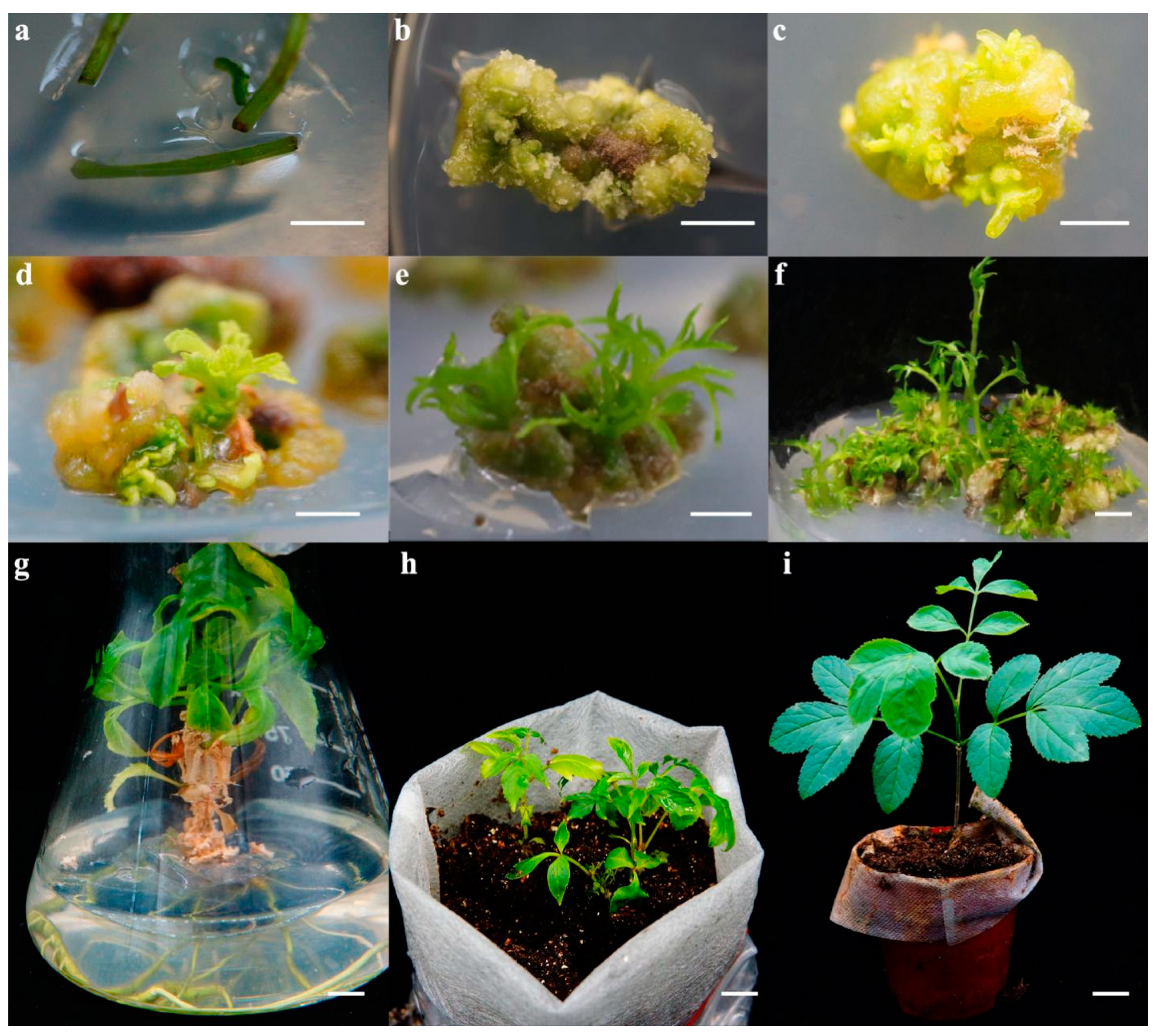
2.2. Adventitious Bud Induction of Hypocotyl, Root, Cotyledon and Stem Segments of F. Mandshurica

The aseptic seedlings that had been germinated for 21 days were selected, and the roots of the embryos were cut off. The hypocotyls, roots and cotyledons were inoculated into separate hormone combination media. Tissue culture that was growing well was selected, and the stem segment was inoculated into the culture medium between the two buds. The specific composition is shown in Table 1. The adventitious buds were formed by light after dark culture for two weeks. The cultures were incubated in growth chamber with 16:8 h light/dark period. The temperature was adjusted at 18 ± 2 °C for dark and 25 ± 2 °C for light periods. Ten tissue materials were treated each time, and the experiments were repeated three times. The callus induction rate at 20 days and the adventitious bud induction rate at 30 days were counted. In the induction process, the 5-aza to c12 medium was added to inhibit the DNA methylation level of the material and observe its effect on the adventitious bud induction rate. Auxin and cell division after the formation of adventitious buds in the stem segments of F. mandshurica culture tissue culture without adding 5-aza (c12 medium) and adding 2.4 mg/L 5-aza for 30 d were also further assessed.
The callus and adventitious bud medium was composed of MSB5 + 30 g/L sucrose + 7 g/L agar + 5 mg/L 6-BA + 8 mg/L TDZ + 2 mg/L-glycine + 0.1 mg/L IBA + 5% coconut water + 0.15 mg/L TSA. The secondary proliferation medium was composed of WPM + 20 g/L sucrose + 7 g/L agar + 8 mg/L 6-BA. The rooting medium was composed of WPM + 20 g/L sucrose + 7 g/L agar + 1.4 mg/L IBA + 0.7 mg/L NAA. After domestication, they were transplanted to a non-woven bag containing peat soil:vermiculite 3:1 mixed matrix. The seedling box was placed in culture conditions at a temperature of 25 °C, humidity 90%, light intensity 80 μmol·m−2·s−1, light-dark alternate culture in a greenhouse with a light time of 16 h.

2.3. Extraction and Analysis of Endogenous Hormones in Plants

Samples were ground to a fine powder in liquid nitrogen with a mortar and pestle, and suspended in potassium phosphate buffer (50 mM, pH = 7.4). After vigorous shaking, they were centrifuged for 30 min at 8000 rpm at 4 °C. Endogenous hormones were extracted from plant extract samples using Mlbio enzyme-linked bio-plant gibberellin (GA3), zeatin nucleoside (ZR), abscisic acid (ABA) and indole acetic acid (IAA) enzyme-linked immunosorbent assay (ELISA) kits. For the determination, three biological replicates were set for each sampling point and three technical replicates were set. Specific methods were as follows: loading, enzyme, incubated with a solution, washed, color, termination, and measurement.

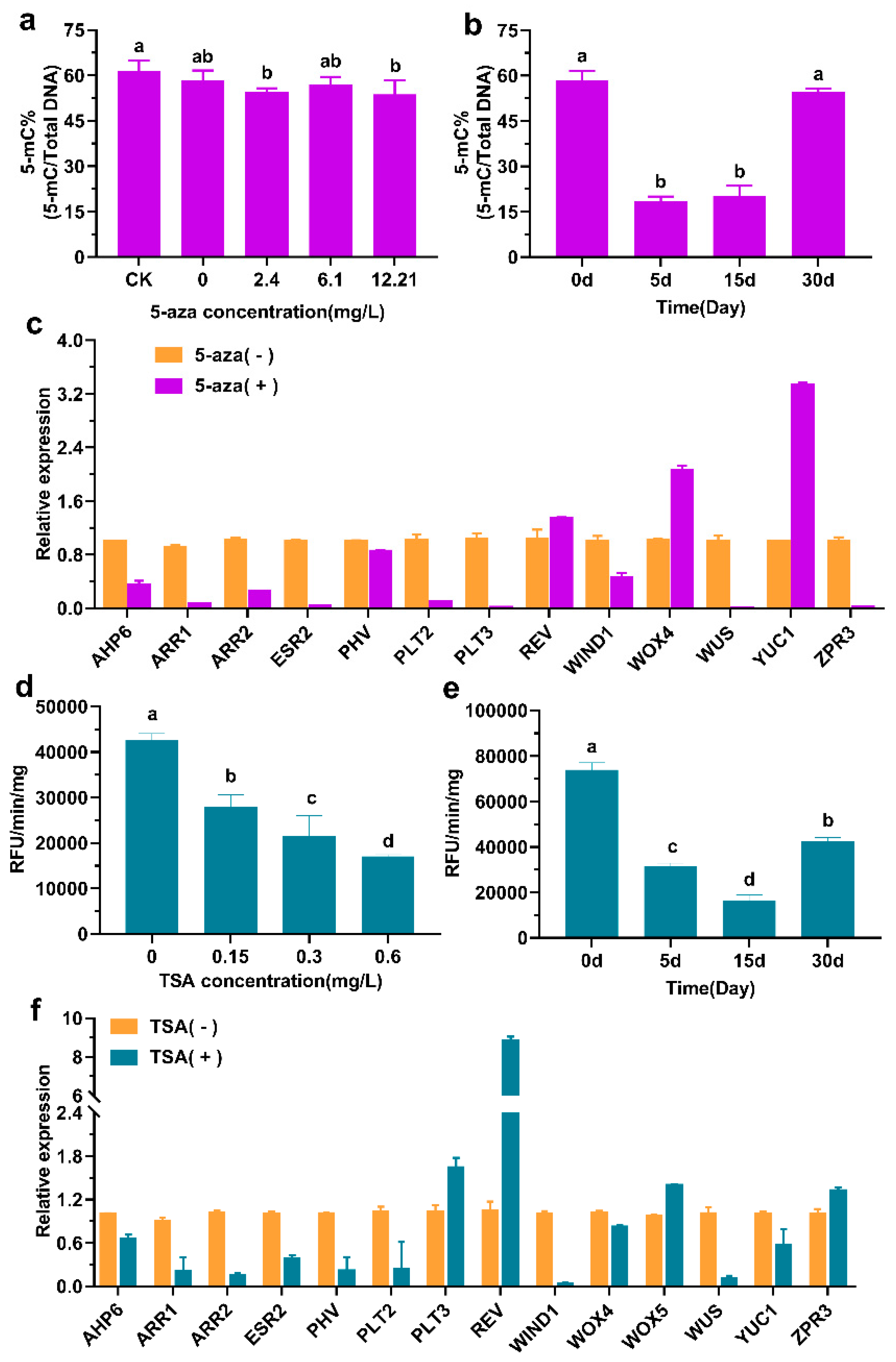
2.4. DNA Demethylation and Histone Acetylation

The total DNA was extracted from the leaves of F. mandshurica by CTAB method. The overall methylation level of DNA was determined using a ZYMO RESEARCH 5-mC DNA ELISA Kit (D5325). Three biological replicates were set for each sampling point and three technical replicates were set. The specific experimental methods were as follows: DNA coating, blocking, adding antibody and incubating at 37 °C for 1 h, color development for 30 min, and ELISA plate reading to measure the absorbance at 405–450 nm.
Histone deacetylase HDAC has the effects of regulating cell proliferation, inducing cell differentiation and apoptosis. In this experiment, the histone deacetylase inhibitor trichostatin A (TSA) was used to cultivate adventitious buds from callus stems of the F. mandshurica. HDAC enzyme activity gradually decreased, and histone acetylation level gradually increased. TSA can advance the induction of adventitious shoots. Enzyme activity assays were performed on the extracted proteins using the BioVision HDAC Active Fluorescence Assay Kit. The specific experimental methods were as follows: 1. Dilute the test sample (10–50 μg nuclear extract or cell lysate) to 85 μL (final volume) with ddH2O in each well (only 85 μL ddH2O is added for the blank control), and the positive control is diluted with 83 μL ddH2O with 2 μL of HeLa nuclear extract, and the negative control had 83 μL of ddH2O with 2 μL of trichostatin A or a sample containing no HDAC activity. 2. Add 10 μL of 10X HDAC Assay Buffer. 3. Add 5 μL of HDAC Fluorescent Substrate and mix well. 4. Incubate for 30 min at 37 °C. 5. Add 10 μL of lysine developer and incubate for 30 min at 37 °C. 6. Read the sample in a fluorescent plate reader (Ex/Em = 350–380 nm/440–460 nm). Histone deacetylase activity is expressed as the relative fluorescence value of each sample protein.

2.5. Quantitative Real-Time PCR Analyses

The RNA of the sample was extracted by the Tris-CTAB method, and the RNA was reverse transcribed into cDNA by TransScript All-in-One First-Strand cDNA Synthesis SuperMix for qPCR (One-Step gDNA Removal), and bioedited from the transcriptome data of F. mandshurica. The fluorescent quantitative PCR primers were designed according to the gene sequence (Table 2) and the quantitative amplification of the Applied Biosystems ABI 7500 quantitative PCR instrument was performed by TransStart Top Green qPCR SuperMix (+Dye II). The reaction system included template cDNA 1 μL (10 ng·μL−1), 2× SYBR Premix Ex taq 10 μL, Rox reference DyeII 0.4 μL, primer F 0.8 μL, primer R 0.8 μL, and ddH2O, added to 20 μL. The reaction procedure was: 95 °C for 1 min; 95 °C for 10 s, 60 °C for 30 s, 72 °C for 35 s, for 40 cycles. The tests were repeated three times per sample. The expression level was calculated by the 2−ΔΔCT method using the F. mandshurica vascular protein gene β-tubulin as an internal reference gene. The experimental data were analyzed by SPSS23 software.

3. Results

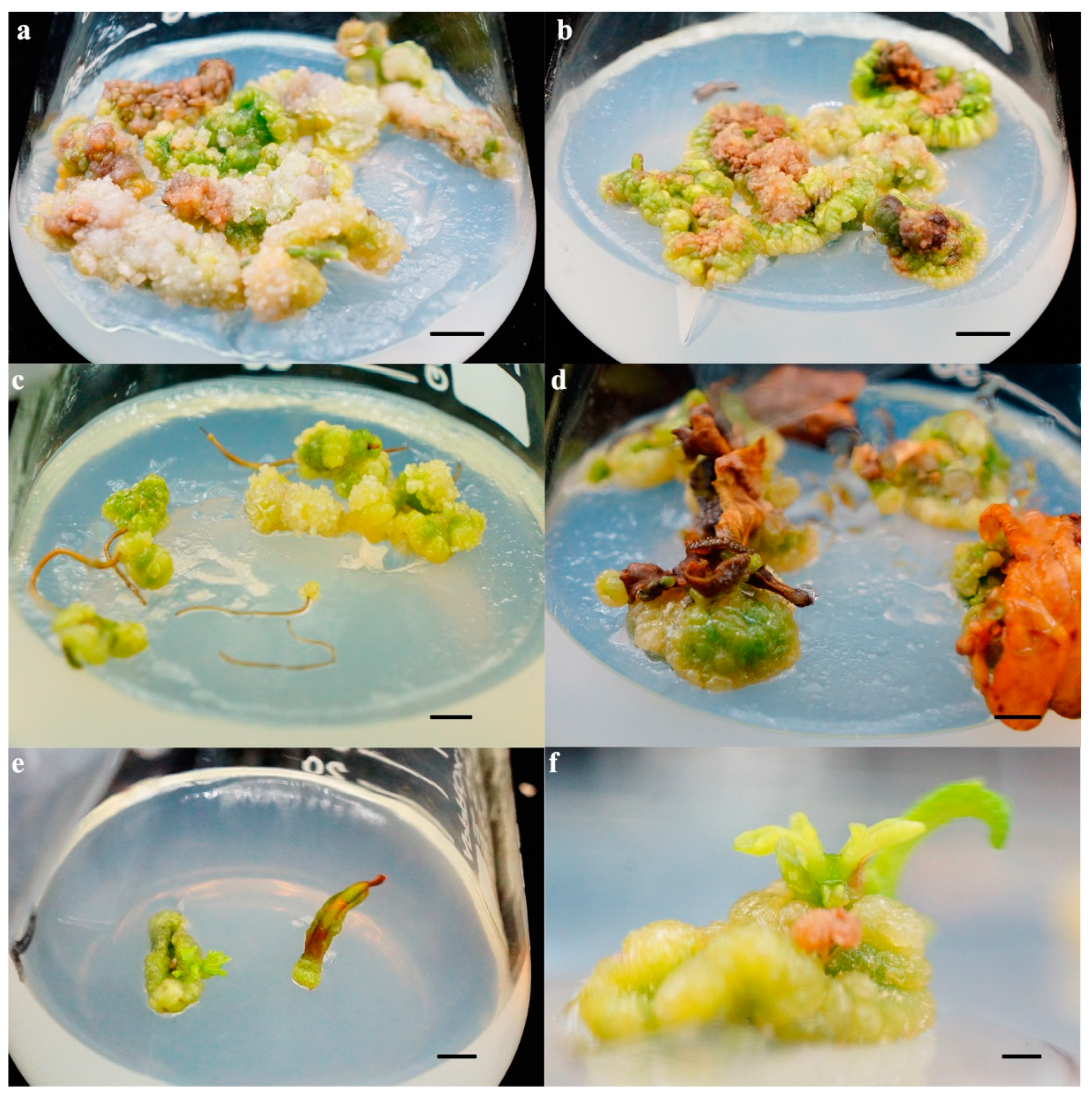
3.1. Effect of Exogenous 6-BA and TDZ Hormone Combination on Adventitious Bud Induction

The effects of different combinations of 6-BA and TDZ hormones on callus and adventitious buds on the use of hypocotyls, cotyledons, roots and stems as explants were explored (Figure 1). The results of 20 hormone treatments on cotyledons and roots used as explants are shown in Table 3. Under a4 medium (Figure 1d), the cotyledon callus induction rate reached 100%, and the texture of the cotyledon callus was hard. The color was dark green and the outer layer was a light green. Under a20 medium (Figure 1c), the highest callus induction rate of the roots was 50.71%. The callus formed by the roots had a velvety texture with canary yellow color and severe browning of the internal callus. Adventitious buds were not induced in all combinations of the two explants.
The results of 22 hormone treatments on hypocotyl used as explants are shown in Table 4. the callus induction rates in the a12 (Figure 1a) and a20 (Figure 1b) media were 93.56% and 88.69%. Germination rates of the a4 (Figure 1e) and a20 (Figure 1f) media were 3.13% and 10.52%. When the concentration of 6-BA was the same, with the increase in TDZ concentration, the callus induction rate also increased, which shows that TDZ played an important role in the induction process of hypocotyl callus. The texture of callus formed by low-concentration 6-BA-induced material was densely opaque green, and the texture surface of callus formed by high-concentration 6-BA-induced material was relatively loose and emerald green. The higher the concentration, the looser the callus. Therefore, a20 medium is suitable as a hypocotyl callus induction and adventitious bud regeneration medium.
The results of 15 hormone treatments on stem sections of tissue culture seedlings as explants are shown in Table 5. The callus induced in the c12 medium was hard inside, and visible protuberances and multiple outgrowths, which subsequently developed into adventitious shoot buds, were observed (Figure 2a–d). The callus induction rate of stem explants in c12 medium was 99.15%, and the bud induction rate was 33.33%. Although callus induction was efficient, the adventitious bud differentiation rate was still not ideal.

3.2. Effects of 5-aza and TSA on Adventitious Bud Differentiation of F. mandshurica

DNA 5-mC content was detected and calculated by the DNA methylation detection kit. The results showed that the addition of 2.4 mg/l 5-aza was most beneficial to the induction of adventitious buds (Figure 3), and the degree of DNA methylation was the lowest (Figure 5a). The DNA methylation level was related to the 5-mC content, where a higher 5-mC content indicates a higher DNA methylation level (Figure 5b). The induction rate of adventitious buds was 50.79%, which was 17.46% higher than that without 5-aza. The callus induced by the stem segment was mostly yellow-green with no signs of aging, and the dense induction of adventitious buds had a significant effect. The expression of genes related to regeneration was analyzed. It was found that the cytokinin pathway gene FmREV, the shoot tip and root tip gene FmWOX4, and the auxin synthesis gene FmYUC1 were significantly increased during regeneration after the addition of 5-aza, which were 1.30, 2.03, 3.33 times, respectively, that of the control (Figure 5c).
The results showed that TSA could inhibit HDAC enzyme activity (Figure 4) and promote the induction of adventitious buds (Figure 5d,e). The regeneration rate of adventitious buds induced by the addition of 0.15 mg/L TSA reached a maximum of 51.11%, which was 17.78% higher than without TSA. The callus induced was emerald green, and the adventitious buds were fairly young. The expression levels of FmPLT3, FmREV, FmWOX5, and FmZPR3 genes were significantly increased after adding TSA, which were 1.64, 8.82, 1.4 and 1.32 times higher than those of the control group (Figure 5f). In conclusion, adding 2.4 mg/L 5-aza or 0.15 mg/L TSA contributed to the induction of adventitious bud differentiation and the dense adventitious buds in the callus of tissue culture seedlings.

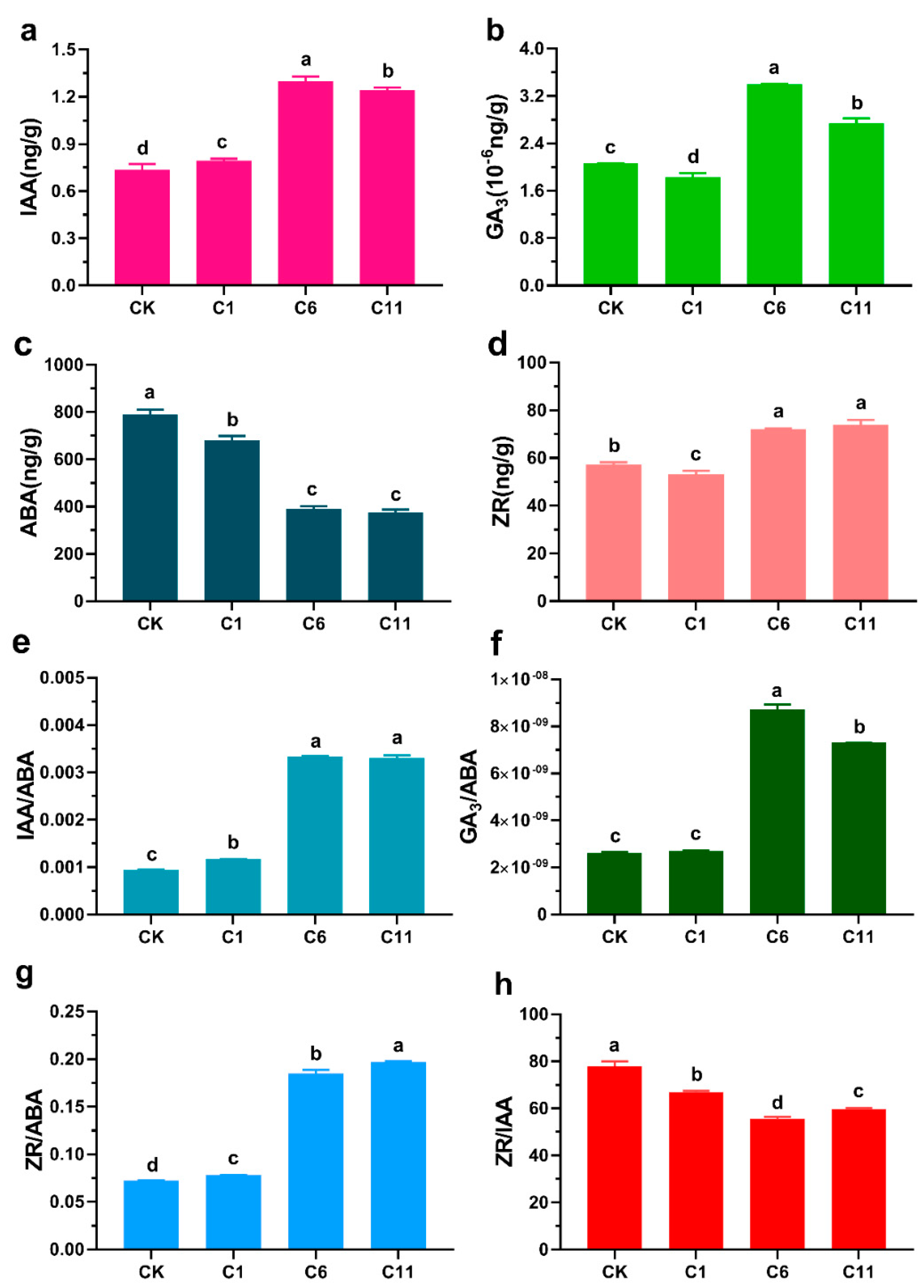
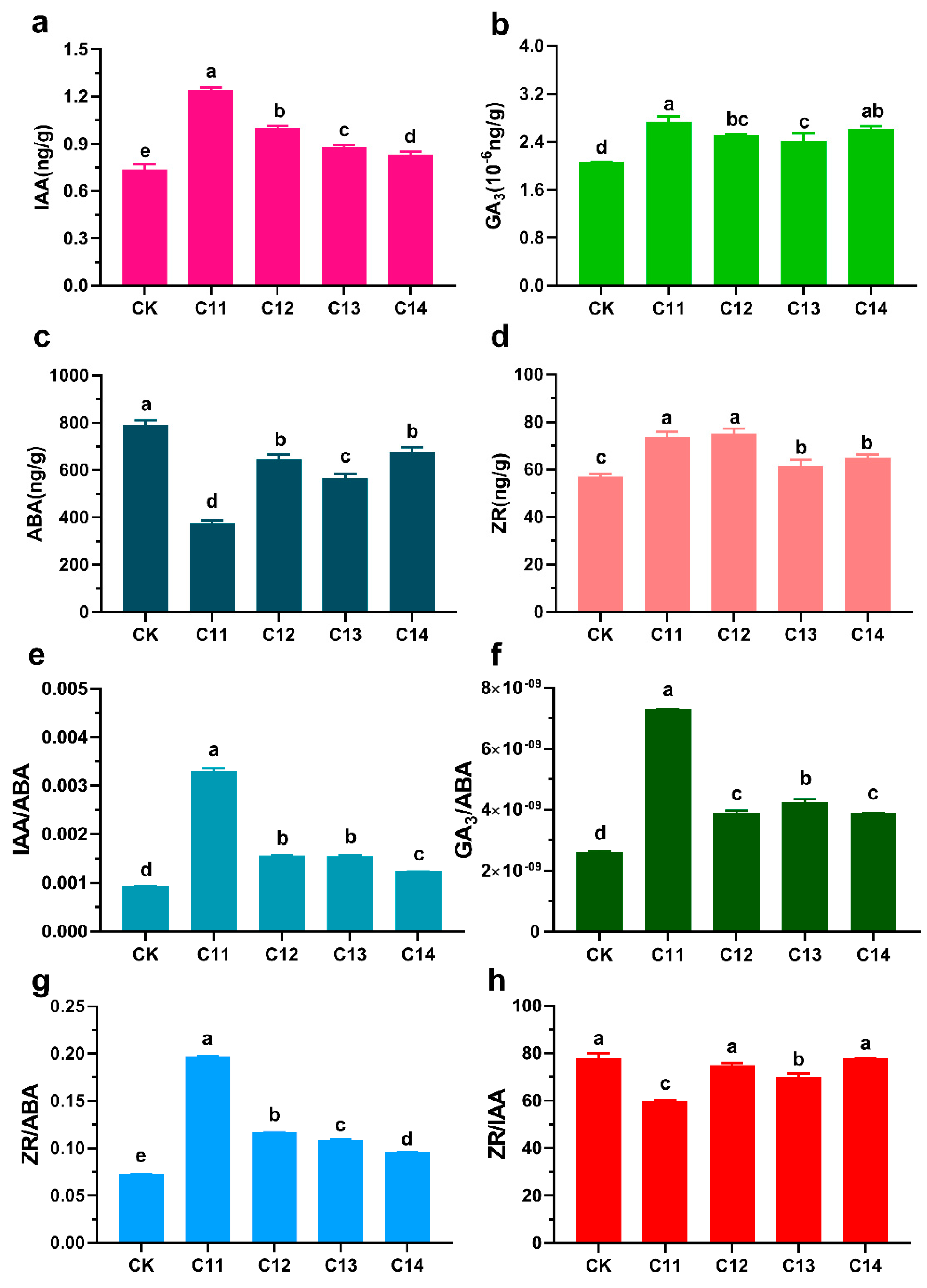
3.3. Effects of TDZ and 6-BA on Endogenous Hormone Levels in the Regeneration of F. mandshurica

After adding TDZ externally, with the increase in TDZ concentration, the content of IAA, ZR and ABA increased (Figure 6a–d). The content of GA3 first increased and then decreased. IAA/ABA, ZR/ABA and GA3/ABA content ratios increased, and ZR/IAA content ratios decreased (Figure 6e–h). It can be seen that exogenous application of 8 mg TDZ can promote the synthesis or accumulation of IAA, Zr and GA3, and inhibit the synthesis or accumulation of ABA. Compared with the control group, IAA accumulation increased 1.69 times from the original level, GA3 increased 1.33 times, ZR increased 1.29 times and ABA decreased 0.48 times. Analysis, combined with Table 5, showed that the change of adventitious bud induction rate was consistent with the change of IAA, ZR, and GA3 hormone levels, indicating that TDZ can increase the levels of IAA, ZR, GA3 hormones and regulate the differentiation of adventitious buds.
As the concentration of exogenous 6-BA gradually increases, the content of endogenous IAA decreases, the content of ZR decreases slightly, the content of ABA increases, and the content of GA3 remains stable (Figure 7a–d). The ratios of IAA/ABA, ZR/ABA and GA3/ABA also reduced. The ratio of ZR/IAA content showed a “W”-shaped fluctuation with the increase in 6-BA concentration (Figure 7e–h). A low concentration of 5–7 mg/L 6-BA stimulated the synthesis or accumulation of IAA, ZR and GA3, and inhibited the synthesis or accumulation of ABA in the 6-BA range. According to the analysis of the adventitious bud induction rate in Table 5, it was found that the adventitious bud induction rate was consistent with the change in IAA hormone levels, indicating that exogenous 6-BA plays a role in regulating the endogenous hormones IAA and ZR.

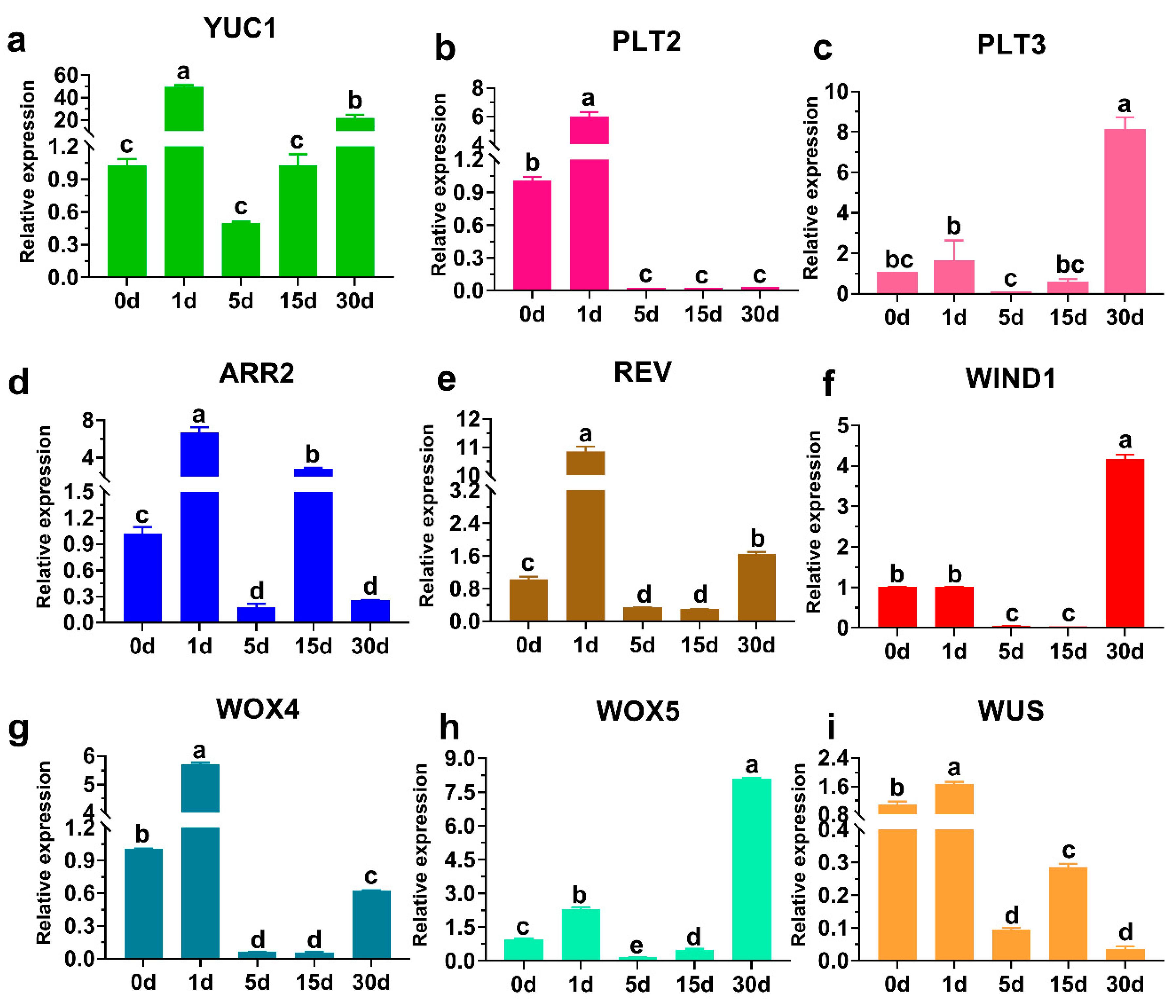
3.4. Analysis of Related Gene Expression Patterns during the Regeneration of F. mandshurica

The expression levels of the FmYUC1, FmWOX4, FmWOX5, FmWUS, FmARR2, FmREV, FmPLT2 and FmPLT3 genes increased significantly in the early stage of adventitious bud induction culture (Figure 8). The expression of FmYUC1, FmPLT2 and FmPLT3 cultivated for 1 d was 48.16, 5.93 and 1.57 times the 0 d cultivated level. At the stage of callus formation at 1–15 d, the ARR2 gene had a higher expression level than the control. The expression levels of FmYUC1, FmWOX4, FmWOX5, FmWUS, FmARR2, FmREV, FmZPR3, FmPLT2 and FmPLT3 were markedly increased at 1 d. The expression of FmWUS, FmWOX4, FmWOX5 and FmARR2 cultivated for 1 d was 1.67, 5.69, 2.28 and 6.56 times the 0 d cultivated level. The expression of FmWIND1, FmYUC1, FmWOX5, FmREV and FmPLT3 was greater than the control group after callus formation, and the adventitious buds were formed at 30 d. The expression of the HD-Zip III transcription factor FmREV cultivated for 1 d was 10 times the 0 d cultivated level. The expression of FmWIND1, FmYUC1, FmWOX5, FmREV and FmPLT3 was higher than that of the control, indicating that these genes are associated with the process of adventitious buds.

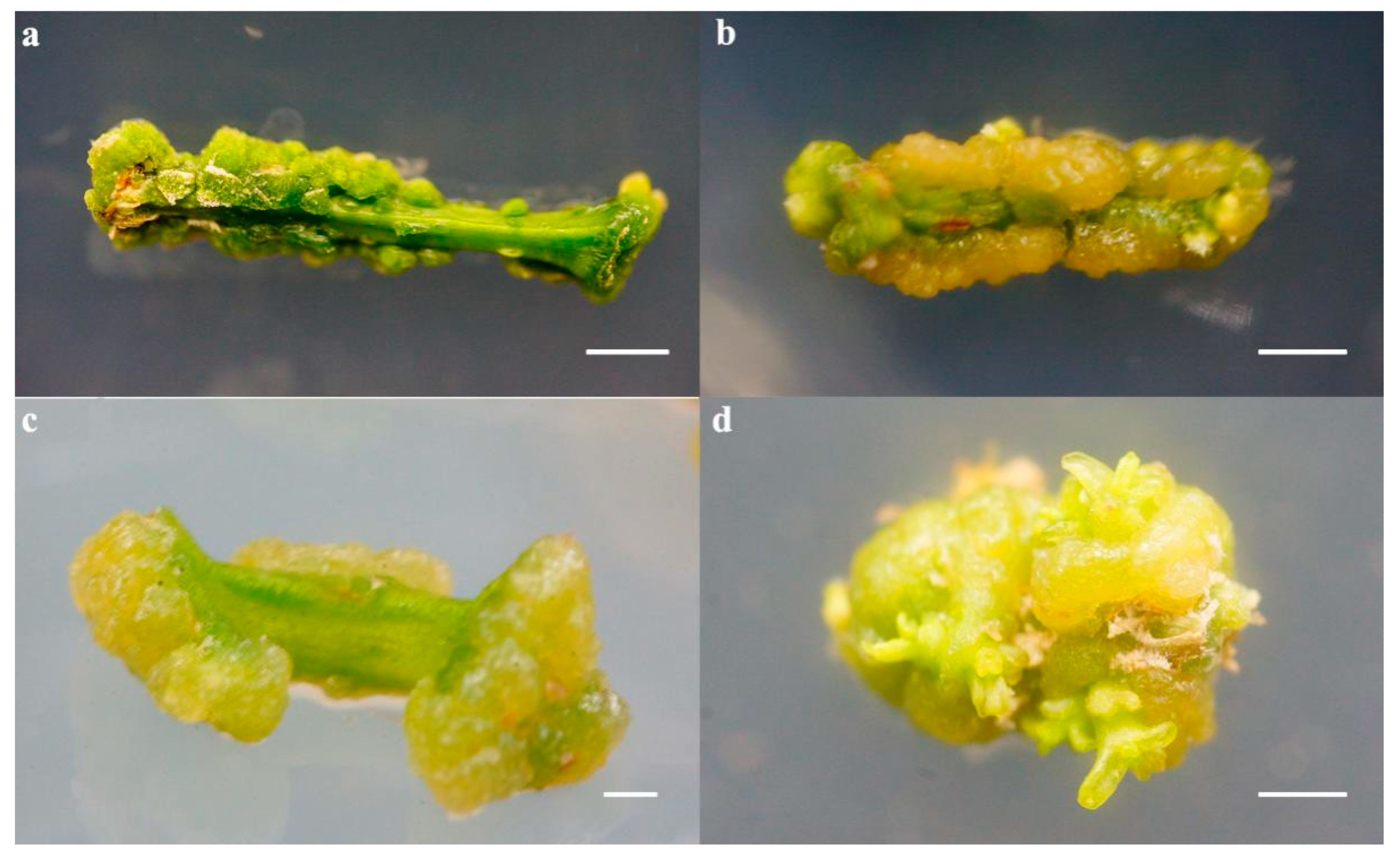
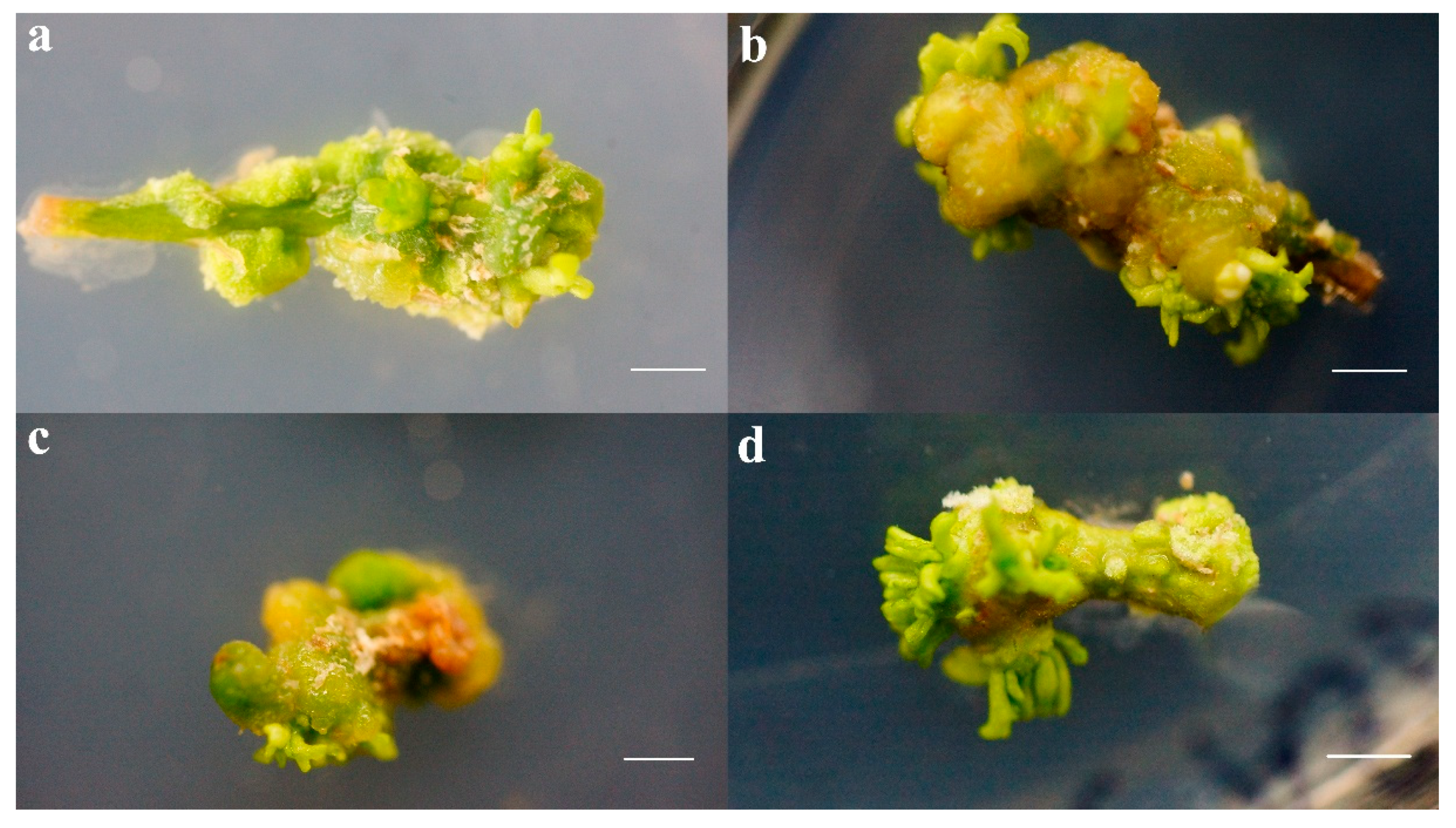
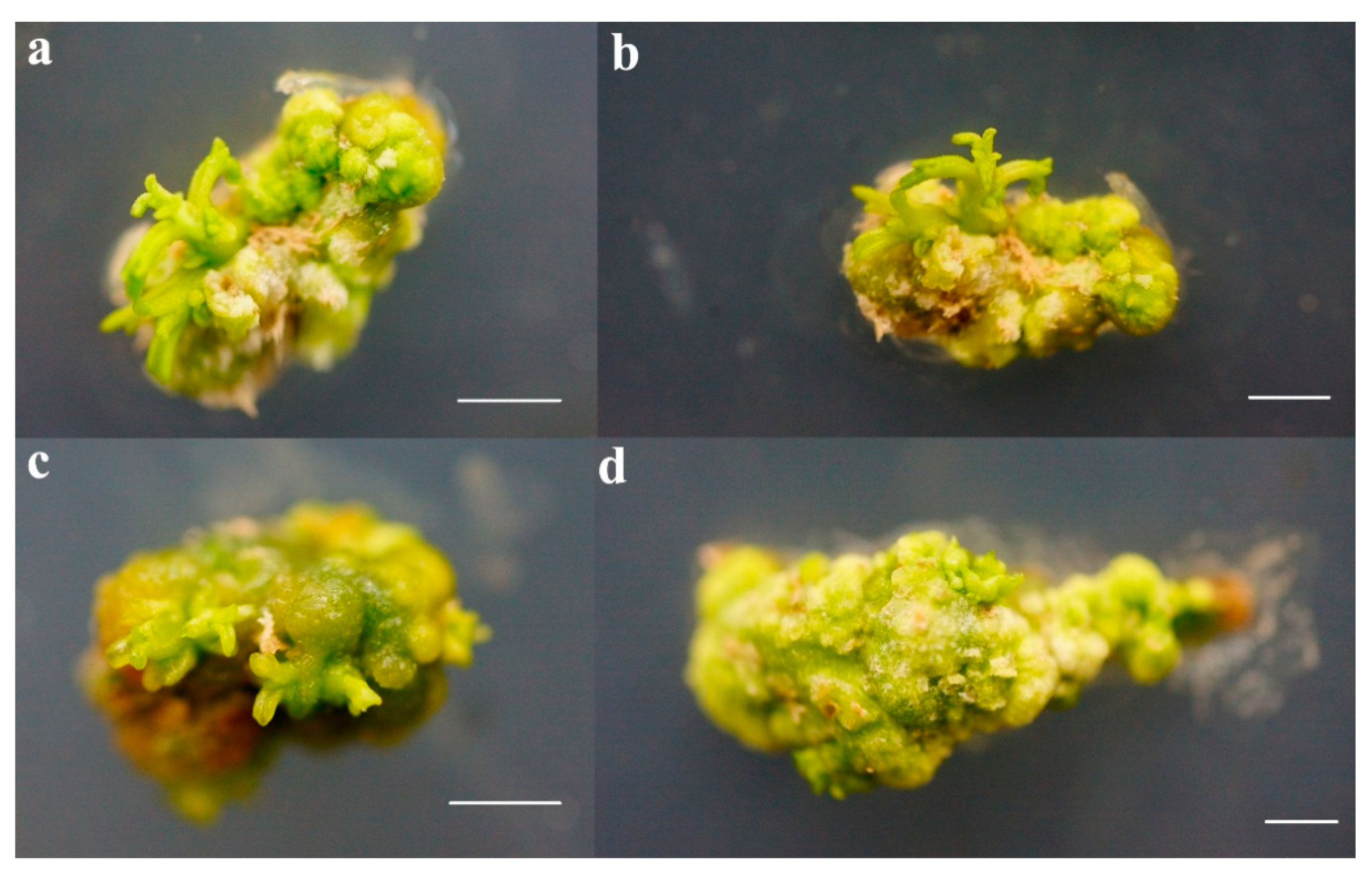
3.5. Establishment of a Callus Regeneration System from Stem Segments of F. mandshurica

Stem tissue of F. mandshurica was used as the explant (Figure 9a). Stem tissue was cultured for 20 d until it formed a callus (Figure 9b). The induction rate of the callus was 99.15%. Stem tissue was cultured for 30 d until it formed adventitious buds (Figure 9c), and the induction rate of the adventitious bud was 51.11%. After transferring to the secondary proliferation medium (Figure 9d-f), the shoots were transferred to the rooting medium for three weeks. The rooting rate was 80.54% (Figure 9g). After alternating light and dark cultivation, the survival rate reached 90% (Figure 9h). Finally, the viable F. mandshurica was transferred into the pot to continue cultivation (Figure 9i), and the in vitro regeneration technique of F. mandshurica was obtained.

4. Discussion

4.1. Effect of the Exogenous Hormones TDZ and 6-BA on Induction of Adventitious Buds of the Callus

The experimental results showed that, at the same TDZ concentration, when 0–8 mg/L 6-BA was added, the higher the concentration of 6-BA, the looser the callus. However, when the adventitious buds induced by cotyledons were used as explants, a higher concentration of 6-BA did not contribute to the formation of a cotyledon callus [23]. The texture of the callus formed by the low-concentration 6-BA-induced material was dense and opaque green, and the texture surface of the callus formed by the high-concentration 6-BA-induced material was relatively loose and emerald green (Figure 1a,b). The callus formed by the hypocotyl was rigid (Figure 1d). At the same 6-BA concentration, with an increase in TDZ concentration, the callus induction rate increased, indicating that TDZ plays a key role in hypocotyl callus induction. This is consistent with the results of Zeng et al., who found that the frequency of bud organ development of explants cultured with TDZ (71.16%) is far higher than that of explants cultured without TDZ (30.42%) [24]. Internal callus tissue began to age and darken after 30 days of culture, and did not contribute to the formation of adventitious buds. In addition, callus induced by using roots as explants was milky white in the lower culture and yellow-green in the upper culture. After subculture, the internal callus was obviously browned, while the external callus could not produce a primordium structure and adventitious buds (Figure 1c). The callus formed by cotyledons has the characteristics of harder and slower growth, and the callus formed by roots has the characteristics of loose and fast growth. This may be why the cotyledons and roots failed to induce adventitious buds.

4.2. Wound Treatment Is Beneficial for Callus Re-Differentiation

An important aspect of organ regeneration is the restoration of cell proliferation and regenerative capacity at the wound site, which results in the formation of callus and subsequent establishment of shoot or root apical meristems [25]. In this study, the research indicates that the callus formed at the wound site of the stem segment differed from the callus formed by the epidermis of the stem segment and was more conducive to the formation and differentiation of adventitious buds (Figure 2). Studies have found that Arabidopsis leaves may produce at least two wound signals after trauma, triggering short- and long-term wound signals, respectively. Short-term wound signals are mainly involved in controlling the fate transition of auxin and regenerative cells, while long-term wound signals primarily regulate the cellular environment at the wound site and maintain auxin levels to form regenerative cells [15]. The expression levels of FmYUC1, FmWOX4, FmWOX5, FmWUS, FmARR2, FmREV, FmPLT2 and FmPLT3 increased to different degrees after initial access to callus induction medium, the auxin synthesis gene YUC1 and the auxin pathway genes. FmPLT2 and FmPLT3 had an expression level of 48.16, 5.93, and 1.57 times the control. The expression levels of the somatic embryogenesis and regeneration genes FmWUS, FmWOX4 and FmWOX5 were 1.67, 5.69 and 2.28 times the control. The expression level of the cytokinin pathway gene b-type ARRs transcription factor FmARR2 in Arabidopsis shoot apical meristems was 6.56 times that of the control [26]. The expression level of the HD-ZIP III transcription factor FmREV was 0.4 and 10.84 times that of the control. At 1–15 d, at the callus formation stage, the expression level of the FmARR2 gene at 15 d was higher than that at 0 d, indicating that the gene is involved in the regulation of callus formation, and the gene responds to cytokinin signals [27]. The results showed that the formation of callus may be regulated by cytokinin signal transduction. The expression of FmWIND1, FmYUC1, FmWOX5, FmREV and FmPLT3 was up-regulated compared with the control during the formation of adventitious buds from 15–30 days. These genes may be involved in the formation of buds. FmREV, FmPLT3 and FmYUC1 belong to the process of bud regeneration. In the regulation of auxin synthesis genes [28], FmWIND1 is involved in the formation of cytokinin pathways and cells in the programming process [29,30], and FmWOX5 can maintain callus embryogenicity [31]. The occurrence of adventitious buds may be affected by the FmWIND1 gene induced by the wound signal, which changes the fate of the cells [32,33,34].

4.3. 5-aza Promotes Callus Re-Differentiation

In the plant genome, DNA methylation is mostly 5-mC and 7-mG. During plant tissue culture, different species, periods, genotypes, hormones, and DNA methylation inhibitors can make DNA methylation levels different [35,36,37,38]. Low DNA methylation levels favor Arabidopsis root regeneration [20]. During the process of callus formation and regeneration of sprouts, DNA methylation presents a variety of trends [39,40,41]. Dedifferentiated maize embryos have higher levels of methylation than normal embryos, and DNA methylation levels increase during callus induction [42]. The molecular mechanism involved in DNA methylation and regeneration is primarily focused on the study of FmWUS, but unfortunately, in this study, there was no change in the expression of the FmWUS gene, which may be that the cell range of FmWUS expression is small, and the expression time is short. The number of adventitious buds of F. mandshurica is small, the expression level of multi-cells may be averaged, and the expression of FmWUS is masked. However, the experimental results showed that the expression levels of FmREV, FmYUC1 and FmWOX4 increased after 5-aza was added, which indicated that 5-aza could promote the expression of these three genes and lead to an increased rate of adventitious bud differentiation.

5. Conclusions

The purpose of this study was to establish and optimize efficient and effective callus induction and an adventitious bud regeneration system. By comparing the differences between callus induction and adventitious bud differentiation of four kinds of explants and combining epigenetic regulation analysis, the regeneration technology of explant callus from stem segments of F. mandshurica were established and optimized. The changes in 5-aza and HDAC enzyme activity in callus formation and adventitious bud differentiation were explored. The level of DNA methylation in the callus formation stage of F. mandshurica decreased, and the adventitious bud formation stage increased again. HDAC enzyme activity was significantly reduced during the induction of adventitious buds, and TSA inhibited the activity of HDAC enzymes to promote adventitious bud regeneration. Analysis of the endogenous hormone content and related gene expression during adventitious bud differentiation showed that exogenous 6-BA and TDZ promote the differentiation of adventitious buds in governing the levels of endogenous IAA and ZR hormones. FmWIND1, FmWOX5 and FmPLT3 advance the formation of adventitious buds.

Author Contributions

L.Y. and Y.Z. designed the experiment and performed technical guidance. L.Y. wrote the manuscript and provided assistance in the data analysis. All authors participated in the experimental process. All authors have read and agreed to the published version of the manuscript.

Funding

This study was funded supported by the Applied Technology Research and Developmental Plan Program of Heilongjiang Province (GA19B201).

Conflicts of Interest

The authors declare no conflict of interest.

References

  1. Gu, J.C.; Wei, X.; Wang, J.; Dong, X.Y.; Wang, Z.Q. Marked differences in standing biomass, length density, anatomy and physiological activity between white and brown roots in fraxinus mandshurica rupr. plantation. Plant Soil 2015, 392, 267–277. [Google Scholar] [CrossRef]
  2. Wang, J.S.; Wu, L.; Zhao, X.H.; Fan, J.; Zhang, C.Y.; von Gadow, K. Influence of ground flora on fraxinus mandshurica seedling growth on abandoned land and beneath forest canopy. Eur. J. For. Res. 2013, 132, 313–324. [Google Scholar] [CrossRef]
  3. Chen, X.H.; Wang, H.T.; Li, X.Y.; Ma, K.; Zhan, Y.G.; Zeng, F.S. Molecular cloning and functional analysis of 4-coumarate:Coa ligase 4(4cl-like 1)from fraxinus mandshurica and its role in abiotic stress tolerance and cell wall synthesis. BMC Plant Biol. 2019, 19, 201. [Google Scholar] [CrossRef] [PubMed] [Green Version]
  4. Liu, W.G.; Fan, X.H.; Wang, J.S.; Zhang, C.Y.; Lu, W.M.; Gadow, K.V. Spectral reflectance response of fraxinus mandshurica leaves to above- and belowground competition. Int. J. Remote Sens. 2012, 33, 5072–5086. [Google Scholar] [CrossRef]
  5. Sun, X.D.; Wang, W.N.; Razaq, M.; Sun, H.L. Effects of stand density on soil organic carbon storage in the top and deep soil layers of fraxinus mandshurica plantations. Austrian J. For. Sci. 2019, 136, 27–44. [Google Scholar]
  6. He, L.M.; Zeng, F.S.; He, Z.L.; Li, Y.H.; Li, S.J.; Zhan, Y.G. Interspecific hybridizations of fraxinus l. (f. Mandshurica x f. Americana and f. Mandshurica x f. Velutina) and heterosis analysis and selection of f1 progenies. Can. J. For. Res. 2019, 49, 1265–1276. [Google Scholar] [CrossRef]
  7. Kong, D.M.; Preece, J.E.; Shen, H.L. Somatic embryogenesis in immature cotyledons of manchurian ash (fraxinus mandshurica rupr.). Plant Cell Tissue Organ. Cult. 2012, 108, 485–492. [Google Scholar] [CrossRef]
  8. Stener, L.G. Genetic evaluation of damage caused by ash dieback with emphasis on selection stability over time. For. Ecol. Manag. 2018, 409, 584–592. [Google Scholar] [CrossRef]
  9. Inoue, T.; Okane, I.; Ishiga, Y.; Degawa, Y.; Hosoya, T.; Yamaoka, Y. The life cycle of hymenoscyphus fraxineus on manchurian ash, fraxinus mandshurica, in Japan. Mycoscience 2019, 60, 89–94. [Google Scholar] [CrossRef]
  10. Du, X.Z.; Wei, X. Definition of fine roots on the basis of the root anatomy, diameter, and branch orders of one-year old fraxinus mandshurica seedlings. J. For. Res. 2018, 29, 1321–1327. [Google Scholar] [CrossRef]
  11. Lee, J.H.; Pijut, P.M. Adventitious shoot regeneration from in vitro leaf explants of fraxinus nigra. Plant Cell Tissue Organ. Cult. 2017, 130, 335–343. [Google Scholar] [CrossRef]
  12. Aida, M.; Beis, D.; Heidstra, R.; Willemsen, V.; Blilou, I.; Galinha, C.; Nussaume, L.; Noh, Y.S.; Amasino, R.; Scheres, B. The plethora genes mediate patterning of the arabidopsis root stem cell niche. Cell 2004, 119, 109–120. [Google Scholar] [CrossRef] [PubMed] [Green Version]
  13. Iwase, A.; Mitsuda, N.; Koyama, T.; Hiratsu, K.; Kojima, M.; Arai, T.; Inoue, Y.; Seki, M.; Sakakibara, H.; Sugimoto, K.; et al. The ap2/erf transcription factor wind1 controls cell dedifferentiation in arabidopsis. Curr. Biol. 2011, 21, 508–514. [Google Scholar] [CrossRef] [PubMed] [Green Version]
  14. Zhou, C.; Guo, J.S.; Feng, Z.H.; Cui, X.H.; Zhu, J. Molecular characterization of a novel ap2 transcription factor thwind1-l from thellungiella halophila. Plant Cell Tissue Organ. Cult. 2012, 110, 423–433. [Google Scholar] [CrossRef]
  15. Chen, L.Q.; Sun, B.B.; Xu, L.; Liu, W. Wound signaling: The missing link in plant regeneration. Plant Signal. Behav. 2016, 11, 4273–4284. [Google Scholar] [CrossRef] [PubMed] [Green Version]
  16. Xin, W.; Wang, Z.C.; Liang, Y.; Wang, Y.H.; Hu, Y.X. Dynamic expression reveals a two-step patterning of wus and clv3 during axillary shoot meristem formation in arabidopsis. J. Plant Physiol. 2017, 214, 1–6. [Google Scholar] [CrossRef]
  17. Kareem, A.; Durgaprasad, K.; Sugimoto, K.; Du, Y.J.; Pulianmackal, A.J.; Trivedi, Z.B.; Abhayadev, P.V.; Pinon, V.; Meyerowitz, E.M.; Scheres, B.; et al. Plethora genes control regeneration by a two-step mechanism. Curr. Biol. 2015, 25, 1017–1030. [Google Scholar] [CrossRef] [Green Version]
  18. Wojcikowska, B.; Botor, M.; Moronczyk, J.; Wojcik, A.M.; Nodzynski, T.; Karcz, J.; Gaj, M.D. Trichostatin a triggers an embryogenic transition in arabidopsis explants via an auxin-related pathway. Front. Plant Sci. 2018, 9, 1353. [Google Scholar] [CrossRef]
  19. Zhao, Q.; Du, Y.L.; Wang, H.T.; Rogers, H.J.; Yu, C.M.; Liu, W.; Zhao, M.Z.; Xie, F.T. 5-azacytidine promotes shoot regeneration during agrobacterium-mediated soybean transformation. Plant Physiol. Biochem. 2019, 141, 40–50. [Google Scholar] [CrossRef]
  20. Shemer, O.; Landau, U.; Candela, H.; Zemach, A.; Williams, L.E. Competency for shoot regeneration from arabidopsis root explants is regulated by DNA methylation. Plant Sci. 2015, 238, 251–261. [Google Scholar] [CrossRef]
  21. Liang, N.S.; Zhan, Y.G.; Yu, L.; Wang, Z.Q.; Zeng, F.S. Characteristics and expression analysis of fmtcp15 under abiotic stresses and hormones and interact with della protein in fraxinus mandshurica rupr. Forests 2019, 10, 343. [Google Scholar] [CrossRef] [Green Version]
  22. Liang, N.S.; Yu, L.; Liu, C.H.; Wang, Z.Q.; Zhao, X.T.; Zhan, Y.G. Molecular cloning and expression under abiotic stresses and hormones of the ethylene response factor vii gene fmrap2.12 from fraxinus mandshurica. J. For. Res. 2019, 30, 1289–1300. [Google Scholar] [CrossRef] [Green Version]
  23. Yang, X.; Yang, X.; Guo, T.; Gao, K.; Zhao, T.; Chen, Z.; An, X. High-efficiency somatic embryogenesis from seedlings of koelreuteria paniculata laxm. Forests 2018, 9, 769. [Google Scholar] [CrossRef] [Green Version]
  24. Zeng, Q.Q.; Han, Z.Q.; Kang, X.Y. Adventitious shoot regeneration from leaf, petiole and root explants in triploid (populus albaxp.Glandulosa)xp.Tomentosa. Plant Cell Tissue Organ. Cult. 2019, 138, 121–130. [Google Scholar] [CrossRef]
  25. Ikeuchi, M.; Shibata, M.; Rymen, B.; Iwase, A.; Bagman, A.M.; Watt, L.; Coleman, D.; Favero, D.S.; Takahashi, T.; Ahnert, S.E.; et al. A gene regulatory network for cellular reprogramming in plant regeneration. Plant Cell Physiol. 2018, 59, 770–782. [Google Scholar] [CrossRef]
  26. Kim, Y.S.; Kim, S.G.; Lee, M.; Lee, I.; Park, H.Y.; Seo, P.J.; Jung, J.H.; Kwon, E.J.; Suh, S.W.; Paek, K.H.; et al. Hd-zip iii activity is modulated by competitive inhibitors via a feedback loop in arabidopsis shoot apical meristem development. Plant Cell 2008, 20, 920–933. [Google Scholar] [CrossRef] [Green Version]
  27. Shull, T.E.; Kurepa, J.; Smalle, J.A. Cytokinin signaling promotes differential stability of type-b arrs. Plant Signal. Behav. 2016, 11, e1169354. [Google Scholar] [CrossRef] [Green Version]
  28. Liu, Y.; Zhang, H.L.; Guo, H.R.; Xie, L.; Zeng, R.Z.; Zhang, X.Q.; Zhang, Z.S. Transcriptomic and hormonal analyses reveal that yuc-mediated auxin biogenesis is involved in shoot regeneration from rhizome in cymbidium. Front. Plant Sci. 2017, 8. [Google Scholar] [CrossRef] [Green Version]
  29. Ikeda, M.; Ohme-Takagi, M. Tcps, wuss, and winds: Families of transcription factors that regulate shoot meristem formation, stem cell maintenance, and somatic cell differentiation. Front. Plant Sci. 2014, 5. [Google Scholar] [CrossRef] [Green Version]
  30. Iwase, A.; Mita, K.; Favero, D.S.; Mitsuda, N.; Sasaki, R.; Kobayshi, M.; Takebayashi, Y.; Kojima, M.; Kusano, M.; Oikawa, A.; et al. Wind1 induces dynamic metabolomic reprogramming during regeneration in brassica napus. Dev. Biol. 2018, 442, 40–52. [Google Scholar] [CrossRef]
  31. Orlowska, A.; Kepczynska, E. Identification of polycomb repressive complex1, trithorax group genes and their simultaneous expression with wuschel, wuschel-related homeobox5 and shoot meristemless during the induction phase of somatic embryogenesis in medicago truncatula gaertn. Plant Cell Tissue Organ. Cult. 2018, 134, 345–356. [Google Scholar] [CrossRef] [Green Version]
  32. Iwase, A.; Mitsuda, N.; Ikeuchi, M.; Ohnuma, M.; Koizuka, C.; Kawamoto, K.; Imamura, J.; Ezura, H.; Sugimoto, K. Arabidopsis wind1 induces callus formation in rapeseed, tomato, and tobacco. Plant Signal. Behav. 2013, 8, e27432. [Google Scholar] [CrossRef] [PubMed] [Green Version]
  33. Iwase, A.; Mita, K.; Nonaka, S.; Ikeuchi, M.; Koizuka, C.; Ohnuma, M.; Ezura, H.; Imamura, J.; Sugimoto, K. Wind1-based acquisition of regeneration competency in arabidopsis and rapeseed. J. Plant Res. 2015, 128, 389–397. [Google Scholar] [CrossRef] [PubMed] [Green Version]
  34. Iwase, A.; Harashima, H.; Ikeuchi, M.; Rymen, B.; Ohnuma, M.; Komaki, S.; Morohashi, K.; Kurata, T.; Nakata, M.; Ohme-Takagi, M.; et al. Wind1 promotes shoot regeneration through transcriptional activation of enhancer of shoot regeneration1 in arabidopsis. Plant Cell 2017, 29, 54–69. [Google Scholar] [CrossRef] [Green Version]
  35. Salaj, T.; Klubicova, K.; Matusova, R.; Salaj, J. Somatic embryogenesis in selected conifer trees pinus nigra arn. And abies hybrids. Front. Plant Sci. 2019, 10. [Google Scholar] [CrossRef] [Green Version]
  36. Ree, J.F.; Polesi, L.G.; Back, F.; Bertolazi, A.A.; Silveira, V.; Guerra, M.P. Aging peach palm (bactris gasipaes kunth) cultures lose embryogenic potential and metabolic cellular function due to continuous culture in hypoxic environments. Plant Cell Tissue Organ. Cult. 2019, 140, 49–67. [Google Scholar] [CrossRef]
  37. Muniandy, K.; Tan, M.H.; Song, B.K.; Ayub, Q.; Rahman, S. Comparative sequence and methylation analysis of chloroplast and amyloplast genomes from rice. Plant Mol. Biol. 2019, 100, 33–46. [Google Scholar] [CrossRef]
  38. Li, J.Y.; Wang, M.J.; Li, Y.J.; Zhang, Q.H.; Lindsey, K.; Daniell, H.; Jin, S.X.; Zhang, X.L. Multi-omics analyses reveal epigenomics basis for cotton somatic embryogenesis through successive regeneration acclimation process. Plant Biotechnol. J. 2019, 17, 435–450. [Google Scholar] [CrossRef] [Green Version]
  39. Hu, L.J.; Li, N.; Zhang, Z.B.; Meng, X.C.; Dong, Q.L.; Xu, C.M.; Gong, L.; Liu, B. Cg hypomethylation leads to complex changes in DNA methylation and transpositional burst of diverse transposable elements in callus cultures of rice. Plant J. 2019, 101, 188–203. [Google Scholar] [CrossRef]
  40. Gulyas, A.; Dobranszki, J.; Kiss, E.; da Silva, J.A.T.; Posta, K.; Hidvegi, N. Changes in DNA methylation pattern of apple long-term in vitro shoot culture and acclimatized plants. J. Plant Physiol. 2019, 239, 18–27. [Google Scholar] [CrossRef] [Green Version]
  41. Osorio-Montalvo, P.; Saenz-Carbonell, L.; De-la-Pena, C. 5-azacytidine: A promoter of epigenetic changes in the quest to improve plant somatic embryogenesis. Int. J. Mol. Sci. 2018, 19, 3182. [Google Scholar] [CrossRef] [PubMed] [Green Version]
  42. Liu, H.J.; Ma, L.L.; Yang, X.R.; Zhang, L.; Zeng, X.; Xie, S.P.; Peng, H.W.; Gao, S.B.; Lin, H.J.; Pan, G.T.; et al. Integrative analysis of DNA methylation, mrnas, and small rnas during maize embryo dedifferentiation. BMC Plant Biol. 2017, 17. [Google Scholar] [CrossRef] [PubMed] [Green Version]
Figure 1. Hypocotyl, root and cotyledon-induced callus and hypocotyl regeneration buds. (a) Hypocotyls induced callus in a12 medium for 20 d (scale 5 mm); (b) Hypocotyls induced callus in a20 medium for 20 d; (c) Root callus induced in a20 medium; (d) Cotyledons induced callus in a4 medium; (e) Hypocotyls induced adventitious buds in a4 medium for 30 d (scale 5 mm); (f) Hypocotyls induced adventitious buds in a20 medium for 30 d (scale 2 mm).
Forests 11 00590 g001
Figure 2. Stem segment induced callus and its regenerated shoots. (a) The callus formed by the epidermis of the stem segment was cultured in C12 for 20 d (scale 5 mm); (b) Cultured for 30 d; (c) The callus formed by the wound of the stem segment was cultured in C12 for 20 d (scale 2 mm). (d) Regenerated shoots formed in 30 d (scale 5 mm).
Forests 11 00590 g002
Figure 3. Adding different concentrations of 5-aza to induce the formation of adventitious buds. (a) Adding 2.4 mg/L 5-aza; (b) Adding 6.1 mg/L 5-aza; (c) Adding 12.2 mg/L 5-aza; (d) Adding 24.42 mg/L 5-aza (scale 2 mm).
Forests 11 00590 g003
Figure 4. Adventitious buds induced by the addition of different concentrations of TSA. (a) Adding 0.15 mg/L TSA; (b) Adding 0.3 mg/L TSA; (c) Adding 0.6 mg/L TSA; (d) Adding 1.2 mg/L TSA (scale is 2 mm).
Forests 11 00590 g004
Figure 5. Epigenetic and related gene expression levels during stem bud regeneration. (a) The 5-aza concentration affects the 5-mC level of shoot bud regeneration during 30 d of culture. (b) Changes in DNA methylation levels at various stages of regeneration of the Fraxinus mandshurica Rupr. (c) 5-aza affects the expression level of regenerative bud-related genes in stem segments. (d) The TSA concentration affects the level of regenerated bud HDAC in shoots of stem segments during 30 d of culture. (e) TSA affects the change in HDAC activity during regeneration. (f) TSA affects the expression level of regenerative bud-related genes in stem segments. Different letters above bars within statistically significant differences between different times of the treatments at the p < 0.05 level according to Duncan’s multiple range test.
Forests 11 00590 g005
Figure 6. Effect of TDZ on endogenous hormone levels in regeneration of F. mandshurica. (ad) CK was the stem tissue, and C1, C6 and C11 were the three endogenous hormones measured under the same culture conditions, and the TDZ concentrations were 6 mg/L, 7 mg/L and 8 mg/L for 30 d. (eh) The relative amount of endogenous hormones. Different letters above bars within statistically significant differences between different times of the treatments at the p < 0.05 level according to Duncan’s multiple range test.
Forests 11 00590 g006
Figure 7. Effect of 6-BA on endogenous hormone levels in regeneration of F. mandshurica. (ad) CK was the stem tissue, and C11, C12, C13 and C14 were the four endogenous hormones measured under the same culture conditions, and the 6-BA concentrations were 0, 5, 6 and 7 mg/L for 30 d. (eh) The relative amount of endogenous hormones. Different letters above bars within statistically significant differences between different times of the treatments at the p < 0.05 level according to Duncan’s multiple range test.
Forests 11 00590 g007
Figure 8. Gene expression of adventitious buds in the stem segments of F. mandshurica. (ai) 0–15 d was used to induce callus in the dark culture stage. After 15–30 d, it was transferred to the light culture stage to induce callus regeneration, and the shoot regeneration time was 30 d. Different letters above bars within statistically significant differences between different times of the treatments at the p < 0.05 level according to Duncan’s multiple range test.
Forests 11 00590 g008
Figure 9. Preliminary establishment of the callus regeneration system of stems of F. mandshurica. (a) Inoculation of stem explants (scale 5 mm); (b) Callus formed at 20 d; (c) at 30 d, young adventitious buds form; (d) at 40 d, clustered buds formed; (e) Adventitious bud elongation; (f) Sub-generation expansion; (g) Rooting (scale 5 mm); (h) Planting (scale 1 cm); (i) Sterilization of sterile F. mandshurica seedlings to survive (scale 3 cm).
Forests 11 00590 g009
Table 1. Adventitious bud induction medium of stem segments.
Media NumberBasic Medium TypeSucrose (g/L)Agar (g/L)6-BA (mg/L)TDZ (mg/L)IBA (mg/L)Coconut (v/v)TSA (mg/L)5-aza (mg/L)
c1MSB5307060.15%00
c2MSB5307560.15%00
c3MSB5307660.15%00
c4MSB5307760.15%00
c5MSB5307860.15%00
c6MSB5307070.15%00
c7MSB5307570.15%00
c8MSB5307670.15%00
c9MSB5307770.15%00
c10MSB5307870.15%00
c11MSB5307080.15%00
c12MSB5307580.15%00
c13MSB5307680.15%00
c14MSB5307780.15%00
c15MSB5307880.15%00
b12MS307580.15%00
d12WPM307580.15%00
T5MSB5307580.15%0.150
T6MSB5307580.15%0.30
T7MSB5307580.15%0.60
T8MSB5307580.15%1.20
A5MSB5307580.15%02.4
A6MSB5307580.15%06.1
A7MSB5307580.15%012.21
A8MSB5307580.15%024.42
Table 2. Real-time PCR primers.
Primer NamePrimer Sequence
qFmTU-F5′-AGGACGCTGCCAACAACTTT-3′
qFmTU-R5′-TTGAGGGGAAGGGTAAATAGTG-3′
qFmAHP6-F5′-GTAAGTCACCACCAATGCCCA-3′
qFmAHP6-R5′-TCAGAGTTACAGAGGGTGATGCC-3′
qFmYUC1-F5′-CCAGCATCACCACCTTCAACAT-3′
qFmYUC1-R5′-CGAACAGACCCTCGGCAAGTA-3′
qFmPLT2-F5′-TGACACTCCGCTTCATAACCA-3′
qFmPLT2-R5′-AACAGATAATCGGAACCACTCG-3′
qFmPLT3-F5′-GGAACATTTGCTACTGAGGAGG-3′
qFmPLT3-R5′-GAGATTATGATGATAGAATGGGGC-3′
qFmARR1-F5′- GGGTTTGATGAGGAGAGTGGG -3′
qFmARR1-R5′- GTCCCATTTAGTCCCCGTTTG -3′
qFmARR2-F5′-CTATGCCTGGGATGACTGGA-3′
qFmARR2-R5′-GGAAAGCCATGATCGTGAAA-3′
qFmESR2-F5′-GGCTTGGAACTTTTGACACG-3′
qFmESR2-R5′-TGAAGCTATACGAACGGACAAG-3′
qFmPHV-F5′-CGGATAAGCAAAACTGGGGGT-3′
qFmPHV-R5′-AGGGTGGTGGCTCCATTATTCA-3′
qFmREV-F5′-CGGTTACTTTCGCCAGCATAC-3′
qFmREV-R5′-GGCTTCATTCCAGGCATTTG-3′
qFmWIND1-F5′-TGCCAATCACAGGATCTAAA-3′
qFmWIND1-R5′-CTGACCAAACTGACACGAAA-3′
qFmWOX4-F5′-TGGATTCTGGGAGCAACATA-3′
qFmWOX4-R5′-AGCCGATTCCTTCTTGTGAT-3′
qFmWOX5-F5′-GGCAATAATGGCGTCAGTGG-3′
qFmWOX5-R5′-CCCTGAATGAGTTGAGTGGAAATA-3′
qFmWUS-F5′- GGAGGAAGTCTTTGTGGTGC -3′
qFmWUS-R5′- GAACACTTTGATTGGAGGTCG -3′
qFmZPR3-F5′-GCAGTGCTTGATTCTCCTGGTT-3′
qFmZPR3-R5′-ATGGAGAGGCTAAACTCAAAGCTG-3′
Table 3. Effect of hormone treatment on cotyledon and root callus induction and adventitious bud induction.
Media Number6-BA (mg/L)TDZ (mg/L)Cotyledon Survival Rate (%)Cotyledon Callus Induction Rate (%)Root Survival Rate (%)Root Callus Induction Rate (%)Adventitious Bud Induction Rate (%)
a10017.5 ± 5.46hi55.00 ± 7.07d25.42 ± 4.28jkl0g0
a200.150 ± 1.57f0g28.48 ± 5.07ijk14.58 ± 7.92e0
a300.559.52 ± 0.49ef41.67 ± 3.27e54.94 ± 1.42bcd23.33 ± 2.35d0
a401100a100a57.71 ± 2.96abc48.49 ± 1.44a0
a51.5012.5 ± 8.92i0g18.06 ± 0.39l0g0
a61.50.188.89 ± 8.17b0g47.17 ± 4.59def10.42 ± 7.5ef0
a71.50.555.56 ± 6.29f44.44 ± 1.02e51.39 ± 8.58cde33.68 ± 4.15c0
a81.5178.42 ± 3.75cd92.71 ± 3.15b61.86 ± 0.69ab27.42 ± 5.54cd0
a92027.27 ± 1.31gh18.18 ± 6.96f64.44 ± 6.81a0g0
a1020.157.14 ± 9.24f0g36.31 ± 2.2ghi4.17 ± 7.48fg0
a1120.575.25 ± 6.75d63.89 ± 8.46c48.33 ± 7.63def14.17 ± 2.68e0
a122187.5 ± 6.5bc91.67 ± 6.29b44.55 ± 6.8efg40.97 ± 0.02b0
a133050 ± 8.05f0g28.65 ± 3.5ijk0g0
a1430.120 ± 5.19hi12.5 ± 3.33f24.64 ± 0.83jkl0g0
a1530.510 ± 6.44i0g33.93 ± 0.16hij0g0
a163170.00 ± 3.57d70 ± 5.11c33.22 ± 0.06hij29.64 ± 0.85cd0
a175032.14 ± 8.89g0g20.35 ± 6.81kl0g0
a1850.133.18 ± 3.79g15.48 ± 4.83f40.24 ± 0.08fgh3.33 ± 9.79fg0
a1950.568.75 ± 7.25de48.75 ± 7.91de27.56 ± 8.77ijk10 ± 7.42ef0
a205150 ± 3.17f16.67 ± 4.99f58.47 ± 8.43abc50.71 ± 0.05a0
Mean ± SE followed by the same letter in the same column were not significantly different by Duncan′s multiple comparison test (p < 0.05).
Table 4. Effects of hormone treatment on hypocotyl callus and adventitious bud induction.
Media Number6-BA (mg/L)TDZ (mg/L)Survival Rate (%)Callus Induction Rate (%)Adventitious Bud Induction Rate (%)
a10026.39 ± 7.01de10 ± 1.2ij0c
a200.130.95 ± 3.58de25 ± 8.41gh0c
a300.582.59 ± 8.48b77.38 ± 4.81b0c
a40196.43 ± 1.39a71.43 ± 9.92b3.13 ± 1.25b
a51.500f0j0c
a61.50.125 ± 6.83e25 ± 6.36gh0c
a71.50.558.33 ± 3.05c53.33 ± 3.1cd0c
a81.5173.86 ± 7.51b87.5 ± 6.24a0c
a9200f0j0c
a1020.136.36 ± 6.73d18.18 ± 4.42hi0c
a1120.578.47 ± 9.12b72.69 ± 2.8b0c
a122195.83 ± 1.77a93.56 ± 2.39a0c
a133028.57 ± 0.72de37.5 ± 4.71ef0c
a1430.152.08 ± 9.97c31.94 ± 1.81fg0c
a1530.530.16 ± 9.87de0j0c
a163162.5 ± 2.03c59.29 ± 0.84c0c
a175030 ± 7.14de20 ± 7.48h0c
a1850.152.05 ± 2.08c27.56 ± 7.24gh0c
a1950.578.61 ± 3.54b61.67 ± 9.79c0c
a205181.75 ± 5.06b88.69 ± 9.64a10.52 ± 2.12a
a215228.64 ± 6.22c46.35 ± 1.67de0c
a225260.34 ± 7.93c40.72 ± 8.65ef0c
Mean ± SE followed by the same letter in the same column were not significantly different by Duncan′s multiple comparison test (p < 0.05).
Table 5. Effect of hormone treatment on stem callus and adventitious bud induction.
Media Number6-BA (mg/L)TDZ (mg/L)Survival Rate (%)Callus Induction Rate (%)Adventitious Bud Induction Rate (%)
c10635.75 ± 1.02f67.98 ± 0.33k0g
c25665.74 ± 3.08cd87.45 ± 0.22h0g
c36678.68 ± 6.79a90.41 ± 0.07f0g
c47667.48 ± 0.44bcd91.34 ± 0.45e4.55 ± 0.15ef
c58679.64 ± 4.3a96.54 ± 0.7c3.33 ± 1.23f
c60745.67 ± 6.27ef74.12 ± 0.84j6.25 ± 2.14de
c75778.14 ± 7.54ab98.54 ± 0.41a3.7 ± 1.33f
c86777.64 ± 5.69ab97.35 ± 0.09b0g
c97764.12 ± 4cd88.97 ± 0.85g11.81 ± 1.49c
c108761.47 ± 1.59d98.74 ± 0.37a16.21 ± 1.19b
c110841.24 ± 8.43ef75.64 ± 0.03i6.94 ± 1.5d
c125874.65 ± 8.32abc99.15 ± 0.27a33.33 ± 1.17a
c136874.51 ± 3.69abc97.64 ± 0.36b10.48 ± 1.45c
c147866.66 ± 9.88cd98.42 ± 0.03a2.86 ± 1.6f
c158850.79 ± 5.99e95.11 ± 0.36d9.72 ± 1.69c
Mean ± SE followed by the same letter in the same column were not significantly different by Duncan′s multiple comparison test (p < 0.05).

