
Impact of Final Irrigation Protocol on the Push-Out Bond Strength of Two Types of Endodontic Sealers

Abstract
:1. Introduction
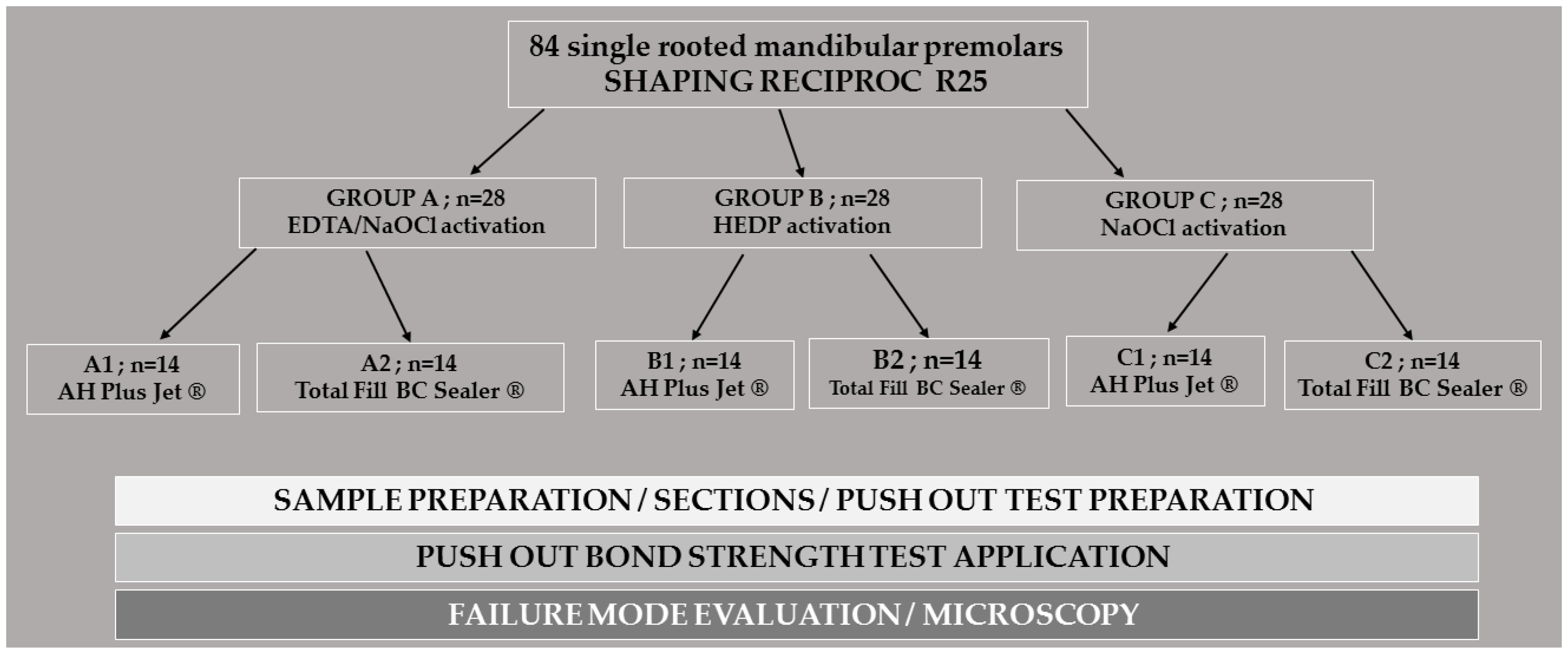
2. Methods
2.1. Sample Size Calculation
2.2. Teeth Selection
2.3. Root Canals Preparation
2.4. Irrigation Protocol
- Group A (n = 28)
- Group B (n = 28)
- Group C (n = 28)
2.5. Root Canal Filling
- A1: 14 root canals filled with single-cone/AH Plus Jet®.
- A2: 14 root canals filled with single-cone/Total Fill BC Sealer®.
- B1: 14 root canals filled with single-cone/AH Plus Jet®.
- B2: 14 root canals filled with single-cone/Total Fill BC Sealer®.
- C1: 14 root canals filled with single-cone/AH Plus Jet®.
- C2: 14 root canals filled with single-cone/Total Fill BC Sealer®.
2.6. Push-Out Test Preparation
2.7. Push-Out Bond Strength Application
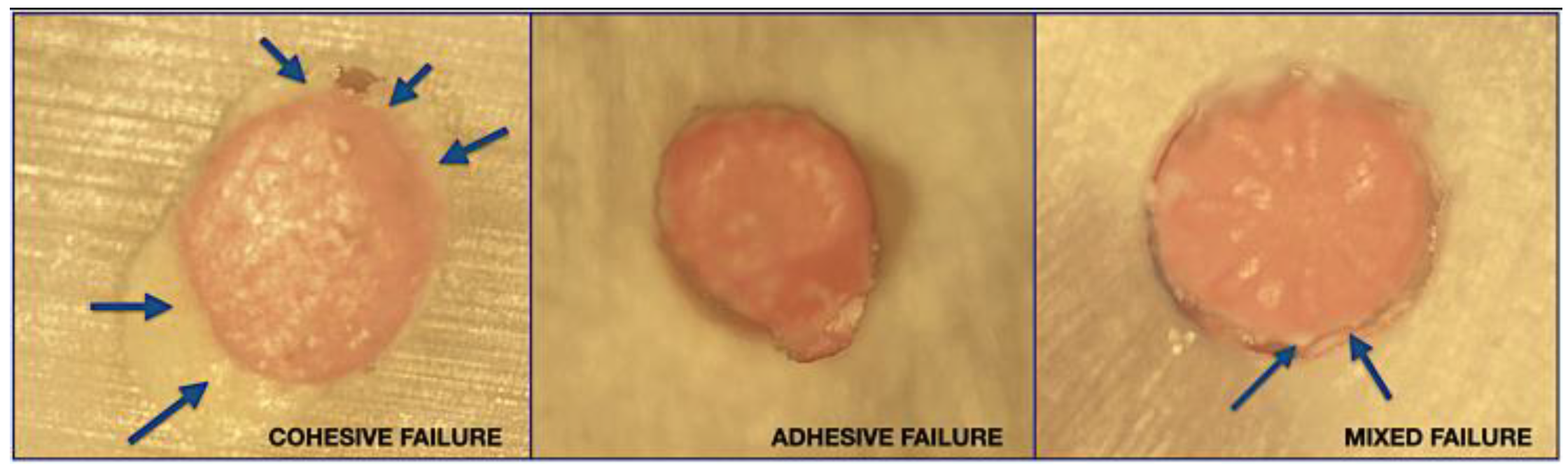
2.8. Failure Mode Evaluation
3. Statistical Analysis
4. Results
4.1. Influence of the Irrigation Protocol
4.2. Root Canal Level and Push out Bond Strength
4.3. Impact of Irrigation Protocol Joined with Root Canal Level on POBS
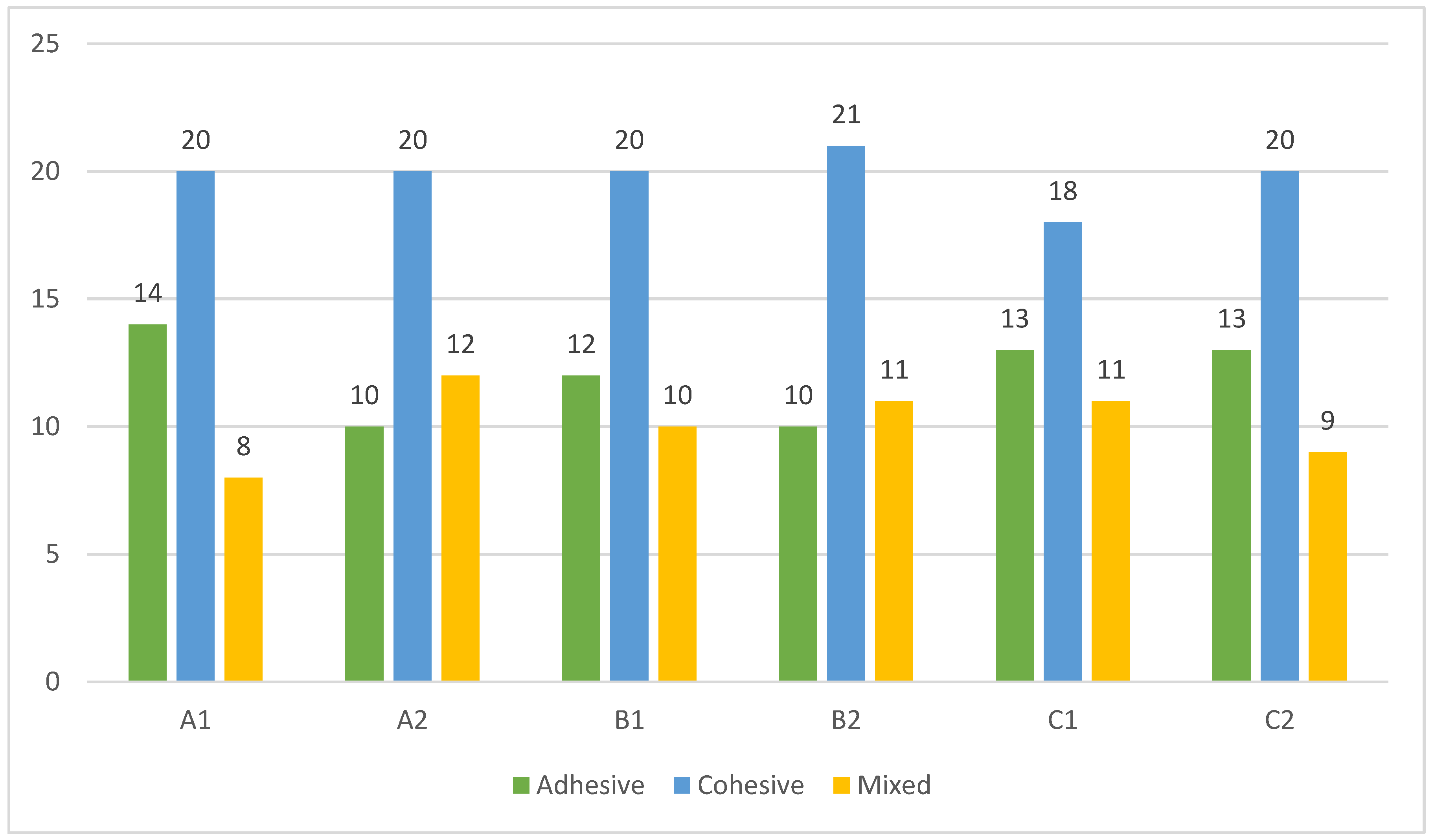
4.4. Failure Mode
5. Discussion
6. Conclusions
Author Contributions
Funding
Institutional Review Board Statement
Informed Consent Statement
Data Availability Statement
Conflicts of Interest
References
- Nair, P.N.R. On the causes of persistent apical periodontitis: A review. Int. Endod. J. 2006, 39, 249–281. [Google Scholar] [CrossRef]
- Celik, D.; Koca, A.T.Ö.; Koşar, T.; Taşdemir, T. The effects of final irrigants on the push-out bond strength of two calcium silicate-based root canal sealers: An in vitro study. Eur. Oral Res. 2021, 55, 146–151. [Google Scholar] [CrossRef] [PubMed]
- Haapasalo, M.; Shen, Y.; Wang, Z.; Gao, Y. Irrigation in endodontics. Br. Dent. J. 2014, 216, 299–303. [Google Scholar] [CrossRef] [PubMed]
- Donnermeyer, D.; Dornseifer, P.; Schäfer, E.; Dammaschke, T. The push-out bond strength of calcium silicate-based endodontic sealers. Head Face Med. 2018, 14, 13. [Google Scholar] [CrossRef] [PubMed]
- Lim, M.; Jung, C.; Shin, D.H.; Cho, Y.B.; Song, M. Calcium silicate-based root canal sealers: A literature review. Restor. Dent. Endod. 2020, 45, e35. [Google Scholar] [CrossRef]
- Camps, J.; Jeanneau, C.; El Ayachi, I.; Laurent, P.; About, I. Bioactivity of a calcium silicate–based endodontic cement (BioRoot RCS): Interactions with human periodontal ligament cells in vitro. J. Endod. 2015, 41, 1469–1473. [Google Scholar] [CrossRef]
- Morgental, R.D.; Vier-Pelisser, F.V.; Oliveira, S.D.D.; Antunes, F.C.; Cogo, D.M.; Kopper, P.M.P. Antibacterial activity of two MTA-based root canal sealers. Int. Endod. J. 2011, 44, 1128–1133. [Google Scholar] [CrossRef]
- Oliveira, D.S.; Cardoso, M.L.; Queiroz, T.F.; Silva, E.J.N.L.; Souza, E.M.; De-Deus, G. Suboptimal push-out bond strengths of calcium silicate-based sealers. Int. Endod. J. 2016, 49, 796–801. [Google Scholar] [CrossRef]
- Sfeir, G.; Zogheib, C.; Patel, S.; Giraud, T.; Nagendrababu, V.; Bukiet, F. Calcium silicate-based root canal sealers: A narrative review and clinical perspectives. Materials 2021, 14, 3965. [Google Scholar] [CrossRef] [PubMed]
- Atmeh, A.R.; Chong, E.Z.; Richard, G.; Festy, F.; Watson, T.F. Dentin-cement interfacial interaction: Calcium silicates and polyalkenoates. J. Dent. Res. 2012, 91, 454–459. [Google Scholar] [CrossRef]
- Eltair, M.; Pitchika, V.; Hickel, R.; Kühnisch, J.; Diegritz, C. Evaluation of the interface between gutta-percha and two types of sealers using scanning electron microscopy (SEM). Clin. Oral Investig. 2018, 22, 1631–1639. [Google Scholar] [CrossRef] [PubMed]
- Guivarc’h, M.; Jeanneau, C.; Giraud, T.; Pommel, L.; About, I.; Azim, A.A.; Bukiet, F. An international survey on the use of calcium silicate-based sealers in non-surgical endodontic treatment. Clin. Oral Investig. 2020, 24, 417–424. [Google Scholar] [CrossRef]
- Collares, F.M.; Portella, F.F.; Rodrigues, S.B.; Celeste, R.K.; Leitune, V.C.B.; Samuel, S.M.W. The influence of methodological variables on the push-out resistance to dislodgement of root filling materials: A meta-regression analysis. Int. Endod. J. 2016, 49, 836–849. [Google Scholar] [CrossRef]
- Shokouhinejad, N.; Gorjestani, H.; Nasseh, A.A.; Hoseini, A.; Mohammadi, M.; Shamshiri, A.R. Push-out bond strength of gutta-percha with a new bioceramic sealer in the presence or absence of smear layer. Aust. Endod. J. 2013, 39, 102–106. [Google Scholar] [CrossRef]
- DeLong, C.; He, J.; Woodmansey, K.F. The effect of obturation technique on the push-out bond strength of calcium silicate sealers. J. Endod. 2015, 41, 385–388. [Google Scholar] [CrossRef]
- Carvalho, N.K.; Prado, M.C.; Senna, P.M.; Neves, A.A.; Souza, E.M.; Fidel, S.R.; Sassone, L.M.; Silva, E.J.N.L. Do smear-layer removal agents affect the push-out bond strength of calcium silicate-based endodontic sealers? Int. Endod. J. 2017, 50, 612–619. [Google Scholar] [CrossRef]
- Rifaat, S.; Rahoma, A.; Alkhalifa, F.; AlQuraini, G.; Alsalman, Z.; Alwesaibi, Z.; Taymour, N. Push-Out Bond Strength of EndoSeal Mineral Trioxide Aggregate and AH Plus Sealers after Using Three Different Irrigation Protocols. Eur. J. Dent. 2022. [Google Scholar] [CrossRef]
- Tuncel, B.; Nagas, E.; Cehreli, Z.; Uyanik, O.; Vallittu, P.; Lassila, L. Effect of endodontic chelating solutions on the bond strength of endodontic sealers. Braz. Oral Res. 2015, 29, 1–6. [Google Scholar] [CrossRef] [PubMed] [Green Version]
- El-Ma’aita, A.M.; Qualtrough, A.J.; Watts, D.C. The effect of smear layer on the push-out bond strength of root canal calcium silicate cements. Dent. Mater. 2013, 29, 797–803. [Google Scholar] [CrossRef] [PubMed]
- Al-Hiyasat, A.S.; Yousef, W.A.A. Push-out bond strength of calcium silicate-based cements in the presence or absence of a smear layer. Int. J. Dent. 2022, 2022, 7724384. [Google Scholar] [CrossRef]
- Zehnder, M.; Schmidlin, P.; Sener, B.; Waltimo, T. Chelation in root canal therapy reconsidered. J. Endod. 2005, 31, 817–820. [Google Scholar] [CrossRef]
- Ballal, N.V.; Ivica, A.; Meneses, P.; Narkedamalli, R.K.; Attin, T.; Zehnder, M. Influence of 1-Hydroxyethylidene-1, 1-Diphosphonic Acid on the Soft Tissue-Dissolving and Gelatinolytic Effect of Ultrasonically Activated Sodium Hypochlorite in Simulated Endodontic Environments. Materials 2021, 14, 2531. [Google Scholar] [CrossRef]
- Tartari, T.; Guimarães, B.M.; Amoras, L.S.; Duarte, M.A.H.; Silva e Souza, P.A.R.; Bramante, C.M. Etidronate causes minimal changes in the ability of sodium hypochlorite to dissolve organic matter. Int. Endod. J. 2015, 48, 399–404. [Google Scholar] [CrossRef]
- Rath, P.P.; Yiu, C.K.; Matinlinna, J.P.; Kishen, A.; Neelakantan, P. The effects of sequential and continuous chelation on dentin. Dent. Mater. 2020, 36, 1655–1665. [Google Scholar] [CrossRef]
- Fernandes Zancan, R.; Hadis, M.; Burgess, D.; Zhang, Z.J.; Di Maio, A.; Tomson, P.; Hungaro Duarte, M.A.; Camilleri, J. A matched irrigation and obturation strategy for root canal therapy. Sci. Rep. 2021, 11, 4666. [Google Scholar] [CrossRef]
- Rebolloso de Barrio, E.; Gancedo-Caravia, L.; García-Barbero, E.; Pérez-Higueras, J.J. Effect of exposure to root canal irrigants on the push-out bond strength of calcium silicate–based cements. Clin. Oral Investig. 2021, 25, 3267–3274. [Google Scholar] [CrossRef]
- Srivastava, A.; Yadav, D.S.; Rao, M.; Rao, H.M.; Arun, A.; Siddique, R. Evaluation of push-out bond strength of BioRoot RCS and AH Plus after using different irrigants: An in vitro study. J. Conserv. Dent. JCD 2020, 23, 26. [Google Scholar] [CrossRef]
- Al-Haddad, A.; Che Ab Aziz, Z.A. Bioceramic-based root canal sealers: A review. Int. J. Biomater. 2016, 2016, 9753210. [Google Scholar] [CrossRef] [Green Version]
- Chen, W.P.; Chen, Y.Y.; Huang, S.H.; Lin, C.P. Limitations of push-out test in bond strength measurement. J. Endod. 2013, 39, 283–287. [Google Scholar] [CrossRef]
- Neelakantan, P.; Ahmed, H.M.A.; Wong, M.C.M.; Matinlinna, J.P.; Cheung, G.S.P. Effect of root canal irrigation protocols on the dislocation resistance of mineral trioxide aggregate-based materials: A systematic review of laboratory studies. Int. Endod. J. 2018, 51, 847–861. [Google Scholar] [CrossRef]
- Suciu, I.; Dimitriu, B.; Ciocardel, M.; Chirila, M.; Amza, O.; Scarlatescu, S.; Preoteasa, C.; Grigorie, M.; Voiculeanu, M. Evaluation of the sealer/gutta-percha ratio on sets of root section surfaces of some extracted teeth sealed using the cold lateral condensation technique. J. Med. Life 2021, 14, 337. [Google Scholar] [CrossRef]
- Mancino, D.; Kharouf, N.; Cabiddu, M.; Bukiet, F.; Haïkel, Y. Microscopic and chemical evaluation of the filling quality of five obturation techniques in oval shaped root canals. Clin. Oral Investig. 2021, 25, 3757–3765. [Google Scholar] [CrossRef]
- Vertucci, F.J. Root canal morphology and its relationship to endodontic procedures. Endod. Top. 2005, 10, 3–29. [Google Scholar] [CrossRef]
- Dabaj, P.; Kalender, A.; Unverdi Eldeniz, A. Push-out bond strength and SEM evaluation in roots filled with two different techniques using new and conventional sealers. Materials 2018, 11, 1620. [Google Scholar] [CrossRef] [Green Version]
- Neelakantan, P.; Varughese, A.A.; Sharma, S.; Subbarao, C.V.; Zehnder, M.; De-Deus, G. Continuous chelation irrigation improves the adhesion of epoxy resin-based root canal sealer to root dentine. Int. Endod. J. 2012, 45, 1097–1102. [Google Scholar]
- Fisher, M.A.; Berzins, D.W.; Bahcall, J.K. An in vitro comparison of bond strength of various obturation materials to root canal dentin using a push-out test design. J. Endod. 2007, 33, 856–858. [Google Scholar]
- Güzel, C.; Uzunoglu, E.; Buzoglu, H.D. Effect of low–surface tension EDTA solutions on the bond strength of resin-based sealer to young and old root canal dentin. J. Endod. 2018, 44, 485–488. [Google Scholar]


| Sub-Groups | Push-Out Bond Strength (Mean ± SD) | Range (Minimum–Maximum) | 95% CI (Lower Bound–Upper Bound) | p-Value |
|---|---|---|---|---|
| A1 (n = 42) | 4.283 ± 3.667 ABC | 0.157–14.961 | 3.140–5.425 | <0.001 * |
| A2 (n = 42) | 9.631 ± 11.108 A | 0.135–41.964 | 6.170–13.093 | |
| B1 (n = 42) | 7.102 ± 7.620 AB | 0.280–27.537 | 4.727–9.477 | |
| B2 (n = 42) | 2.914 ± 2.594 C | 0.205–12.600 | 2.105–3.722 | |
| C1 (n = 42) | 4.105 ± 3.793 BC | 0.335–15.071 | 2.923–5.287 | |
| C2 (n = 42) | 7.697 ± 8.137 AB | 0.341–33.892 | 5.162–10.233 |
| Root Levels | Push-Out Bond Strength (Mean ± SD) | Range (Minimum–Maximum) | 95% CI (Lower Bound–Upper Bound) | p-Value |
|---|---|---|---|---|
| Coronal (n = 84) | 0.980 ± 0.635 C | 0.135–2.634 | 0.842–1.117 | <0.001 * |
| Middle (n = 84) | 3.839 ± 2.063 B | 0.829–11.086 | 3.391–4.286 | |
| Apical (n = 84) | 13.048 ± 8.433 A | 1.043–41.964 | 11.218–14.878 |
| Root Levels | Coronal Level | Middle Level | Apical Level | p-Value | |
|---|---|---|---|---|---|
| Sub-Groups | |||||
| A1: EDTA/AH Plus Jet | 0.938 ± 0.583 ABb | 4.012 ± 2.136 a | 7.897 ± 3.362 Ba | <0.001 * | |
| A2: EDTA/Total Fill BC Sealer | 1.361 ± 0.672 Ac | 4.718 ± 2.692 b | 22.815 ± 9.726 Aa | <0.001 * | |
| B1: HEDP/AH Plus Jet | 1.318 ± 0.824 ABb | 3.346 ± 2.102 b | 16.642 ± 5.367 Aa | <0.001 * | |
| B2: HEDP/Total Fill BC Sealer | 0.588 ± 0.244 Bb | 2.805 ± 0.925 a | 5.349 ± 2.842 Ba | <0.001 * | |
| C1: NaOCl/AH Plus Jet | 0.761 ± 0.442 ABb | 3.503 ± 1.461 a | 8.051 ± 3.722 Ba | <0.001 * | |
| C2: NaOCl/Total Fill BC Sealer | 0.911 ± 0.589 ABc | 4.649 ± 2.202 b | 17.533 ± 6.314 Aa | <0.001 * | |
| p-value | 0.009 * | 0.165 | <0.001 * | ||
Disclaimer/Publisher’s Note: The statements, opinions and data contained in all publications are solely those of the individual author(s) and contributor(s) and not of MDPI and/or the editor(s). MDPI and/or the editor(s) disclaim responsibility for any injury to people or property resulting from any ideas, methods, instructions or products referred to in the content. |
© 2023 by the authors. Licensee MDPI, Basel, Switzerland. This article is an open access article distributed under the terms and conditions of the Creative Commons Attribution (CC BY) license (https://creativecommons.org/licenses/by/4.0/).
Share and Cite
Sfeir, G.; Bukiet, F.; Hage, W.; El Hachem, R.; Zogheib, C. Impact of Final Irrigation Protocol on the Push-Out Bond Strength of Two Types of Endodontic Sealers. Materials 2023, 16, 1761. https://doi.org/10.3390/ma16051761
Sfeir G, Bukiet F, Hage W, El Hachem R, Zogheib C. Impact of Final Irrigation Protocol on the Push-Out Bond Strength of Two Types of Endodontic Sealers. Materials. 2023; 16(5):1761. https://doi.org/10.3390/ma16051761
Chicago/Turabian StyleSfeir, Germain, Frédéric Bukiet, Wajih Hage, Roula El Hachem, and Carla Zogheib. 2023. "Impact of Final Irrigation Protocol on the Push-Out Bond Strength of Two Types of Endodontic Sealers" Materials 16, no. 5: 1761. https://doi.org/10.3390/ma16051761