
Stainless Steel in Municipal Sewage—How to Recognize Favorable Corrosion Conditions


Abstract
:1. Introduction
2. Materials and Methods
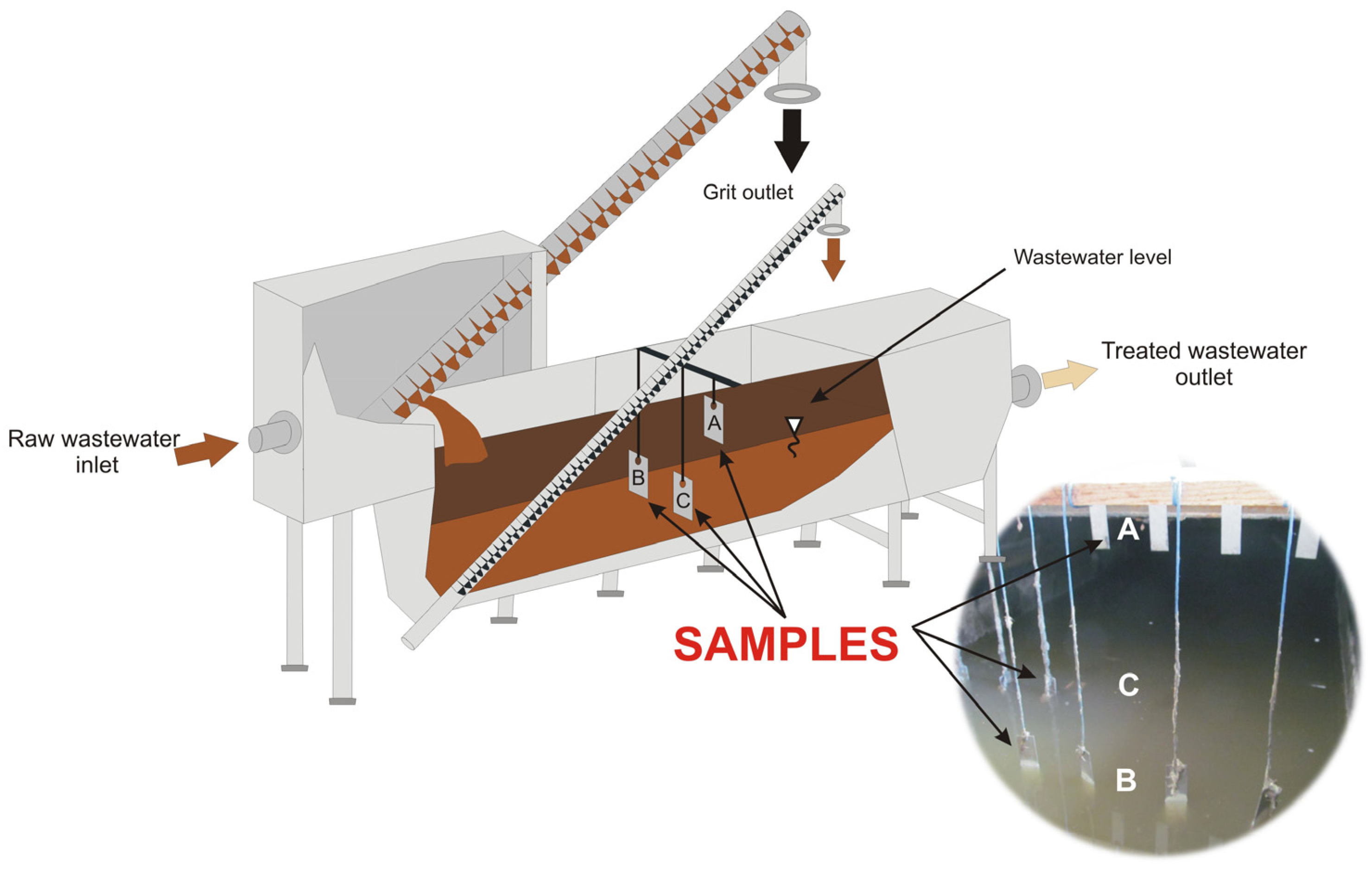
2.1. Characteristics of the Test Facility
2.2. Description of the Experiment
2.3. Material Characterization
2.4. Bacteria Identification
3. Results
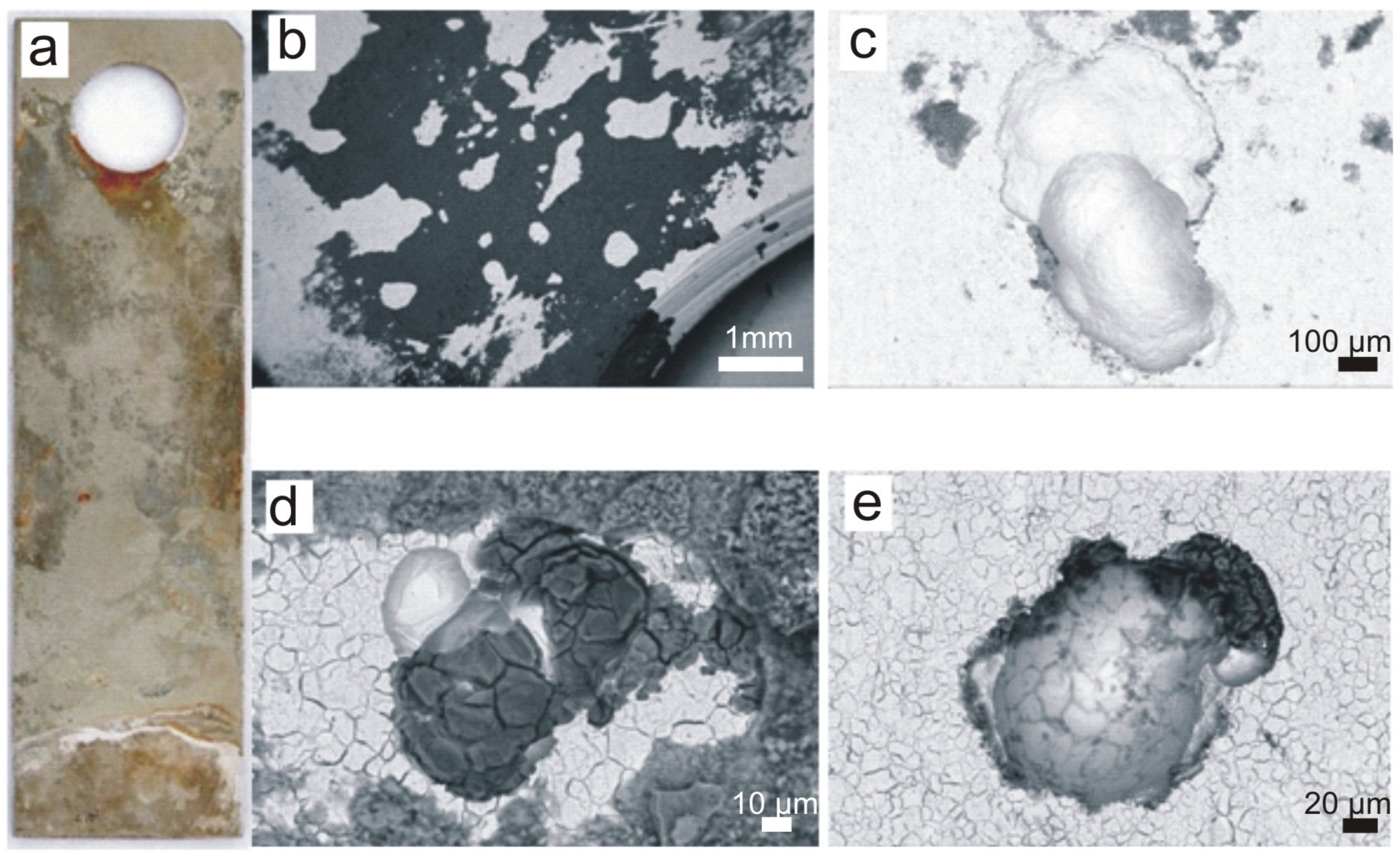
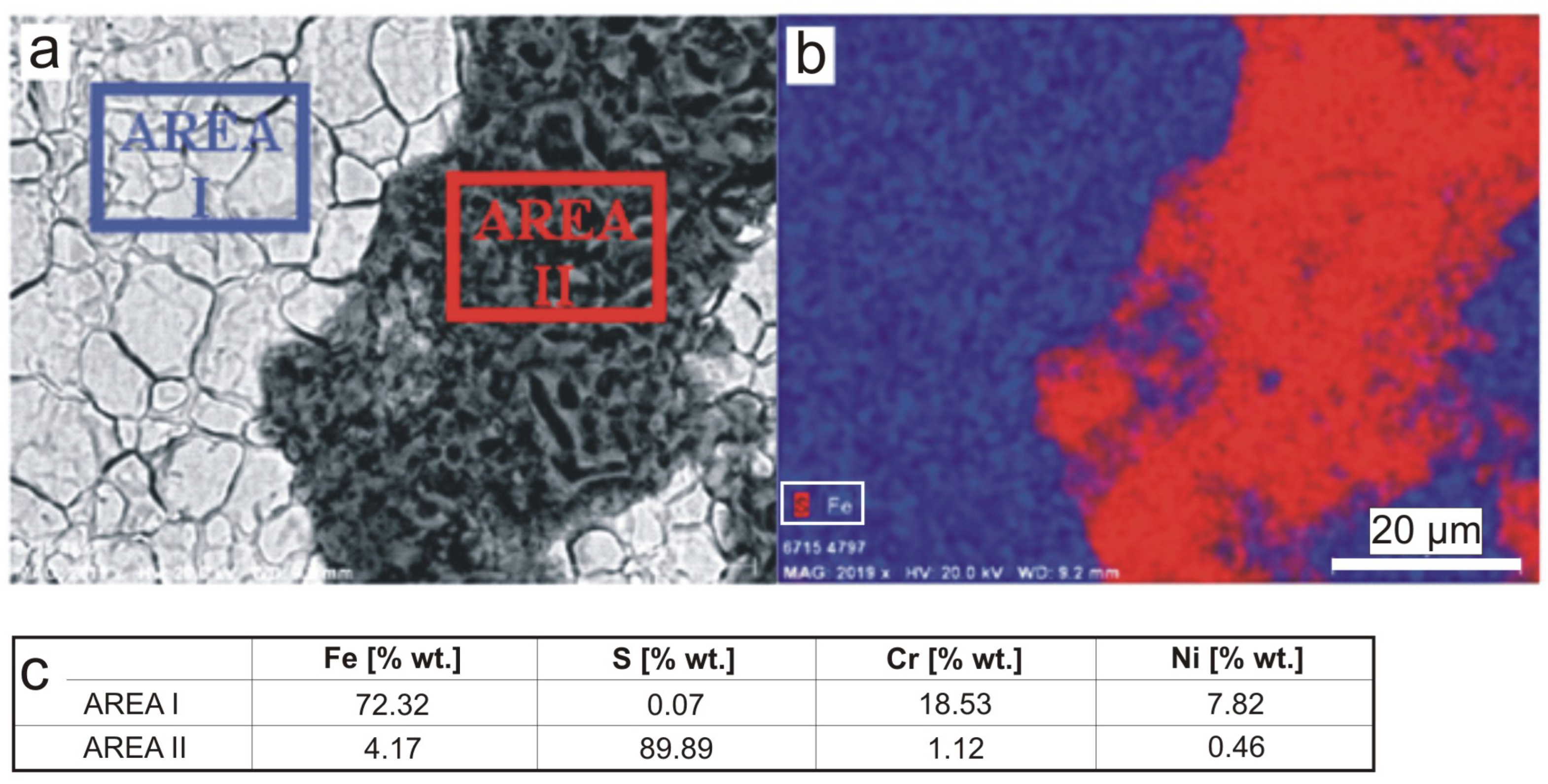
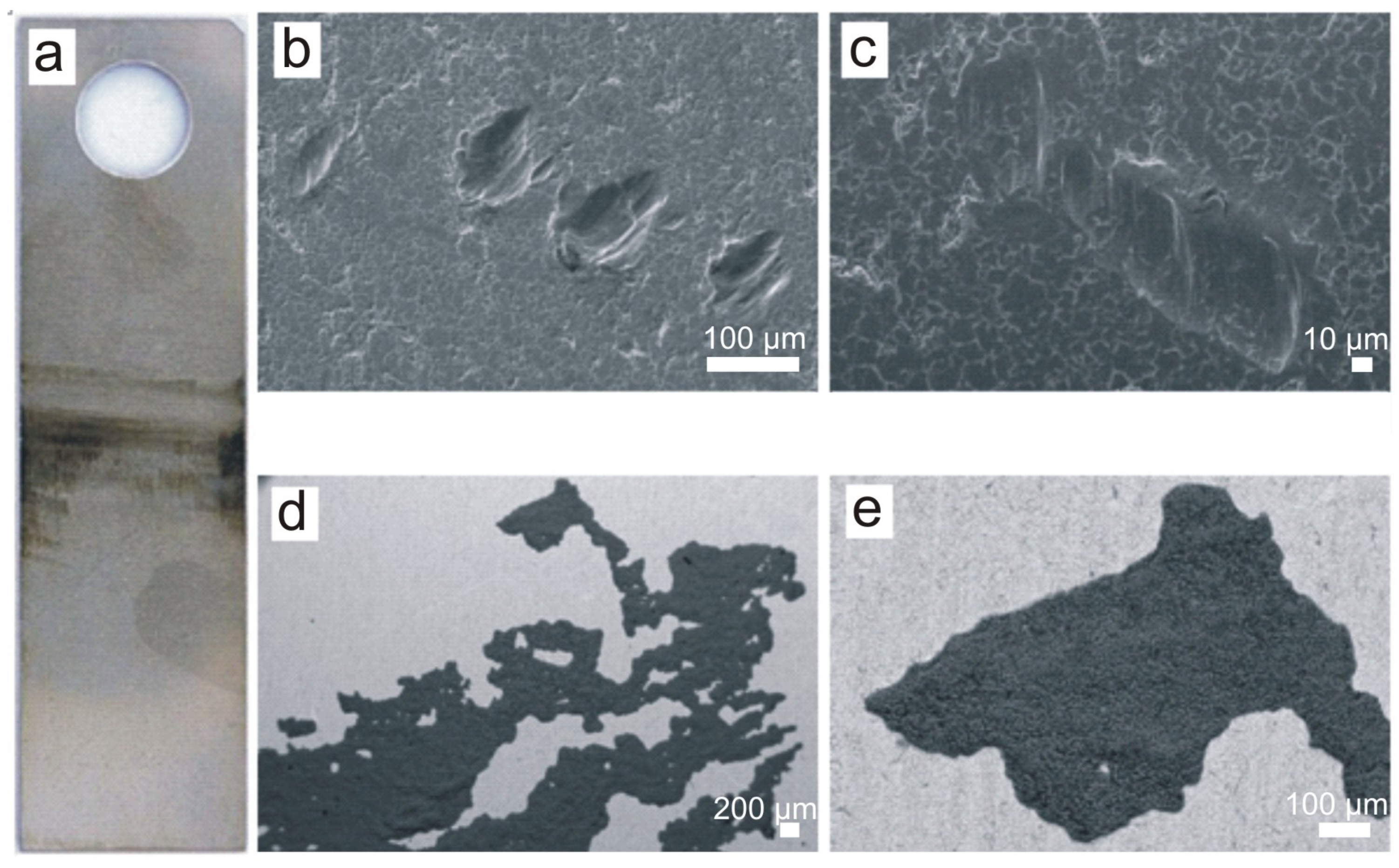
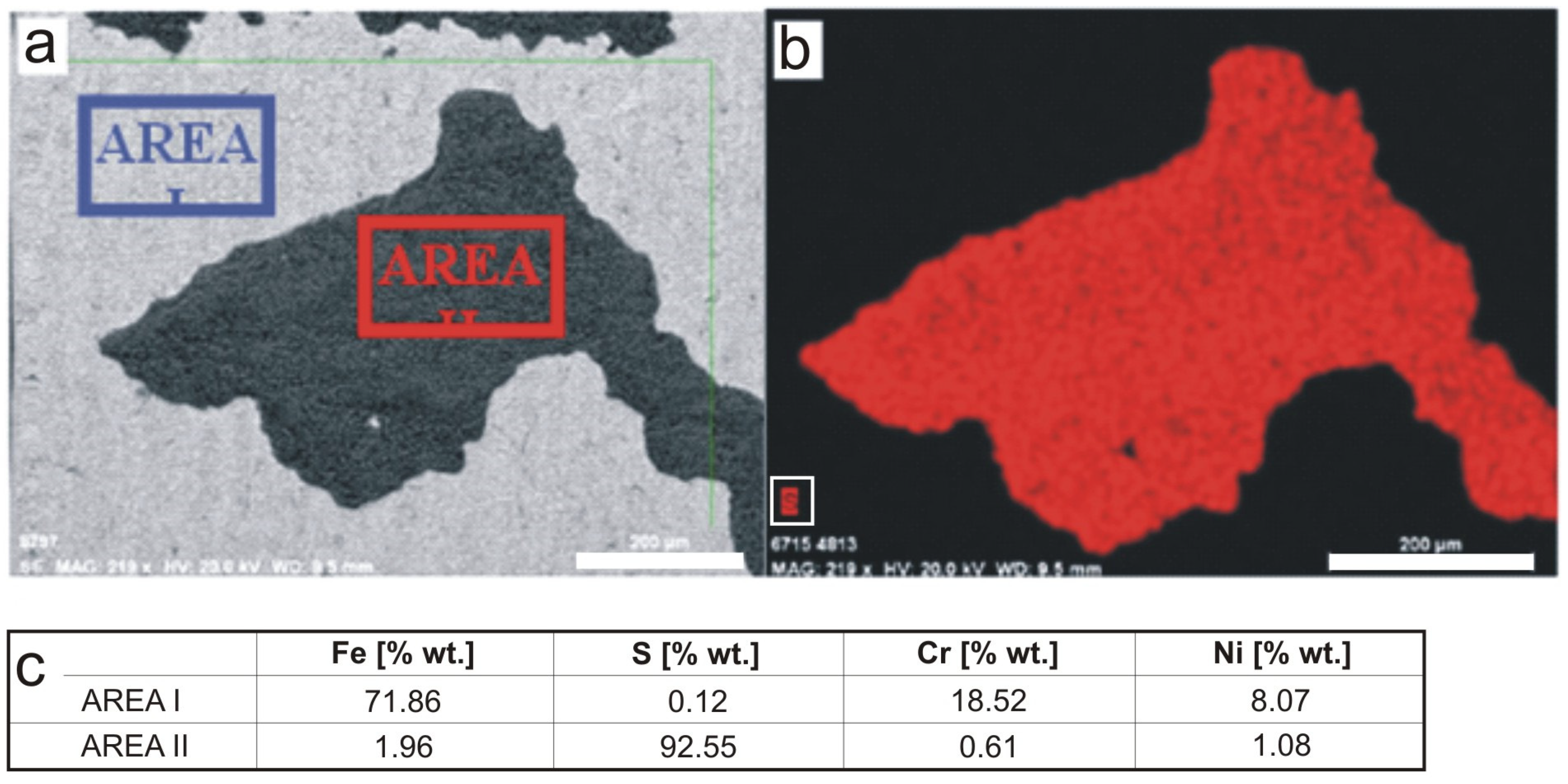
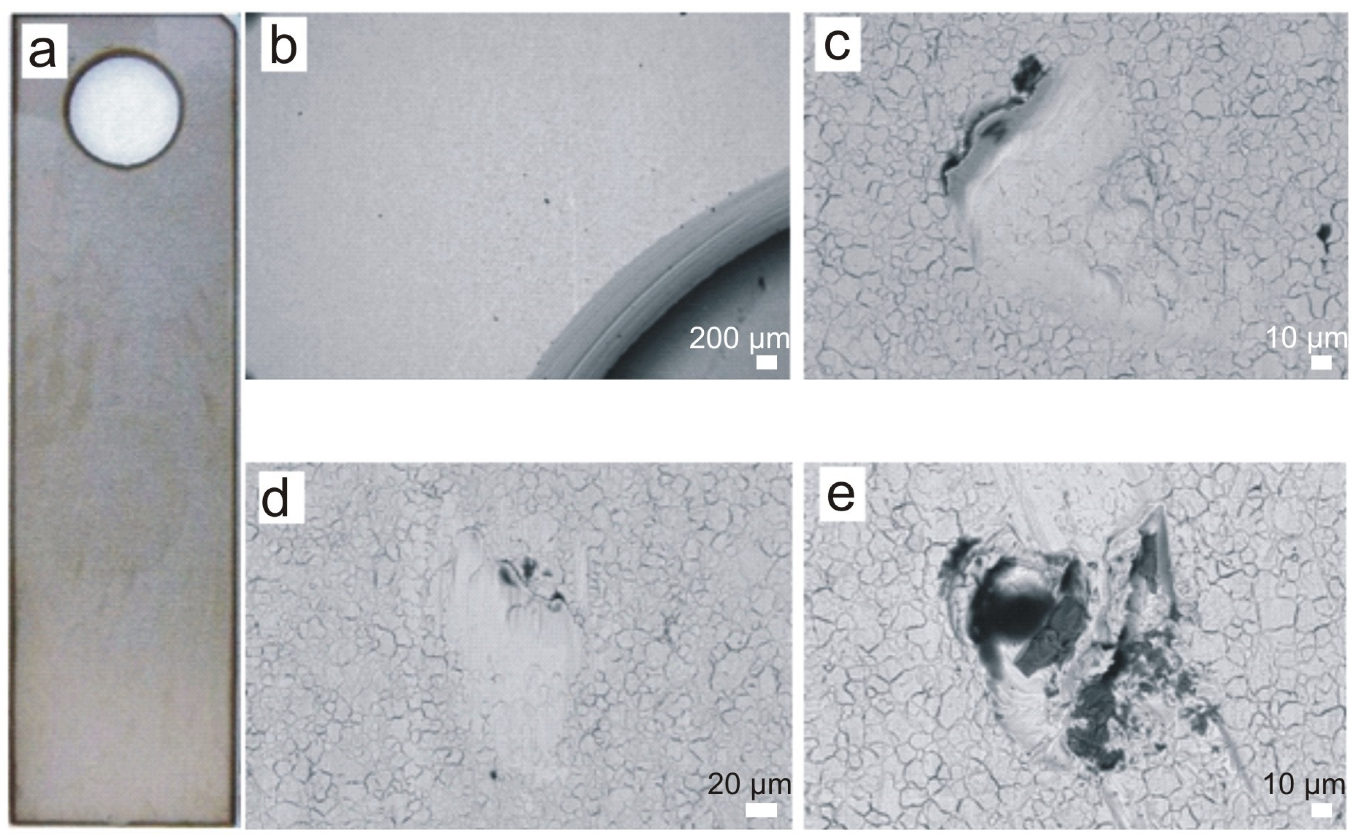
3.1. SEM Analysis
3.2. Surface Roughness, Weight Loss of Samples and Pitting Geometry
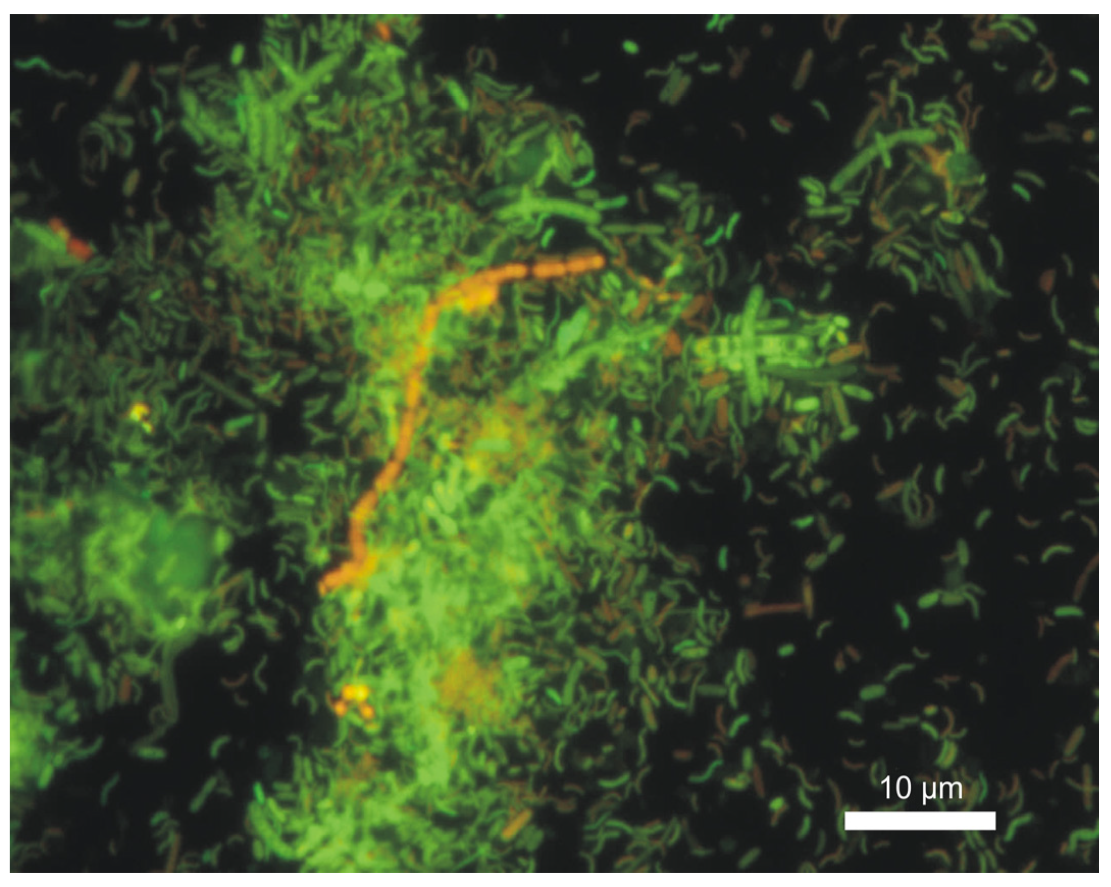
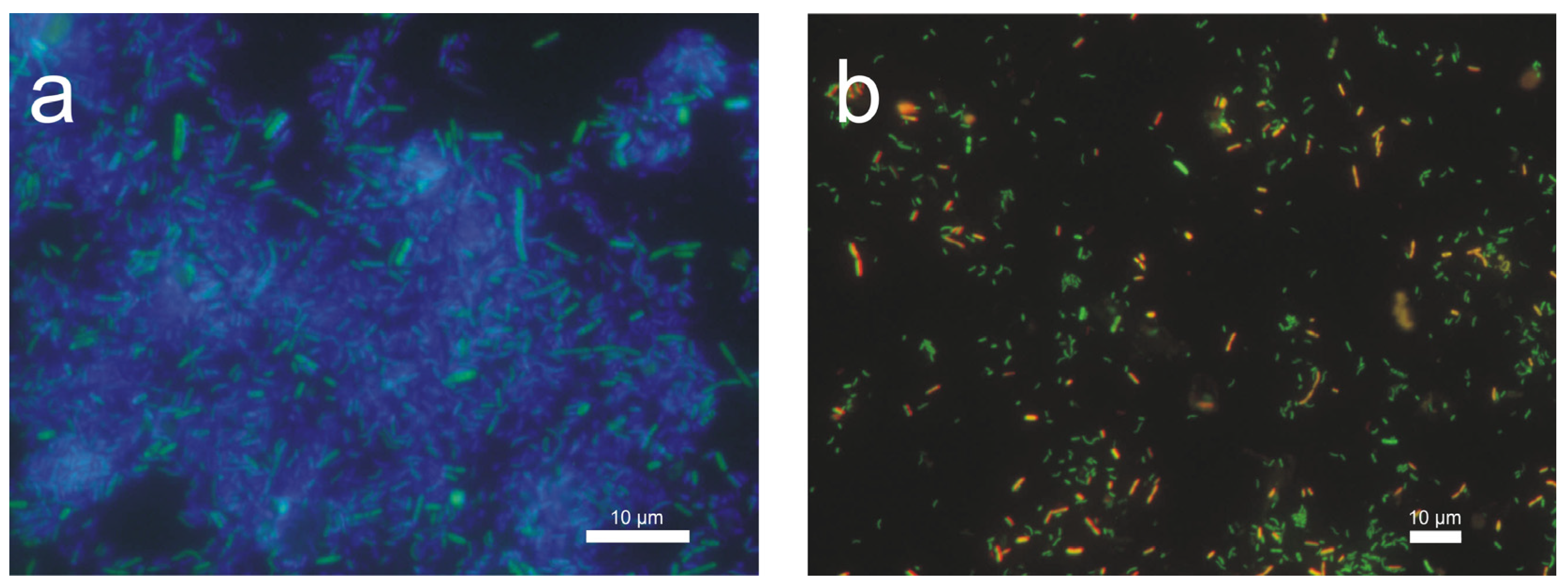
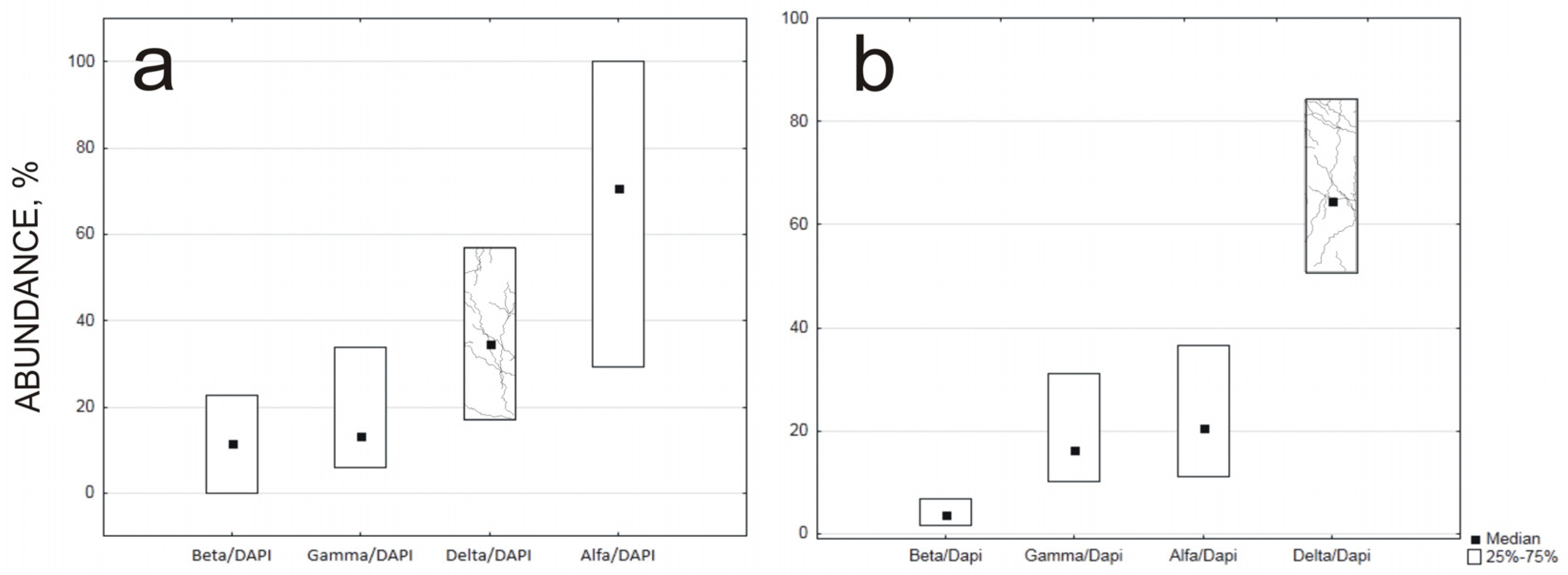
3.3. Microbial Identification and Quantitative Assessment
4. Discussion
5. Conclusions
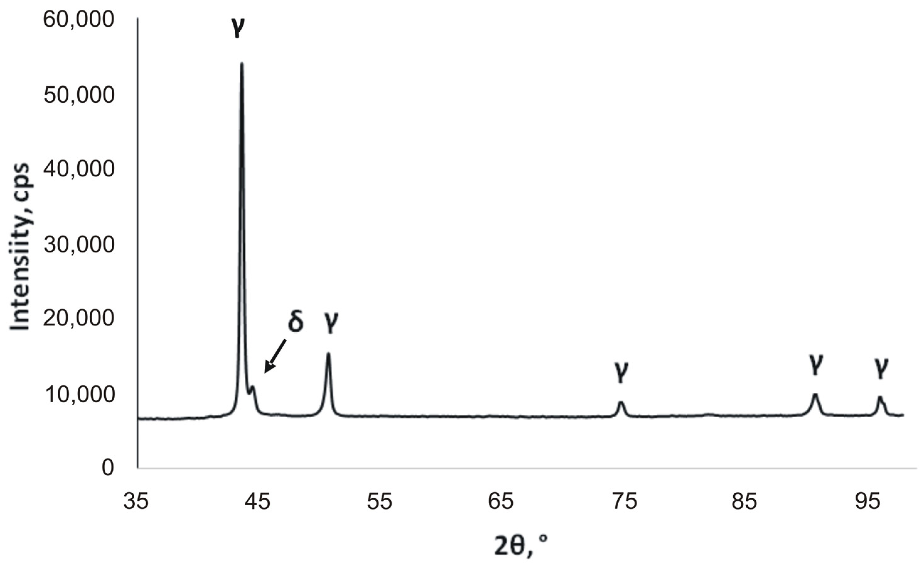
- According to this study, delta ferrite can have a negative effect on the properties of stainless steel placed above the surface of the wastewater (A-series) and can affect pitting corrosion.
- The results showed that in a humid environment containing H2S, this can lead to the intensification of corrosion from the center of corrosion, propagating deep into the pits after the damage of the passive layer under favorable conditions of temperature, humidity, insufficiently efficient ventilation and the impact of microorganisms.
- The proposed approach to microbiological assessment by determining the classes of bacteria can be a useful tool for evaluating wastewater for aggressiveness. The early identification of bacteria can also reduce financial losses of technical facilities.
- As a result of the FISH analysis, the δ-proteobacteria class dominated in the sludge taken from the steel samples submerged in wastewater. This confirms the presence of bacteria involved in the reduction of sulfur or sulfate compounds, resulting in the formation of hydrogen sulfide, which is the main cause of increased corrosion of steel plates hung above the surface of the wastewater.
Author Contributions
Funding
Institutional Review Board Statement
Informed Consent Statement
Data Availability Statement
Conflicts of Interest
References
- Barton, L.L.; Hamilton, W.A. Sulphate-Reducing Bacteria: Environmental and Engineered Systems, 1st ed.; Cambridge University Press: Cambridge, UK, 2007. [Google Scholar]
- Woyciechowski, P.; Łukowski, P.; Szmigiera, E.; Adamczewski, G.; Chilmon, K.; Spodzieja, S. Concrete Corrosion in a Wastewater Treatment Plant–A Comprehensive Case Study. Constr. Build. Mater. 2021, 303, 124388. [Google Scholar] [CrossRef]
- Opiekun, Z.; Orłowicz, A.; Kupiec, B. Corrosion of AlSi 304L Austenitic Stainless Steel in Non-Stationary Aqueous HCl+ NaCl Solution. Arch. Foundry Eng. 2014, 14, 165–168. [Google Scholar]
- Mathers, G. Welding of Austenitic Stainless Steel. Retrieved from the Welding Institute. 2019. Available online: https://www.twi-global.com (accessed on 20 March 2023).
- Graham, C.D.; Lorenz, B.E. Delta Ferrite Is Ubiquitous in Type 304 Stainless Steel: Consequences for Magnetic Characterization. J. Magn. Magn. Mater. 2018, 458, 15–18. [Google Scholar] [CrossRef]
- Soleymani, S.; Ojo, O.A.; Richards, N. Effect of Composition on the Formation of Delta Ferrite in 304L Austenitic Stainless Steels During Hot Deformation. J. Mater. Eng. Perform. 2015, 24, 499–504. [Google Scholar] [CrossRef]
- Matsuda, F.; Nakagawa, H.; Uehara, T.; Katayama, S.; Arata, Y.A. New Explanation for Role of Delta-Ferrite Improving Weld Solidification Crack Susceptibility in Austenitic Stainless Steel (Materials, Metallurgy, Weldability). Trans. JWRI 1979, 8, 105–112. [Google Scholar]
- Rhouma, A.B.; Amadou, T.; Sidhom, H.; Braham, C. Correlation between Microstructure and Intergranular Corrosion Behavior of Low Delta-Ferrite Content AISI 316L Aged in the Range 550–700 C. J. Alloys Compd. 2017, 708, 871–886. [Google Scholar] [CrossRef]
- Eghbali, F.; Moayed, M.H.; Davoodi, A.; Ebrahimi, N. Critical Pitting Temperature (CPT) Assessment of 2205 Duplex Stainless Steel in 0.1 M NaCl at Various Molybdate Concentrations. Corros. Sci. 2011, 53, 513–522. [Google Scholar] [CrossRef]
- Mohammed-Ali, H.B.; Street, S.R.; Attallah, M.M.; Davenport, A.J. Effect of Microstructure on the Morphology of Atmospheric Corrosion Pits in Type 304L Stainless Steel. Corrosion 2018, 74, 1373–1384. [Google Scholar] [CrossRef]
- Rahimi, E.; Kosari, A.; Hosseinpour, S.; Davoodi, A.; Zandbergen, H.; Mol, J.M. Characterization of the Passive Layer on Ferrite and Austenite Phases of Super Duplex Stainless Steel. Appl. Surf. Sci. 2019, 496, 143634. [Google Scholar] [CrossRef]
- Ziadi, I.; Alves, M.M.; Taryba, M.; El-Bassi, L.; Hassairi, H.; Bousselmi, L.; Montemor, M.F.; Akrout, H. Microbiologically influenced corrosion mechanism of 304L stainless steel in treated urban wastewater and protective effect of silane-TiO2 coating. Bioelectrochemistry 2020, 132, 107413. [Google Scholar] [CrossRef]
- Moreno, D.A.; Ibars, J.R.; Polo, J.L.; Bastidas, J.M. EIS monitoring study of the early microbiologically influenced corrosion of AISI 304L stainless steel condenser tubes in freshwater. J. Solid State Electrochem. 2014, 18, 377–388. [Google Scholar] [CrossRef]
- Ziadi, I.; El-Bassi, L.; Bousselmi, L.; Akrout, H. Characterization of the biofilm grown on 304L stainless steel in urban wastewaters: Extracellular polymeric substances (EPS) and bacterial consortia. Biofouling 2020, 36, 977–989. [Google Scholar] [CrossRef] [PubMed]
- Inaba, Y.; Xu, S.; Vardner, J.T.; West, A.C.; Banta, S. Microbially influenced corrosion of stainless steel by Acidithiobacillus ferrooxidans supplemented with pyrite: Importance of thiosulfate. Appl. Environ. Microbiol. 2019, 85, e01381-19. [Google Scholar] [CrossRef]
- Lee, J.S.; Little, B.J. A mechanistic approach to understanding microbiologically influenced corrosion by metal-depositing bacteria. Corrosion 2019, 75, 6–11. [Google Scholar] [CrossRef] [PubMed]
- Lochyński, P.; Domańska, M.; Kasprzyk, K. Corrosion of the chromium-nickel steel screenings and grit separator. Ochr. Przed Koroz. 2019, 62, 225–235. [Google Scholar]
- Amann, R.I.; Ludwig, W.; Schleifer, K.H. Phylogenetic Identification and in Situ Detection of Individual Microbial Cells without Cultivation. Microbiol. Rev. 1995, 59, 143–169. [Google Scholar] [CrossRef]
- Domanska, M.; Kaminska, J. Quantification of Proteobacteria with Fluorescence in Situ Hybridization and Next-Generation Sequencing. Environ. Eng. Manag. J. 2022, 21, 981–993. [Google Scholar] [CrossRef]
- Nielsen, P.; Daims, H. FISH Handbook for Biological Wastewater Treatment; IWA Publishing: London, UK, 2009. [Google Scholar]
- Osoba, L.O.; Elemuren, R.A.; Ekpe, I.C. Influence of Delta Ferrite on Corrosion Susceptibility of AISI 304 Austenitic Stainless Steel. Cogent Eng. 2016, 3, 1150546. [Google Scholar] [CrossRef]
- Salgado-Dávalos, V.; Osorio-Avilés, S.; Kamaraj, S.K.; Vega-Alvarado, L.; Juárez, K.; Silva-Martínez, S.; Alvarez-Gallegos, A. Sediment Microbial Fuel Cell Power Boosted by Natural Chitin Degradation and Oxygen Reduction Electrocatalysts. CLEAN Soil Air Water 2021, 49, 2000465. [Google Scholar] [CrossRef]
- Kushkevych, I.; Hýžová, B.; Vítězová, M.; Rittmann, S.K.-M. Microscopic Methods for Identification of Sulfate-Reducing Bacteria from Various Habitats. Int. J. Mol. Sci. 2021, 22, 4007. [Google Scholar] [CrossRef]
- McIlroy, S.J.; Saunders, A.M.; Albertsen, M.; Nierychlo, M.; McIlroy, B.; Hansen, A.A.; Karst, S.M.; Nielsen, J.L.; Nielsen, P.H. MiDAS: The Field Guide to the Microbes of Activated Sludge. Database 2015, 2015, bav062. [Google Scholar] [CrossRef] [PubMed]
- Kong, Y.; Xia, Y.; Nielsen, J.L.; Nielsen, P.H. Structure and Function of the Microbial Community in a Full-Scale Enhanced Biological Phosphorus Removal Plant. Microbiology 2007, 153, 4061–4073. [Google Scholar] [CrossRef] [PubMed]
- Fischer, D.A.; Daille, L.; Aguirre, J.; Galarce, C.; Armijo, F.; De la Iglesia, R.; Pizarro, G.; Vargas, I.; Walczak, M. Corrosion of Stainless Steel in Simulated Tide of Fresh Natural Seawater of South East Pacific. Int. J. Electrochem. Sci 2016, 11, 6873–6885. [Google Scholar] [CrossRef]
- Mele, C.; Bozzini, B. A Simple and Safe Method to Implement Corrosion Experiments with 1 Bar of H2S. Corros. Eng. Sci. Technol. 2017, 52, 325–331. [Google Scholar] [CrossRef]
- Von Sperling, M. Wastewater Characteristics, Treatment and Disposal; IWA Publishing: London, UK, 2007; ISBN 1-84339-161-9. [Google Scholar]
- Jensen, H.S.; Nielsen, A.H.; Hvitved-Jacobsen, T.; Vollertsen, J. Survival of Hydrogen Sulfide Oxidizing Bacteria on Corroded Concrete Surfaces of Sewer Systems. Water Sci. Technol. 2008, 57, 1721–1726. [Google Scholar] [CrossRef] [PubMed]
- Yuan, S.; Liang, B.; Zhao, Y.; Pehkonen, S. Surface Chemistry and Corrosion Behaviour of 304 Stainless Steel in Simulated Seawater Containing Inorganic Sulphide and Sulphate-Reducing Bacteria. Corros. Sci. 2013, 74, 353–366. [Google Scholar] [CrossRef]
- Guo, L.; Street, S.R.; Mohammed-Ali, H.B.; Ghahari, M.; Mi, N.; Glanvill, S.; Du Plessis, A.; Reinhard, C.; Rayment, T.; Davenport, A.J. The Effect of Relative Humidity Change on Atmospheric Pitting Corrosion of Stainless Steel 304L. Corros. Sci. 2019, 150, 110–120. [Google Scholar] [CrossRef]
- Cheng, C.-Q.; Klinkenberg, L.-I.; Ise, Y.; Zhao, J.; Tada, E.; Nishikata, A. Pitting Corrosion of Sensitised Type 304 Stainless Steel under Wet–Dry Cycling Condition. Corros. Sci. 2017, 118, 217–226. [Google Scholar] [CrossRef]
- Zhang, L.; Tang, X.; Wang, Z.; Li, T.; Zhang, Z.; Lu, M. The Corrosion Behavior of 316L Stainless Steel in H2S Environment at High Temperatures. Int. J. Electrochem. Sci. 2017, 12, 8806–8819. [Google Scholar] [CrossRef]
- Newman, R. 2001 WR Whitney Award Lecture: Understanding the Corrosion of Stainless Steel. Corrosion 2001, 57, 1030–1041. [Google Scholar] [CrossRef]
- Nielsen, P.H.; Raunkjær, K.; Hvitved-Jacobsen, T. Sulfide Production and Wastewater Quality in Pressure Mains. Water Sci. Technol. 1998, 37, 97–104. [Google Scholar] [CrossRef]
- Ziadi, I.; Akrout, H.; Hassairi, H.; El-Bassi, L.; Bousselmi, L. Investigating the Biocorrosion Mechanism of 304L Stainless Steel in Raw and Treated Urban Wastewaters. Eng. Fail. Anal. 2019, 101, 342–356. [Google Scholar] [CrossRef]
- Muszyński, A.; Tabernacka, A.; Miłobędzka, A. Long-Term Dynamics of the Microbial Community in a Full-Scale Wastewater Treatment Plant. Int. Biodeterior. Biodegrad. 2015, 100, 44–51. [Google Scholar] [CrossRef]
- Dworkin, M.; Falkow, S.; Rosenberg, E.; Schleifer, K.-H.; Stackebrandt, E. The Prokaryotes: Volume 4: Bacteria: Firmicutes, Cyanobacteria; Springer: New York, NY, USA, 2006; ISBN 0-387-30744-3. [Google Scholar]
- Fernández, N.; Díaz, E.E.; Amils, R.; Sanz, J.L. Analysis of Microbial Community during Biofilm Development in an Anaerobic Wastewater Treatment Reactor. Microb. Ecol. 2008, 56, 121–132. [Google Scholar] [CrossRef] [PubMed]


| Fe | C | Ni | Cr | Mn | Si | N | P | S |
|---|---|---|---|---|---|---|---|---|
| [wt.%] | ||||||||
| Bal. | 0.037 | 8.04 | 18.13 | 1.28 | 0.42 | 0.057 | 0.029 | 0.002 |
| Probe | Sequence | Formamide [%] | Fluorescent Dye | Examples of Mechanism and Bacteria Categories from Each Class |
|---|---|---|---|---|
| ALF968 1 | 5′-GGT AAG GTT CTG CGC GTT-3′ | 20 | Green | Alphaproteobacteria |
| nitrite-oxidizing bacteria Nitrobacter denitrifying (nitrate-reducing) bacteria Paracoccus denitrificans | ||||
| BET42a | 5′-GCC TTC CCA CTT CGT TT-3′ [a] | 35 | Red | Betaproteobacteria |
| ammonia-oxidizing bacteria Nitrosomonas iron-oxidizing bacteria Gallionella ferruginea | ||||
| Gam42a | 5′-GCC TTC CCA CTT CGT TT-3′ [a] | 35 | Red | Gammaproteobacteria |
| S-oxidizing (sulfur-oxidizing) bacteria Acidithiobacillus thiooxidans | ||||
| DELTA495a 2 | 5′-AGT TAG CCG GTG CTT CCT-3′ [a] | 35 | Green | Deltaproteobacteria |
| sulfur-reducing bacteria Desulfuromonas iron- and manganese—reducing bacteria sulfate-reducing microbes Desulfovibrio aestuarii |
Disclaimer/Publisher’s Note: The statements, opinions and data contained in all publications are solely those of the individual author(s) and contributor(s) and not of MDPI and/or the editor(s). MDPI and/or the editor(s) disclaim responsibility for any injury to people or property resulting from any ideas, methods, instructions or products referred to in the content. |
© 2023 by the authors. Licensee MDPI, Basel, Switzerland. This article is an open access article distributed under the terms and conditions of the Creative Commons Attribution (CC BY) license (https://creativecommons.org/licenses/by/4.0/).
Share and Cite
Lochyński, P.; Domańska, M.; Dziedzic, R.; Hamal, K. Stainless Steel in Municipal Sewage—How to Recognize Favorable Corrosion Conditions. Materials 2023, 16, 6637. https://doi.org/10.3390/ma16206637
Lochyński P, Domańska M, Dziedzic R, Hamal K. Stainless Steel in Municipal Sewage—How to Recognize Favorable Corrosion Conditions. Materials. 2023; 16(20):6637. https://doi.org/10.3390/ma16206637
Chicago/Turabian StyleLochyński, Paweł, Magdalena Domańska, Robert Dziedzic, and Kamila Hamal. 2023. "Stainless Steel in Municipal Sewage—How to Recognize Favorable Corrosion Conditions" Materials 16, no. 20: 6637. https://doi.org/10.3390/ma16206637