
Photodegradation of Bamboo: A Study on Changes in Mechanical Performances
 , , and
Abstract
:1. Introduction
2. Materials and Methods
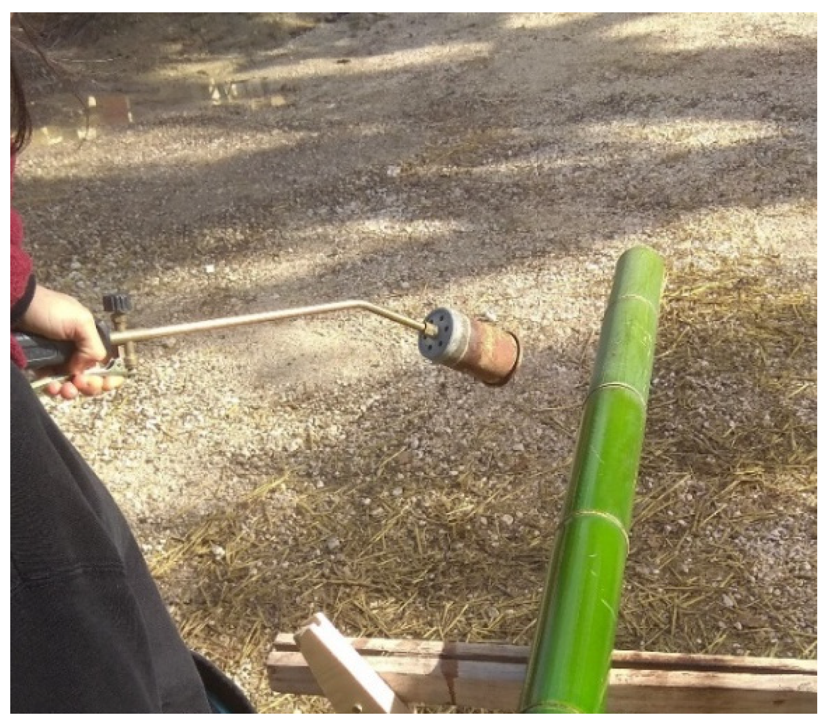
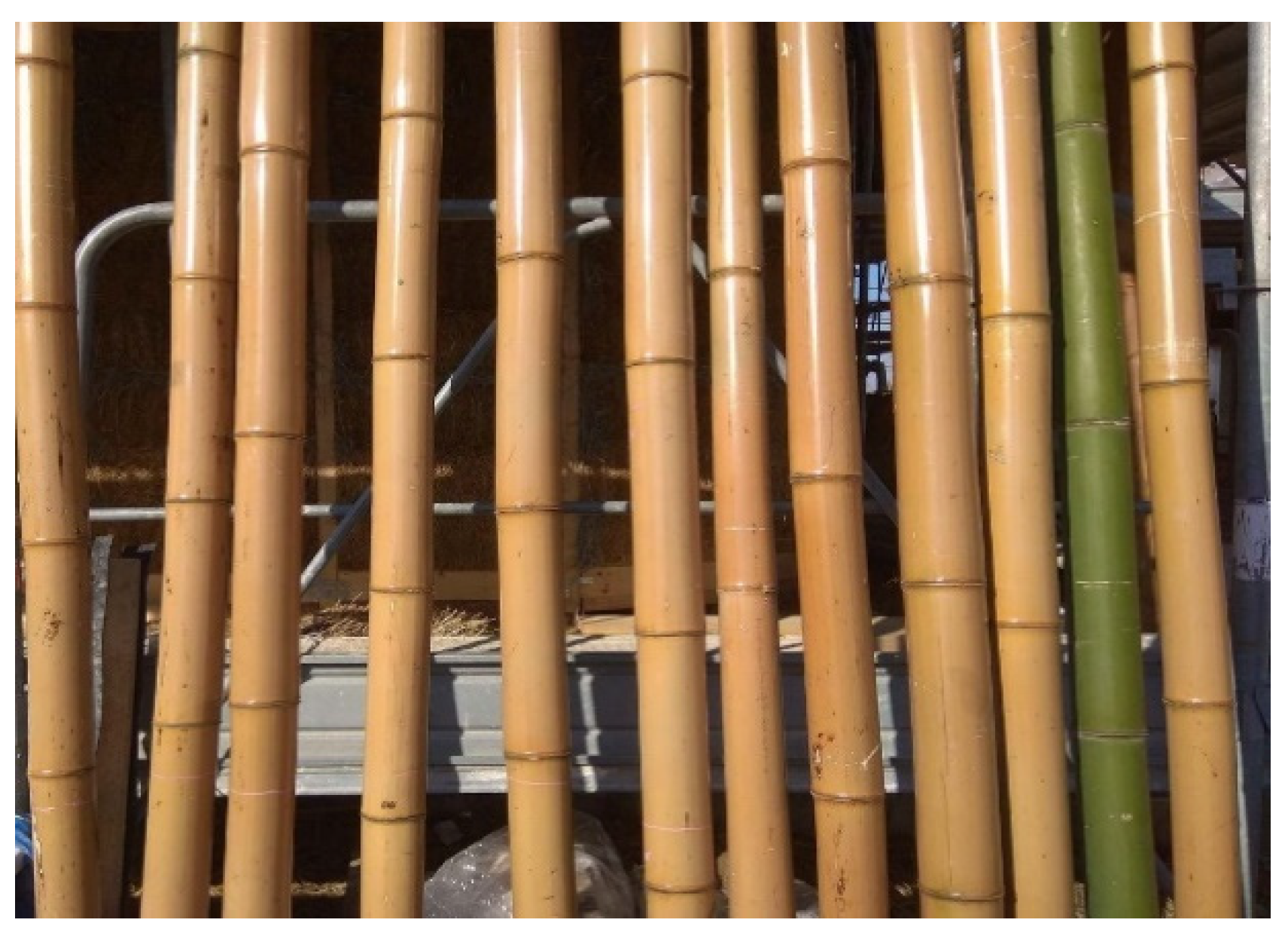
2.1. Materials
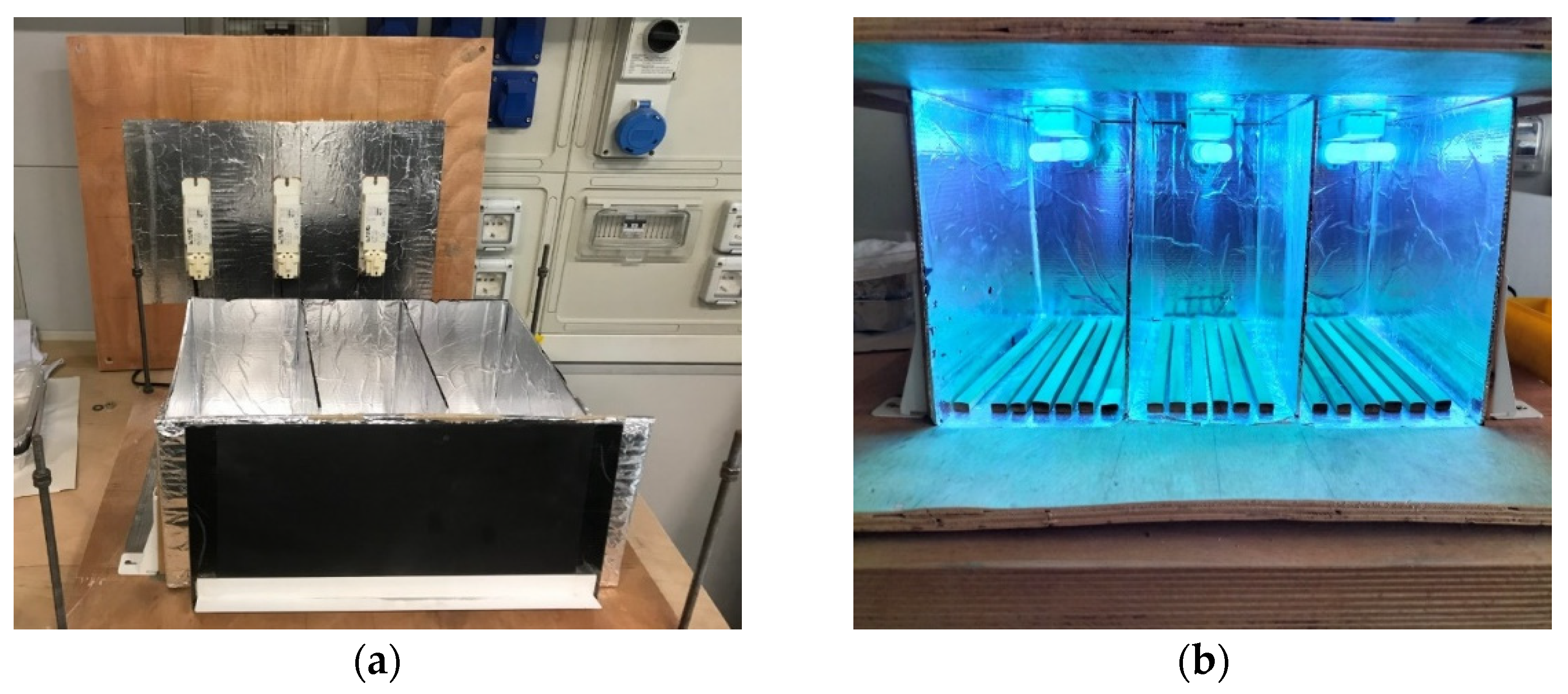
2.2. UV Aging
2.3. Microscopic Analysis
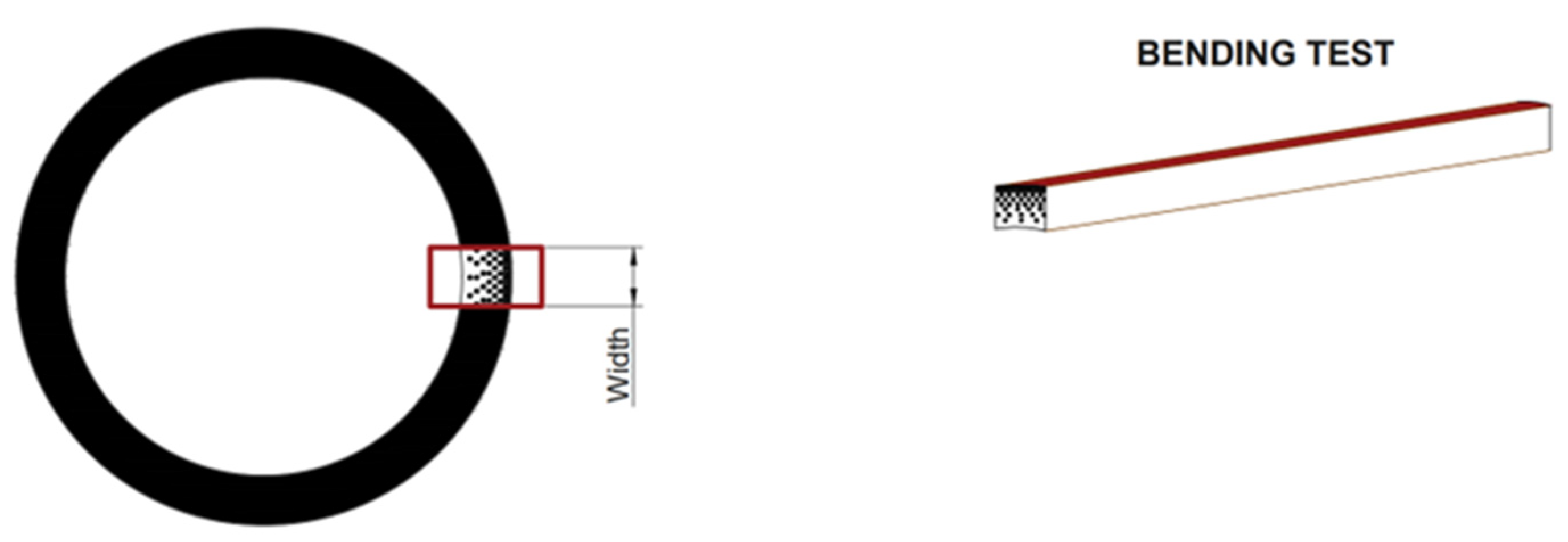
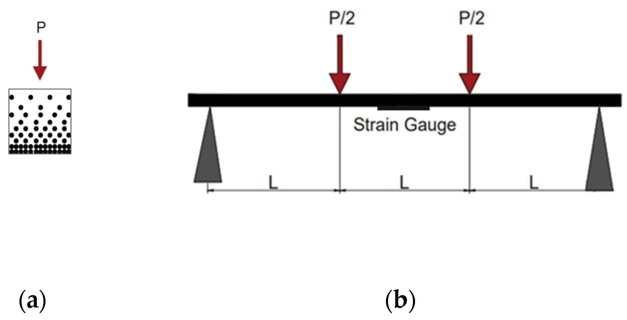
2.4. Bending Tests
3. Results
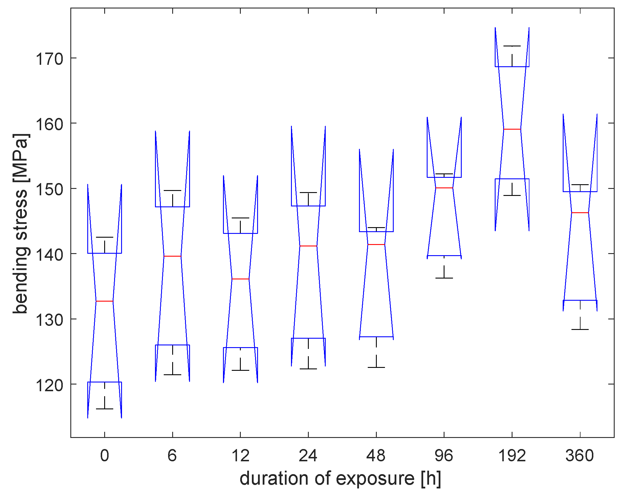
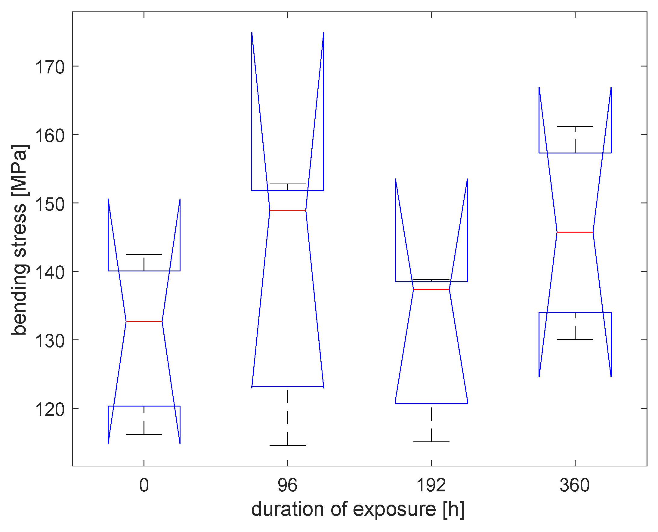
3.1. Bending Tests
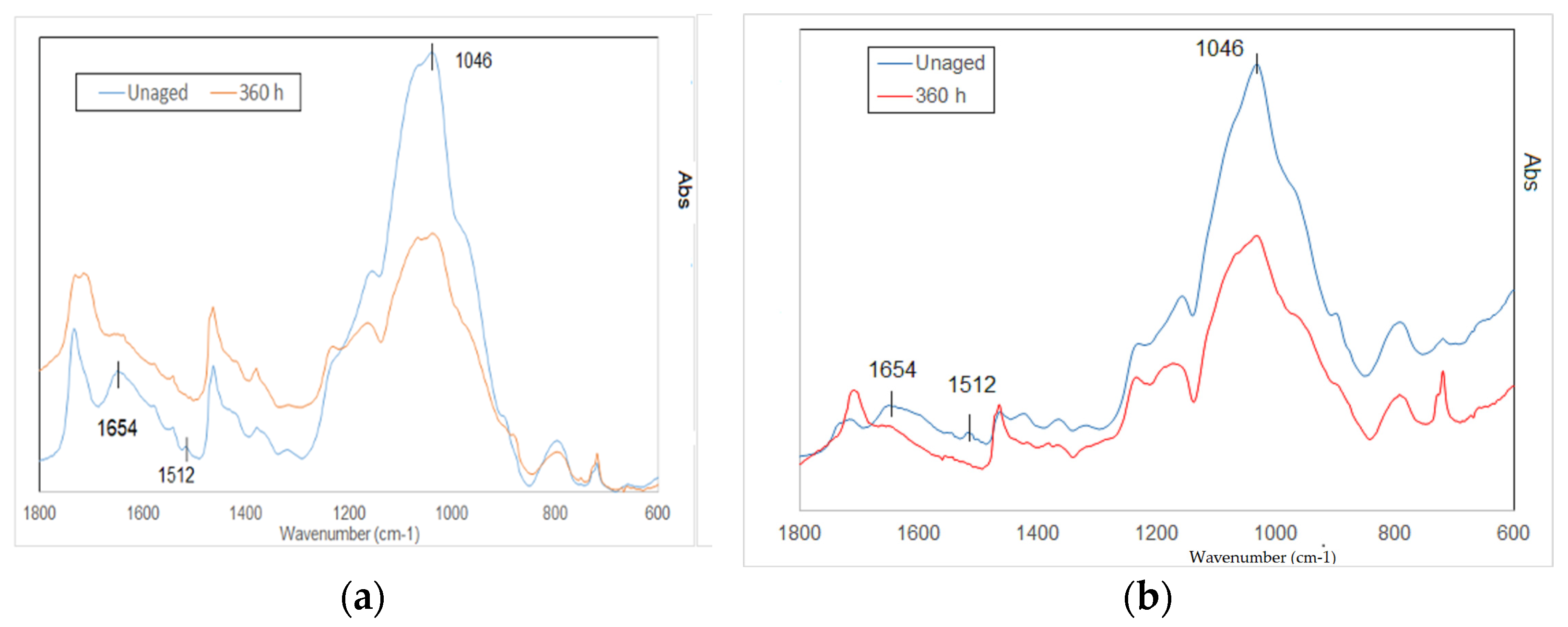
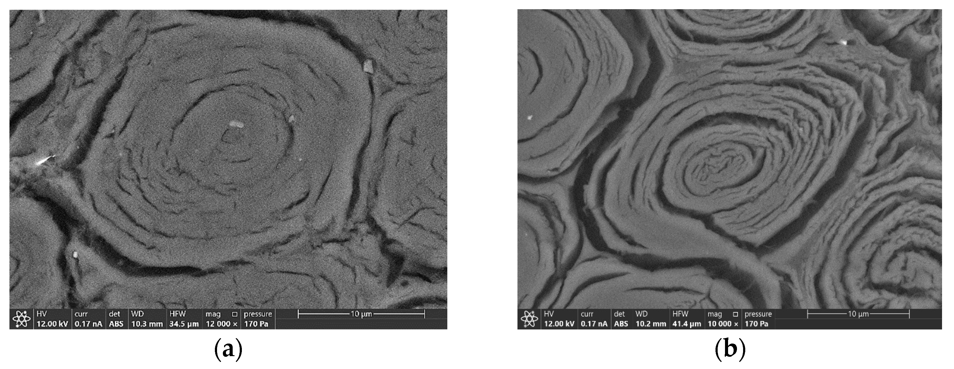
3.2. IR and Microscopical Analysis
4. Conclusions
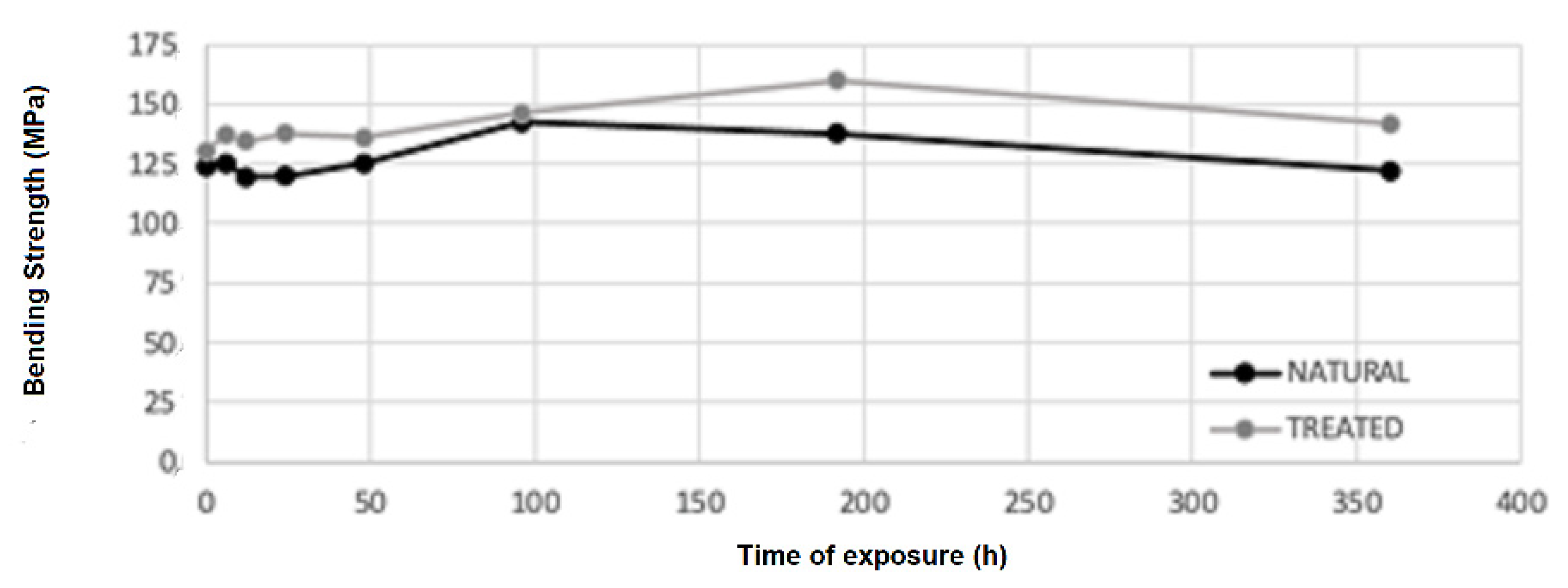
- After 48 h of exposure, bending strength starts growing, reaching an increment of 31% (149 MPa), concerning the initial value (113 MPa), at 96 h of exposure.
- After 96 h, bending strength starts declining slightly from the higher value but remains higher than the initial strength; after 360 h of exposure, it is 8% of the initial one (122 MPa).
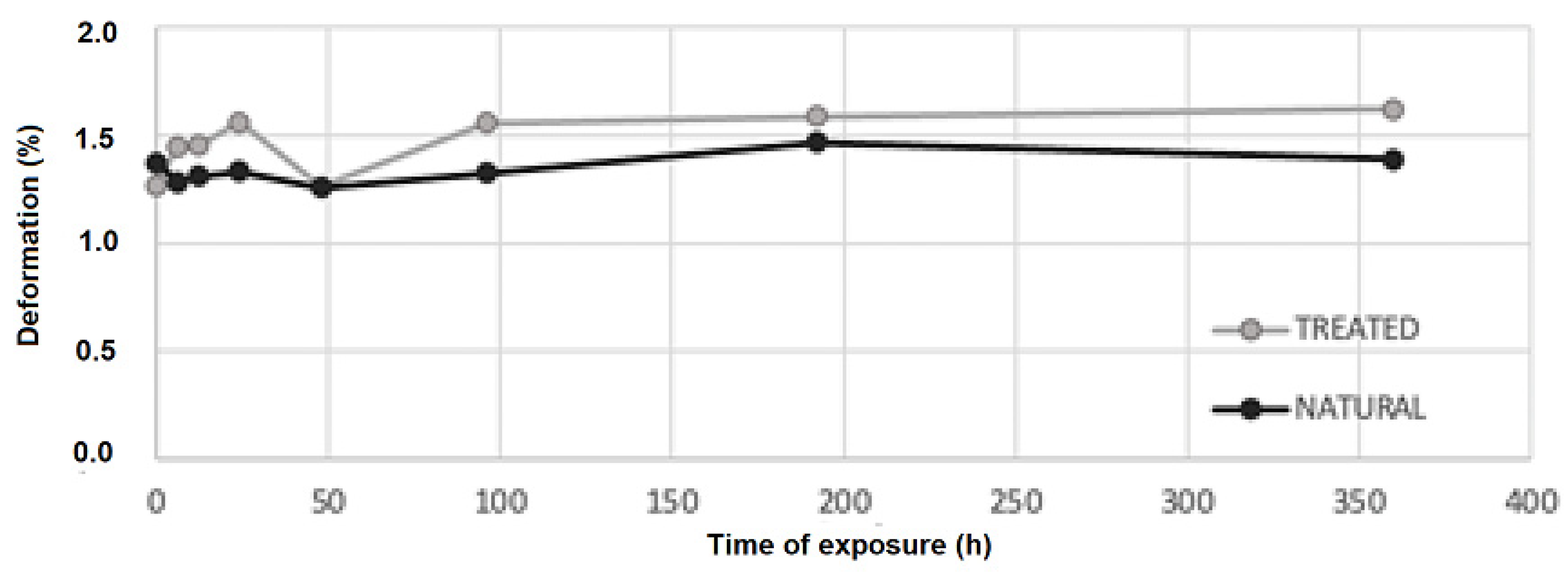
- There are no significant changes in deformation at different times of exposure.
- Modifications of the chemical features of the material have been analyzed with FTIR spectroscopy and a progressive degradation of lignin is reported.
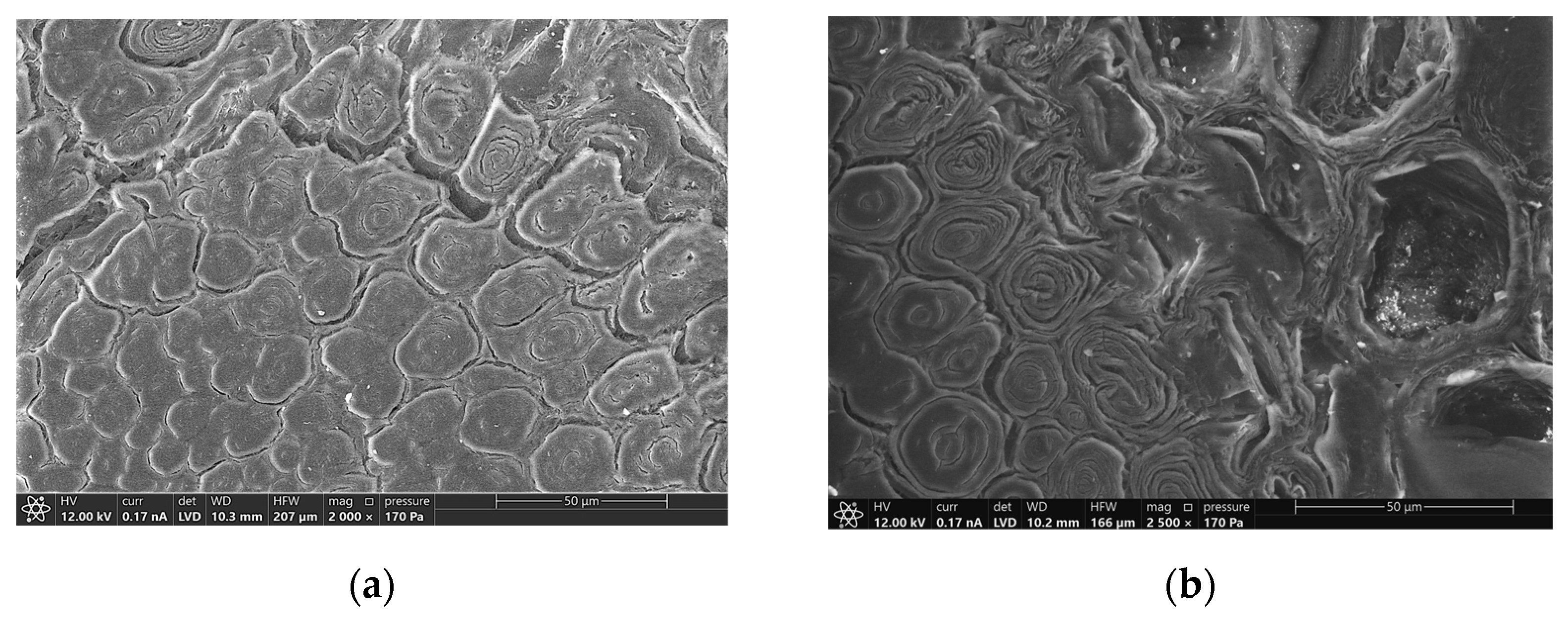
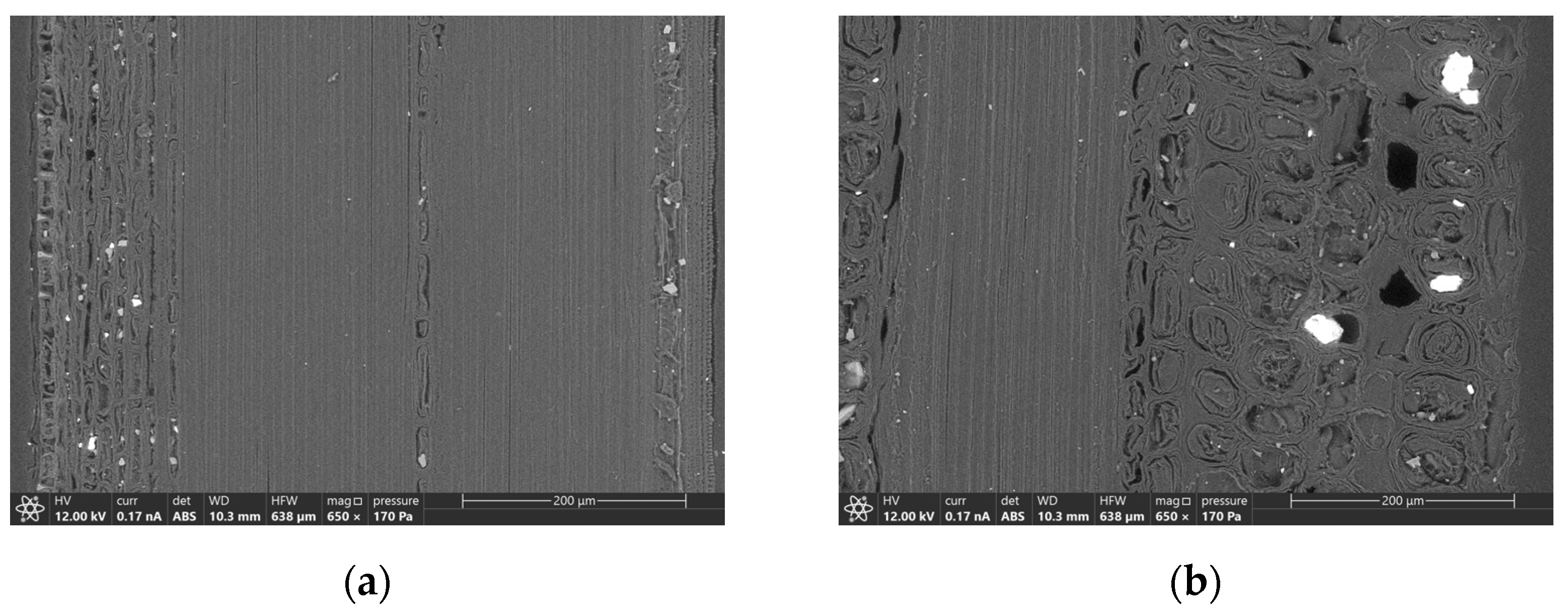
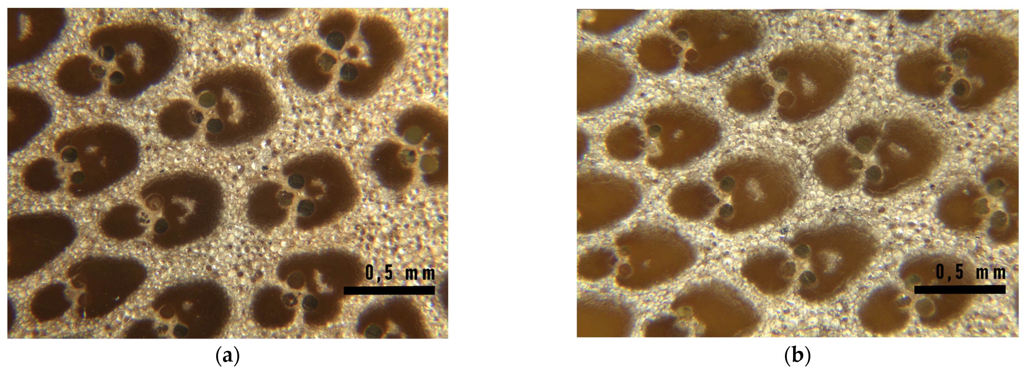
- Modifications of the morphological features have been analyzed by ESEM and optical microscopy observations, and cracks in the fiber walls are highlighted from micrographs, as reported in [10]. No effects have been found on the fiber length.
- After 48 h of exposure, bending strength starts growing, reaching an increment of 23% (160 MPa) concerning the initial value (130 MPa) at 192 h of exposure.
- After 192 h, bending strength starts declining slightly from the higher value but remains higher than the initial strength, as it is around the 8% after 360 h of exposure (142 MPa).
- There are no significant changes in deformation at different times of exposure.
- Modifications of the chemical features of the material have been analyzed with FTIR spectroscopy and a progressive degradation of lignin is reported for the virgin samples.
Author Contributions
Funding
Data Availability Statement
Acknowledgments
Conflicts of Interest
References
- Azadeha, A.; Ghavami, K.; Savastano, H., Jr.; Filho, T.; Barbosa, N.P. Static Flexural Behavior of Bamboo as a Functionally Graded Material and the Effect of heat on dynamic flexural modulus. J. Build. Eng. 2020, 34, 101949. [Google Scholar] [CrossRef]
- Zhang, Y.M.; Yu, Y.L.; Yu, W.J. Effect of thermal treatment on the physical and mechanical properties of Phyllostachys pubescen bamboo. Eur. J. Wood Prod. 2013, 71, 61–67. [Google Scholar] [CrossRef]
- Nguyen, C.T.; Wagenfür, A.; Phuong, L.X.; Dai, V.H.; Bremer, M.; Fischer, S. The effects of thermal modification on the properties of two vietnamese bamboo species. BioResources 2012, 7, 5355–5366. [Google Scholar] [CrossRef] [Green Version]
- Zhong, Y.; Ren, H.Q.; Jiang, Z.H. Effects of Temperature on the Compressive Strength Parallel to the Grain of Bamboo Scrimbe. Materials 2016, 9, 436. [Google Scholar] [CrossRef] [PubMed] [Green Version]
- Lee, C.H.; Yang, T.H.; Cheng, Y.W.; Lee, C.J. Effects of thermal modification on the surface and chemical properties of moso bamboo. Constr. Build. Mat. 2018, 178, 59–71. [Google Scholar] [CrossRef]
- Ramful, R.; Sunthar, T.P.M.; Marin, E.; Zhu, W.; Pezzoli, G. Investigating the effect of smoke treatment on hygroscopic characteristic of Bamboo by FTIR and Raman spectroscopy. Materials 2022, 15, 1544. [Google Scholar] [CrossRef] [PubMed]
- Zhang, Y.; Yu, Y.; Lu, Y.; Yu, W.; Wang, S. Effects of heat treatment on surface physicochemical properties and sorption behavior of bamboo (Phyllostachys edulis). Constr. Build. Mat. 2021, 2823, 122683. [Google Scholar] [CrossRef]
- Silviana, S.; Darmawan, A.; Dalanta, F.; Subagio, A.; Hermawan, F.; Santoso, H.M. Superhydrophobic coating derived from geothermal silica to enhance material durability of bamboo using haxanethylsilazane (HMDS) and trimethylchlorosilane (TMCS). Materials 2021, 14, 530. [Google Scholar] [CrossRef] [PubMed]
- Ayadi, N.; Lejeune, F.; Charrier El Bouhtoury, F.; Charrier, B.; Merlin, A. Color stability of heat-treated wood during artificial weathering. Eur. J. Wood Wood Prod. 2003, 61, 221–226. [Google Scholar] [CrossRef]
- Wang, X.Q.; Ren, H.Q. Surface deterioration of moso bamboo (Phyllostachys pubescens) induced by exposure to artificial sunlight. J. Wood Sci. 2009, 55, 47–52. [Google Scholar] [CrossRef]
- Yu, H.X.; Pan, X.; Xu, M.P.; Yang, W.M.; Zhuang, J.W. Surface chemical changes analysis of UV-light irradiated Moso bamboo (Phyllostachys pubescens Mazel). R. Soc. Open Sci. 2018, 5, 180110. [Google Scholar] [CrossRef] [PubMed] [Green Version]
- Wang, X.; Ren, H. Comparative study of the photo-discoloration of moso bamboo (Phyllostachys pubescens Mazel) and two wood species. Appl. Surf. Sci. 2008, 254, 7029–7034. [Google Scholar] [CrossRef]
- ISO/DIS 22157-1; Bamboo Structures—Determination of Physical and Mechanical Properties, Part 1: Test Method. ISO: Geneva, Switzerland, 2019.
- UNI 11842-2021; Bamboo—Determination of the Physical and Mechanical Properties of the Culms. UNI: Rome, Italy, 2021.
- Greco, S.; Molari, L. Flexural Behaviour of Five Species of Italian Bamboo. In Construction Technologies and Architecture; Trans Tech Publications Ltd.: Bäch, Switzerland, 2018; pp. 723–729. [Google Scholar] [CrossRef]
- Moro, D.; Ulian, G.; Valdrè, G. SEM-EDS nanoanalysis of mineral composite materials: A Monte Carlo approach. Comp. Struct. 2021, 259, 113227. [Google Scholar] [CrossRef]
- Fabiani, M.; Greco, S.; Mentrasi, L.; Molari, L.; Valdrè, G. Thermal treatment of bamboo with flame: Influence on the mechanical characteristics. Adv. Bamboo Sci. 2023. submitted. [Google Scholar]
- Meng, F.; Yu, Y.; Zhang, Y.; Yu, W.; Gao, J. Surface chemical composition analysis of heat-treated bamboo. Appl. Surf. Sci. 2016, 371, 383–390. [Google Scholar] [CrossRef]


| Hours of Exposure | Natural Samples | Treated Samples | ||
|---|---|---|---|---|
| σb,ult [MPa] | ε [%] | σb,ult [MPa] | ||
| 0 | 113.26 (18.94) | 0.98 (0.16) | 130.48 (13.30) | 1.27 (0.17) |
| 6 | 125.12 (11.44) | 1.27 (0.02) | 136.92 (14.31) | 1.45 (0.09) |
| 12 | 119.45 (15.14) | 1.23 (0.1) | 134.55 (11.76) | 1.45 (0.17) |
| 24 | 119.75 (14.09) | 1.36 (0.04) | 137.62 (13.86) | 1.56 (0.13) |
| 48 | 125.30 (10.46) | 1.3 (0.05) | 135.99 (11.71) | 1.26 (0.2) |
| 96 | 149.72 (12.55) | 1.2 (0.17) | 146.18 (8.67) | 1.56 (0.14) |
| 192 | 137.69 (17.33) | 1.51 (0.13) | 159.95 (11.49) | 1.59 (0.09) |
| 360 | 122.20 (26.70) | 1.39 (0.04) | 141.74 (11.78) | 1.62 (0.13) |
| Hours of Exposure | σb,ult [MPa] (St Dev) | |||
|---|---|---|---|---|
| UV Rays | Oven | |||
| Natural | Treated | Natural | Treated | |
| 96 | 149.72 (12.55) | 146.18 (8.67) | 123.25 (9.03) | 138.00 (21.03) |
| 192 | 137.69 (17.33) | 159.95 (11.49) | 120.62 (20.30) | 130.45 (13.31) |
| 360 | 122.20 (26.70) | 141.74 (11.78) | 132.10 (6.16) | 145.67 (15.54) |
| 6 h | 12 h | 24 h | 48 h | 96 h | 192 h | 360 h | |
|---|---|---|---|---|---|---|---|
| 0 h | 0.997 | 0.999 | 0.995 | 0.999 | 0.759 | 0.125 | 0.941 |
| 6 h | − | 1.000 | 1.000 | 1.000 | 0.978 | 0.346 | 0.999 |
| 12 h | − | 1.000 | 1.000 | 0.931 | 0.245 | 0.995 | |
| 24 h | − | 1.000 | 0.986 | 0.381 | 0.999 | ||
| 48 h | − | 0.964 | 0.304 | 0.999 | |||
| 96 h | − | 0.853 | 0.999 | ||||
| 192 h | − | 0.614 |
| 96 h | 192 h | 360 h | |
|---|---|---|---|
| 0 h | 0.919 | 1.000 | 0.668 |
| 96 h | 0.918 | 0.951 | |
| 192 h | 0.667 |
Disclaimer/Publisher’s Note: The statements, opinions and data contained in all publications are solely those of the individual author(s) and contributor(s) and not of MDPI and/or the editor(s). MDPI and/or the editor(s) disclaim responsibility for any injury to people or property resulting from any ideas, methods, instructions or products referred to in the content. |
© 2022 by the authors. Licensee MDPI, Basel, Switzerland. This article is an open access article distributed under the terms and conditions of the Creative Commons Attribution (CC BY) license (https://creativecommons.org/licenses/by/4.0/).
Share and Cite
Greco, S.; Manzi, S.; Molari, L.; Saccani, A.; Ulian, G.; Valdrè, G. Photodegradation of Bamboo: A Study on Changes in Mechanical Performances. Materials 2023, 16, 285. https://doi.org/10.3390/ma16010285
Greco S, Manzi S, Molari L, Saccani A, Ulian G, Valdrè G. Photodegradation of Bamboo: A Study on Changes in Mechanical Performances. Materials. 2023; 16(1):285. https://doi.org/10.3390/ma16010285
Chicago/Turabian StyleGreco, Silvia, Stefania Manzi, Luisa Molari, Andrea Saccani, Gianfranco Ulian, and Giovanni Valdrè. 2023. "Photodegradation of Bamboo: A Study on Changes in Mechanical Performances" Materials 16, no. 1: 285. https://doi.org/10.3390/ma16010285