Communication

Seed Density as a New Predictive Index of Seed Migration in Brachytherapy for Prostate Cancer Using Iodine-125 Loose Seed

1
Department of Radiology, Gifu Municipal Hospital, Gifu 5008513, Japan
2
Department of Radiology, Gifu University Graduate School of Medicine, Gifu 5011194, Japan
3
Gifu University Institute for Advanced Study, Gifu 5011193, Japan
4
Department of Urology, Gifu University Graduate School of Medicine, Gifu 5011194, Japan
5
Department of Urology, Gifu Prefectural General Medical Center, Gifu 5008717, Japan
6
Department of Radiology, Yamaguchi University Graduate School of Medicine, Ube 7558505, Japan
*
Author to whom correspondence should be addressed.
Curr. Oncol. 2023, 30(4), 4060-4066; https://doi.org/10.3390/curroncol30040308
Submission received: 24 February 2023 / Revised: 29 March 2023 / Accepted: 3 April 2023 / Published: 4 April 2023
(This article belongs to the Special Issue Radiotherapy for Genitourinary Cancer)

Abstract

:
Aim: This study aimed to examine the usefulness of seed density as a predictor of seed migration in patients with prostate cancer who received brachytherapy using Iodine-125 loose seed. Methods: From May 2006 to April 2016, 320 patients with localized prostate cancer underwent transperineal brachytherapy using iodine-125 loose seeds. Among them, 202 (63.1%) patients received brachytherapy monotherapy and 118 (36.9%) received combined brachytherapy and external beam radiotherapy. Seed density was calculated using the following formula: seed density = implanted seed number/prostate volume. All patients underwent radiography of the chest, abdomen and pelvis, and computed tomography at 1 day, 1 month, and 1 year after brachytherapy to evaluate the presence of seed migration. Results: In total, the number of implanted seeds was 21,876. Seed migration was detected in 92 (28.8%) patients. Of a total of 21,876 seeds, 144 (0.66%) showed migration. The number of needles, number of seeds, and seed density were significantly higher in the group with migration than in the group without migration (p = 0.05). The ROC cutoff values for prostate volume, number of needles, number of seeds, and seed density were 20.9 cc, 21, 65, and 3.0, respectively. In the univariate analysis, prostate volume, number of needles, number of seeds, seed density, and treatment modality were all significant factors in predicting migration (p = 0.05). In the multivariate analysis, seed density and treatment modality were significant factors in predicting migration (p = 0.05). Conclusion: Seed density is useful for predicting seed migration. In cases with seed density > 3.0, it is necessary to take measures such as considering the use of stranded seeds.

1. Introduction

Prostate cancer is the second most common male cancer worldwide, with a global estimated number of new cases in 2020 of 1,414,259. In contrast, in the number of cancer-related deaths, prostrate cancer was ranked fifth among males [1]. Patients with localized prostate cancer have many treatment options, such as surgery, radiotherapy, and androgen deprivation therapy [2]. Radiotherapy includes intensity-modulated radiotherapy, stereotactic body radiotherapy, particle therapy, and low-dose rate and high-dose rate brachytherapy [3,4,5,6,7,8]. Low-dose rate brachytherapy with iodine-125 has been demonstrated to be an effective and safe treatment [2,7,9].
The migration rate of loose seeds was reported to be higher than that of stranded seeds [10,11]. However, a high degree of freedom in placement is one of the benefits of loose seeds.
Implanted seed numbers and inserted needle numbers have been reported as predictors of migration in brachytherapy for prostate cancer using loose seeds [12,13,14].
It is thought that the insertion of many needles or the placement of many seeds in the limited volume of the prostate facilitates migration. However, even with the same number of seed placements, the possibility of migration is expected to differ among cases with large and small prostate volumes. Therefore, we devised an index, a seed density, that considers not only the number of implanted seeds but also the prostate volume.
This study aimed to examine the usefulness of seed density as a predictor of migration.

2. Materials and Methods

The present study was approved by the Human Research Committee of the Institutional Review Board of our hospital and complied with the guidelines of the Health Insurance Portability and Accountability Act of 1996 and the Declaration of Helsinki. Due to the retrospective nature of the study, the requirement for informed consent was waived.

2.1. Patients

From May 2006 to April 2016, 320 patients with localized prostate cancer underwent transperineal brachytherapy using iodine-125 loose seeds. Patient characteristics are shown in Table 1. The median patient age was 66 years (range, 50–78 years). The median PSA level at pretreatment was 6.3 ng/mL (range, 1.7–55.0 ng/mL). A total of 187 (58.4%), 100 (31.3%), 11 (3.4%), 19 (5.9%), and 3 (0.9%) patients had cT1c, T2a, T2b, T2c, and T3a, respectively. The patients had a Gleason Score (GS) of ≤6 (n = 151 [47.2%]), 7 (n = 144 [45.0%]), 8 (n = 18 [5.6%]), and 9 (n = 7 [2.2%]). Among them, 202 (63.1%) patients received brachytherapy monotherapy and 118 (36.9%) received combined brachytherapy and external beam radiotherapy. The radioactive source strength varied from 0.31 to 0.38 mCi per seed.

2.2. Pre-Plan

One month before the procedure, computed tomography (CT) and magnetic resonance imaging (MRI) of the pelvis were performed as a pre-plan to simulate implantation. An order was placed for the number of seeds used in the pre-plan, plus a few extra seeds. Treatment was planned using a peripheral approach. The prescribed doses were 145 Gy and 114 Gy for monotherapy and combined therapy, respectively. VariSeed (Varian Medical Systems, Palo Alto, CA, USA) was used both in calculating the preoperative prostate volume and in planning seed placement. Extraprostatic seed placement was not planned.

2.3. Brachytherapy

Treatment was performed using standard techniques. All patients underwent lumbar spinal anesthesia. Using transrectal ultrasound as a guide, the needles were inserted transperineally into the peripheral position of the prostate and peripheral seeds were implanted. Next, the needles were placed inside the prostate and internal seeds were also placed.

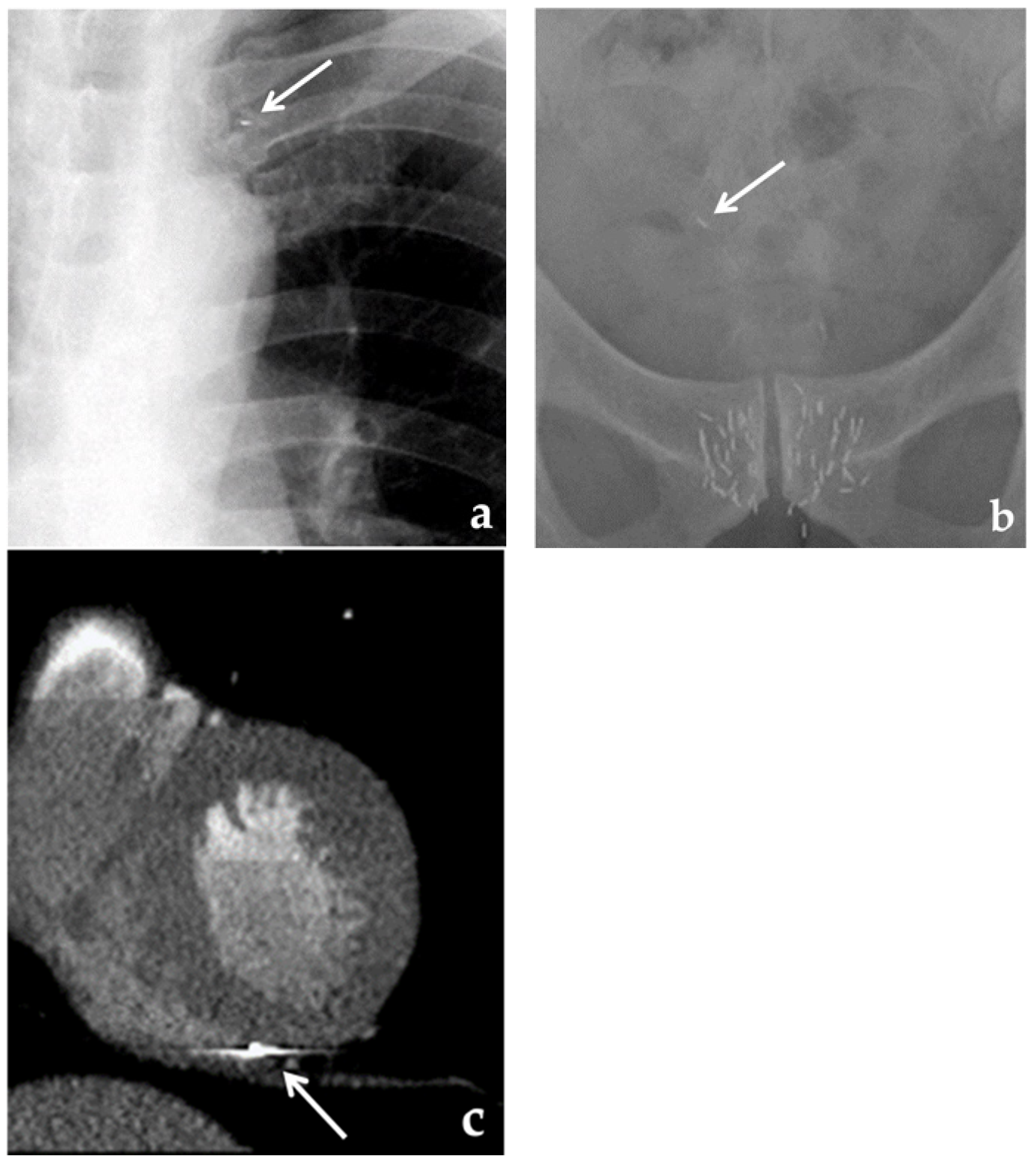
2.4. Evaluation of Migration

All patients underwent radiography (orthogonal chest radiographs, an abdominal radiograph, and a posteroanterior pelvic radiograph) and CT at 1 day, 1 month, and 1 year after brachytherapy to evaluate the presence of seed migration. In this study, seed migration was defined as an event in which seeds enter a blood vessel and are transported out of the prostate by blood flow. Hence, the cases in which the seeds moved to the periprostatic tissue were not counted as migration. In other words, the cases in which the seeds were detected radiographically out of the anatomical location were counted as migration.

2.5. Statistics

Seed density was calculated using the following formula: seed density = implanted seed number / prostate volume. The Mann–Whitney U test was used to compare the variables between the groups with and without migration.
A p value < 0.05 was considered statistically significant.
Cut-off values for predicting the occurrence of migration were determined using receiver operating characteristic (ROC) for prostate volume, number of needles, number of seeds, and seed density. These variables were divided into two groups based on the cut-off values determined by ROC. Univariate and multivariate logistic regression analyses were performed to predict the occurrence of migration using these variables.

3. Results

In total, the number of implanted seeds was 21,876. The median prostate volume was 24.5 cc (range, 11.3–54.5). The median number of needles inserted was 22 (range, 13–35). The median number of seeds per patient was 66 (range, 38–108).
Seed migration was detected in 58 patients (18.1%) 1 day after brachytherapy. One month later, 31 additional cases showed migration for a total of 89 cases (27.8%). One year later, 3 additional cases of migration were observed for a total of 92 cases (28.8%). At 1 year, the location of migrated seeds in the pelvis occurred in 89 cases (27.8%), in the lung in 47 cases (14.7%), and others in 8 cases (2.5%). Others included two kidneys, one right atrium, one spinal canal, one left diaphragm, one peritoneum, one right thigh, and one unknown site (Figure 1).
Of a total of 21,876 seeds, 144 (0.66%) showed migration.
Table 2 summarizes the median number of variables of patients stratified by the results of seed migration. The number of needles, number of seeds, and seed density were significantly higher in the group with migration than in the group without migration (p = 0.000324, 0.0000351, and 0.0142, respectively). The participants in the group with migration tended to have smaller prostate volumes than those in the group without migration (p = 0.0651).
The results of univariate and multivariate analysis are summarized in Table 3. The ROC cutoff values (AUC, sensitivity, and specificity) for prostate volume, number of needles, number of seeds, and seed density were 20.9 cc (0.566, 0.772, 0.368), 21 (0.631, 0.707, 0.504), 65 (0.648, 0.739, 0.535), and 3.0 (0.588, 0.402, 0.772), respectively. In the univariate analysis, prostate volume, number of needles, number of seeds, seed density, and treatment modality were all significant factors in predicting migration (p = 0.0167, 0.000705, 0.0000139, 0.00342 and 0.0000038, respectively). In the multivariate analysis, seed density (p = 0.0232) and treatment modality (p = 0.028) were significant factors in predicting migration.

4. Discussion

Seed migration may occasionally be observed during brachytherapy with loose seeds. Migration was reported to be observed in 22.2–36.2% [12,13,14]. Migration to the vertebral venous plexus has also been reported in rare cases [15]. Our data also showed migration in 28.6% of the samples, which is comparable to findings in previous reports. In recent years, stranded seeds with a low risk of migration have also become available.
However, loose seeds, which have a high degree of freedom in terms of seed placement, are still used. In brachytherapy using iodine-125 for prostate cancer, a large number of seeds are conventionally placed; therefore, even if a small number of seeds migrate, the negative effect on the dose distribution is small [16,17]. Migration is often observed in the lungs; however, migrated seeds rarely cause adverse events [18]. However, on rare occasions, migrated seeds can cause symptomatic adverse events, such as radiation pneumonitis and myocardial infarction [19,20].
It has been reported that migration is significantly more likely to occur when there are many inserted needles and implanted seeds [12,13,14]. Intuitively, it is easy to understand that when many seeds are placed in the limited space of the prostate, some of the seeds do not remain in the prostate and are prone to migration. It has been reported that extraprostatic placement is a risk factor for migration [21]; however, extraprostatic placement is inevitable with excessive seed placement. However, these risk factors do not include host-side characteristics. Even when the same number of seeds is implanted, seed placement is more difficult in cases where the prostate gland has a smaller volume than in a larger one. Similarly, it is naturally more difficult to place a large number of seeds than a small number of seeds in cases with the same prostate volume. Monotherapy usually requires more seeds than combined therapy, which makes seed implantation more difficult. We devised seed density as an index that considers both the number of seed placements and prostate volume. This measures the number of seeds per unit volume of the prostate. In the ROC analysis, the cutoff value of seed density for predicting the presence or absence of migration was 3.0. Univariate analysis revealed that prostate volume, number of needles, number of seeds, seed density, and treatment modality were all significant factors. However, only seed density and treatment modality remained significant factors in the multivariate analysis. Seed density is considered to be more useful than previously reported predictors of migration, such as the numbers of seeds and needles. In cases with a seed density > 3, it is necessary to take measures such as considering the use of stranded seeds.

5. Conclusions

We examined the usefulness of seed density as a new predictor of migration after brachytherapy that considers both the number of seed placements and prostate volume. In the ROC analysis, the cutoff value of seed density for predicting the presence or absence of migration was 3.0. Univariate analysis revealed that prostate volume, number of needles, number of seeds, seed density, and treatment modality were all significant factors. However, only seed density and treatment modality remained significant factors in the multivariate analysis. The seed density that we formulated might help predict seed migration and help develop treatment strategies.

Author Contributions

Conceptualization, T.Y., H.T. and M.M.; methodology, T.Y., T.M., Y.N., C.M., F.H. and H.T.; investigation, T.Y. and H.T.; resources, T.Y., H.T., K.I., M.N., T.K. and M.M.; data curation, T.Y. and H.T.; supervision, M.M.; writing—original draft preparation, T.Y. and H.T.; writing—review and editing, T.M., Y.N., C.M., F.H. and M.M. All authors have read and agreed to the published version of the manuscript.

Funding

This study received no external funding.

Institutional Review Board Statement

The study protocol was approved by the Institutional Review Board of Gifu University (number: 2022-018). All procedures performed in studies involving human participants were in accordance with the ethical standards of the institutional and/or national research committee and with the 1964 Helsinki declaration and its later amendments or comparable ethical standards.

Informed Consent Statement

For this type of study, formal consent is not required. Pursuant to the provisions of the Ethics Committee and the ethics guideline in Japan, written consent was not required in exchange for public disclosure of study information in the case of retrospective and/or observational study using a material such as the existing documentation.

Data Availability Statement

The data presented in this study are available on request from the corresponding author. The data are not publicly available due to privacy and ethical reasons.

Conflicts of Interest

The authors declare no conflict of interest.

References

  1. World Health Organization. GLOBOCAN 2020. International Agency for Research on Cancer. Available online: https://gco.iarc.fr/today/online-analysis-pie?v=2020&mode=cancer&mode_population=continents&population=900&populations=900&key=total&sex=1&cancer=39&type=0&statistic=5&prevalence=0&population_group=0&ages_group%5B%5D=0&ages_group%5B%5D=17&nb_items=7&group_cancer=1&include_nmsc=1&include_nmsc_other=1&half_pie=0&donut=0; (accessed on 30 November 2022).
  2. Grimm, P.; Billiet, I.; Bostwick, D.; Dicker, A.P.; Frank, S.; Immerzeel, J.; Keyes, M.; Kupelian, P.; Lee, W.R.; Machtens, S.; et al. Comparative analysis of prostate-specific antigen free survival outcomes for patients with low, intermediate and high risk prostate cancer treatment by radical therapy. Results from the Prostate Cancer Results Study Group. BJU Int. 2012, 109 (Suppl. 1), 22–29. [Google Scholar] [CrossRef] [PubMed]
  3. Tanaka, H.; Yamaguchi, T.; Hachiya, K.; Kamei, S.; Ishihara, S.; Hayashi, M.; Ogawa, S.; Nishibori, H.; Goshima, S.; Matsuo, M. Treatment outcomes and late toxicities of intensity-modulated radiation therapy for 1091 Japanese patients with localized prostate cancer. Rep. Pract. Oncol. Radiother. 2018, 23, 28–33. [Google Scholar] [CrossRef] [PubMed]
  4. Tree, A.C.; Ostler, P.; van der Voet, H.; Chu, W.; Loblaw, A.; Ford, D.; Tolan, S.; Jain, S.; Martin, A.; Staffurth, J.; et al. Intensity-modulated radiotherapy versus stereotactic body radiotherapy for prostate cancer (PACE-B): 2-year toxicity results from an open-label, radomised, phase 3, non-inferiority trial. Lancet Oncol. 2022, 23, 1308–1320. [Google Scholar] [CrossRef] [PubMed]
  5. Ha, B.; Cho, K.H.; Lee, K.H.; Joung, J.Y.; Kim, Y.J.; Lee, S.U.; Kim, H.; Suh, Y.G.; Moon, S.H.; Lim, Y.K.; et al. Long-term results of a phase II study of hypofractionated proton therapy for prostate cancer: Moderate versus extreme hypofractionation. Radiat. Oncol. 2019, 14, 4. [Google Scholar] [CrossRef] [PubMed]
  6. Sato, H.; Kasuya, G.; Ishikawa, H.; Nomoto, A.; Ono, T.; Nakajima, M.; Isozaki, Y.; Yamamoto, N.; Iwai, Y.; Nemoto, K.; et al. Long-term clinical outcomes after 12-fractionated carbon-ion radiotherapy for localized prostate cancer. Cancer Sci. 2021, 112, 3598–3606. [Google Scholar] [CrossRef] [PubMed]
  7. Tanaka, N.; Asakawa, I.; Hasegawa, M.; Fujimoto, K. Low-dose-rate brachytherapy for prostate cancer: A 15-year experience in Japan. Int. J. Urol. 2020, 27, 17–23. [Google Scholar] [CrossRef] [PubMed]
  8. Yamazaki, H.; Masui, K.; Suzuki, G.; Nakamura, S.; Yoshida, K.; Kotsuma, T.; Tanaka, E.; Otani, K.; Yoshioka, Y.; Ogawa, K. Comparison of three moderate fractionated schedules employed in high-dose-rate brachytherapy monotherapy for clinically localized prostate cancer. Radiother. Oncol. 2018, 129, 370–376. [Google Scholar] [CrossRef] [PubMed]
  9. Stone, N.N.; Stock, R.G. Complication following permanent prostate brachytherapy. Eur. Urol. 2002, 41, 427–433. [Google Scholar] [CrossRef] [PubMed]
  10. Wei, G.; Jiang, P.; Li, C.; Wei, S.; Jiang, Y.; Sun, H.; Wang, J. A review on permanent implants for prostate brachytherapy with comparison between stranded and loose seed. Jpn. J. Radiol. 2022, 40, 135–146. [Google Scholar] [CrossRef] [PubMed]
  11. Nuver, T.T.; Hilgers, G.C.; Kattevilder, R.A.J.; Westendorp, H. Local seed displacement from Day 0 to Day 30 in I-125 permanent prostate brachytherapy: A detailed, computed tomography-based analysis. Brachytherapy 2022, 21, 208–215. [Google Scholar] [CrossRef] [PubMed]
  12. Ankem, M.K.; DeCarvalho, V.S.; Harangozo, A.M.; Hartanto, V.H.; Perrotti, M.; Han, K.; Shih, W.J.; Malka, E.; White, E.C.; Maggio, R.; et al. Implications of radioactive seed migration to the lungs after prostate brachytherapy. Urology 2002, 59, 555–559. [Google Scholar] [CrossRef] [PubMed]
  13. Nakano, M.; Yorozu, A.; Saito, S.; Sugawara, A.; Maruo, S.; Kojima, S.; Kikuchi, T.; Fukushima, M.; Dokiya, T.; Yamanaka, H. Seed migration after transperineal interstitial prostate brachytherapy with I-125 free seeds: Analysis of its incidence and risk factors. Jpn. J. Radiol. 2012, 30, 635–641. [Google Scholar]
  14. Sugawara, A.; Nakashima, J.; Shigematsu, N.; Kunieda, E.; Kubo, A. Prediction of seed migration after transperineal interstitial prostate brachytherapy with -125 free seeds. Brachytherapy 2009, 8, 52–56. [Google Scholar] [CrossRef] [PubMed]
  15. Nakano, M.; Uno, H.; Gotoh, T.; Kubota, Y.; Ishihara, S.; Deguchi, T.; Hayashi, S.; Matsuo, M.; Tanaka, O.; Hoshi, H. Migration of prostate brachytherapy seeds to the vertebral venous plexus. Brachytherapy 2006, 5, 127–130. [Google Scholar] [CrossRef] [PubMed]
  16. Sugawara, A.; Nakashima, J.; Kunieda, E.; Nagata, H.; Mizuno, R.; Seki, S.; Shiraishi, Y.; Kouta, R.; Oya, M.; Shigematsu, N. Incidence of seed migration to the chest, abdomen, and pelvis after transperineal interstitial prostate brachytherapy with loose (125)I seeds. Radiat. Oncol. 2011, 6, 130. [Google Scholar] [CrossRef] [PubMed] [Green Version]
  17. Nakano, M.; Yorozu, A.; Saito, S.; Sugawara, A.; Maruo, S.; Kojima, S.; Kikuchi, T.; Fukushima, M.; Dokiya, T.; Yamanaka, H. Seed migration after transperineal interstitial prostate brachytherapy by using loose seeds: Japanese prostate cancer outcomes study permanent iodin-125 seed implantation (J-POPS) multi-institutional cohort study. Radiat. Oncol. 2015, 10, 228. [Google Scholar] [CrossRef] [PubMed] [Green Version]
  18. Stone, N.N.; Stoch, R.G. Reduction of pulmonary migration of permanent interstitial sources in patients undergoing prostate brachytherapy. Urology 2005, 66, 119–123. [Google Scholar] [CrossRef] [PubMed]
  19. Miura, N.; Kusuhara, Y.; Numata, K.; Shirato, A.; Hashine, K.; Sumiyoshi, Y.; Kataoka, M.; Takechi, S. Radiation pneumonitis caused by a migrated brachytherapy seed lodged in the lung. Jpn. J. Clin. Oncol. 2008, 38, 623–625. [Google Scholar] [CrossRef] [PubMed] [Green Version]
  20. Zhu, A.X.; Wallner, K.E.; Frivold, G.P.; Ferry, D.; Jutzy, K.R.; Foster, G.P. Prostate brachytherapy seed migration to the right coronary artery associated with an acute myocardial infarction. Brachytherapy 2006, 5, 262–265. [Google Scholar] [CrossRef] [PubMed]
  21. Eshleman, J.S.; Davis, B.J.; Pisansky, T.M.; Wilson, T.M.; Haddock, M.G.; King, B.F.; Darby, C.H.; Lajoie, W.N.; Oberg, A.L. Radioactive seed migration to the chest after transperineal interstitial prostate brachytherapy: Extraprostatic seed placement correlates with migration. Int. J. Radiat. Oncol. Biol. Phys. 2004, 59, 419–425. [Google Scholar] [CrossRef] [PubMed]
Figure 1. Seed migration after brachytherapy. A follow-up orthogonal chest radiograph showed that a seed had migrated to the left upper lung (solid arrow) (a). A follow-up posteroanterior pelvic radiograph showed that a seed had migrated to the intra pelvic area (solid arrow) (b). Contrast-enhanced CT showed that the seed had migrated to the right ventricle (solid arrow) (c).
Curroncol 30 00308 g001
Table 1. Patient characteristics (N = 320).
CharacteristicPatients (%)Median (Range)
Age (years) 66 (50–78)
Tumor stage
T1c187 (58.4)
T2a100 (31.3)
T2b11 (3.4)
T2c19 (5.9)
T3a3 (0.9)
Pretreatment PSA (ng/mL) 6.3 (1.7–55.0)
<4.011 (3.4)
≥4.0, <10.0257 (80.3)
≥10.052 (16.3)
Gleason score
3 + 3151 (47.2)
3 + 4105 (32.8)
4 + 339 (12.2)
3 + 52 (0.6)
4 + 416 (5.0)
4 + 56 (1.9)
5 + 41 (0.3)
Prostate volume (cc) 24.5 (11.3–54.5)
Needles inserted 22 (13–35)
Seeds implanted 66 (38–108)
Seed density 2.7 (1.5–4.8)
Treatment modality
Monotherapy202 (63.1)
Combined therapy118 (36.9)
Table 2. Median number of variables of patients stratified by results of seed migration.
Seed Migration (+)
(n = 92)
Seed Migration (-)
(n = 228)
p Value
Prostate volume (cc)26.224.20.0651
Needles22200.000324 *
Seeds75630.0000351 *
Seed density2.782892.6663110.0142*
* Significant difference in values was found between the group with migration and the group without migration (p < 0.05).
Table 3. Results of univariate and multivariate analysis.
Univariate AnalysesMultivariate Analyses
Odds Ratio95% CIp ValueOdds Ratio95% CIp Value
Prostate volume (cc)20.9< vs. ≥20.91.971.13–3.440.0167 *1.330.55–3.220.531
Needles inserted≥21 vs. 21<2.451.46–4.110.000705 *1.470.76–2.860.256
Seeds implanted≥65 vs. 65<3.261.91–5.560.0000139 *1.560.67–3.670.306
Seed density≥3.0 vs. 3.0<2.181.29–3.660.00342 *2.101.11–3.980.0232 †
Treatment modalityMono vs. combined4.232.29–7.800.0000038 *2.281.09–4.760.028 †
Note.—95% CI: 95% confidence interval. * Significant factors for predicting migration in the univariate analysis (p < 0.05). † Significant factors for predicting migration in the multivariate analysis (p < 0.05).
Disclaimer/Publisher’s Note: The statements, opinions and data contained in all publications are solely those of the individual author(s) and contributor(s) and not of MDPI and/or the editor(s). MDPI and/or the editor(s) disclaim responsibility for any injury to people or property resulting from any ideas, methods, instructions or products referred to in the content.

