Impact of the Extremities Positioning on the Set-Up Reproducibility for the Total Marrow Irradiation Treatment

 , ,
, ,  , , , , ,
  , ,
, ,
Abstract
:1. Introduction
2. Materials and Methods
2.1. Simulation and Target Volume Definition
2.2. Immobilization System
2.3. Treatment Planning
2.4. Image-Guided Radiotherapy
2.5. Study Design
2.6. Statistical Analysis
3. Results
4. Discussion
5. Conclusions
Supplementary Materials
Author Contributions
Funding
Institutional Review Board Statement
Informed Consent Statement
Data Availability Statement
Conflicts of Interest
References
- Wong, J.Y.C.; Filippi, A.R.; Dabaja, B.S.; Yahalom, J.; Specht, L. Total Body Irradiation: Guidelines from the International Lymphoma Radiation Oncology Group (ILROG). Int. J. Radiat. Oncol. Biol. Phys. 2018, 101, 521–529. [Google Scholar] [CrossRef] [PubMed]
- Peters, C.; Dalle, J.-H.; Locatelli, F.; Poetschger, U.; Sedlacek, P.; Buechner, J.; Shaw, P.J.; Staciuk, R.; Ifversen, M.; Pichler, H.; et al. Total Body Irradiation or Chemotherapy Conditioning in Childhood ALL: A Multinational, Randomized, Noninferiority Phase III Study. JCO 2021, 39, 295–307. [Google Scholar] [CrossRef]
- Blaise, D.; Maraninchi, D.; Michallet, M.; Reiffers, J.; Jouet, J.P.; Milpied, N.; Devergie, A.; Attal, M.; Sotto, J.J.; Kuentz, M.; et al. Long-Term Follow-up of a Randomized Trial Comparing the Combination of Cyclophosphamide with Total Body Irradiation or Busulfan as Conditioning Regimen for Patients Receiving HLA-Identical Marrow Grafts for Acute Myeloblastic Leukemia in First Complete Remission. Blood 2001, 97, 3669–3671. [Google Scholar] [CrossRef] [PubMed] [Green Version]
- Bunin, N.; Aplenc, R.; Kamani, N.; Shaw, K.; Cnaan, A.; Simms, S. Randomized Trial of Busulfan vs Total Body Irradiation Containing Conditioning Regimens for Children with Acute Lymphoblastic Leukemia: A Pediatric Blood and Marrow Transplant Consortium Study. Bone Marrow Transplant. 2003, 32, 543–548. [Google Scholar] [CrossRef] [PubMed] [Green Version]
- Sieker, K.; Fleischmann, M.; Trommel, M.; Ramm, U.; Licher, J.; Bug, G.; Martin, H.; Serve, H.; Rödel, C.; Balermpas, P. Twenty Years of Experience of a Tertiary Cancer Center in Total Body Irradiation with Focus on Oncological Outcome and Secondary Malignancies. Strahlenther. Onkol. 2022, 198, 547–557. [Google Scholar] [CrossRef]
- Oertel, M.; Martel, J.; Mikesch, J.-H.; Scobioala, S.; Reicherts, C.; Kröger, K.; Lenz, G.; Stelljes, M.; Eich, H.T. The Burden of Survivorship on Hematological Patients—Long-Term Analysis of Toxicities after Total Body Irradiation and Allogeneic Stem Cell Transplantation. Cancers 2021, 13, 5640. [Google Scholar] [CrossRef]
- Wong, J.Y.C.; Filippi, A.R.; Scorsetti, M.; Hui, S.; Muren, L.P.; Mancosu, P. Total Marrow and Total Lymphoid Irradiation in Bone Marrow Transplantation for Acute Leukaemia. Lancet Oncol. 2020, 21, e477–e487. [Google Scholar] [CrossRef]
- Schultheiss, T.E.; Wong, J.; Liu, A.; Olivera, G.; Somlo, G. Image-Guided Total Marrow and Total Lymphatic Irradiation Using Helical Tomotherapy. Int. J. Radiat. Oncol. Biol. Phys. 2007, 67, 1259–1267. [Google Scholar] [CrossRef]
- Hui, S.K.; Kapatoes, J.; Fowler, J.; Henderson, D.; Olivera, G.; Manon, R.R.; Gerbi, B.; Mackie, T.R.; Welsh, J.S. Feasibility Study of Helical Tomotherapy for Total Body or Total Marrow Irradiation. Med. Phys. 2005, 32, 3214–3224. [Google Scholar] [CrossRef]
- Wong, J.Y.C.; Rosenthal, J.; Liu, A.; Schultheiss, T.; Forman, S.; Somlo, G. Image-Guided Total-Marrow Irradiation Using Helical Tomotherapy in Patients With Multiple Myeloma and Acute Leukemia Undergoing Hematopoietic Cell Transplantation. Int. J. Radiat. Oncol. Biol. Phys. 2009, 73, 273–279. [Google Scholar] [CrossRef] [Green Version]
- Wilkie, J.R.; Tiryaki, H.; Smith, B.D.; Roeske, J.C.; Radosevich, J.A.; Aydogan, B. Feasibility Study for Linac-Based Intensity Modulated Total Marrow Irradiation. Med. Phys. 2008, 35, 5609–5618. [Google Scholar] [CrossRef]
- Yeginer, M.; Roeske, J.C.; Radosevich, J.A.; Aydogan, B. Linear Accelerator-Based Intensity-Modulated Total Marrow Irradiation Technique for Treatment of Hematologic Malignancies: A Dosimetric Feasibility Study. Int. J. Radiat. Oncol. Biol. Phys. 2011, 79, 1256–1265. [Google Scholar] [CrossRef]
- Aydogan, B.; Mundt, A.J.; Roeske, J.C. Linac-Based Intensity Modulated Total Marrow Irradiation (IM-TMI). Technol. Cancer Res. Treat. 2006, 5, 513–519. [Google Scholar] [CrossRef]
- Fogliata, A.; Cozzi, L.; Clivio, A.; Ibatici, A.; Mancosu, P.; Navarria, P.; Nicolini, G.; Santoro, A.; Vanetti, E.; Scorsetti, M. Preclinical Assessment of Volumetric Modulated Arc Therapy for Total Marrow Irradiation. Int. J. Radiat. Oncol. Biol. Phys. 2011, 80, 628–636. [Google Scholar] [CrossRef]
- Han, C.; Schultheisss, T.E.; Wong, J.Y.C. Dosimetric Study of Volumetric Modulated Arc Therapy Fields for Total Marrow Irradiation. Radiother. Oncol. 2012, 102, 315–320. [Google Scholar] [CrossRef]
- Aydogan, B.; Yeginer, M.; Kavak, G.O.; Fan, J.; Radosevich, J.A.; Gwe-Ya, K. Total Marrow Irradiation With RapidArc Volumetric Arc Therapy. Int. J. Radiat. Oncol. Biol. Phys. 2011, 81, 592–599. [Google Scholar] [CrossRef]
- Shahid, T.; Mandal, S.; Biswal, S.S.; De, A.; Mukherjee, M.; Roy Chowdhury, S.; Chakrapani, A.; George, K.; Bhattacharya, J.; Soren, P.; et al. Preclinical Validation and Treatment of Volumetric Modulated Arc Therapy Based Total Bone Marrow Irradiation in HalcyonTM Ring Gantry Linear Accelerator. Radiat. Oncol. 2022, 17, 145. [Google Scholar] [CrossRef]
- Bao, Z.; Zhao, H.; Wang, D.; Gong, J.; Zhong, Y.; Xiong, Y.; Deng, D.; Xie, C.; Liu, A.; Wang, X.; et al. Feasibility of a Novel Dose Fractionation Strategy in TMI/TMLI. Radiat. Oncol. 2018, 13, 248. [Google Scholar] [CrossRef]
- Haraldsson, A.; Engellau, J.; Lenhoff, S.; Engelholm, S.; Bäck, S.; Engström, P.E. Implementing Safe and Robust Total Marrow Irradiation Using Helical Tomotherapy—A Practical Guide. Phys. Med. 2019, 60, 162–167. [Google Scholar] [CrossRef] [Green Version]
- Mancosu, P.; Navarria, P.; Castagna, L.; Reggiori, G.; Sarina, B.; Tomatis, S.; Alongi, F.; Nicolini, G.; Fogliata, A.; Cozzi, L.; et al. Interplay Effects between Dose Distribution Quality and Positioning Accuracy in Total Marrow Irradiation with Volumetric Modulated Arc Therapy. Med. Phys. 2013, 40, 111713. [Google Scholar] [CrossRef]
- Mancosu, P.; Navarria, P.; Muren, L.P.; Castagna, L.; Reggiori, G.; Clerici, E.; Sarina, B.; Bramanti, S.; De Philippis, C.; Tomatis, S.; et al. Development of an Immobilization Device for Total Marrow Irradiation. Pract. Radiat. Oncol. 2021, 11, e98–e105. [Google Scholar] [CrossRef] [PubMed]
- Sarina, B.; Mancosu, P.; Navarria, P.; Bramanti, S.; Mariotti, J.; De Philippis, C.; Clerici, E.; Franzese, C.; Mannina, D.; Valli, V.; et al. Nonmyeloablative Conditioning Regimen Including Low-Dose Total Marrow/Lymphoid Irradiation Before Haploidentical Transplantation with Post-Transplantation Cyclophosphamide in Patients with Advanced Lymphoproliferative Diseases. Transplant. Cell. Ther. 2021, 27, 492.e1–492.e6. [Google Scholar] [CrossRef] [PubMed]
- Mancosu, P.; Navarria, P.; Castagna, L.; Roggio, A.; Pellegrini, C.; Reggiori, G.; Fogliata, A.; Lobefalo, F.; Castiglioni, S.; Alongi, F.; et al. Anatomy Driven Optimization Strategy for Total Marrow Irradiation with a Volumetric Modulated Arc Therapy Technique. J. Appl. Clin. Med. Phys. 2012, 13, 138–147. [Google Scholar] [CrossRef] [PubMed]
- Zuro, D.; Madabushi, S.S.; Brooks, J.; Chen, B.T.; Goud, J.; Salhotra, A.; Song, J.Y.; Parra, L.E.; Pierini, A.; Sanchez, J.F.; et al. First Multimodal, Three-Dimensional, Image-Guided Total Marrow Irradiation Model for Preclinical Bone Marrow Transplantation Studies. Int. J. Radiat. Oncol. Biol. Phys. 2021, 111, 671–683. [Google Scholar] [CrossRef] [PubMed]
- Brooks, J.; Zuro, D.; Song, J.Y.; Madabushi, S.S.; Sanchez, J.F.; Guha, C.; Kortylewski, M.; Chen, B.T.; Gupta, K.; Storme, G.; et al. Longitudinal Preclinical Imaging Characterizes Extracellular Drug Accumulation After Radiation Therapy in the Healthy and Leukemic Bone Marrow Vascular Microenvironment. Int. J. Radiat. Oncol. Biol. Phys. 2022, 112, 951–963. [Google Scholar] [CrossRef]
- Miften, M.; Olch, A.; Mihailidis, D.; Moran, J.; Pawlicki, T.; Molineu, A.; Li, H.; Wijesooriya, K.; Shi, J.; Xia, P.; et al. Tolerance Limits and Methodologies for IMRT Measurement-Based Verification QA: Recommendations of AAPM Task Group No. 218. Med. Phys. 2018, 45, e53–e83. [Google Scholar] [CrossRef] [Green Version]
- Mancosu, P.; Navarria, P.; Reggiori, G.; Cozzi, L.; Fogliata, A.; Gaudino, A.; Lobefalo, F.; Paganini, L.; Palumbo, V.; Sarina, B.; et al. In-Vivo Dosimetry with Gafchromic Films for Multi-Isocentric VMAT Irradiation of Total Marrow Lymph-Nodes: A Feasibility Study. Radiat. Oncol. 2015, 10, 86. [Google Scholar] [CrossRef] [Green Version]
- Lambri, N.; Dei, D.; Hernandez, V.; Castiglioni, I.; Clerici, E.; Crespi, L.; De Philippis, C.; Loiacono, D.; Navarria, P.; Reggiori, G.; et al. Automatic Planning of the Lower Extremities for Total Marrow Irradiation Using Volumetric Modulated Arc Therapy. Strahlenther. Onkol. 2022, 199, 412–419. [Google Scholar] [CrossRef]
- Lambri, N.; Dei, D.; Hernandez, V.; Castiglioni, I.; Clerici, E.; De Philippis, C.; Loiacono, D.; Navarria, P.; Reggiori, G.; Rusconi, R.; et al. Evaluation of Plan Complexity and Dosimetric Plan Quality of Total Marrow and Lymphoid Irradiation Using Volumetric Modulated Arc Therapy. J. Appl. Clin. Med. Phys. 2023. [Google Scholar]
- Dei, D.; Lambri, N.; Stefanini, S.; Vernier, V.; Ricardo, C.B.; Crespi, L.; Clerici, E.; Bellu, L.; De Philippis, C.; Loiacono, D.; et al. Internal Guidelines for Reducing Lymph Node Contour Variability in Total Marrow and Lymph Node Irradiation. Cancers 2023, 15, 1536. [Google Scholar] [CrossRef]


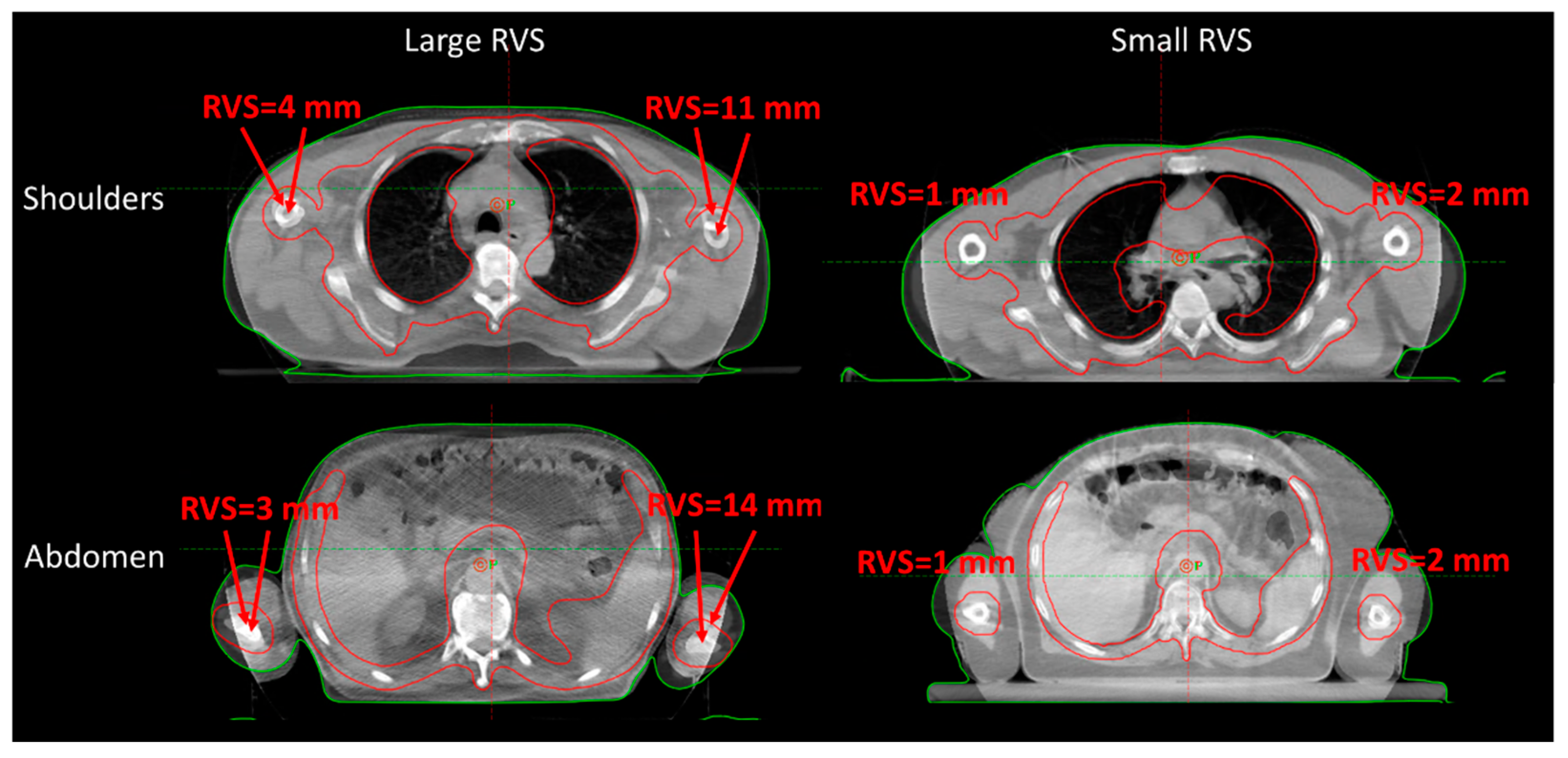
| Isocenter Position | OVS [mm] | Left Extremity RVS [mm] | Right Extremity RVS [mm] |
|---|---|---|---|
| Head | 3.8 (2.3, 5.1) | - | - |
| Shoulders | 2.8 (1.6, 4.2) | 2.9 (0, 6.2) | 5.1 (0, 8.0) |
| Abdomen/Arms | 4.7 (3.0, 6.4) | 7.0 (4.5, 10.0) | 7.3 (3.3, 9.8) |
| Hip | 4.1 (2.5, 6.4) | 3.8 (0, 8.9) | 5.1 (1.2, 8.9) |
| Legs | 4.1 (2.4, 6.3) | 2.0 (0, 5.8) | 2.1 (0, 6.0) |
| Feet | 4.4 (2.6, 6.4) | 7.5 (3.0, 11.0) | 7.5 (3.6, 10.4) |
| Total | 4.0 (2.4, 6.0) | 4.8 (0, 8.0) | 5.1 (0, 8.4) |
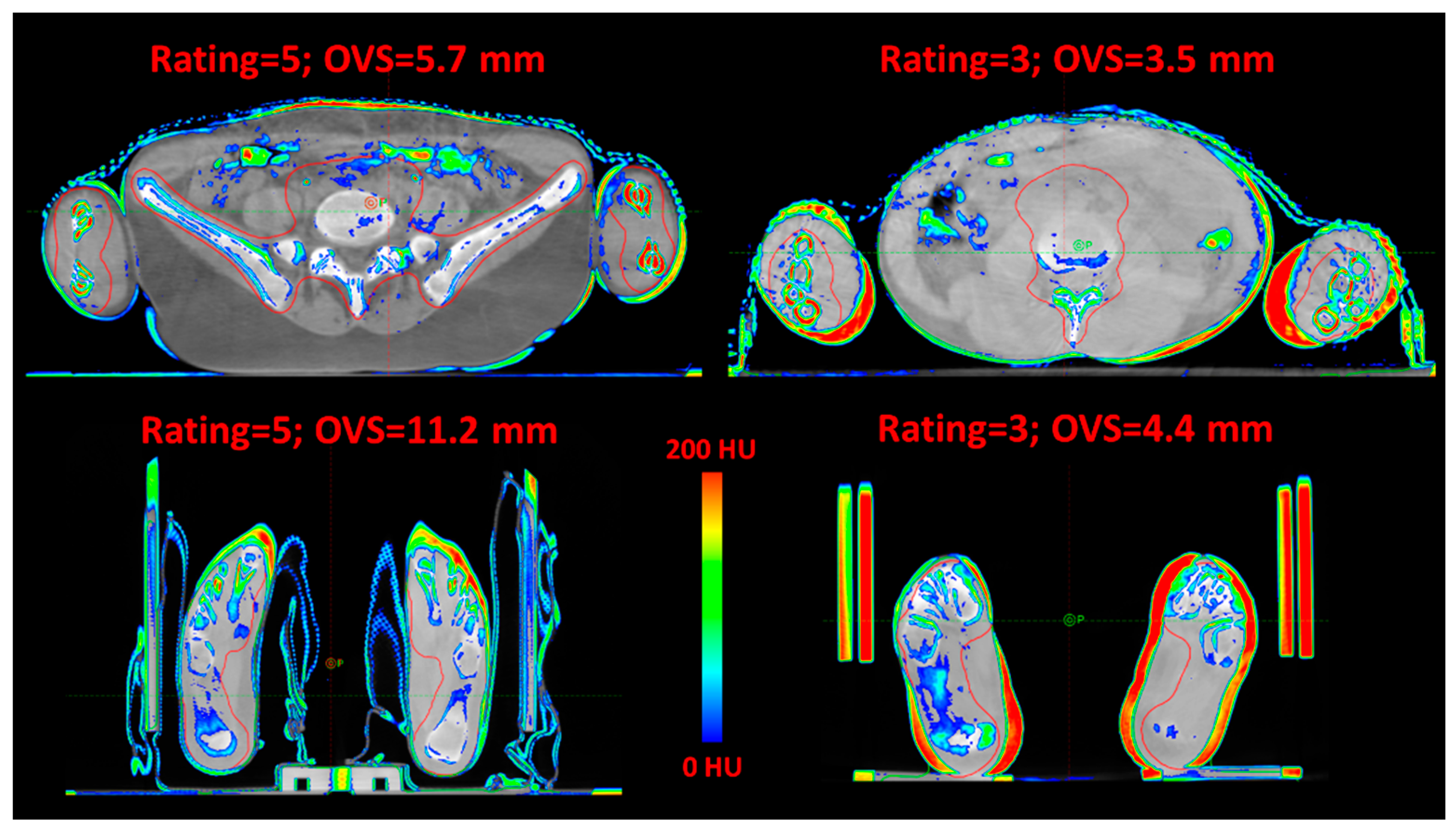
| OVS (mm) | ||||||
|---|---|---|---|---|---|---|
| Rating | Head | Shoulders | Abdomen/Arms | Hip | Legs | Feet |
| 2 | - | - | 3.5, 7.3, 17.6 | 4.7, 6.1, 22.5 | 2.4, 13.8 | 1.7, 3.0 |
| 3 | 2.7 (1.9, 3.6) | 2.7 (1.8, 3.2) | 5.4 * (4.6, 7.5) | 4.8 (3.0, 8.7) | 4.5 (2.3, 6.1) | 4.3 (2.6, 7.1) |
| 4 | 5.8 (3.9, 10.8) | 3.7 (2.0, 5.4) | 4.4 * (2.7, 5.8) | 4.4 † (2.9, 6.7) | 3.6 (2.2, 5.3) | 4.7 (2.9, 7.4) |
| 5 | 3.8 (2.4, 5.1) | 2.7 (1.3, 3.7) | 4.0 (3.3, 5.8) | 3.2 † (2.0, 5.0) | 4.3 (2.7, 7.1) | 3.8 (2.6, 4.8) |
| Extremities Immobilization Method | OVS (mm) | Left Extremity RVS (mm) | Right Extremity RVS (mm) |
|---|---|---|---|
| Arms leaning on the frame | 4.5 (3.1, 6.2) | 6.0 † (3.0, 9.0) | 5.9 * (0, 8.9) |
| Arms above the body | 5.2 (3.0, 6.5) | 8.0 † (5.0, 10.0) | 7.5 * (5.0, 11.0) |
| With feet cushions | 4.1 (2.8, 5.3) | 1.8 ‡ (0, 6.3) | 2.6 ** (0, 7.3) |
| Without feet cushions | 4.5 (2.4, 7.2) | 8.3 ‡ (5.9, 11.0) | 8.2 ** (5.9, 12.0) |
Disclaimer/Publisher’s Note: The statements, opinions and data contained in all publications are solely those of the individual author(s) and contributor(s) and not of MDPI and/or the editor(s). MDPI and/or the editor(s) disclaim responsibility for any injury to people or property resulting from any ideas, methods, instructions or products referred to in the content. |
© 2023 by the authors. Licensee MDPI, Basel, Switzerland. This article is an open access article distributed under the terms and conditions of the Creative Commons Attribution (CC BY) license (https://creativecommons.org/licenses/by/4.0/).
Share and Cite
Lambri, N.; Antonetti, S.L.; Dei, D.; Bellu, L.; Bramanti, S.; Brioso, R.C.; Carlo-Stella, C.; Castiglioni, I.; Clerici, E.; Crespi, L.; et al. Impact of the Extremities Positioning on the Set-Up Reproducibility for the Total Marrow Irradiation Treatment. Curr. Oncol. 2023, 30, 4067-4077. https://doi.org/10.3390/curroncol30040309
Lambri N, Antonetti SL, Dei D, Bellu L, Bramanti S, Brioso RC, Carlo-Stella C, Castiglioni I, Clerici E, Crespi L, et al. Impact of the Extremities Positioning on the Set-Up Reproducibility for the Total Marrow Irradiation Treatment. Current Oncology. 2023; 30(4):4067-4077. https://doi.org/10.3390/curroncol30040309
Chicago/Turabian StyleLambri, Nicola, Simone Leopoldo Antonetti, Damiano Dei, Luisa Bellu, Stefania Bramanti, Ricardo Coimbra Brioso, Carmelo Carlo-Stella, Isabella Castiglioni, Elena Clerici, Leonardo Crespi, and et al. 2023. "Impact of the Extremities Positioning on the Set-Up Reproducibility for the Total Marrow Irradiation Treatment" Current Oncology 30, no. 4: 4067-4077. https://doi.org/10.3390/curroncol30040309