Article

Comparing the Ability of Secretory Signal Peptides for Heterologous Expression of Anti-Lipopolysaccharide Factor 3 in Chlamydomonas reinhardtii

1
Guangdong Technology Research Center for Marine Algal Bioengineering, College of Life Sciences and Oceanography, Shenzhen University, Shenzhen 518060, China
2
Shenzhen Engineering Laboratory for Marine Algal Biological Development and Application, College of Life Sciences and Oceanography, Shenzhen University, Shenzhen 518060, China
3
Laboratory of Marine Bioresource & Eco-Environmental Science, College of Life Sciences and Oceanography, Shenzhen University, Shenzhen 518060, China
4
Guangdong Provincial Key Laboratory for Plant Epigenetics, College of Life Sciences and Oceanography, Shenzhen University, Shenzhen 518060, China
*
Author to whom correspondence should be addressed.
Mar. Drugs 2023, 21(6), 346; https://doi.org/10.3390/md21060346
Submission received: 4 April 2023 / Revised: 26 May 2023 / Accepted: 31 May 2023 / Published: 4 June 2023
(This article belongs to the Special Issue Biotechnology of Algae)

Abstract

:
Anti-lipopolysaccharide factor 3 (ALFPm3) possesses a wide antimicrobial spectrum and high antibacterial and viral activities for broad application prospects in the aquaculture industry. However, the application of ALFPm3 is limited by its low production in nature, as well as its low activity when expressed in Escherichia coli and yeast. Although it has been proven that its secretory expression can be used to produce antimicrobial peptides with strong antimicrobial activity, there is no study on the high-efficiency secretory expression of ALFPm3 in Chlamydomonas reinhardtii. In this study, signal peptides ARS1 and CAH1 were fused with ALFPm3 and inserted into the pESVH vector to construct pH-aALF and pH-cALF plasmids, respectively, that were transformed to C. reinhardtii JUV using the glass bead method. Subsequently, through antibiotic screening, DNA-PCR, and RT-PCR, transformants expressing ALFPm3 were confirmed and named T-JaA and T-JcA, respectively. The peptide ALFPm3 could be detected in algal cells and culture medium by immunoblot, meaning that ALFPm3 was successfully expressed in C. reinhardtii and secreted into the extracellular environment. Moreover, ALFPm3 extracts from the culture media of T-JaA and T-JcA showed significant inhibitory effects on the growth of V. harveyi, V. alginolyticus, V. anguillarum, and V. parahaemolyticus within 24 h. Interestingly, the inhibitory rate of c-ALFPm3 from T-JcA against four Vibrio was 2.77 to 6.23 times greater than that of a-ALFPm3 from T-JaA, indicating that the CAH1 signal peptide was more helpful in enhancing the secreted expression of the ALFPm3 peptide. Our results provided a new strategy for the secretory production of ALFPm3 with high antibacterial activity in C. reinhardtii, which could improve the application potentiality of ALFPm3 in the aquaculture industry.

1. Introduction

The abuse of antibiotics leads to the frequent occurrence of drug-resistant microorganisms and brings irreversible losses in the aquaculture industry [1,2]. The prohibition of the addition of antibiotics in breeding feed is a consensus. Antimicrobial peptides (AMPs) originally found in insects [3] have become the perfect substitutes for antibiotics due to their broad antimicrobial spectrum, strong antibacterial activity, and low susceptibility to drug resistance [4,5,6]. As one cluster of the known AMPs, anti-lipopolysaccharide factors (ALF) firstly found in Limulus polyphemus displayed high antibacterial activity [7]. In recent years, it was reported that the anti-lipopolysaccharide factor 3 (ALFPm3) from Penaeus monodon with a lipopolysaccharide binding domain (LBD) had strong antibacterial activity against common aquaculture pathogenic bacteria, such as Vibrio harveyi, Vibrio parahaemolyticus, and Staphylococcus aureus, at a very low concentration (0.77 µM), showing its potential for application in the aquaculture industry [8,9,10]. ALFPm3 is commonly produced via genetic engineering due to its low yield from natural sources. However, ALFPm3 is difficult to express in Escherichia coli because of its high antimicrobial activity against E. coli [8,11]. Although ALFPm3 could be expressed in P. pastoris, its antibacterial activity is affected by excessive glycosylation in P. pastoris [12,13]. With lots of advantages for industry, such as short growth cycle, posttranslational modification, low and cheap nutritional requirements, and efficient large-scale culture techniques [14,15,16], microalgae have been received widespread attention for their potential to be a factory to produce valuable components. Currently, studies on microalgae are mainly focused on Chlamydomonas reinhardtii, Chlorella vulgaris, Phaeodactylum tricornutum, and so on. However, in Chlorella and P. tricornutum, the expression of exogenous genes is unstable and the transformation efficiency is low [17,18]. Differently, as a model species, genetic manipulation toll kits were well developed in C. reinhardtii [19]. In addition, C. reinhardtii has the advantages of high photosynthesis efficiency, no endotoxin, and reliable industrial cultivation system [20]. It was reported that several medicinal proteins, such as the RBD vaccine [21,22], IFN-α [23], and human interleukin 2 [24], were successfully expressed in C. reinhardtii, indicating that C. reinhardtii is an ideal platform for the production of pharmaceutical proteins.
Until now, the genetic transformation of foreign genes in the nuclei, chloroplasts, and mitochondria of C. reinhardtii has been reported. However, target products were mainly accumulated within cells. In general, the extracellular secretory expression system can help secrete proteins into culture media, providing convenience for protein isolation, purification, and industrial application [25,26]. Moreover, glycosylation modification during the secretion process also facilitates the stability of the protein structure [27,28]. It is a pity that only a few studies on the secretion of proteins have been conducted on C. reinhardtii. Nowadays, secretory expression is mainly achieved through signal peptides (SPs), which are located at the N-terminal of proteins and carry protein secretory information to mediate the secretion process. Different SPs perform differently in the secretion and production of recombinant proteins. For example, a 10-fold difference on the secretion of the mCherry fluorescent protein was observed among more than 2000 signal peptides [29]. Even more significantly, a 100-fold difference in the protein secretion in CHO cells was found among 17 signal peptides [30]. Therefore, finding effective SPs is important for the development of the secretory expression system in C. reinhardtii.
Previously, it was reported that over 95% of the xylanase 1 produced in C. reinhardtii was successfully secreted into the culture medium by inserting arylsulfatase 1 (ARS1) into the coding region of xyn1 [27]. Additionally, the secretion of luciferase in C. reinhardtii was increased by nearly 84% when using carbonic anhydrase 1 (CAH1) [31]. Therefore, ARS1 and CAH1 signal peptides have a great potential in mediating the secretion of ALFPm3 in C. reinhardtii.
In this study, ARS1 and CAH1 were fused with ALFPm3 to construct plasmids pH-aALF and pH-cALF, respectively, which were then transferred into the nuclear genome of C. reinhardtii using the glass bead method. Transformants were identified through DNA-PCR, RT-PCR, and immunoblot analysis. Furthermore, their ability to inhibit aquaculture pathogenic bacteria was also evaluated. Results showed that ALFPm3 could be secreted into medium with the help of the ARS1 signal peptide and the CAH1 signal peptide. The protein extracts from the medium showed high antibacterial activity towards aquaculture pathogenic bacteria such as Vibrio harveyi, Vibrio alginolyticus, Vibrio anguillarum, and Vibrio parahaemolyticus. This study provides a new idea for the highly efficient expression and secretion of AMPs in C. reinhardtii.

2. Results

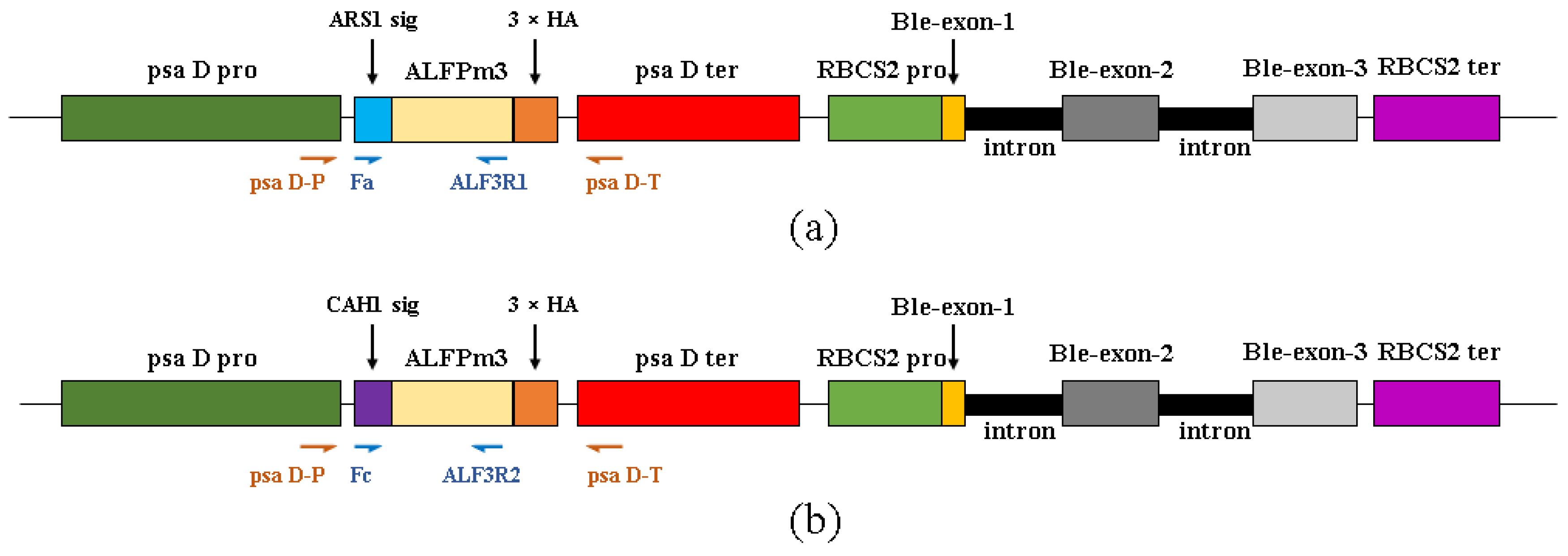
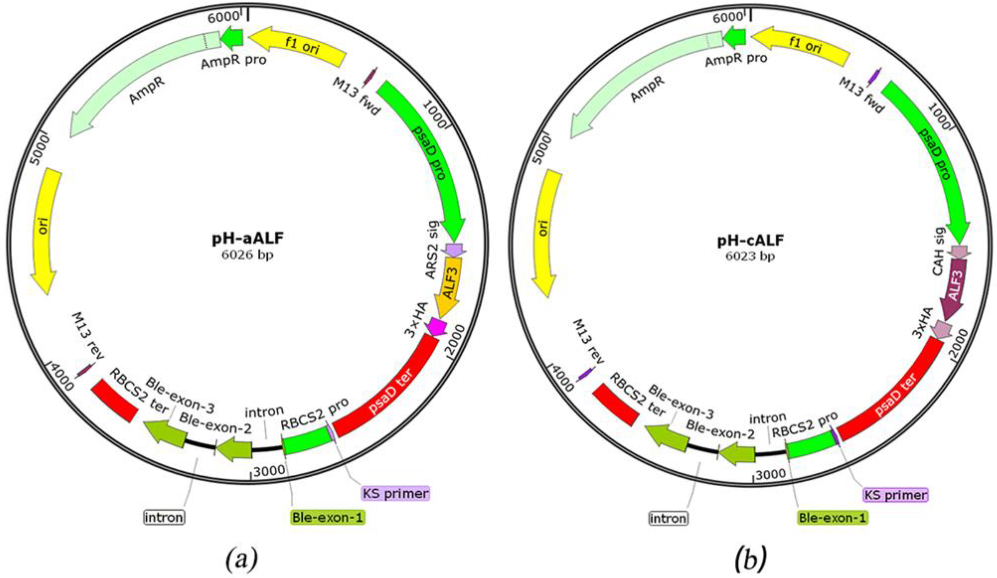
2.1. Design of ALFPm3 Expression Cassette

Based on the codon preference of the C. reinhardtii nuclear genome, the ALFPm3 gene from P. monodon (Genbank number JQ256520) was optimized and its GC content was increased from 56% to 68%. The signal peptides ARS1 (21aa: MGALAVFAVACLAAVASVAHA) and CAH1 (23aa: MARTGALLLVALALAGCAQACIY) were fused with ALFPm3 at the N-terminal, respectively. The 3 × HA tag was connected to the C-terminal of ALFPm3 for subsequent protein identification. The psaD promoter and psaD terminator with high efficiency in expressing heterologous genes in C. reinhardtii [32,33] were used to express target gene. The expression cassette of the ble given transformants with the bleomycin/zeocin-resistance was used as the selection maker [19,34]. To screen positive transformants at molecular level, the psaD-P and psaD-T primer set, located in the psaD promoter and psaD terminator region, was used. The Fa and ALF3R1 and the Fc and ALF3R2 primer sets, located in the signal peptide region and ALFPm3 coding region, were used for RT-PCR to confirm the expression of ALFPm3 in transformants (Figure 1).

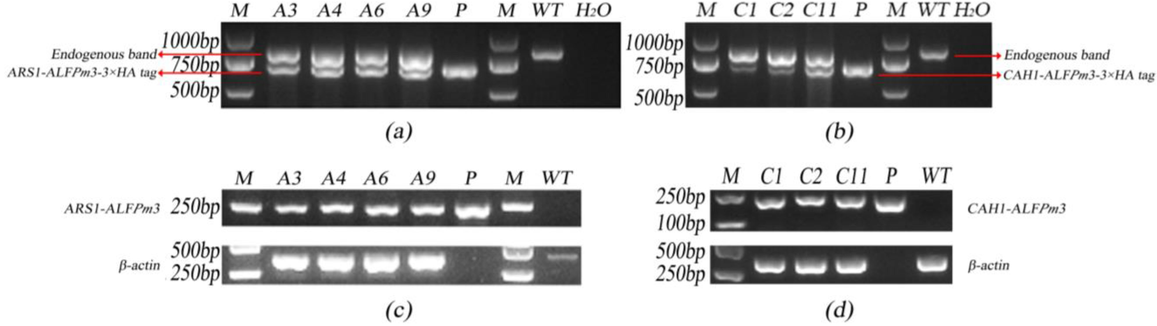
2.2. Screening of Transgenic C. reinhardtii

The transformation of C. reinhardtii JUV was carried out using the glass bead method. After cultivation for three to four weeks, green colonies were visible and were transferred to the new TAP agar medium containing 100 µg/mL ampicillin and 10 µg/mL zeocin. Candidates of algae transformed with plasmid pH-aALF were named T-JaA, and those with plasmid pH-cALF were named T-JcA. It was found that the transformation frequency was 2.5 × 10⁻⁵ in T-JaA and 3.7 × 10⁻⁵ in T-JcA.
As a common endogenous promoter of microalgae, the native psaD promoter/terminator is responsible for initiating the transcription of photosystem I complex related proteins in C. reinhardtii [33]. Therefore, PCR with primers psaD-P and psaD-T could generate an 813 bp endogenous gene fragment in the genome of C. reinhardtii while generating additional 679 bp and 676 bp fragments in T-JaA and T-JcA, respectively. As expected, according to the genomic PCR results, positive transformants could detect target bands at both 679/676 bp and 813 bp, indicating that the target genes were successfully inserted into the genome of C. reinhardtii JUV (Figure 2a,b).
The expression of ALFPm3 in T-JaA and T-JcA was identified by RT-PCR using primer pairs Fa/ALF3R1 and Fc/ALF3R2, respectively. PCR fragments at 222 bp and 203 bp were expected in T-JaA and T-JcA, respectively. As expected, the results of RT-PCR showed that target fragments could be detected as the positive control (Figure 2c,d), indicating the successful expression of ALFPm3 at the transcription level in C. reinhardtii JUV.

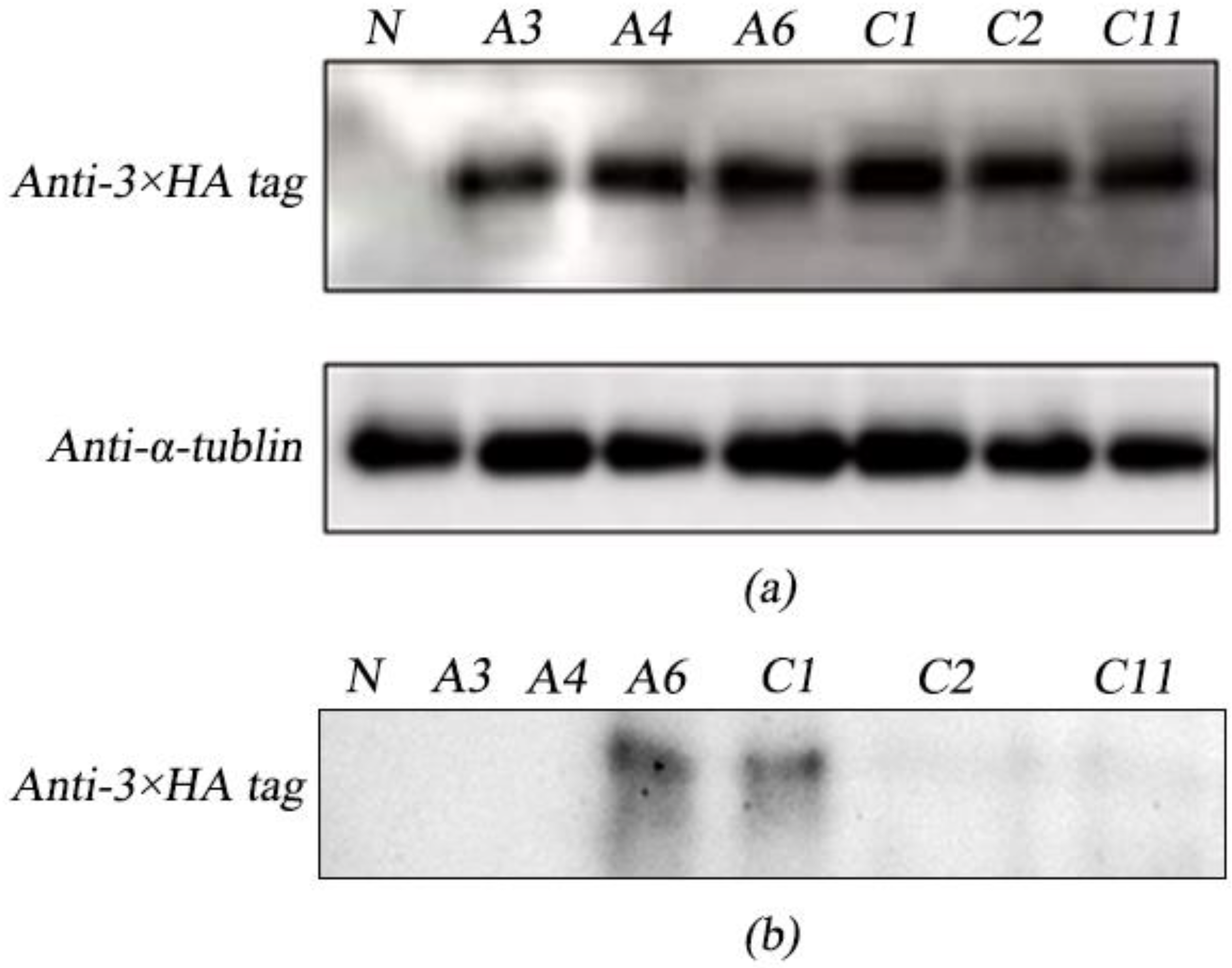
2.3. Analysis of Intracellular and Extracellular Expression of ALFPm3 in Transgenic

C. reinhardtii 

The existence of ARS1-ALFPm3 and CAH1-ALFPm3 fusion proteins in cells was evaluated by immunoblot. Results showed that the proteins ARS1-ALFPm3 (a-ALFPm3) and CAH1-ALFPm3 (c-ALFPm3) were detected in T-JaA (A3, A4, A6) and T-JcA (C1, C2, C11), respectively, which was evidenced by the detection of 3 × HA using anti-3 × HA tag antibody at the expected size of about 16 kD and no detection of that in in the wide type of C. reinhardtii JUV (WT) (Figure 3a). Meanwhile, the protein of β-actin was detected in all the wild-type and positive transformants using anti-α-tublin antibody (Figure 3a), indicating that ARS1-ALFPm3 and CAH1-ALFPm3 fusion proteins were successfully expressed in transgenic C. reinhardtii JUV.
Subsequently, the extracellular secretion of fusion proteins a-ALFPm3 and c-ALFPm3 was analyzed in culture medium. Results showed that only proteins extracted from the culture medium of T-JaA6 (A6) and T-JcA1 (C1) generated the hybrid signal at 16 kD using anti-3 × HA tag antibody (Figure 3b), indicating that the ALFPm3 peptide was successfully secreted in vitro when fused with signal peptides ARS1 and CAH1. However, the target protein was not detected in the culture medium of A3, A4, C2, and C11, even though target proteins were found within cells (Figure 3a). A6 and C1 were used for subsequent bacteriostatic analysis.

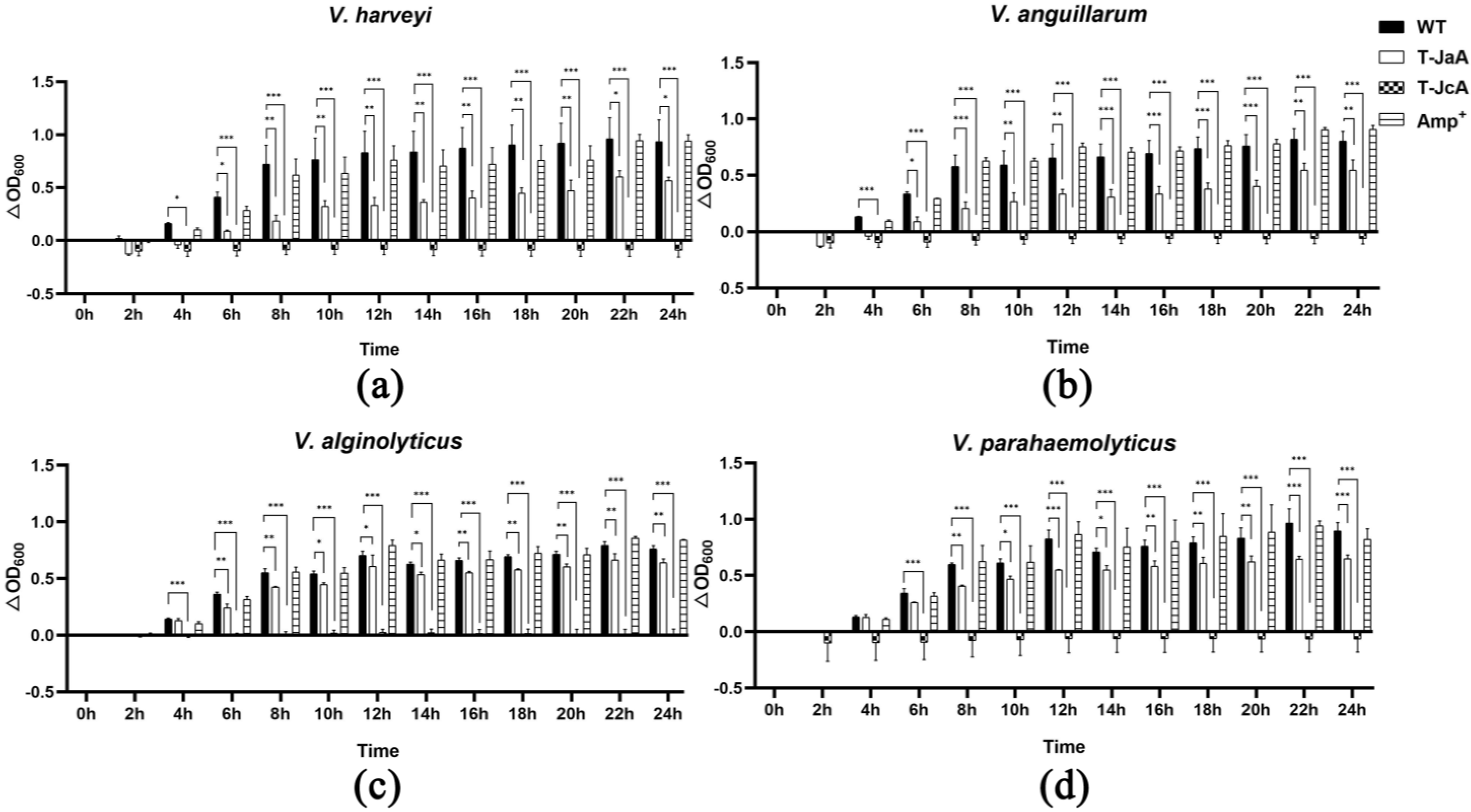
2.4. Both a-ALFPm3 and c-ALFPm3 Showed High Antibacterial Activity

For the antibacterial analysis, 100 mL of culture medium of T-JaA6 and T-JcA1 was collected and freeze dried for protein extraction. The concentration of protein extracts from T-JaA6 (containing a-ALFPm3) was 0.98 µg/µL, and that from T-JcA1 (containing c-ALFPm3) was 2.02 µg/µL. Compared with the wild type, protein extracts containing a-ALFPm3 and c-ALFPm3 had significant inhibitory effects on the growth of V. harveyi, V. alginolyticus, V. anguillarum, and V. parahaemolyticus within 24 h (Figure 4). After 24 h, except for V. alginolyticus with a bacteriostatic rate of 15.79%, the bacteriostatic rate of a-ALFPm3 against aquaculture pathogenic bacteria exceeded 30%. The highest bacteriostatic rate, 39.67%, was found against V. harveyi. It is noted that c-ALFPm3 performed much better than a-ALFPm3, which had a bacteriostatic rate of over 98% against all four aquaculture pathogenic bacteria. Similarly, the highest bacteriostatic rate was found against V. harveyi, reaching 110.07%. The two types of ALFPm3 had the strongest inhibitory effect on V. harveyi and the weakest inhibitory effect on V. alginolyticus. Although the concentration of ampicillin used in the study was as high as 2 mg/mL, its inhibitory efficiency was only −0.69%, −9.90%, −12.77%, and 8.48% against V. harveyi, V. alginolyticus, V. anguillarum, and V. parahaemolyticus, respectively, after 24 h. The antibacterial activity of a-ALFPm3 and c-ALFPm3 against V. parahaemolyticus was 3.88 and 12.67 times as that of ampicillin, demonstrating the strong antibacterial activity of a-ALFPm3 and c-ALFPm3. Indeed, the bacteriostatic effect of ALFPm3 was also affected by the type of signal peptide, and after 24 h, the inhibitory rate of c-ALFPm3 against V. harveyi, V. alginolyticus, V. anguillarum, and V. parahaemolyticus was 2.77, 6.23, 3.32, and 3.26 times that of a-ALFPm3, indicating that the CAH1 signal peptide was more effective in secreting the ALFPm3 peptide in C. reinhardtii than the ARS1 signal peptide.

3. Discussion

Prokaryotic expression systems such as E. coli were not generally used to express AMPs, since those AMPs with high antibacterial activity might kill E.coli [35,36,37]. Given the previous studies, P. pastoris successfully expressed and secreted some AMPs, such as rFcALF2 [38], rMnALF4 [39], Mytichitin-A [40], and rPaDef [41]. However, the secretory expression of rALFPm3 in P. pastoris was only found in a small quantity outside the cell [42]. Additionally, the activity of recombinant proteins secreted by P. pastoris was affected, for example, compared with the natural proteins Mycithin-A [40], CecropinB2 [43], and Ch-Penaeidin [44] secreted by P. pastoris, which had no or low antibacterial activity. Although the secretion of AMPs from C. reinhardtii includes many natural advantages, such as glycosylation for proteins to stabilize their structure and biological activities [45,46], the utilization of algae as a byproduct [47], and the large-scale cultivation in photoreactors for industrial production [48,49], there are few reports about the secretion of AMPs by C. reinhardtii. In previous studies, the signal peptides ARS1 and CAH1 were confirmed to have a secretory expression function and could be used in various areas such as in the delivery of targeted drugs [50] and the secretion of human intrinsic factors [51], xylanase 1 [27], and luciferase [31]. In this study, “ARS1-ALFPm3” and “CAH1-ALFPm3” expression cassettes were designed. Based on genomic PCR, RT-PCR, and immunoblot analyses, transgenic algal cells that could express and secrete ALFPm3 peptide in C. reinhardtii were obtained. It was noted that, according to the immunoblot assay using the medium supernatant, only one out of three transformants could secrete the protein into the medium, which might be because of the random integration of foreign DNA into the nucleus of C. reinhardtii [52]. In previous studies, it was observed that foreign DNA could be cleaved during transformation [53,54]. The resulting fragments could induce complex situations, including deletions and inversions of genomic DNA flanking the foreign DNA [54,55,56] and the integrated concatemers of identical molecules in transformants with multiple copies of foreign DNA [54,57]. The above events might cause gene silencing or transcript instability [57,58,59], thereby affecting the expression and secretion of the target protein [60]. Compared with ALFPm3 secreted by P. pastoris [42], a-ALFPm3 and c-ALFPm3 secreted by C. reinhardtii in this study exhibited a strong inhibitory ability against V. harveyi, V. alginolyticus, V. anguillarum, and V. parahaemolyticus within 24 h, and the inhibition rate was much higher than that of ampicillin (2 mg/mL).
Differences in signal peptides affect the secretion yield and activity of recombinant proteins distinctly by influencing the folding state of the peptide chain [61,62], translocation efficiency [63], and protein stability [64,65]. In the study of signal peptides mediating the secretion and expression of NHases in B. subtilis, it was found that the values of the highest secretion were nearly 4.32 those of the lowest secretion [66]. The human–mouse chimeric CMV-IgM was successfully expressed and secreted in CHO cells by five signal peptides, and its secretory efficiency was more than 6.72-fold different [67]. According to the previous research, when fused with different signal peptides, the secretion yield of mCherry fluorescent proteins in C. reinhardtii was up to 10-fold different [29]. In this study, comparing a-ALFPm3 with c-ALFPm3, the secretion yield of c-ALFPm3 was 2.06 times that of a-ALFPm3, and the inhibition rate of c-ALFPm3 was also much higher than that of a-ALFPm3, indicating that both the signal peptides could secrete the ALFPm3 peptide out of C. reinhardtii efficiently, and the CAH1 signal peptide performed better than the ARS1 signal peptide. In particular, ALFPm3, derived from T-JcA1, had an inhibitory rate of over 98% against four pathogenic bacteria within 24 h, indicating that the strategy of “CAH1-ALFPm3” constructed in this study had a high potential value of AMPs.
This work provides a new idea to produce ALFPm3 with high antibacterial activity. In the future, further improvements in the secretion efficiency of T-JaA and T-JcA will be conducted, and the application value of ALFPm3 in the aquaculture industry will be further explored.

4. Materials and Methods

4.1. Bacterial Strains, Algal Strain, and Culture Conditions

The C. reinhardtii cell-wall-deficient strain JUV was purchased from the Chlamydomonas Resource Center (Duke University, Durham, NC, USA). It is suitable for cultivation in triethyl phosphate (TAP) liquid medium or TAP agar solid medium with 100 µg/mL ampicillin (Biosharp, Anhui, China) at a temperature of 22–25 °C and a light intensity of 90 μE·m−2·s−1. Transgenic algae were screened and cultivated on TAP liquid or agar solid media containing 100 µg/mL ampicillin and 10 µg/mL zeocin.
Bacterial strains of V. harveyi, V. alginolyticus, V. anguillarum, and V. parahaemolyticus were kindly provided by Professor Huang Jianhua from the South China Sea Fisheries Research Institute of the Chinese Academy of Fishery Sciences. Bacteria were cultured on LB medium under 37 °C at 200 rpm.

4.2. Plasmid Construction and Genetic Transformation

ARS1 signal peptide [27] and CAH1 signal peptide [31] were selected to construct the fusion protein with ALFPm3 from P. monodon. The fusion fragments were named ARS1-ALFPm3 and CAH1-ALFPm3, respectively. In order to improve the transformation efficiency and enhance the heterologous expression of C. reinhardtii, the coding sequence of ALFPm3 (Genbank number: JQ256520) was optimized based on the codon bias of the genome of C. reinhardtii. Subsequently, fusion fragments tailed with 3 × HA tag were inserted into the expression vector pESVH to obtain the plasmid pH-aALF (Figure 5a) and the plasmid pH-cALF (Figure 5b). The “ARS1-ALFPm3-3 × HA tag” and “CAH1-ALFPm3-3 × HA tag” expression cassettes were driven by the pasD promoter and the pasD terminator. The RBCS2 promoter and the RBCS2 terminator were used to drive the expression of sh-ble given the transformants with zeocin resistance. The transformation of C. reinhardtii JUV was performed using the glass bead method according to the literature [68]. Transformed C. reinhardtii cells were selected on a TAP agar plate containing 100 µg/mL ampicillin and 10 µg/mL zeocin at 22 °C, with continuous light intensity 90 μE·m−2·s−1 for 2–3 weeks until green colonies appeared. The number of colonies was recorded and used to calculate the transformation frequency.

4.3. Genomic PCR and RT-PCR Analysis

Genomic PCR and RT-PCR were performed to identify the positive transformants [69] using 2 × M5 HiPer plus Taq HiFi PCR mix (Mei5bio, Beijing, China) as recommended. For genomic PCR, algal cells were collected from 2 mL cell culture by centrifugation at 5000 rpm for 10 min and then subjected to genomic DNA extraction using the Ultra DNA Isolation kit (Beibei Biotechnology Co., Ltd., Zhengzhou, China). Then, 2 microliters of extracted DNA was used in the 20 μL PCR reaction system with the primer pair psaD-P/psaD-T (Table 1). The PCR program was 95 °C for 3 min, 35 cycles of 94 °C for 25 s, 58 °C for 25 s, 72 °C for 40 s, and finally extended at 72 °C for 5 min. For RT-PCR, the total RNA was extracted with an RNA fast 200 kit (Fastagen, Shanghai, China), and the first stranded cDNA was synthesized using a Hifair® III 1st Strand cDNA Synthesis SuperMix for qRCR (gDNA digest plus) (Yeasen, Shanghai, China), as recommended by the instructions. The RT-PCR reaction system was in a 20 μL column containing 4 μL cDNA as the template. The amplification primer pair for T-JaA was Fa/ALF3R1, and that for T-JcA was Fc/ALF3R2. The primer pair actin-F/actin-R was selected to amplify β-actin as the internal control (Table 1). The RT-PCR program was 95 °C for 3 min, 35 cycles of 95 °C for 30 s, 58 °C for 30 s, 72 °C for 40 s, and finally extended at 72 °C for 5 min. Finally, all PCR products were analyzed via 1.5% agarose gel electrophoresis.

4.4. Protein Extraction and Immunoblot Analysis

To evaluate the presence of the protein ALFPm3 in T-JaA and T-JcA, the total proteins in cells and the secretory protein in culture supernatant were extracted and subjected to immunoblot analysis. Algal cells were pelleted by centrifugation and total intracellular proteins were extracted based on the reference literature [9]. The extracellular protein from culture medium was extracted as follows: the culture medium was collected by centrifugation and then freeze dried. The obtained powder was finally dissolved with 1 × PBS (Biyuntian Biotechnology Co., Ltd., Shanghai, China).
The protein was separated by 12.5% SDS-PAGE (GenScript, Piscataway, NJ, USA) and transferred to a PVDF membrane (Merck, Rahway, NJ, USA). The hybridization was performed first with Anti-HA.11 Epitope Tag Antibody (1:2000) (Biogene, Beverly Hills, CA, USA) and then with Anti-mouse IgG, HRP-linked Antibody (1:2000) (Cell Signaling Technology, Boston, MA, USA) as per the instructions. Finally, BeyoECL Moon (Biyuntian Biotechnology Co., Ltd., Shanghai, China) was used for the stain development, and Odyssey® Fc (Gene Company Limited, Hong Kong, China) was used to detect chemiluminescence signals.

4.5. Antibacterial Assay

T-JaA6 and T-JcA1 algal cells were cultivated at 22–25 °C with continuous light intensity at 90 μE·m−2·s−1 until the cell density reached 1 × 10⁶ cells/mL. Total proteins were extracted from culture supernatant as described above and dissolved in 1 × PBS for subsequent antibacterial assay. The antibacterial assay was tested on four common aquaculture pathogenic bacteria including V. harveyi, V. alginolyticus, V. anguillarum, and V. parahaemolyticus. The tested strains were cultured with 3 mL fresh LB overnight at 37 °C, 200 rpm. Then, 30 μL overnight culture was inoculated to 3 mL of fresh LB and incubated for 1 h at 37 °C. Subsequently, the mixture was diluted 1000 times with fresh LB liquid medium for the antibacterial assay. The 50 μL protein extracts from the culture supernatant of T-JaA and T-JcA were mixed with 150 μL bacteria culture in a sterile 96-well plate. At the same time, 10 μL 2 mg/mL Ampicillin and 50 μL protein extracts from the culture supernatant of C. reinhardtii JUV were set as the positive and negative control, respectively. The total 200 μL mixtures were incubated at 37 °C for 24 h. During this period, the absorbance value of the mixture at 600 nm was measured every 2 h. The growth rate (∆OD600) of the bacteria in each treatment group was obtained by comparing the OD600 value at different time points with that at 0 h. The bacteriostatic rate at each time point was calculated as (%) = (ΔOD600 of WT − Δ OD600 of Experimental group)/ΔOD600 of WT × 100.

4.6. Statistical Analysis

Each experiment had three independent replicates, and the software GraphPad Prism 8 was used to perform statistical analysis and make charts. In this study, the t-test was used to analyze the statistical differences between the treatment groups. p < 0.05 was considered to have a significant difference.

5. Conclusions

In this study, the signal peptides ARS1 and CAH1 were fused with ALFPm3, respectively, and inserted into the pESVH vector to construct pH-aALF and pH-cALF plasmids, which were transformed to C. reinhardtii JUV using the glass bead method. The positive transformants were obtained after bleomycin resistance selection, DNA-PCR, and RT-PCR analysis. The peptide ALFPm3 was detected within cells and culture supernatant, demonstrating the successful expression and secretion of ALFPm3 using C. reinhardtii. In addition, ALFPm3 extracted from the culture supernatant of T-JaA and T-JcA showed the significant inhibition of the growth of V. harveyi, V. alginolyticus, V. anguillarum, and V. parahaemolyticus within 24 h and performed much better than ampicillin (2 mg/mL), indicating the superior antibacterial activity of ALFPm3. Interestingly, we found the inhibitory rate of c-ALFPm3 against the tested Vibrio was 2.77 to 6.23 times as that of a-ALFPm3, showing the superiority of the CAH1 signal peptide for the secretory expression of ALFPm3 in C. reinhardtii. This study provided a new strategy for the highly efficient secretion of ALFPm3 with high antibacterial activity in C. reinhardtii.

Author Contributions

Conceptualization, C.W.; methodology, C.W., Y.O. and H.Z.; investigation, Y.O. and R.C.; data curation, Y.O. and H.Z.; writing—original draft preparation, Y.O. and H.Z.; writing—review and editing, C.W. and D.H.; project administration, C.W. All authors have read and agreed to the published version of the manuscript. .

Funding

This work was supported financially by the National key research and development plan special project for synthetic biology (2018YFA0902500), the Shenzhen Scientific Project (JCYJ20190808114216058), and the Guangdong Natural Science Foundation (2022A1515010285).

Institutional Review Board Statement

Not applicable.

Data Availability Statement

The original data are available from the correspondent author on request.

Acknowledgments

We thank Yiwen Lin for detecting the ALFPm3 in transgenic C. reinhardtii.

Conflicts of Interest

The authors declare no conflict of interest.

References

  1. Bombaywala, S.; Mandpe, A.; Paliya, S.; Kumar, S. Antibiotic resistance in the environment: A critical insight on its occurrence, fate, and eco-toxicity. Environ. Sci. Pollut. Res. 2021, 28, 24889–24916. [Google Scholar] [CrossRef] [PubMed]
  2. Cabello, F.C.; Godfrey, H.P.; Buschmann, A.H.; Dölz, H.J. Aquaculture as yet another environmental gateway to the development and globalisation of antimicrobial resistance. Lancet Infect. Dis. 2016, 16, e127–e133. [Google Scholar] [CrossRef]
  3. Boman, H.G.; Faye, I.; Gan, R.; Gudmundsson, G.H.; Lidholm, D.A.; Lee, J.Y.; Xanthopoulos, K.G. Insect immunity: A gene system for antibacterial proteins. Memórias Inst. Oswaldo Cruz 1987, 82, 115–124. [Google Scholar] [CrossRef] [PubMed] [Green Version]
  4. Hancock, R.E.; Brown, K.L.; Mookherjee, N. Host defence peptides from invertebrates—Emerging antimicrobial strategies. Immunobiology 2006, 211, 315–322. [Google Scholar] [CrossRef]
  5. Tornesello, A.L.; Borrelli, A.; Buonaguro, L.; Buonaguro, F.M.; Tornesello, M.L. Antimicrobial Peptides as Anticancer Agents: Functional Properties and Biological Activities. Molecules 2020, 25, 2850. [Google Scholar] [CrossRef] [PubMed]
  6. Hancock, R.E.; Diamond, G. The role of cationic antimicrobial peptides in innate host defences. Trends Microbiol. 2000, 8, 402–410. [Google Scholar] [CrossRef]
  7. Tanaka, S.; Nakamura, T.; Morita, T.; Iwanaga, S. Limulus anti-LPS factor: An anticoagulant which inhibits the endotoxin-mediated activation of Limulus coagulation system. Biochem. Biophys. Res. Commun. 1982, 105, 717–723. [Google Scholar] [CrossRef]
  8. Jaree, P.; Tassanakajon, A.; Somboonwiwat, K. Effect of the anti-lipopolysaccharide factor isoform 3 (ALFPm3) from Penaeus monodon on Vibrio harveyi cells. Dev. Comp. Immunol. 2012, 38, 554–560. [Google Scholar] [CrossRef]
  9. Li, A.; Huang, R.; Wang, C.; Hu, Q.; Li, H.; Li, X. Expression of Anti-Lipopolysaccharide Factor Isoform 3 in Chlamydomonas reinhardtii Showing High Antimicrobial Activity. Mar. Drugs 2021, 19, 239. [Google Scholar] [CrossRef]
  10. Suraprasit, S.; Methatham, T.; Jaree, P.; Phiwsaiya, K.; Senapin, S.; Hirono, I.; Lo, C.F.; Tassanakajon, A.; Somboonwiwat, K. Anti-lipopolysaccharide factor isoform 3 from Penaeus monodon (ALFPm3) exhibits antiviral activity by interacting with WSSV structural proteins. Antivir. Res. 2014, 110, 142–150. [Google Scholar] [CrossRef] [Green Version]
  11. Somboonwiwat, K.; Bachère, E.; Rimphanitchayakit, V.; Tassanakajon, A. Localization of anti-lipopolysaccharide factor (ALFPm3) in tissues of the black tiger shrimp, Penaeus monodon, and characterization of its binding properties. Dev. Comp. Immunol. 2008, 32, 1170–1176. [Google Scholar] [CrossRef]
  12. Puxbaum, V.; Mattanovich, D.; Gasser, B. Quo vadis? The challenges of recombinant protein folding and secretion in Pichia pastoris. Appl. Microbiol. Biotechnol. 2015, 99, 2925–2938. [Google Scholar] [CrossRef] [PubMed]
  13. Rabert, C.; Weinacker, D.; Pessoa, A., Jr.; Farías, J.G. Recombinants proteins for industrial uses: Utilization of Pichia pastoris expression system. Braz. J. Microbiol. 2013, 44, 351–356. [Google Scholar] [CrossRef] [PubMed] [Green Version]
  14. Specht, E.; Miyake-Stoner, S.; Mayfield, S. Micro-algae come of age as a platform for recombinant protein production. Biotechnol. Lett. 2010, 32, 1373–1383. [Google Scholar] [CrossRef] [Green Version]
  15. Ferrer-Miralles, N.; Saccardo, P.; Corchero, J.L.; Garcia-Fruitós, E. Recombinant Protein Production and Purification of Insoluble Proteins. Methods Mol. Biol. 2022, 2406, 1–31. [Google Scholar] [CrossRef] [PubMed]
  16. Dehghani, J.; Adibkia, K. Towards a new avenue for producing therapeutic proteins: Microalgae as a tempting green biofactory. Biotechnol. Adv. 2020, 40, 107499. [Google Scholar] [CrossRef]
  17. Yang, B.; Liu, J. Chlorella species as hosts for genetic engineering and expression of heterologous proteins: Progress, challenge and perspective. Biotechnol. J. 2016, 11, 1244–1261. [Google Scholar] [CrossRef] [PubMed]
  18. Liu, J.; Chen, F. Biology and Industrial Applications of Chlorella: Advances and Prospects. Adv. Biochem. Eng. Biotechnol. 2016, 153, 1–35. [Google Scholar] [CrossRef]
  19. Lumbreras, V.; Stevens, D.R. Efficient foreign gene expression in Chlamydomonas reinhardtii mediated by an endogenous intron. Plant J. 1998, 14, 441–447. [Google Scholar] [CrossRef]
  20. Scaife, M.A.; Nguyen, G.T.D.T. Establishing Chlamydomonas reinhardtii as an industrial biotechnology host. Plant J. 2015, 82, 532–546. [Google Scholar] [CrossRef] [PubMed]
  21. Govea-Alonso, D.O.; Malla, A. An Algae-Made RBD from SARS-CoV-2 Is Immunogenic in Mice. Pharmaceuticals 2022, 15, 1298. [Google Scholar] [CrossRef] [PubMed]
  22. Rosales-Mendoza, S.; García-Silva, I.; González-Ortega, O.; Sandoval-Vargas, J.M.; Malla, A.; Vimolmangkang, S. The Potential of Algal Biotechnology to Produce Antiviral Compounds and Biopharmaceuticals. Molecules 2020, 25, 4049. [Google Scholar] [CrossRef]
  23. El-Ayouty, Y.; El-Manawy, I.; Nasih, S.; Hamdy, E.; Kebeish, R. Engineering Chlamydomonas reinhardtii for Expression of Functionally Active Human Interferon-α. Mol. Biotechnol. 2019, 61, 134–144. [Google Scholar] [CrossRef] [PubMed]
  24. Dehghani, J.; Adibkia, K.; Movafeghi, A.; Pourseif, M.M.; Omidi, Y. Designing a new generation of expression toolkits for engineering of green microalgae; robust production of human interleukin-2. Bioimpacts 2020, 10, 259–268. [Google Scholar] [CrossRef] [PubMed]
  25. Pollock, J.; Ho, S.V.; Farid, S.S. Fed-batch and perfusion culture processes: Economic, environmental, and operational feasibility under uncertainty. Biotechnol. Bioeng. 2013, 110, 206–219. [Google Scholar] [CrossRef] [PubMed]
  26. Tyo, K.E.J.; Liu, Z.; Magnusson, Y.; Petranovic, D.; Nielsen, J. Impact of protein uptake and degradation on recombinant protein secretion in yeast. Appl. Microbiol. Biotechnol. 2014, 98, 7149–7159. [Google Scholar] [CrossRef]
  27. Rasala, B.A.; Lee, P.A.; Shen, Z.; Briggs, S.P.; Mendez, M.; Mayfield, S.P. Robust Expression and Secretion of Xylanase1 in Chlamydomonas reinhardtii by Fusion to a Selection Gene and Processing with the FMDV 2A Peptide. PLoS ONE 2012, 7, e43349. [Google Scholar] [CrossRef] [Green Version]
  28. Ramos-Martinez, E.M.; Fimognari, L.; Sakuragi, Y. High-yield secretion of recombinant proteins from the microalga Chlamydomonas reinhardtii. Plant Biotechnol. J. 2017, 15, 1214–1224. [Google Scholar] [CrossRef] [Green Version]
  29. Molino, J.V.D.; de Carvalho, J.C.M. Comparison of secretory signal peptides for heterologous protein expression in microalgae: Expanding the secretion portfolio for Chlamydomonas reinhardtii. PLoS ONE 2018, 13, e0192433. [Google Scholar] [CrossRef]
  30. Kober, L.; Zehe, C.; Bode, J. Optimized signal peptides for the development of high expressing CHO cell lines. Biotechnol. Bioeng. 2013, 110, 1164–1173. [Google Scholar] [CrossRef]
  31. Lauersen, K.J.; Berger, H.; Mussgnug, J.H.; Kruse, O. Efficient recombinant protein production and secretion from nuclear transgenes in Chlamydomonas reinhardtii. J. Biotechnol. 2013, 167, 101–110. [Google Scholar] [CrossRef]
  32. Kumar, A.; Falcao, V.R.; Sayre, R.T. Evaluating nuclear transgene expression systems in Chlamydomonas reinhardtii. Algal Res. 2013, 2, 321–332. [Google Scholar] [CrossRef]
  33. Fischer, N.; Rochaix, J.-D. The flanking regions of PsaD drive efficient gene expression in the nucleus of the green alga Chlamydomonas reinhardtii. Mol. Genet. Genom. 2001, 265, 888–894. [Google Scholar] [CrossRef] [PubMed]
  34. Baier, T.; Wichmann, J.; Kruse, O.; Lauersen, K. Intron-containing algal transgenes mediate efficient recombinant gene expression in the green microalga Chlamydomonas reinhardtii. Nucleic Acids Res. 2018, 46, 6909–6919. [Google Scholar] [CrossRef] [PubMed]
  35. Liu, C.-C.; Chung, C.-P.; Lin, C.-Y.; Sung, H.-H. Function of an anti-lipopolysaccharide factor (ALF) isoform isolated from the hemocytes of the giant freshwater prawn Macrobrachium rosenbergii in protecting against bacterial infection. J. Invertebr. Pathol. 2014, 116, 1–7. [Google Scholar] [CrossRef]
  36. Ponprateep, S.; Somboonwiwat, K.; Tassanakajon, A. Recombinant anti-lipopolysaccharide factor isoform 3 and the prevention of vibriosis in the black tiger shrimp, Penaeus monodon. Aquaculture 2009, 289, 219–224. [Google Scholar] [CrossRef]
  37. Anju, M.V.; Archana, K.; Anooja, V.V.; Athira, P.P.; Neelima, S.; Singh, I.S.B.; Philip, R. A novel anti-lipopolysaccharide factor from blue swimmer crab Portunus pelagicus and its cytotoxic effect on the prokaryotic expression host, E. coli on heterologous expression. J. Genet. Eng. Biotechnol. 2023, 21, 22. [Google Scholar] [CrossRef]
  38. Yang, H.; Li, S.; Li, F.; Yu, K.; Yang, F.; Xiang, J. Recombinant Expression of a Modified Shrimp Anti-Lipopolysaccharide Factor Gene in Pichia pastoris GS115 and Its Characteristic Analysis. Mar. Drugs 2016, 14, 152. [Google Scholar] [CrossRef] [Green Version]
  39. Tang, T.; Liu, J.; Li, S.; Li, H.; Liu, F. Recombinant expression of an oriental river prawn anti-lipopolysaccharide factor gene in Pichia pastoris and its characteristic analysis. Fish Shellfish. Immunol. 2020, 98, 414–419. [Google Scholar] [CrossRef]
  40. Meng, D.-M.; Dai, H.-X.; Gao, X.-F.; Zhao, J.-F.; Guo, Y.-J.; Ling, X.; Dong, B.; Zhang, Z.-Q.; Fan, Z.-C. Expression, purification and initial characterization of a novel recombinant antimicrobial peptide Mytichitin—A in Pichia pastoris. Protein Expr. Purif. 2016, 127, 35–43. [Google Scholar] [CrossRef]
  41. Meng, D.-M.; Zhao, J.-F.; Ling, X.; Dai, H.-X.; Guo, Y.-J.; Gao, X.-F.; Dong, B.; Zhang, Z.-Q.; Meng, X.; Fan, Z.-C. Recombinant expression, purification and antimicrobial activity of a novel antimicrobial peptide PaDef in Pichia pastoris. Protein Expr. Purif. 2017, 130, 90–99. [Google Scholar] [CrossRef] [PubMed]
  42. Somboonwiwat, K.; Marcos, M.; Tassanakajon, A.; Klinbunga, S.; Aumelas, A.; Romestand, B.; Gueguen, Y.; Boze, H.; Moulin, G.; Bachère, E. Recombinant expression and anti-microbial activity of anti-lipopolysaccharide factor (ALF) from the black tiger shrimp Penaeus monodon. Dev. Comp. Immunol. 2005, 29, 841–851. [Google Scholar] [CrossRef]
  43. Lai, W.-S.; Kan, S.-C.; Lin, C.-C.; Shieh, C.-J.; Liu, Y.-C. Antibacterial Peptide CecropinB2 Production via Various Host and Construct Systems. Molecules 2016, 21, 103. [Google Scholar] [CrossRef] [Green Version]
  44. Li, L.; Wang, J.-X.; Zhao, X.-F.; Kang, C.-J.; Liu, N.; Xiang, J.-H.; Li, F.-H.; Sueda, S.; Kondo, H. High level expression, purification, and characterization of the shrimp antimicrobial peptide, Ch-penaeidin, in Pichia pastoris. Protein Expr. Purif. 2004, 39, 144–151. [Google Scholar] [CrossRef] [PubMed]
  45. Mathieu-Rivet, E.; Kiefer-Meyer, M.-C. Protein N-glycosylation in eukaryotic microalgae and its impact on the production of nuclear expressed biopharmaceuticals. Front Plant Sci. 2014, 5, 359. [Google Scholar] [CrossRef] [PubMed]
  46. Lingg, N.; Zhang, P.; Song, Z.; Bardor, M. The sweet tooth of biopharmaceuticals: Importance of recombinant protein glycosylation analysis. Biotechnol. J. 2012, 7, 1462–1472. [Google Scholar] [CrossRef]
  47. Gangl, D.; Zedler, J.A.Z.; Rajakumar, P.D.; Martinez, E.M.R.; Riseley, A.; Włodarczyk, A.; Purton, S.; Sakuragi, Y.; Howe, C.J.; Jensen, P.E.; et al. Biotechnological exploitation of microalgae. J. Exp. Bot. 2015, 66, 6975–6990. [Google Scholar] [CrossRef] [PubMed] [Green Version]
  48. Gimpel, J.A.; Hyun, J.S.; Schoepp, N.G.; Mayfield, S.P. Production of recombinant proteins in microalgae at pilot greenhouse scale. Biotechnol. Bioeng. 2015, 112, 339–345. [Google Scholar] [CrossRef]
  49. Zedler, J.A.Z.; Gangl, D.; Guerra, T.; Santos, E.; Verdelho, V.V.; Robinson, C. Pilot-scale cultivation of wall-deficient transgenic Chlamydomonas reinhardtii strains expressing recombinant proteins in the chloroplast. Appl. Microbiol. Biotechnol. 2016, 100, 7061–7070. [Google Scholar] [CrossRef]
  50. Murugan, C.; Venkatesan, S.; Kannan, S. Cancer Therapeutic Proficiency of Dual-Targeted Mesoporous Silica Nanocomposite Endorses Combination Drug Delivery. ACS Omega 2017, 2, 7959–7975. [Google Scholar] [CrossRef] [Green Version]
  51. Lima, S.; Webb, C.L.; Deery, E.; Robinson, C.; Zedler, J.A.Z. Human Intrinsic Factor Expression for Bioavailable Vitamin B12 Enrichment in Microalgae. Biology 2018, 7, 19. [Google Scholar] [CrossRef] [PubMed] [Green Version]
  52. Zhang, R.; Patena, W.; Armbruster, U.; Gang, S.S.; Blum, S.R.; Jonikas, M.C. High-Throughput Genotyping of Green Algal Mutants Reveals Random Distribution of Mutagenic Insertion Sites and Endonucleolytic Cleavage of Transforming DNA. Plant Cell 2014, 26, 1398–1409. [Google Scholar] [CrossRef] [Green Version]
  53. Meslet-Cladière, L.; Vallon, O. Novel Shuttle Markers for Nuclear Transformation of the Green Alga Chlamydomonas reinhardtii. Eukaryot. Cell 2011, 10, 1670–1678. [Google Scholar] [CrossRef] [PubMed] [Green Version]
  54. Dent, R.M.; Haglund, C.M.; Chin, B.L.; Kobayashi, M.C.; Niyogi, K.K. Functional Genomics of Eukaryotic Photosynthesis Using Insertional Mutagenesis of Chlamydomonas reinhardtii. Plant Physiol. 2005, 137, 545–556. [Google Scholar] [CrossRef] [PubMed] [Green Version]
  55. Li, X.; Zhang, R.; Patena, W.; Gang, S.S.; Blum, S.R.; Ivanova, N.; Yue, R.; Robertson, J.M.; Lefebvre, P.A.; Fitz-Gibbon, S.T.; et al. An Indexed, Mapped Mutant Library Enables Reverse Genetics Studies of Biological Processes in Chlamydomonas reinhardtii. Plant Cell 2016, 28, 367–387. [Google Scholar] [CrossRef] [PubMed] [Green Version]
  56. Matsuo, T.; Okamoto, K.; Onai, K.; Niwa, Y.; Shimogawara, K.; Ishiura, M. A systematic forward genetic analysis identified components of the Chlamydomonas circadian system. Genes Dev. 2008, 22, 918–930. [Google Scholar] [CrossRef] [Green Version]
  57. Cerutti, H.; Johnson, A.M.; Gillham, N.W.; Boynton, J.E. A Eubacterial Gene Conferring Spectinomycin Resistance on Chlamydomonas reinhardtii: Integration into the Nuclear Genome and Gene Expression. Genetics 1997, 145, 97–110. [Google Scholar] [CrossRef] [PubMed]
  58. Yamasaki, T.; Miyasaka, H.; Ohama, T. Unstable RNAi Effects Through Epigenetic Silencing of an Inverted Repeat Transgene in Chlamydomonas reinhardtii. Genetics 2008, 180, 1927–1944. [Google Scholar] [CrossRef] [Green Version]
  59. Koblenz, B.; Lechtreck, K.-F. The NIT1 Promoter Allows Inducible and Reversible Silencing of Centrin in Chlamydomonas reinhardtii. Eukaryot. Cell 2005, 4, 1959–1962. [Google Scholar] [CrossRef] [Green Version]
  60. Eichler-Stahlberg, A.; Weisheit, W.; Ruecker, O.; Heitzer, M. Strategies to facilitate transgene expression in Chlamydomonas reinhardtii. Planta 2009, 229, 873–883. [Google Scholar] [CrossRef]
  61. Sardis, M.F.; Tsirigotaki, A. Preprotein Conformational Dynamics Drive Bivalent Translocase Docking and Secretion. Structure 2017, 25, 1056–1067.e1056. [Google Scholar] [CrossRef] [PubMed] [Green Version]
  62. Smets, D.; Smit, J.; Xu, Y.; Karamanou, S.; Economou, A. Signal Peptide-rheostat Dynamics Delay Secretory Preprotein Folding. J. Mol. Biol. 2022, 434, 167790. [Google Scholar] [CrossRef]
  63. Freudl, R. Signal peptides for recombinant protein secretion in bacterial expression systems. Microb. Cell Fact. 2018, 17, 52. [Google Scholar] [CrossRef] [PubMed] [Green Version]
  64. Zalucki, Y.M.; Gittins, K.L.; Jennings, M.P. Secretory signal sequence non-optimal codons are required for expression and export of β-lactamase. Biochem. Biophys. Res. Commun. 2008, 366, 135–141. [Google Scholar] [CrossRef] [PubMed]
  65. Zalucki, Y.M.; Jennings, M.P. Experimental confirmation of a key role for non-optimal codons in protein export. Biochem. Biophys. Res. Commun. 2007, 355, 143–148. [Google Scholar] [CrossRef] [PubMed]
  66. Shen, J.; Cai, X.; Liu, Z.; Zheng, Y. High-Throughput Screening of Signal Peptide Library with Novel Fluorescent Probe. Chembiochem 2022, 23, 202100523. [Google Scholar] [CrossRef] [PubMed]
  67. Cui, Y.; Tian, X. Effects of different signal peptides on the secretion of human-mouse chimeric CMV-IgM. Shengwu Gongcheng Xuebao 2020, 36, 1223. [Google Scholar]
  68. Neupert, J.; Shao, N.; Lu, Y.; Bock, R. Genetic Transformation of the Model Green Alga Chlamydomonas reinhardtii. Methods Mol. Biol. 2012, 847, 35–47. [Google Scholar] [CrossRef]
  69. Cao, M.; Fu, Y.; Guo, Y.; Pan, J. Chlamydomonas (Chlorophyceae) colony PCR. Protoplasma 2009, 235, 107–110. [Google Scholar] [CrossRef]
Figure 1. The diagram of the “ARS1-ALFPm3” and “CAH1-ALFPm3” expression cassettes. (a) “ARS1-ALFPm3” expression cassette in plasmid pH-aALF. (b) “CAH1-ALFPm3” expression cassette in plasmid pH-cALF. PsaD pro: the psaD promoter, PsaD ter: the psaD terminator, RBCS2 pro: the RBCS2 promoter, RBCS2 ter: the RBCS2 terminator, Ble-exon-1,2,3: the three exons of ble gene, intron: the intron of RBCS2 gene, ARS1 sig: ARS1 signal peptide, CAH1 sig: CAH1 signal peptide, 3 × HA: 3 × HA tag for protein identification. PsaD-P and psaD-T were primers used for genomic PCR. Fa and ALF3R1 were primers used for RT-PCR of T-JaA. Fc and ALF3R2 were primers used for RT-PCR of T-JcA.
Marinedrugs 21 00346 g001
Figure 2. Genomic PCR analysis and RT-PCR analysis of transgenic algae. (a) Genomic PCR analysis of T-JaA. (b) Genomic PCR analysis of T-JcA. (c) RT PCR analysis of T-JaA. (d) RT PCR analysis of T-JcA. Endogenous band: Genomic PCR analysis of the endogenous gene of C. reinhardtii. ARS1-ALFPm3-3 × HA tag: Genomic PCR analysis of ARS1-ALFPm3-3 × HA tag gene in T-JaA. CAH1-ALFPm3-3 × HA tag: Genomic PCR analysis of CAH1-ALFPm3-3 × HA tag gene in T-JcA. ARS1-ALFPm3: RT-PCR analysis of ARS1-ALFPm3 gene in T-JaA. CAH1-ALFPm3: RT-PCR analysis of CAH1-ALFPm3 gene in T-JcA. β-actin: RT-PCR analysis of β-actin gene. M: DL 2000 DNA ladder marker. P: Positive control. WT: C. reinhardtii JUV. H2O: blank control. A3, A4, A6, A9: Candidates of T-JaA. C1, C2, C11: Candidates of T-JcA.
Marinedrugs 21 00346 g002
Figure 3. The immunoblot analysis of intracellular and secretory proteins extracted from genetically engineered algae T-JaA and T-JcA. (a) Immunoblot analysis of proteins from algal cells of T-JaA and T-JcA. (b) Immunoblot analysis of proteins from the culture medium of T-JaA and T-JcA. Anti-3 × HA tag: protein extract was incubated with mouse anti-3 × HA tag antibody for testing the existence of 3 × HA tag proteins. Anti-α-tublin: protein extract was incubated with mouse anti-α-tublin tag antibody for testing the existence of α-tublin. N: Negative control. A3, A4, A6: Candidates of T-JaA. C1, C2, C11: Candidates of T-JcA.
Marinedrugs 21 00346 g003
Figure 4. The effects of protein extracts from T-JaA6 and T-JcA1 on the growth of aquaculture pathogenic bacteria. The data were analyzed statistically. (a) V. harveyi. (b) V. anguillarum. (c) V. alginolyticus. (d) V. parahaemolyticus. WT: culture supernatant of C. reinhardtii JUV. T-JaA6: culture supernatant of T-JaA6. T-JcA1: culture supernatant of T-JcA1. Amp+: ampicillin (2 mg/mL).*, **, ***: statistically difference at the level of 0.05, 0.01, and 0.001, respectively.
Marinedrugs 21 00346 g004
Figure 5. The sketch map of constructed plasmid used in this study. (a) The plasmid of pH-aALF. (b) The plasmid of pH-cALF.
Marinedrugs 21 00346 g005
Table 1. Sequence of primers used in this study.
NameSequence ( 5′–3′)Target GeneExpected Product (bp)
psaD-PGGGAATTGGAGGTACGACCGAGATARS1-ALFPm3
CAH1-ALFPm3
679
676
psaD-TAGCTCCGATCCCGTATCAATCAGC
FaCGCGCTGGCTGTGTTCGARS1-ALFPm3222
ALF3R1GTCCAGCCGGGGCACCACATG
FcCGCGCACCGGGGCACTCCTGCAH1-ALFPm3203
ALF3R2ACATGCGGCCCTTGTAGTACACCT
actin-FACCCCGTGCTGCTGACTGβ-actin351
actin-RACGTTGAAGGTCTCGAACA
Disclaimer/Publisher’s Note: The statements, opinions and data contained in all publications are solely those of the individual author(s) and contributor(s) and not of MDPI and/or the editor(s). MDPI and/or the editor(s) disclaim responsibility for any injury to people or property resulting from any ideas, methods, instructions or products referred to in the content.

