Article

Fucose-Rich Sulfated Polysaccharides from Two Vietnamese Sea Cucumbers Bohadschia argus and Holothuria (Theelothuria) spinifera: Structures and Anticoagulant Activity

by
Nadezhda E. Ustyuzhanina
1,*,
Maria I. Bilan
1,
Andrey S. Dmitrenok
1,
Eugenia A. Tsvetkova
1,
Sofya P. Nikogosova
1,
Cao Thi Thuy Hang
2,
Pham Duc Thinh
2,
Dinh Thanh Trung
2,
Tran Thi Thanh Van
2,
Alexander S. Shashkov
1,
Anatolii I. Usov
1,* and
Nikolay E. Nifantiev
1
1
The Laboratory of Glycoconjugate Chemistry, N.D. Zelinsky Institute of Organic Chemistry, Russian Academy of Sciences, Leninsky Prospect 47, 119991 Moscow, Russia
2
Chemical Analysis and Technology Development Department, NhaTrang Institute of Technology Research and Application, Vietnam Academy of Science and Technology, 02 Hung Vuong Street, Nhatrang 650000, Vietnam
*
Authors to whom correspondence should be addressed.
Mar. Drugs 2022, 20(6), 380; https://doi.org/10.3390/md20060380
Submission received: 10 May 2022 / Revised: 31 May 2022 / Accepted: 2 June 2022 / Published: 6 June 2022

Abstract

:
Fucosylated chondroitin sulfates (FCSs) FCS-BA and FCS-HS, as well as fucan sulfates (FSs) FS-BA-AT and FS-HS-AT were isolated from the sea cucumbers Bohadschia argus and Holothuria (Theelothuria) spinifera, respectively. Purification of the polysaccharides was carried out by anion-exchange chromatography on DEAE-Sephacel column. Structural characterization of polysaccharides was performed in terms of monosaccharide and sulfate content, as well as using a series of non-destructive NMR spectroscopic methods. Both FCSs were shown to contain a chondroitin core [→3)-β-d-GalNAc-(1→4)-β-d-GlcA-(1→]n bearing sulfated fucosyl branches at O-3 of every GlcA residue in the chain. These fucosyl residues were different in pattern of sulfation: FCS-BA contained Fuc2S4S, Fuc3S4S and Fuc4S at a ratio of 1:8:2, while FCS-HS contained these residues at a ratio of 2:2:1. Polysaccharides differed also in content of GalNAc4S6S and GalNAc4S units, the ratios being 14:1 for FCS-BA and 4:1 for FCS-HS. Both FCSs demonstrated significant anticoagulant activity in clotting time assay and potentiated inhibition of thrombin, but not of factor Xa. FS-BA-AT was shown to be a regular linear polymer of 4-linked α-L-fucopyranose 3-sulfate, the structure being confirmed by NMR spectra of desulfated polysaccharide. In spite of considerable sulfate content, FS-BA-AT was practically devoid of anticoagulant activity. FS-HS-AT cannot be purified completely from contamination of some FCS. Its structure was tentatively represented as a mixture of chains identical with FS-BA-AT and other chains built up of randomly sulfated alternating 4- and 3-linked α-L-fucopyranose residues.

1. Introduction

Two types of fucose-rich sulfated polysaccharides are known as components of marine invertebrates belonging to the class Holothuroidea (sea cucumbers). Unique fucosylated chondroitin sulfates (FCSs) have been found exclusively in the body walls of sea cucumbers. Molecules of these biopolymers have been shown to contain a linear core [→3)-β-d-GalNAc-(1→4)-β-d-GlcA-(1→]n identical to the backbone of vertebrate chondroitin sulfates [1,2]. This chondroitin core usually contains α-l-fucosyl branches attached to O-3 of GlcA. Depending on the species of sea cucumber, FCSs may contain four types of GalNAc units (non-sulfated and sulfated at O-4, at O-6, or both at O-4 and O-6) [3], as well as GlcA, not only fucosylated at O-3, but also sulfated at O-3 or both at O-2 and O-3 [4,5]. In addition, disaccharide branches attached to O-3 of GlcA were found side by side with monofucosyl branches in several FCSs. Thus, FCS from Holothuria (Ludwigothuria) grisea was shown to contain the branch α-l-Fuc-(1→2)-α-l-Fuc3S-(1→ [6]. It should be noted that this structure was suggested after reinvestigation and correction of data described previously [7,8]. Similar difucoside branches were detected in other FCSs, examples are α-l-Fuc-(1→2)-α-l-Fuc3S4S-(1→ in FCS from Eupentacta fraudatrix [5], α-l-Fuc2S4S-(1→3)-α-l-Fuc4S-(1→ in FCS from Stichopus japonicas [3] and α-l-Fuc-(1→3)-α-l-Fuc4S-(1→ in FCS from Holothuria lentiginosa [9]. Recently more complex branches containing galactose or galactosamine residues have been found in several FCSs, examples are α-d-Gal4S(6S)-(1→2)-α-l-Fuc3S-(1→ in FCS from Thelenota ananas [10], α-d-GalNAc-(1→2)-α-l-Fuc3S4S-(1→ in FCS from Acaudina molpadioides [11] and α-d-GalNAcS-(1→2)-α-l-Fuc3S-(1→ in FCS from Holothuria nobilis [12]. There is some evidence that branches may be attached not only to O-3 of GlcA, but also to O-4 or O-6 of GalNAc residues of the backbone [13,14,15]. These examples demonstrate high structural diversity of FCSs. Since investigation of FCSs is connected with their promising biological activities [16,17,18,19], it is clear that different biological properties of FCS should depend on their fine structural characteristics, such as degree and position of sulfation, nature and position of branches and molecular mass distribution.
Another type of fucose-rich sulfated polysaccharides of sea cucumbers is represented by fucan sulfates (FSs), which are similar in many respects to FSs of sea urchins, but differ considerably from much more complex fucoidans of brown algae [20]. The simplest structures are polymers of 3- or 4-linked monosulfated α-l-fucose residues, examples being highly regular polysaccharides [-3)-α-l-Fuc2S-(1-]n from Stichopus horrens [21,22] and Stichopus herrmanni [23]. Similar 3-linked FS, with a non-regular distribution of sulfate groups, was isolated from Acaudina leicoprocta [24], whereas 4-linked FSs, namely, [-4)-α-l-Fuc3S-(1-]n and [-4)-α-l-Fuc2S-(1-]n, were found in Holothuria fuscopunctata and Thelenota ananas, respectively [22]. Structures of linear FSs are often represented as regular molecules built up of differently sulfated tetrasaccharide repeating units. Such polysaccharides, containing 3-linked backbone, were isolated from Isostichopus badionotus [25], Acaudina molpadioides [26], Thelenota ananas [27], Pearsonothuria graeffei [28], Holothuria tubulosa [29], Holothuria polii [30] and Holothuria hilla [31]. The structure of FS from Holothuria albiventer was shown to have a sulfated hexasaccharide repeating units [32], whereas sulfation pattern in FS from Holothuria floridana deviates considerably from the norm [33]. More complicated FSs contain other interfucoside linkages and/or branched carbohydrate chains. Thus, polysaccharides from Holothuria edulis and Ludwigothurea grisea contain backbones of 3-linked tetrafucosides connected by 1-2-linkages, where 2-substituted residues are additionally fucosylated (partially, as in Ludwigothurea grisea, or completely, as in Holothuria edulis) at position 4 [34]. Branched FS from Apostichopus japonicus contains a backbone of 3-linked α-l-Fuc2S, where every trisaccharide carries a non-sulfated 3-linked difucoside branch at-O-4 [35]. Recently some evidence has appeared on the possible presence of several structurally different FS in the same holothurian species. Thus, Holothuria fuscopunctata contains, in addition to 4-linked polymer of α-l-fucose 3-sulfate, another FS with tetrasaccharide repeating units -3)-α-l-Fuc2S,4S-(1-4)-α-l-Fuc-(1-3)-α-l-Fuc2S-(1-4)-α-l-Fuc-(1- with alternating 1-3 and 1-4 interfucoside linkages [36]. A new branched FS was isolated from Pattalus mollis. The polysaccharide contained a backbone of 4-linked α-l-Fuc2S, where every third residue was glycosylated at O-3 by α-l-Fuc4S or α-l-Fuc3S [37]. Further investigation of the same species made it possible to find, in addition to branched FS, the simultaneous presence of linear components, namely, randomly sulfated 3-linked fucan together with two differently sulfated 4-linked FS [38].
Natural fucose-enriched sulfated polysaccharides exerted excellent anticoagulant, antithrombotic, antivirus and anticancer activity together with many other biological actions [15,16,17,18,19]. Holothurian FCSs and FSs are isolated and characterized in order to find new biopolymers of practical importance and to establish distinct structure–activity correlations in these unique biologically active biopolymers. The Vietnamese coastal waters may be regarded as a rich source of marine invertebrates, including sea cucumbers, which contain both FCSs and FSs. For example, a novel FS with anticancer activity has been isolated from the Vietnamese sea cucumber Stichopus variegatus [39]. In the present communication we describe the isolation and structural characterization of sulfated polysaccharides from two Vietnamese holothurian species, Bohadschia argus and Holothuria spinifera (Figure S1). Some preliminary data on anticoagulant activity of these polysaccharides were obtained as well. It should be noted that FCS from Bohadschia argus has been carefully investigated previously [40], whereas accompanying FS and both FCS and FS of Holothuria spinifera have not been described in the literature. FCS isolated from H. spinifera was structurally similar to the corresponding FCS of B. argus. Both polysaccharides demonstrated anticoagulant activity in vitro, comparable with that of LMWH (enoxaparin). Surprisingly FS, isolated from H. spinifera, was practically inactive in these tests, in spite of its rather high sulfate content.

2. Results and Discussion

Crude extracts of sulfated polysaccharides were obtained from the body walls of sea cucumbers Bohadschia argus and Holothuria spinifera (Figure S2) by conventional solubilization of biomass in the presence of papain [8] followed by treatment of the extract with hexadecyltrimethylammonium bromide to precipitate the sulfated components, which were then transformed into water-soluble sodium salts by dissolving in 2 M NaCl and precipitation with ethanol, giving rise to crude sulfated polysaccharides SP-BA and SP-HS, respectively. Both crude extracts were subjected to anion-exchange chromatography on DEAE-Sephacel column. The fractions obtained as the result of chromatographic resolution are listed in Table 1.
Isolation of FCS from Bohadschia argus has been described previously, and the polysaccharide was used to obtain oligosaccharides acting as anticoagulants by intrinsic factor Xase complex inhibition [40]. According primarily to NMR spectral data, this polysaccharide had a typical FCS structure that was wholly 3-O-fucosylated GlcA and 4,6-disulfated GalNAc in the core with Fuc3S4S (~95%) and Fuc2S4S (~5%) as branches. In our work three fractions appeared (FCS-BA1, FCS-BA2, and FCS-BA3) by elution with water, 0.75 M and 1.0 M NaCl, respectively. These preparations were obtained in comparable yields of about 10% and contained GlcA, GalNAc, Fuc and sulfate in ratios near to the majority of known holothurian FCSs. Detection of minor Gal and GlcN in hydrolysates may be explained by the possible presence of small amounts of other GAGs, which could not be eliminated by anion-exchange chromatography. Appearance of a part of FCS eluted with water, and hence not absorbed on anion-exchanger, may probably be explained by the high molecular weight of FCS-BA1 (cf. [38]), whereas FCS-BA2 and FCS-BA3, having similar NMR spectra, were eluted separately, probably due to slightly different position of sulfation. All three FCS fractions had similar behavior in agarose gel electrophoresis (Figure S3). Fraction eluted with 1.0 M NaCl and designated as FCS-BA (Table 1) was used further for structural analysis.
The structure of FCS-BA was characterized using 1D and 2D NMR spectroscopy (Figure 1, Figure 2, Figure S4, Table S1). The presence of Fuc, GalNAc and GlcA units in both polysaccharides was confirmed by the characteristic values of chemical shifts of C-6 for Fuc (δ 17.2 ppm) and GlcA (δ 176.0 ppm), as well as of C-2 for GalNAc (δ 52.7 ppm) in 13C NMR spectrum (Figure 1). The anomeric region in 1H NMR spectrum contained several low-field signals indicating the presence of different fucosyl branches (Figure 2). These spectra gave no doubts that FCS-BA belongs to the well-known class of holothurian FCSs [1,2,41,42]. It should be emphasized that very small intensity of C-6 signal in 13C NMR spectrum (δ 62.3 ppm, Figure 1) means that practically all the GalNAc residues are 4,6-disulfated. The ratio between residues B and C (Figure 3) was found to be 14:1. According to intensities of anomeric signals of sulfated Fuc residues it is possible to conclude that Fuc3S,4S (E, δ 5.34 ppm) predominates considerably over the two other branches (D, δ 5.68 ppm, and F, δ 5.40 ppm) in the polysaccharide molecule (Figure 3, the calculated ratio D:E:F = 1:8:2).
Chromatography of SP-HS gave rise to FCS-HS, which was eluted, as expected, with 1.0 M NaCl. Its electrophoretic mobility in agarose gel was identical to FCS-BA (Figure S3). According to NMR spectra (Figure 1 and Figure 2), which are very similar to those of FCS-BA, the polysaccharide undoubtedly belongs to FCSs. There are some minor structural differences between these two samples. Thus, the more intense signal at δ 62.3 ppm (Figure 1) corresponds to more substantial content of GalNAc residues non-sulfated at C-6 (the ratio between residues B and C, Figure 3, was found to be 4:1), whereas signals at δ 5.68 ppm and δ 5.34 ppm, having practically equal intensities (Figure 2), show the more substantial content of Fuc2S,4S in FCS-HS. The molar ratio between differently sulfated fucose residues D:E:F (Figure 3) was calculated as 2:2:1.
A rather unusual property of SP-BA is its incomplete solubility in water. The non-soluble gel-like fraction separated by centrifugation was treated with dilute acid in very mild conditions, giving rise to precipitate, which was shown to be mainly a protein. Neutralization of mother liquor followed by gel chromatography afforded FS-BA-AT (Table 1). Application of 1D and 2D NMR experiments (COSY, HSQC, and ROESY) led to assigning all the signals in NMR spectra (Figure 4 and Figure 5, Figure S5, Table S2) and to establish the structure of FS-BA-AT as a regular linear polymer of 4-linked fucose 3-sulfate (Figure 6). As mentioned above, similar polysaccharide was isolated previously from Holothuria fuscopunctata [22]. The signal assignments presented in [22] and our data are given in Table S2 for comparison. The systematic differences up to 2 ppm in carbon chemical shifts between spectra of two polymers probably arise due to alternative conditions of signal registration used in these two works. The structure of linear backbone built up of 4-linked α-L-fucopyranose in FS-BA-AT was confirmed by NMR spectra of its desulfated preparation FS-BA-AT-DS (Table S2).
Preparation SP-HS was also partially soluble in water, but in this case a rather small insoluble fraction (12%) was not investigated further. Anion-exchange chromatography afforded the main fraction FS-HS, not absorbed on the column and eluted with water. To improve the resolution of spectral signals, this fraction was treated with dilute acid, as above, to give FS-HS-AT. The NMR spectra of FS-HS-AT were shown to be rather complicated for detailed analysis due to overlapping of many important signals (Figure 4, Figure 5, Figure S6). This complexity may be explained by the random distribution of sulfates along the polymeric chains, since sulfation at every position causes a change in chemical shifts not only in its own residue, but also in both neighboring glycosylated and glycosylating residues. Nevertheless, the assignment of many signals and the corresponding correlations in the anomeric region of the spectra has been suggested (Table S3) based on the assumption about the simultaneous presence of two types of polymers containing randomly sulfated repeating units shown in Figure 6. Further attempts to resolve the complex preparation FS-HS-AT are now in progress.
Since FCSs and FSs are known to demonstrate anticoagulant and antithrombotic activities [16,43], we have studied three isolated samples FCS-BA, FCS-HS and FS-BA-AT as anticoagulant agents in vitro. Low molecular weight heparin (enoxaparin) was used as standard (Figure 7). In the clotting time assay (APTT-test) the effects of branched FCS-BA and FCS-HS were higher than that of enoxaparin, while linear FS-BA-AT was almost inactive at the same concentrations (Figure 7A). The values of 2APTT (the concentrations that led to two-time increasing of clot formation) were 2.6 ± 0.1 μg/mL for FCS-HS, 3.1 ± 0.1 μg/mL for FCS-BA, and 3.8 ± 0.2 μg/mL for enoxaparin. The active samples FCS-BA and FCS-HS were studied further in the experiments with purified proteins thrombin and factor Xa (Figure 7B,C). It was shown that in the presence of anti-thrombin III (ATIII) FCS-BA and FCS-HS potentiate the inhibition of thrombin, although their effect was slightly lower than that of enoxaparin (Figure 7B). This activity may be explained by specific formation of the ternary complex between thrombin, antithrombin III and FCS. The possibility of such complexation was demonstrated by computer docking studies in one of our previous works [44]. At the same time both samples had very low activity against factor Xa (Figure 7C).

3. Materials and Methods

3.1. General Methods

Procedures for determination of neutral monosaccharides, amino sugars, uronic acids and sulfate content of polysaccharides have been described previously [45,46,47]. Solvolytic desulfation [45] was used to prepare FS-HS-AT-DS from FS-HS-AT. Molecular weights of polysaccharides were evaluated by chromatographic comparison with standard pullulans [48].

3.2. Isolation of Polysaccharides

Wild sea cucumbers Holothuria (Theelothuria) spinifera (Theel, 1886) and Bohadschia argus (Jaeger, 1833) with size range of 20–30 cm in length and 300–500 g in weight were collected in October 2020 at the coast-line of Nhatrang Bay, Vietnam. The samples were stored in sea water and transported to the laboratory in a day. After removing the viscera and washing, the fresh sea cucumber body walls were treated with 96% ethanol for 3 to 5 days, followed by soaking in acetone overnight at room temperature. Finally, the defatted samples were chopped and air-dried. Sulfated polysaccharides were isolated from the body wall of the sea cucumber using a method of Pham Duc Thinh et al., 2018 [39] (Figure 2). The defatted residue (50 g) was incubated with papain (10 g) in 1 L of 0.1 M sodium acetate buffer (pH 6) containing 5 mM EDTA and 5 mM cysteine at 60 °C for 24 h. The obtained mixture was heated at 100 °C for 20 min to inactivate the enzyme and to remove a small precipitate. The supernatant was treated with aqueous 10% hexadecyltrimethylammonium bromide solution (Cetavlon) up to complete precipitation of sulfated polysaccharides and left overnight at 4 °C. The resulting precipitate was centrifuged, washed with distilled water, dissolved in a mixture of 2 M NaCl and EtOH (4:1), precipitated with 3 volumes of 96% ethanol, and left at 4 °C for 24 h. The precipitate was centrifuged, washed with ethanol, dissolved in water, dialyzed, concentrated under a vacuum, and lyophilized to obtain crude sulfated polysaccharide SP-HS from Holothuria spinifera (yield 3,63%) and SP-BA from Bohadschia argus (yield 3,57%). Composition of crude polysaccharides is given in Table 1.
A suspension of 400 mg of SP-BA in 90 mL of water was stirred at room temperature for several hours and centrifuged at 6.000 rpm for 30 min. The gel-like precipitate was washed several times with water and lyophilized to give FS-BA-ins. The supernatant was placed on a column (3 × 10 cm) with DEAE-Sephacel (Saint Louis, MO, USA) in Cl¯-form and eluted with water followed by NaCl solution of increasing concentration (0.5, 0.75, 1.0 and 1.5 M), each time up to the absence of a positive reaction of eluate for carbohydrates [49]. Fractions were desalted on Sephadex G-15 (Saint Louis, MO, USA) column and lyophilized. Fractions eluted with water, 0.75 M NaCl and 1.0 M NaCl with the yields of 7.3, 11.8 and 11.8%, respectively, had similar composition and NMR spectra. The latter fraction was designated as FCS-BA and used further for structural analysis. Similar treatment of SP-HS gave rise to a rather small water-insoluble fraction (yield 12%), which was not investigated further. Chromatography of the water-soluble part of a sample on DEAE-Sephacel afforded the main fraction FS-HS eluted with water (yield 34.7%) and the second considerable fraction FCS-HS eluted with 1.0 M NaCl (yield 24.5%, Table 1).

3.3. Dilute Acid Treatment of Samples

A suspension of 170 mg of FS-BA-ins in 20 mL of 0.1 M HCl was stirred at 50 °C for 3 h, the insoluble material was centrifuged, washed and lyophilized to give a preparation (35.3%) containing 77.6% of protein and no carbohydrates. A viscous supernatant was neutralized by NaHCO3, desalted on Sephadex G-15 and chromatographed on DEAE-Sephacel, as above. The main polysaccharide fraction eluted with 1 M NaCl was desalted and lyophilized to afford FS-BA-AT (Table 1). Preparation FS-HS-AT was obtained after similar mild acid treatment of FS-HS (Table 1).

3.4. Agarose Gel Electrophoresis (PAGE)

Polysaccharides, heparin and enoxaparin (Clexan, Sanofi, Paris, France) (15 μg) were applied to 0.6% agarose gel prepared in 60 mM 1,3 diaminopropan-acetate buffer pH 9.0. Electrophoresis was run at 100 V in 60 mM 1,3 diaminopropan-acetate buffer pH 9.0 for 120 min. After migration gel was soaked in 0.1% cetyltrimethylammonium bromide for 30 min, dried, stained with 0.003% Stains-all in formamide-isopropanol-water (5:25:70) overnight in the dark and destained with water.

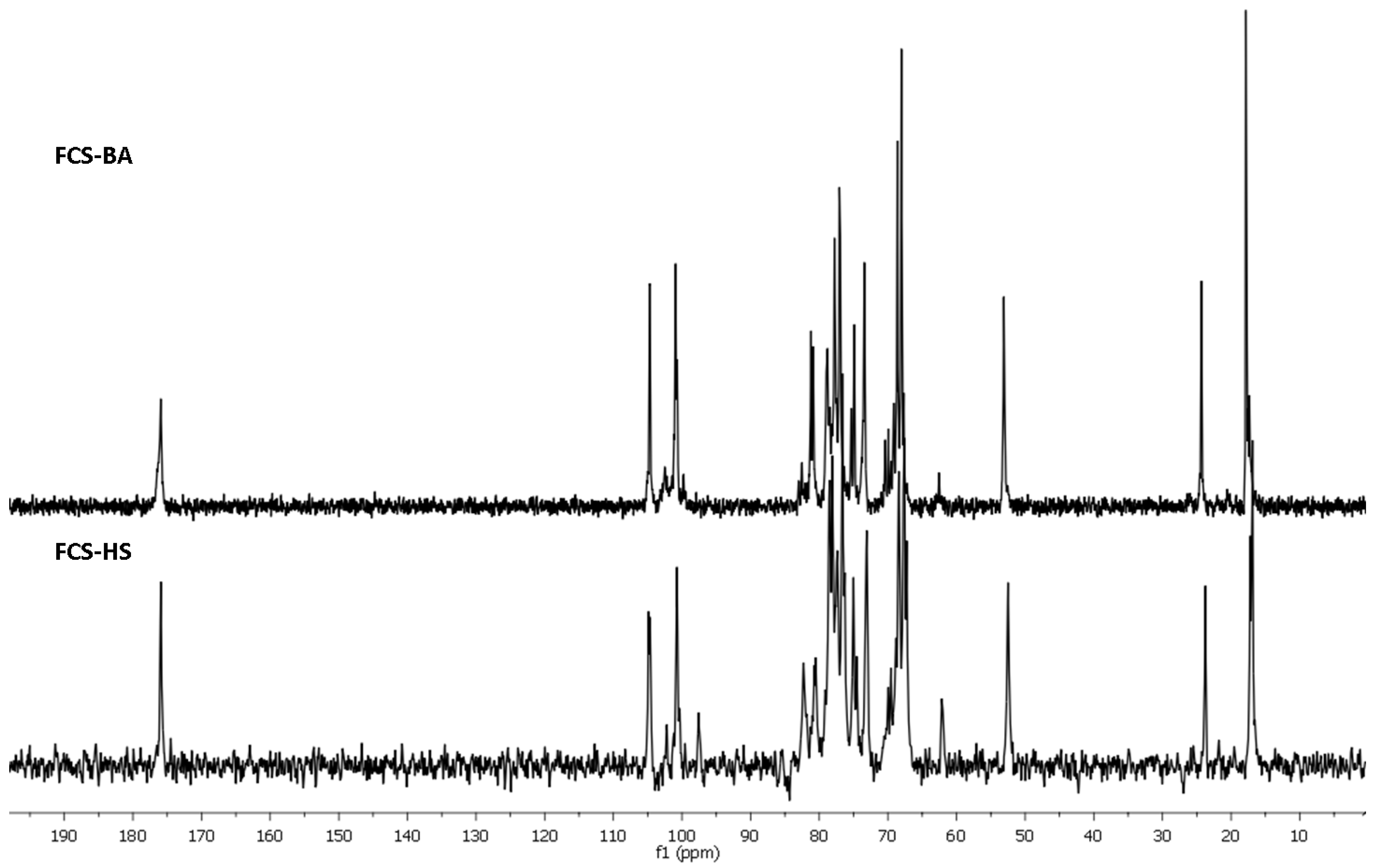
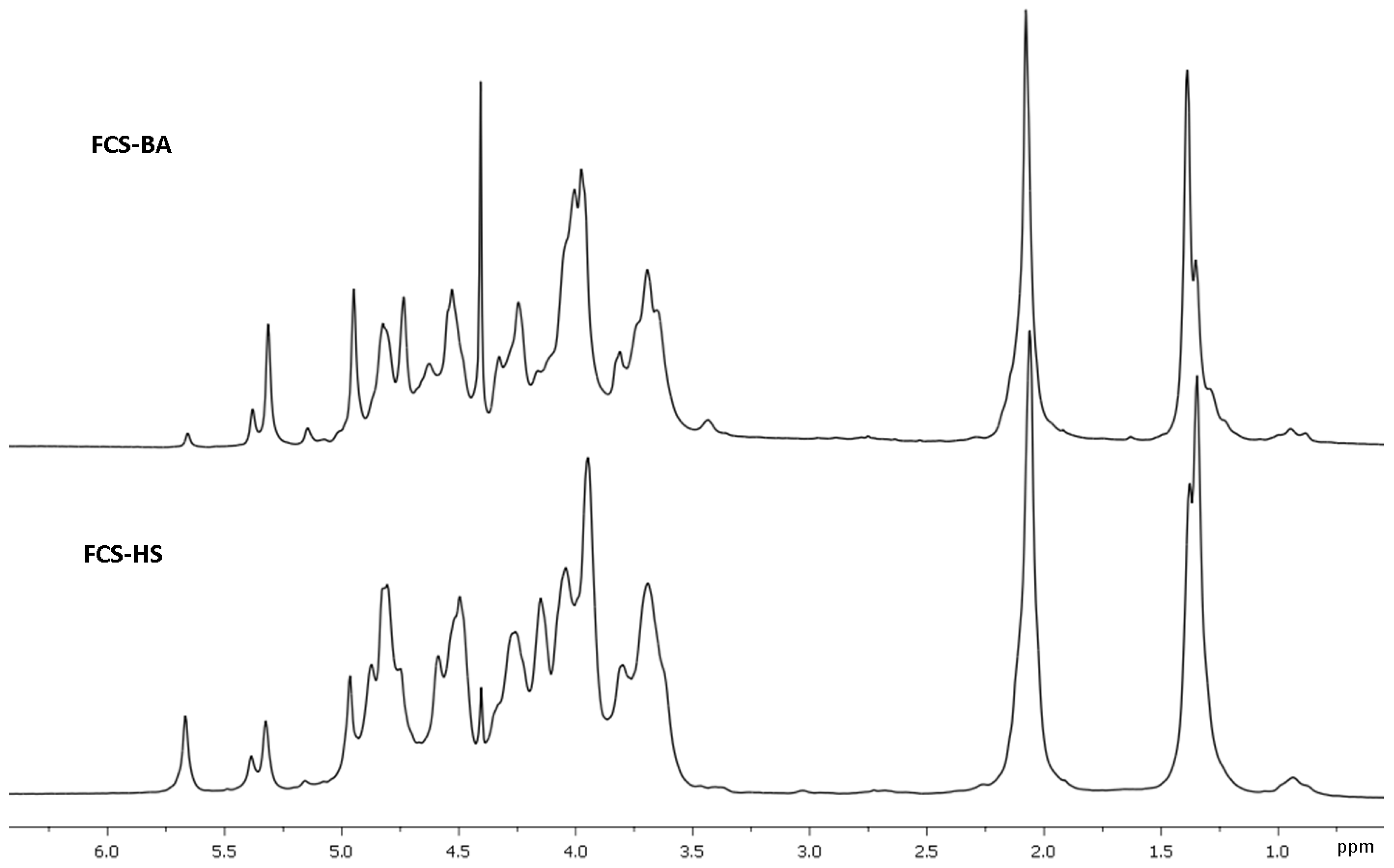
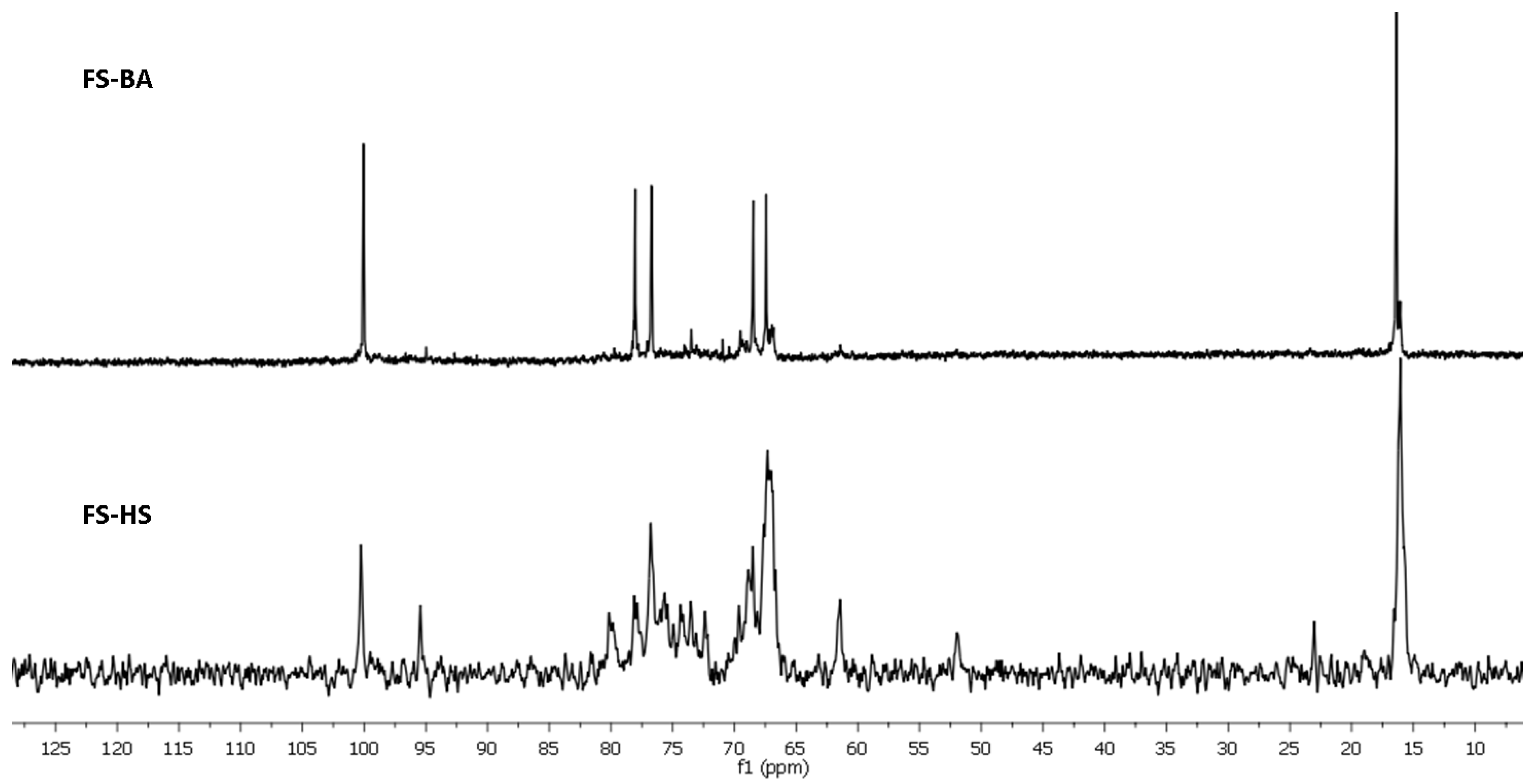
3.5. NMR Spectroscopy

The NMR spectra were recorded using the facilities of Zelinsky Institute Shared Center. Sample preparation and the conditions of experiments were described previously [50].

3.6. Anticoagulant Activity Measured in Clotting Time Test

The activated partial thromboplastin time assay for samples FCS-BA, FCS-HS, FS-BA and enoxaparin (Clexane®, Sanofi, Paris, France) was performed using Coatron®M2 coagulation analyzer (TECO, Munich, Germany) as described previously [4,51]. Briefly, a solution (10 µL) with concentration of 75 μg/mL, 37.5 μg/mL or 19.0 μg/mL of a polysaccharide sample (FCS-BA, FCS-HS, FS-BA, or enoxaparin) in purified water was added to 50 µL of normal plasma (Cormay, Poland). The mixture was incubated for 2 min at 37 °C, then 50 µL of APTT reagent (Cormay, Poland) was added, and the mixture was incubated again for 3 min at 37 °C. Then 50 µL of CaCl2 solution (0.025M) was added, and the time of clot formation was recorded. Purified water instead of a saccharide solution was used as a control.

3.7. Effect of Polysaccharides on Thrombin or Factor Xa Inactivation by Antithrombin III

Both experiments were carried out for samples FCS-BA, FCS-HS, and enoxaparin (Clexane®, Sanofi) at 37 °C in a 96-well plate using MultiscanGo (Thermo, Stockholm, Sweden) as described previously [15]. Briefly, a solution of polysaccharide sample (FCS-BA, FCS-HS, or enoxaparin) (20 µL) with concentrations of 500, 50, 5, 0.5, 0.05 μg/mL in Tris-HCl buffer (0.15 µM, pH 8.4) was added to 50 µL of a solution of ATIII (0.2 U/mL, Renam, Moscow, Russia) in Tris-HCl buffer. After 3 min incubation an aqueous solution of thrombin (50 µL, 20 U/mL, Renam, Russia) was added, and the mixture was incubated for 2 min. Then a chromogenic substrate (50 µL, 2 mM, Renam, Russia) was added, and the mixture was kept for 2 min. Absorbance of p-nitroaniline (405 nm) was measured.
To 50 µL of a solution of ATIII (0.5 U/mL) in Tris-HCl buffer a solution of a polysaccharide sample (FCS-BA, FCS-HS or enoxaparin) (20 µL) with concentrations of 500, 50, 5, 0.5, 0.05 μg/mL in Tris-HCl buffer was added. After 3 min incubation an aqueous solution of factor Xa (50 µL, 2 U/mL, Renam, Russia) was added, and the mixture was incubated for 2 min. Then a chromogenic substrate (50 µL, 2 mM, Renam, Russia) was added, and the mixture was kept for 2 min. Absorbance of p-nitroaniline (405 nm) was measured.

3.8. Statistical Analysis

All biological experiments were performed in quadruplicate (n = 4). The results are presented as Mean ± SD. Statistical significance was determined with Student’s t test. The p values less than 0.05 were considered as significant.

4. Conclusions

Fucosylated chondroitin sulfate FCS-BA and fucan sulfate FS-BA were isolated from the sea cucumber Bohadschia argus. Similar preparations FCS-HS and FS-HS were obtained from Holothuria (Theelothuria) spinifera. The main components of both FCSs were GlcA, GalNAc, Fuc and sulfate. Based on the data of NMR spectroscopy, both polysaccharides were shown to contain chondroitin core [→3)-β-d-GalNAc-(1→4)-β-d-GlcA-(1→]n bearing sulfated fucosyl branches at O-3 of every GlcA residue in the chain. These fucosyl residues were different in pattern of sulfation: FCS-BA contained Fuc2S4S, Fuc3S4S and Fuc4S in a ratio of 1:8:2, while FCS-HS included Fuc2S4S, Fuc3S4S and Fuc4S in a ratio of 2:2:1. Moreover, the polysaccharides differ also in GalNAc4S6S and GalNAc4S units content, the ratios being 14:1 for FCS-BA and 4:1 for FCS-HS. Both polysaccharides demonstrated significant anticoagulant activity in clotting time assay. This activity is probably connected with the ability of these FCSs to potentiate inhibition of thrombin by formation of ternary complex with thrombin and antithrombin III. Such complexation was confirmed previously by computer docking experiments [44]. At the same time activity of these FCSs as inhibitors of Xa was rather low. Fucan sulfate FS-BA was shown to be a linear polymer of 4-linked α-L-fucopyranose 3-sulfate, structure being confirmed by NMR spectra of the native polysaccharide and its desulfated derivative. It is interesting to mention that FS-BA, despite its rather substantial sulfate content, was practically devoid of anticoagulant activity. FS-HS had much more complex NMR spectra, and its structure was tentatively represented as a polysaccharide containing fragments which coincide with FS-BA, together with other fragments built up of randomly sulfated alternating 4- and 3-linked α-L-fucopyranose residues.

Supplementary Materials

The following are available online at https://www.mdpi.com/article/10.3390/md20060380/s1, Figure S1: Sea cucumbers (A) Bohadschia argus (Jaeger, 1833) and (B) Holothuria (Theelothuria) spinifera (Theel, 1886). Figure S2: Agarose gel electrophoresis of polysaccharides. Figure S3. The HSQC NMR spectrum of FCS-BA. Table S1. The data of the 1H and 13C NMR spectra (chemical shifts, ppm) of fucosylated chondroitin sulfates. Figure S4. The HSQC NMR spectrum of FS-BA-AT. Table S2. The data of 1H and 13C NMR spectra (chemical shifts, ppm) of sulfated fucan FS-BA-AT and its desulfated derivative FS-BA-AT-DS. Figure S5. The HSQC NMR spectrum of FS-HS-AT. Table S3. 13C and 1H chemical shifts (δ, ppm) of anomeric atoms in the NMR spectra of fucan sulfate FS-HS-AT (see Figure 6).

Author Contributions

Conceptualization, A.I.U., N.E.N., P.D.T., T.T.T.V. and N.E.U.; methodology, A.I.U., M.I.B. and N.E.U.; analysis, M.I.B., E.A.T., N.E.U., D.T.T., C.T.T.H., A.S.D. and A.S.S.; investigation, M.I.B., N.E.U., S.P.N., A.S.D., A.S.S., T.T.T.V. and A.I.U.; resources, D.T.T. and C.T.T.H.; writing—original draft preparation, N.E.U. and A.I.U.; writing—review and editing, all authors; supervision, A.I.U., N.E.N. and P.D.T. All authors have read and agreed to the published version of the manuscript.

Funding

This work was supported by the Russian Foundation for Basic Research, Russia (Project 21-53-54004 Viet_a) and the Vietnam Academy of Science and Technology (Grant number QTRU01.07/21-22).

Conflicts of Interest

The authors declare no conflict of interest. The funders had no role in the design of the study; in the collection, analyses, or interpretation of data; in the writing of the manuscript, or in the decision to publish the results.

References

  1. Pomin, V.H. Holothurian fucosylated chondroitin sulfates. Mar. Drugs 2014, 12, 232–254. [Google Scholar] [CrossRef] [PubMed] [Green Version]
  2. Ustyuzhanina, N.E.; Bilan, M.I.; Nifantiev, N.E.; Usov, A.I. New insight on the structural diversity of holothurian fucosylated chondroitin sulfates. Pure Appl. Chem. 2019, 91, 1065–1071. [Google Scholar] [CrossRef]
  3. Gong, P.-X.; Li, Q.-Y.; Wu, Y.-C.; Lu, W.-Y.; Zeng, J.; Li, H.-J. Structural elucidation and antidiabetic activity of fucosylated chondroitin sulfate from sea cucumber Stichopus japonicas. Carbohydr. Polym. 2021, 262, 117969. [Google Scholar] [CrossRef] [PubMed]
  4. Ustyuzhanina, N.E.; Bilan, M.I.; Dmitrenok, A.S.; Shashkov, A.S.; Kusaykin, M.I.; Stonik, V.A.; Nifantiev, N.E.; Usov, A.I. Structure and biological activity of a fucosylated chondroitin sulfate from the sea cucumber Cucumaria japonica. Glycobiology 2016, 26, 449–459. [Google Scholar] [CrossRef] [Green Version]
  5. Ustyuzhanina, N.E.; Bilan, M.I.; Dmitrenok, A.S.; Nifantiev, N.E.; Usov, A.I. Two fucosylated chondroitin sulfates from the sea cucumber Eupentacta fraudatrix. Carbohydr. Polym. 2017, 164, 8–12. [Google Scholar] [CrossRef]
  6. Santos, G.R.C.; Porto, A.C.O.; Soares, P.A.G.; Vilanova, E.; Mourão, P.A.S. Exploring the structure of fucosylated chondroitin sulfate through bottom-up nuclear magnetic resonance and electrospray ionization-high-resolution mass spectrometry approach. Glycobiology 2017, 27, 625–634. [Google Scholar] [CrossRef] [Green Version]
  7. Vieira, R.P.; Mourão, P.A.S. Occurrence of a unique fucose-branched chondroitin sulfate in the body wall of a sea cucumber. J. Biol. Chem. 1988, 263, 18176–18183. [Google Scholar] [CrossRef]
  8. Vieira, R.P.; Mulloy, B.; Mourão, P.A.S. Structure of a fucose-branched chondroitin sulfate from sea cucumber. Evidence for the presence of 3-O-sulfo-β-D-glucuronosyl residues. J. Biol. Chem. 1991, 266, 13530–13536. [Google Scholar] [CrossRef]
  9. Soares, P.A.G.; Ribeiro, K.A.; Valente, A.P.; Capillé, N.V.; Oliveira, S.-N.M.C.G.; Tovar, A.M.F.; Pereira, M.S.; Vilanova, E.; Mourão, P.A.S. A unique fucosylated chondroitin sulfate type II with strikingly homogeneous and neatly distributed α-fucose branches. Glycobiology 2018, 28, 565–579. [Google Scholar] [CrossRef] [PubMed]
  10. Yin, R.; Zhou, L.; Gao, N.; Lin, L.; Sun, H.; Chen, D.; Cai, Y.; Zuo, Z.; Hu, K.; Huang, S.; et al. Unveiling the disaccharide-branched glycosaminoglycan and anticoagulant potential of its derivatives. Biomacromolecules 2021, 22, 1244–1255. [Google Scholar] [CrossRef]
  11. Mao, H.; Cai, Y.; Li, S.; Sun, H.; Lin, L.; Pan, Y.; Yang, W.; He, Z.; Chen, R.; Zhou, L.; et al. A new fucosylated glycosaminoglycan containing disaccharide branches from Acaudina molpadioides: Unusual structure and anti-intrinsic tenase activity. Carbohydr. Polym. 2020, 245, 116503. [Google Scholar] [CrossRef]
  12. Li, S.; Zhong, W.; Pan, Y.; Lin, L.; Cai, Y.; Mao, H.; Zhang, T.; Li, S.; Chen, R.; Zhou, L.; et al. Structural characterization and anticoagulant analysis of the novel branched fucosylated glycosaminoglycan from the sea cucumber Holothuria nobilis. Carbohydr. Polym. 2021, 269, 118290. [Google Scholar] [CrossRef]
  13. Yang, J.; Wang, Y.; Jiang, T.; Lv, Z. Novel branch patterns and anticoagulant activity of glycosaminoglycan from sea cucumber Apostichopus japonicus. Int. J. Biol. Macromol. 2015, 72, 911–918. [Google Scholar] [CrossRef]
  14. Ustyuzhanina, N.E.; Bilan, M.I.; Dmitrenok, A.S.; Shashkov, A.S.; Nifantiev, N.E.; Usov, A.I. The structure of a fucosylated chondroitin sulfate from the sea cucumber Cucumaria frondosa. Carbohydr. Polym. 2017, 165, 7–12. [Google Scholar] [CrossRef]
  15. Ustyuzhanina, N.E.; Bilan, M.I.; Dmitrenok, A.S.; Silchenko, A.S.; Grebnev, B.B.; Stonik, V.A.; Nifantiev, N.E.; Usov, A.I. Fucosylated chondroitin sulfates from the sea cucumbers Paracaudina chilensis and Holothuria hilla: Structures and anticoagulant activity. Mar. Drugs 2020, 18, 540. [Google Scholar] [CrossRef]
  16. Li, H.; Yuan, Q.; Lv, K.; Ma, H.; Gao, C.; Liu, Y.; Zhang, S.; Zhao, L. Low-molecular-weight fucosylated glycosaminoglycan and its oligosaccharides from sea cucumber as novel anticoagulants: A review. Carbohydr. Polym. 2021, 251, 117034. [Google Scholar] [CrossRef]
  17. Lu, W.; Yang, Z.; Chen, J.; Wang, D.; Zhang, Y. Recent advances in antiviral activities and potential mechanisms of sulfated polysaccharides. Carbohydr. Polym. 2021, 272, 118526. [Google Scholar] [CrossRef]
  18. Fonseca, R.J.C.; Mourão, P.A.S. Pharmacological activities of sulfated fucose-rich po0lysaccharides after oral administration: Perspectives for the development of new carbohydrate-based drugs. Mar. Drugs 2021, 19, 425. [Google Scholar] [CrossRef]
  19. Xu, H.; Zhou, Q.; Liu, B.; Chen, F.; Wang, M. Holothurian fucosylated chondroitin sulfates and their potential benefits for human health: Structures and biological activities. Carbohydr. Polym. 2022, 275, 118691. [Google Scholar] [CrossRef]
  20. Pereira, M.S.; Mulloy, D.; Mourão, P.A.S. Structure and anticoagulant activity of sulfated fucans. Comparison between the regular, repetitive, and linear fucans from echinoderms with the more heterogeneous and branched polymers from brown algae. J. Biol. Chem. 1999, 274, 7656–7667. [Google Scholar] [CrossRef] [Green Version]
  21. Ustyuzhanina, N.E.; Bilan, M.I.; Dmitrenok, A.S.; Borodina, E.Y.; Nifantiev, N.E.; Usov, A.I. A highly regular fucan sulfate from the sea cucumber Stichopus horrens. Carbohydr. Res. 2018, 456, 5–9. [Google Scholar] [CrossRef]
  22. Shang, S.; Mou, R.; Zhang, Z.; Gao, N.; Lin, L.; Li, Z.; Wu, M.; Zhao, J. Structural analysis and anticoagulant activities of three highly regular fucan sulfates as novel intrinsic factor Xase inhibitors. Carbohydr. Polym. 2018, 195, 257–266. [Google Scholar] [CrossRef]
  23. Li, X.; Li, S.; Liu, J.; Lin, L.; Sun, H.; Yang, W.; Cai, Y.; Gao, N.; Zhou, L.; Qin, H.; et al. A regular fucan sulfate from Stichopus herrmanni and its peroxide depolymerization: Structure and anticoagulant activity. Carbohydr. Polym. 2021, 256, 117513. [Google Scholar] [CrossRef] [PubMed]
  24. He, W.; Sun, H.; Su, L.; Zhou, D.; Zhang, X.; Shanggui, D.; Chen, Y. Structure and anticoagulant activity of a sulfated fucan from the sea cucumber Acaudina leucoprocta. Int. J. Biol. Macromol. 2020, 164, 87–94. [Google Scholar] [CrossRef]
  25. Chen, S.; Hu, Y.; Ye, X.; Li, G.; Yu, G.; Xue, C.; Chai, W. Sequence determination and anticoagulant and antithrombotic activities of a novel sulfated fucan isolated from the sea cucumber Isostichopus badionotus. Biochim. Biophys. Acta 2012, 1820, 989–1000. [Google Scholar] [CrossRef]
  26. Yu, L.; Ge, L.; Xue, C.; Chang, Y.; Zhang, C.; Xu, X.; Wang, Y. Structural study of fucoidan from sea cucumber Acaudina molpadioides: A fucoidan containing novel tetrafucose repeating unit. Food Chem. 2014, 142, 197–200. [Google Scholar] [CrossRef] [PubMed]
  27. Yu, L.; Xue, C.; Chang, Y.; Xu, X.; Ge, L.; Liu, G.; Wang, Y. Structure elucidation of fucoidan composed of a novel tetrafucose repeating unit from sea cucumber Thelenota ananas. Food Chem. 2014, 146, 113–119. [Google Scholar] [CrossRef]
  28. Hu, Y.; Li, S.; Li, J.; Ye, X.; Ding, T.; Liu, D.; Chen, J.; Ge, Z.; Chen, S. Identification of a highly sulfated fucoidan from sea cucumber Pearsonothuria graeffei with well-repeated tetrasaccharides units. Carbohydr. Polym. 2015, 134, 808–816. [Google Scholar] [CrossRef] [PubMed]
  29. Chang, Y.; Hu, Y.; Yu, L.; McClements, D.J.; Xu, X.; Liu, G.; Xue, C. Primary structure and chain conformation of fucoidan extracted from sea cucumber Holothuria tubulosa. Carbohydr. Polym. 2016, 136, 1091–1097. [Google Scholar] [CrossRef] [PubMed] [Green Version]
  30. Li, C.; Niu, Q.; Li, S.; Zhang, X.; Liu, C.; Cai, C.; Li, G.; Yu, G. Fucoidan from sea cucumber Holothuria polii: Structural elucidation and stimulation of hematopoietic activity. Int. J. Biol. Macromol. 2020, 154, 1123–1131. [Google Scholar] [CrossRef]
  31. Chen, G.; Yu, L.; Zhang, Y.; Chang, Y.; Liu, Y.; Shen, J.; Xue, C. Utilizing heterologously overexpressed endo-1,3-fucanase to investigate the structure of sulfated fucan from sea cucumber (Holothuria hilla). Carbohydr. Polym. 2021, 272, 118480. [Google Scholar] [CrossRef]
  32. Cai, Y.; Yang, W.; Yin, R.; Zhou, L.; Li, Z.; Wu, M.; Zhao, J. An anticoagulant fucan sulfate with hexasaccharide repeating units from the sea cucumber Holothuria albiventer. Carbohydr. Res. 2018, 464, 12–18. [Google Scholar] [CrossRef] [PubMed]
  33. An, Z.; Zhang, Z.; Zhang, X.; Yang, H.; Li, H.; Liu, M.; Shao, Y.; Zhao, X.; Zhang, H. Oligosaccharide mapping analysis by HILIC-ESI-HCD-MS/MS for structural elucidation of fucoidan from sea cucumber Holothuria floridana. Carbohydr. Polym. 2022, 275, 118694. [Google Scholar] [CrossRef]
  34. Wu, M.; Xu, L.; Zhao, L.; Xiao, C.; Gao, N.; Luo, L.; Yang, L.; Li, Z.; Chen, L.; Zhao, J. Structural analysis and anticoagulant activities of the novel sulfated fucan possessing a regular well-defined repeating unit from sea cucumber. Mar. Drugs 2015, 13, 2063–2084. [Google Scholar] [CrossRef] [Green Version]
  35. Yu, L.; Xue, C.; Chang, Y.; Hu, Y.; Xu, X.; Ge, L.; Liu, G. Structure and rheological characteristics of fucoidan from sea cucumber Apostichopus japonicus. Food Chem. 2015, 180, 71–76. [Google Scholar] [CrossRef]
  36. Gao, N.; Chen, R.; Mou, R.; Xiang, J.; Zhou, K.; Li, Z.; Zhao, J. Purification, structural characterization and anticoagulant activities of four sulfated polysaccharides from sea cucumber Holothuria fuscopunctata. Int. J. Biol. Macromol. 2020, 164, 3421–3428. [Google Scholar] [CrossRef] [PubMed]
  37. Zheng, W.; Zhou, L.; Lin, L.; Cai, Y.; Sun, H.; Zhao, L.; Gao, N.; Yin, R.; Zhao, J. Physicochemical characteristics and anticoagulant activities of the polysaccharides from sea cucumber Pattalus mollis. Mar. Drugs 2019, 17, 198. [Google Scholar] [CrossRef] [PubMed] [Green Version]
  38. Ma, Y.; Gao, N.; Zuo, Z.; Li, S.; Zheng, W.; Shi, X.; Liu, Q.; Ma, T.; Yin, R.; Li, X.; et al. Five distinct fucan sulfates from sea cucumber Pattalus mollis: Purification, structural characterization and anticoagulant activities. Int. J. Biol. Macromol. 2021, 186, 535–543. [Google Scholar] [CrossRef]
  39. Pham Duc, T.; Ly, B.M.; Usoltseva, R.V.; Shevchenko, N.M.; Rasin, A.B.; Anastyuk, S.D.; Malyarenko, O.S.; Zvyagintseva, T.N.; San, P.T.; Ermakova, S.P. A novel sulfated fucan from Vietnamese sea cucumber Stichopus variegatus: Isolation, structure and anticancer activity in vitro. Int. J. Biol. Macromol. 2018, 117, 1101–1109. [Google Scholar]
  40. Yin, R.; Zhou, L.; Gao, N.; Li, Z.; Zhao, L.; Shang, F.; Wu, M.; Zhao, J. Oligosaccharides from depolymerized fucosylated glycosaminoglycan: Structures and minimum size for intrinsic factor Xase complex inhibition. J. Biol. Chem. 2018, 293, 14089–14099. [Google Scholar] [CrossRef] [Green Version]
  41. Pomin, V.H. NMR structural determination of unique invertebrate glycosaminoglycans endowed with medical properties. Carbohydr. Res. 2015, 413, 41–50. [Google Scholar] [CrossRef]
  42. Ustyuzhanina, N.E.; Bilan, M.I.; Nifantiev, N.E.; Usov, A.I. Structural analysis of holothurian fucosylated chondroitin sulfates: Degradation versus non-destructive approach. Carbohydr. Res. 2019, 476, 6–11. [Google Scholar] [CrossRef]
  43. Mourão, P.A.S. Perspective on the use of sulfated polysaccharides from marine organisms as a source of new antithrombotic drugs. Mar. Drugs 2015, 13, 2770–2784. [Google Scholar] [CrossRef] [PubMed] [Green Version]
  44. Gerbst, A.G.; Ustyuzhanina, N.E.; Nifantiev, N.E. Computational study of the possible formation of the ternary complex between thrombin, antithrombin III and fucosylated chondroitin sulfates. Mendeleev Commun. 2015, 25, 420–421. [Google Scholar] [CrossRef]
  45. Bilan, M.I.; Grachev, A.A.; Ustuzhanina, N.E.; Shashkov, A.S.; Nifantiev, N.E.; Usov, A.I. Structure of a fucoidan from the brown seaweed Fucus evanescens C.Ag. Carbohydr. Res. 2002, 337, 719–730. [Google Scholar] [CrossRef]
  46. Bilan, M.I.; Zakharova, A.N.; Grachev, A.A.; Shashkov, A.S.; Nifantiev, N.E.; Usov, A.I. Polysaccharides of algae: 60. Fucoidan from the Pacific brown alga Analipus japonicus (Harv.) Winne (Ectocarpales, Scytosiphonaceae). Russ. J. Bioorg. Chem. 2007, 33, 38–46. [Google Scholar] [CrossRef] [PubMed]
  47. Usov, A.I.; Bilan, M.I.; Klochkova, N.G. Polysaccharides of algae: 48. Polysaccharide composition of several calcareous red algae: Isolation of alginate from Corallina pilulifera P. et R. (Rhodophyta, Corallinaceae). Bot. Mar. 1995, 38, 43–51. [Google Scholar] [CrossRef]
  48. Guo, X.; Condra, M.; Kimura, K.; Berth, G.; Dautzenberg, H.; Dubin, P.L. Determination of molecular weight of heparin by size exclusion chromatography with universal calibration. Anal. Biochem. 2003, 312, 33–39. [Google Scholar] [CrossRef] [Green Version]
  49. Dubois, M.; Gilles, K.A.; Hamilton, J.K.; Rebers, P.A.; Smith, F. Colorimetric method for determination of sugars and related substances. Anal Chem. 1956, 28, 350–356. [Google Scholar] [CrossRef]
  50. Ustyuzhanina, N.E.; Bilan, M.I.; Dmitrenok, A.S.; Tsvetkova, E.A.; Shashkov, A.S.; Stonik, V.A.; Nifantiev, N.E.; Usov, A.I. Structural characterization of fucosylated chondroitin sulfates from sea cucumbers Apostichopus japonicus and Actinopyga mauritiana. Carbohydr. Polym. 2016, 153, 399–405. [Google Scholar] [CrossRef]
  51. Ustyuzhanina, N.E.; Bilan, M.I.; Dmitrenok, A.S.; Borodina, E.Y.; Stonik, V.A.; Nifantiev, N.E.; Usov, A.I. A highly regular fucosylated chondroitin sulfate from the sea cucumber Massinium magnum: Structure and effects on coagulation. Carbohydr. Polym. 2017, 167, 20–26. [Google Scholar] [CrossRef] [PubMed]
Figure 1. The 13C NMR spectra of fucosylated chondroitin sulfates FCS-BA and FCS-HS.
Marinedrugs 20 00380 g001
Figure 2. Fragments of 1H NMR spectra of fucosylated chondroitin sulfates FCS-BA and FCS-HS.
Marinedrugs 20 00380 g002
Figure 3. Repeating blocks of fucosylated chondroitin sulfates FCS-BA and FCS-HS. Unit A bears Fuc2S4S (D), whereas unit A’ bears Fuc3S4S (E) or Fuc4S (F).
Marinedrugs 20 00380 g003
Figure 4. The 13C NMR spectra of sulfated fucans FS-BA and FS-HS.
Marinedrugs 20 00380 g004
Figure 5. The 1H NMR spectra of sulfated fucans FS-BA and FS-HS.
Marinedrugs 20 00380 g005
Figure 6. Repeating blocks of fucans FS-BA-AT and FS-HS-AT.
Marinedrugs 20 00380 g006
Figure 7. Anticoagulant activity of polysaccharides FCS-BA (blue), FCS-HS (red), FS-BA-AT (green) and enoxaparin (dotted line). (A) APTT assay, (B) Anti-IIa-activity in the presence of ATIII, (C) Anti-Xa-activity in the presence of ATIII. n = 4, p < 0.05.
Marinedrugs 20 00380 g007
Table 1. Characteristics of crude polysaccharide preparations SP-BA and SP-HS and the fractions obtained by their chromatography on DEAE-Sephacel and then used for structural analysis (composition in molar ratios relative to fucose).
SampleFucGalGlcNAcGalNAcUA, Na-saltSO3NaMolecular Weight, kDa (Dispercity)
SP-BA1.000.140.070.300.302.05
FCS-BA1.000.09n.d.0.780.834.0432 (1.55)
FS-BA-AT1.000.090.040.060.041.2855 (1.35)
SP-HS1.000.120.080.30.212.44
FCS-HS1.000.120.040.741.103.4430 (1.62)
FS-HS-AT1.000.090.040.180.181.69
Publisher’s Note: MDPI stays neutral with regard to jurisdictional claims in published maps and institutional affiliations.

