
The Anti-Inflammatory and Skin Barrier Function Recovery Effects of Schisandra chinensis in Mice with Atopic Dermatitis
 and
and 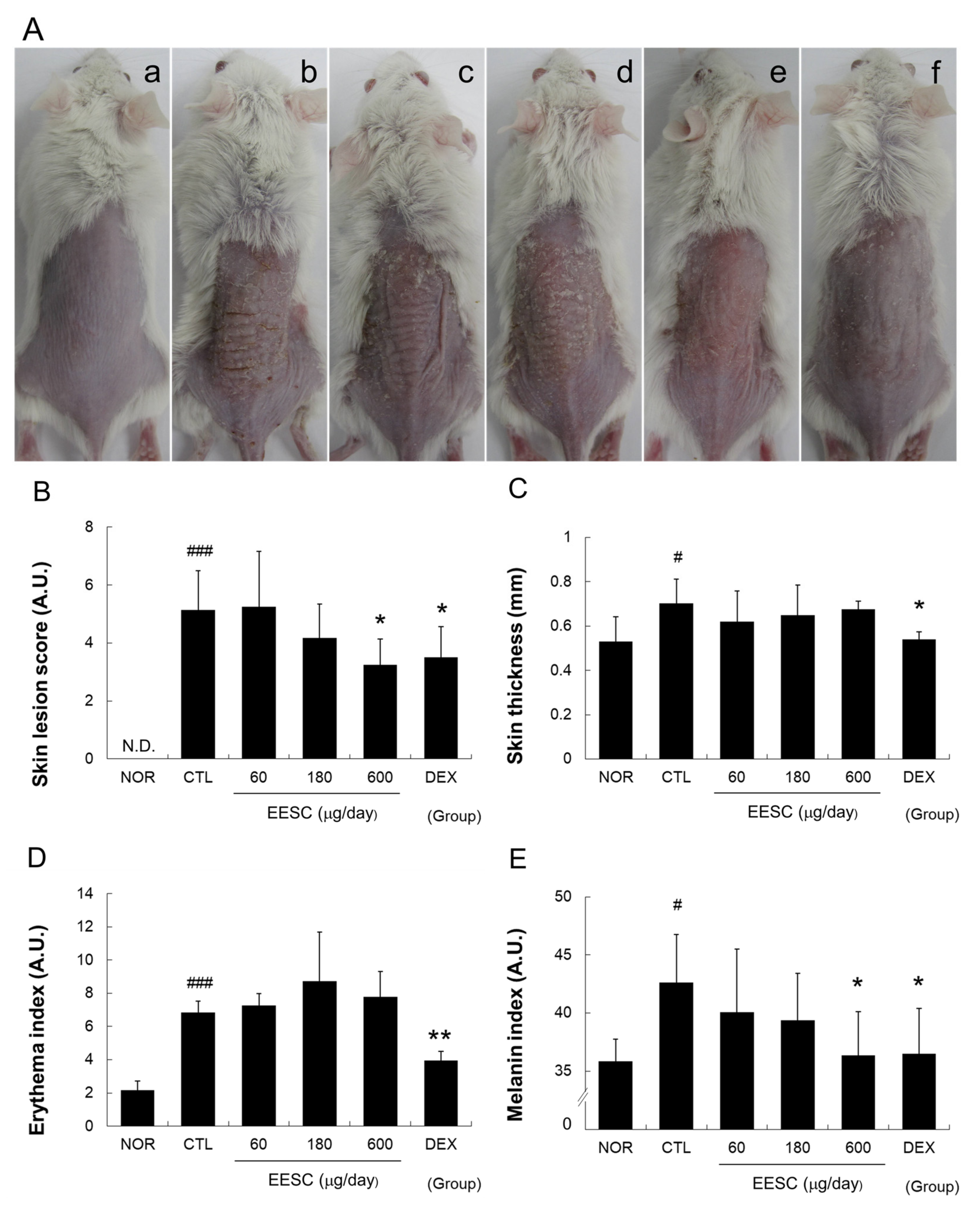
Abstract
:1. Introduction
2. Materials and Methods
2.1. Preparation of the Schisandra Chinensis Extract
2.2. Animals
2.3. Experimental Design and the Induction of AD
2.4. Assessment of Skin Lesion Severity
2.5. Skin Thicknesses and Color
2.6. Skin Water Content and Water-Holding Capacity
2.7. Histopathological Examination
2.8. Total RNA Isolation and Quantitative PCR
2.9. Cytokine Level Measurements
2.10. Body Weight and Spleen/Body Weight Ratio
2.11. Statistical Analysis
3. Results
3.1. EESC Relieved Skin Lesions and Reduced the Melanin Index
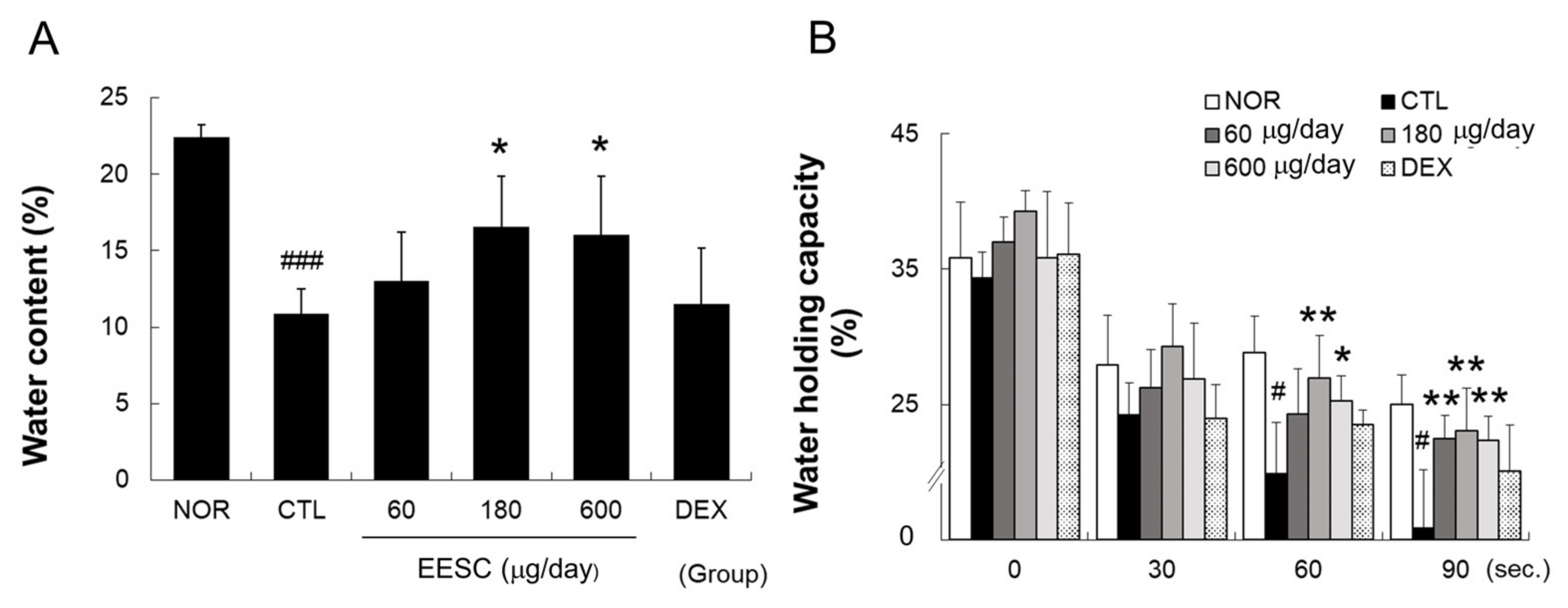
3.2. EESC Improved the Skin Water Content and Water-Holding Capacity
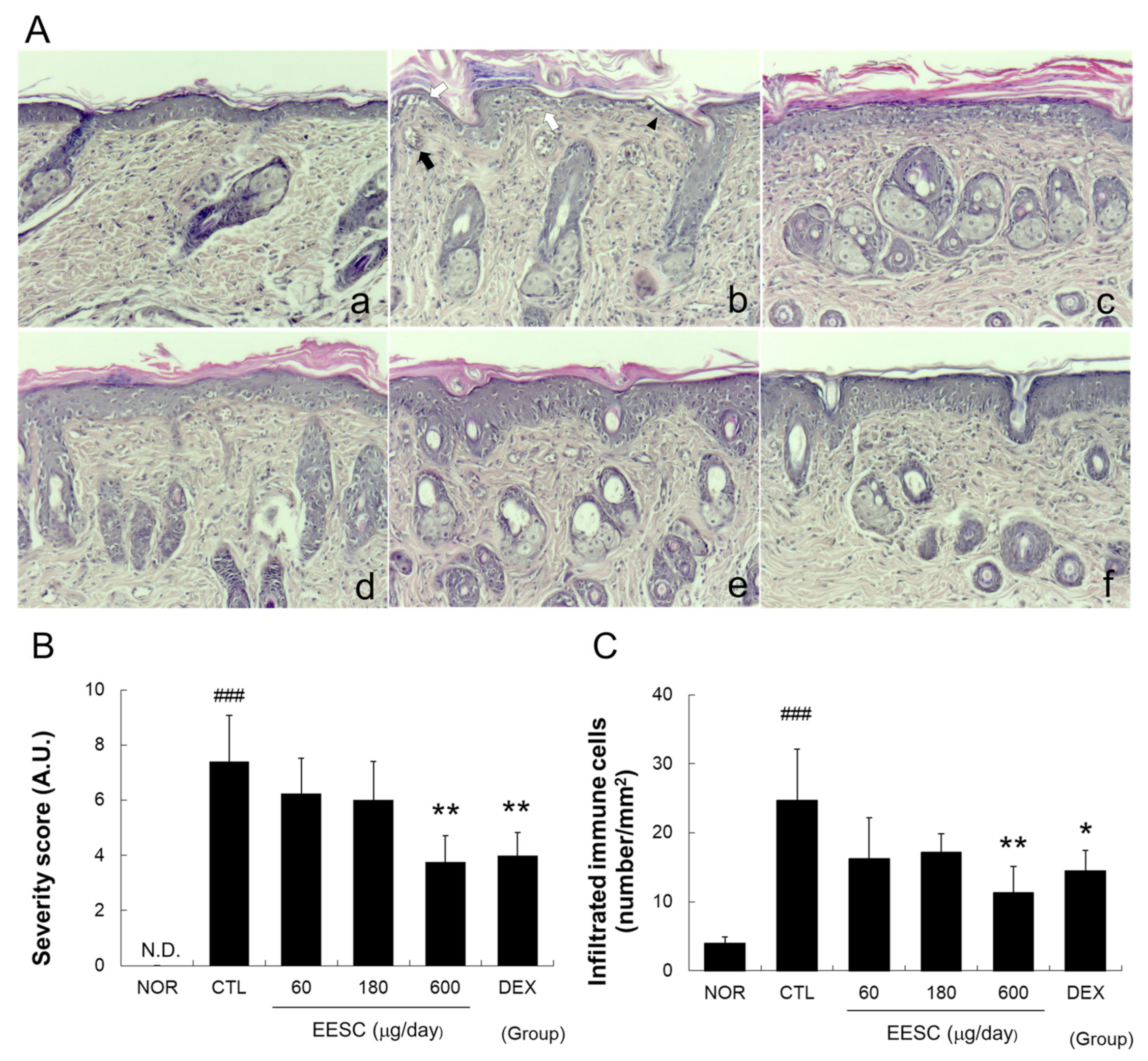
3.3. EESC Prevented Histopathological Abnormalities in Inflamed Tissue
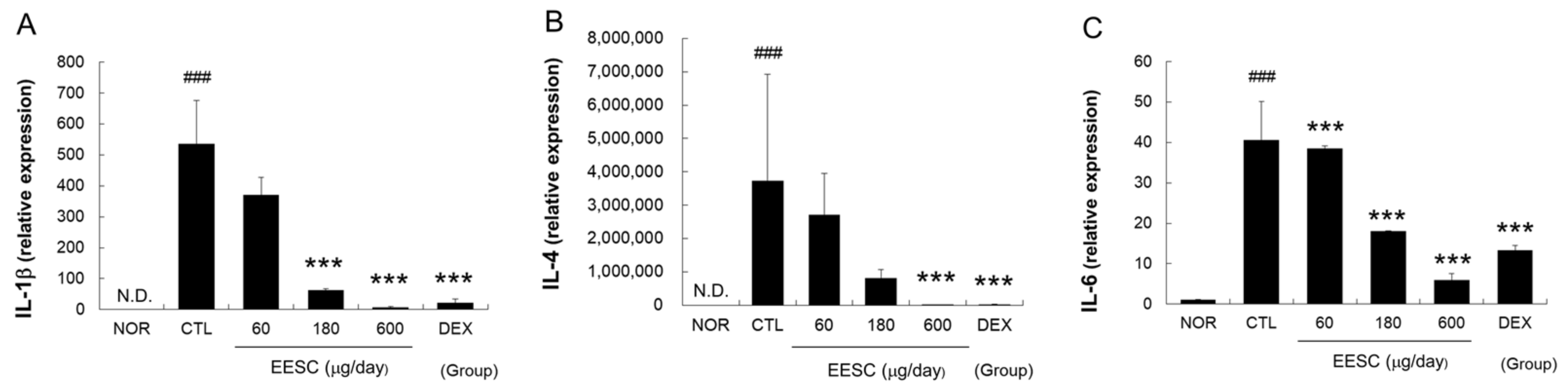
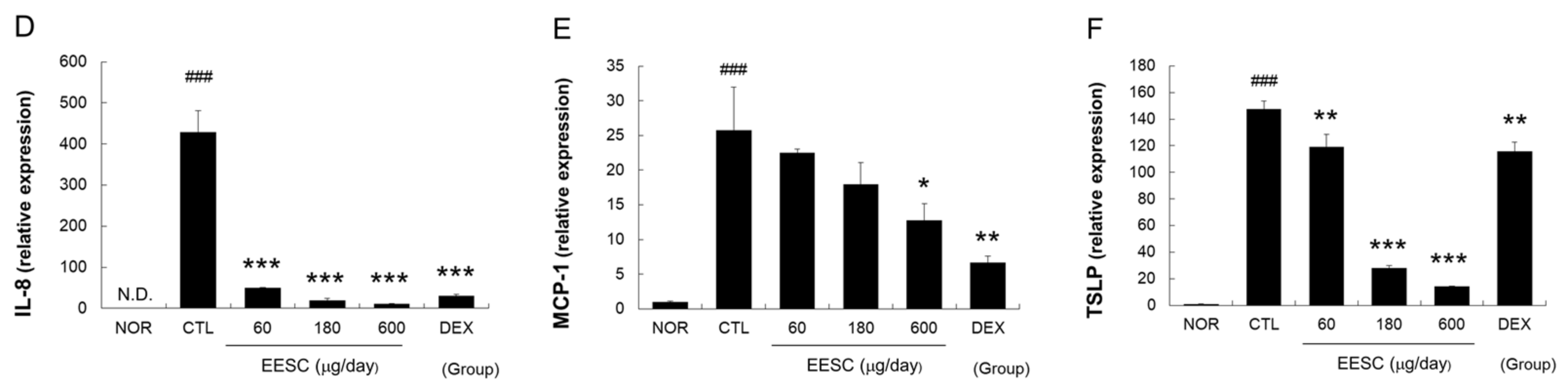
3.4. EESC Suppressed MC-903-Induced Increases in Cytokine and Chemokine mRNA Levels
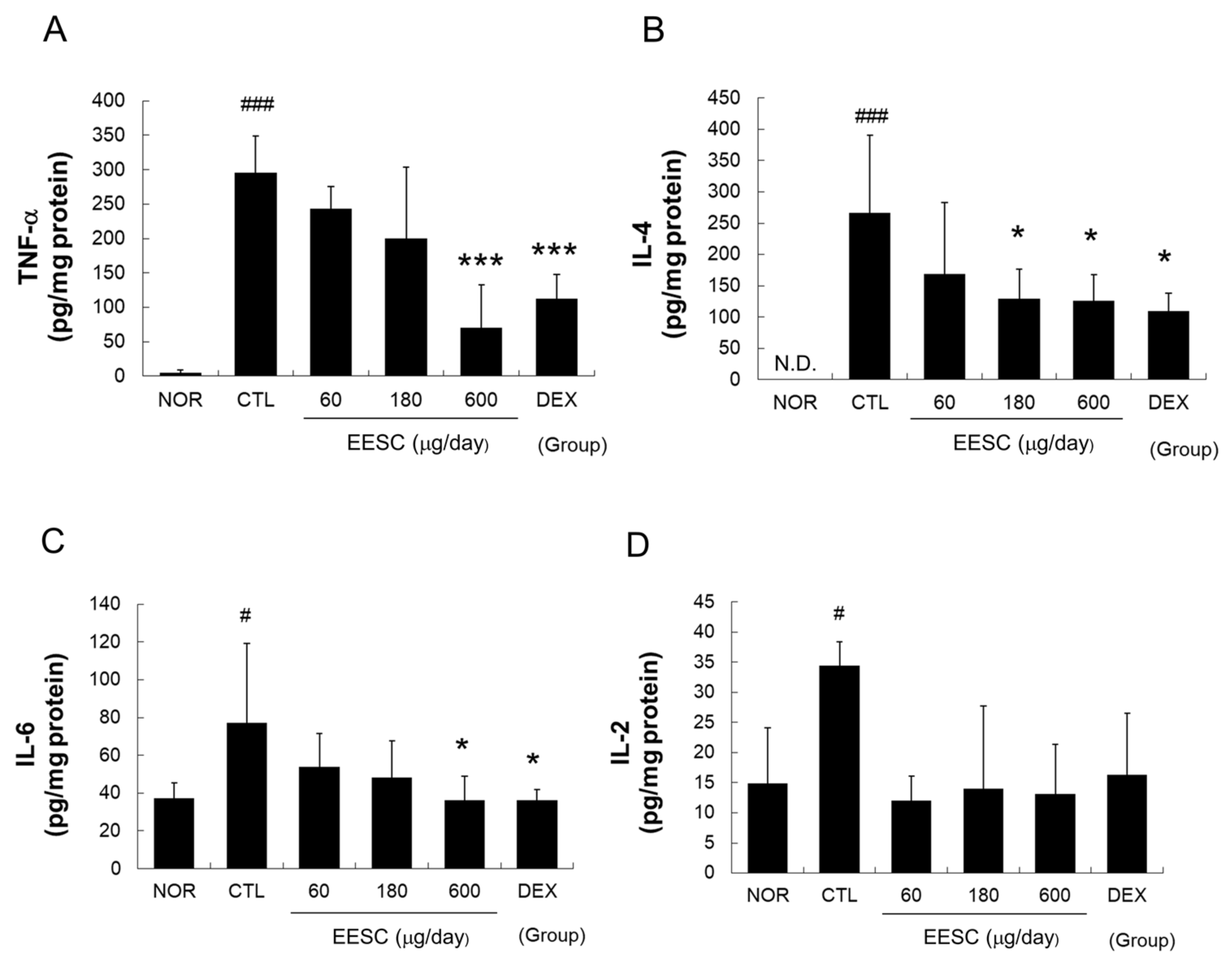
3.5. EESC Suppressed the MC-903-Induced Production of Tumor Necrosis Factor (TNF)-α, IL-4, and IL-6 in Inflamed Tissue
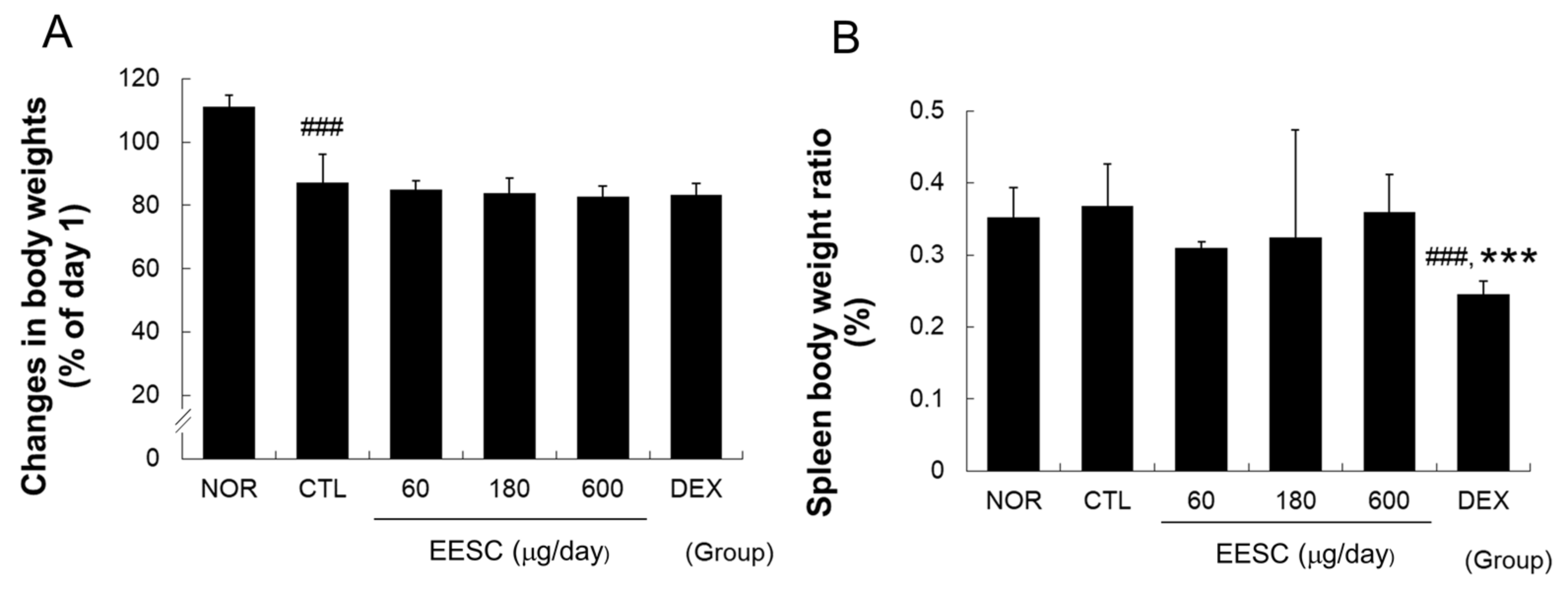
3.6. EESC Did Not Affect the Weight Gain or Spleen/Body Weight Ratio in AD Mice
4. Discussion
5. Conclusions
Supplementary Materials
Author Contributions
Funding
Institutional Review Board Statement
Informed Consent Statement
Data Availability Statement
Conflicts of Interest
References
- Kim, C.M.; Shin, M.K.; An, D.G.; Lee, K.S. The encyclopedia of oriental herbal medicine. Seoul Jeongdam. 1997, 1, 3058–3066. [Google Scholar]
- Kopustinskiene, D.M.; Bernatoniene, J. Antioxidant Effects of Schisandra chinensis Fruits and Their Active Constituents. Antioxidants 2021, 10, 620. [Google Scholar] [CrossRef] [PubMed]
- Zagórska-Dziok, M.; Wójciak, M.; Ziemlewska, A.; Nizioł-Łukaszewska, Z.; Hoian, U.; Klimczak, K.; Szczepanek, D.; Sowa, I. Evaluation of the Antioxidant, Cytoprotective and Antityrosinase Effects of Schisandra chinensis Extracts and Their Applicability in Skin Care Product. Molecules 2022, 27, 8877. [Google Scholar] [CrossRef]
- Choi, H.; Seo, E.; Yeon, M.; Kim, M.S.; Hur, H.J.; Oh, B.C.; Jun, H.S. Anti-Aging Effects of Schisandrae chinensis Fructus Extract: Improvement of Insulin Sensitivity and Muscle Function in Aged Mice. Evid. Based Complement. Altern. Med. 2019, 2019, 5642149. [Google Scholar] [CrossRef] [Green Version]
- Han, N.R.; Moon, P.D.; Kim, N.R.; Kim, H.Y.; Jeong, H.J.; Kim, H.M. Schisandra chinensis and Its Main Constituent Schizandrin Attenuate Allergic Reactions by Down-Regulating Caspase-1 in Ovalbumin-Sensitized Mice. Am. J. Chin. Med. 2017, 45, 159–172. [Google Scholar] [CrossRef]
- Kang, Y.S.; Han, M.H.; Hong, S.H.; Park, C.; Hwang, H.J.; Kim, B.W.; Kyoung, K.H.; Choi, Y.W.; Kim, C.M.; Choi, Y.H. Anti-inflammatory Effects of Schisandra chinensis (Turcz.) Baill Fruit Through the Inactivation of Nuclear Factor-κB and Mitogen-activated Protein Kinases Signaling Pathways in Lipopolysaccharide-stimulated Murine Macrophages. J. Cancer Prev. 2014, 19, 279–287. [Google Scholar] [CrossRef]
- Lee, J.E.; Choi, Y.W.; Im, D.S. Inhibitory effect of α-cubebenoate on atopic dermatitis-like symptoms by regulating Th2/Th1/Th17 balance in vivo. J. Ethnopharmacol. 2022, 291, 115162. [Google Scholar] [CrossRef]
- Lee, K.P.; Kang, S.; Park, S.J.; Kim, J.M.; Lee, J.M.; Lee, A.Y.; Chung, H.Y.; Choi, Y.W.; Lee, Y.G.; Im, D.S. Anti-allergic effect of α-cubebenoate isolated from Schisandra chinensis using in vivo and in vitro experiments. J. Ethnopharmacol. 2015, 173, 361–369. [Google Scholar] [CrossRef]
- Kang, J.; Lee, S.; Kim, N.; Dhakal, H.; Kwon, T.K.; Kim, E.N.; Jeong, G.S.; Kim, S.H. Gomisin M2 Ameliorates Atopic Dermatitis-like Skin Lesions via Inhibition of STAT1 and NF-κB Activation in 2,4-Dinitrochlorobenzene/Dermatophagoides farinae Extract-Induced BALB/c Mice. Molecules 2021, 26, 4409. [Google Scholar] [CrossRef] [PubMed]
- Hanifin, J.M.; Cooper, K.D.; Ho, V.C.; Kang, S.; Krafchik, B.R.; Margolis, D.J.; Schachner, L.A.; Sidbury, R.; Whitmore, S.E.; Sieck, C.K.; et al. Guidelines of care for atopic dermatitis. Br. J. Dermatol. 2004, 50, 391–404. [Google Scholar] [CrossRef]
- Frazier, W.; Bhardwaj, N. Atopic Dermatitis: Diagnosis and Treatment. Am. Fam. Physician 2020, 101, 590–598. [Google Scholar] [PubMed]
- Wen, M.C.; Wei, C.H.; Hu, Z.Q.; Srivastava, K.; Ko, J.; Xi, S.T.; Mu, D.Z.; Du, J.B.; Li, G.H.; Wallenstein, S.; et al. Efficacy and tolerability of anti-asthma herbal medicine intervention in adult patients with moderate-severe allergic asthma. J. Allergy Clin. Immunol. 2005, 116, 517–524. [Google Scholar] [CrossRef] [PubMed]
- Egawa, G.; Kabashima, K. Multifactorial skin barrier deficiency and atopic dermatitis: Essential topics to prevent the atopic march. J. Allergy Clin. Immunol. 2016, 138, 350–358. [Google Scholar] [CrossRef] [PubMed] [Green Version]
- Kim, J.; Kim, B.E.; Leung, D.Y. Pathophysiology of atopic dermatitis: Clinical implications. Allergy Asthma Proc. 2019, 40, 84–92. [Google Scholar] [CrossRef]
- David Boothe, W.; Tarbox, J.A.; Tarbox, M.B. Atopic Dermatitis: Pathophysiology. In Advances in Experimental Medicine and Biology; Springer: Berlin, Germany, 2017; Volume 1027, pp. 21–37. [Google Scholar] [CrossRef]
- Yosipovitch, G.; Papoiu, A.D.P. What causes itch in atopic dermatitis? Curr. Allergy Asthma Rep. 2008, 8, 306–311. [Google Scholar] [CrossRef]
- Yang, G.; Seok, J.K.; Kang, H.C.; Cho, Y.Y.; Lee, H.S.; Lee, J.Y. Skin Barrier Abnormalities and Immune Dysfunction in Atopic Dermatitis. Int. J. Mol. Sci. 2020, 21, 2867. [Google Scholar] [CrossRef] [Green Version]
- Jang, S.K.; Kang, Y.H.; Kang, Y.T.; Oh, S.Y.; Kim, S.Y.; Lyu, J.H.; Kim, H.Y. The ethanol extract of Caragana sinica ameliorated skin lesions in mice with contact dermatitis. Pharmacogn. Mag. 2022, 18, 201–206. [Google Scholar] [CrossRef]
- Moosbrugger-Martinz, V.; Schmuth, M.; Dubrac, S. A Mouse Model for Atopic Dermatitis Using Topical Application of Vitamin D3 or of Its Analog MC903. Methods Protoc. 2017, 1559, 91–106. [Google Scholar] [CrossRef]
- Lyu, J.H.; Lee, G.S.; Kim, K.H.; Kim, H.W.; Cho, S.I.; Jeong, S.I.; Kim, H.J.; Ju, Y.S.; Kim, H.K.; Sadikot, R.T.; et al. Ent-kaur-16-en-19-oic Acid, isolated from the roots of Aralia continentalis, induces activation of Nrf2. J. Ethnopharmacol. 2011, 137, 1442–1449. [Google Scholar] [CrossRef] [Green Version]
- Li, M.; Hener, P.; Zhang, Z.; Kato, S.; Metzger, D.; Chambon, P. Topical vitamin D3 and low-calcemic analogs induce thymic stromal lymphopoietin in mouse keratinocytes and trigger an atopic dermatitis. Proc. Natl. Acad. Sci. USA 2006, 103, 11736–11741. [Google Scholar] [CrossRef]
- Robinson, M.; Visscher, M.; Laruffa, A.; Wickett, R. Natural moisturizing factors (NMF) in the stratum corneum (SC). I. Effects of lipid extraction and soaking. J. Cosmet. Sci. 2010, 61, 13–22. [Google Scholar] [PubMed]
- Shin, H.S.; Ahn, S.S.; Yeo, H.J.; Jeong, Y.J.; Shin, S.Y. Ethanolic Extracts of Ageratum houstonium, Bupleurum falcatum, and Schisandra chinensis Inhibit Interleukin-4-Induced Expression of Itching Factors in HaCaT Keratinocytes. J. Cosmet. Sci. 2021, 72, 580–599. [Google Scholar]
- Nagappan, A.; Jung, D.Y.; Kim, J.H.; Jung, M.H. Protective Effects of Gomisin N against Hepatic Cannabinoid Type 1 Receptor-Induced Insulin Resistance and Gluconeogenesis. Int. J. Mol. Sci. 2018, 19, 968. [Google Scholar] [CrossRef] [Green Version]
- Chae, J.K.; Subedi, L.; Jeong, M.; Park, Y.U.; Kim, C.Y.; Kim, H.; Kim, S.Y. Gomisin N Inhibits Melanogenesis through Regulating the PI3K/Akt and MAPK/ERK Signaling Pathways in Melanocytes. Int. J. Mol. Sci. 2017, 18, 471. [Google Scholar] [CrossRef] [PubMed] [Green Version]
- Hu, Y.; Zeng, H.; Huang, J.; Jiang, L.; Chen, J.; Zeng, Q. Traditional Asian Herbs in Skin Whitening: The Current Development and Limitations. Front. Pharmacol. 2020, 11, 982. [Google Scholar] [CrossRef] [PubMed]
- Eckert, E. Histopathological and Immunohistological Aspects of Atopic Eczema. In Handbook of Atopic Eczema; Springer: Berlin/Heidelberg, Germany, 1991; pp. 127–131. [Google Scholar] [CrossRef]
- Bernard, M.; Carrasco, C.; Laoubi, L.; Guiraud, B.; Rozières, A.; Goujon, C.; Duplan, H.; Bessou-Touya, S.; Nicolas, J.F.; Vocanson, M.; et al. IL-1β induces thymic stromal lymphopoietin and an atopic dermatitis-like phenotype in reconstructed healthy human epidermis. J. Pathol. 2017, 242, 234–245. [Google Scholar] [CrossRef] [PubMed]
- Engebretsen, K.A.; Thyssen, J.P. Skin Barrier Function and Allergens. In Current Problems in Dermatology; Karger: Basel, Switzerland, 2016; Volume 49, pp. 90–102. [Google Scholar] [CrossRef]
- Akdis, C.A.; Arkwright, P.D.; Brüggen, M.C.; Busse, W.; Gadina, M.; Guttman-Yassky, E.; Kabashima, K.; Mitamura, Y.; Vian, L.; Wu, J.; et al. Type 2 immunity in the skin and lungs. Allergy 2020, 75, 1582–1605. [Google Scholar] [CrossRef]
- Ilves, T.; Tiitu, V.; Suttle, M.M.; Saarinen, J.V.; Harvima, I.T. Epidermal Expression of Filaggrin/Profilaggrin Is Decreased in Atopic Dermatitis: Reverse Association with Mast Cell Tryptase and IL-6 but Not with Clinical Severity. Dermatitis 2015, 26, 260–267. [Google Scholar] [CrossRef]
- Sebastiani, S.; Albanesi, C.; De, P.O.; Puddu, P.; Cavani, A.; Girolomoni, G. The role of chemokines in allergic contact dermatitis. Arch. Dermatol. Res. 2002, 293, 552–559. [Google Scholar] [CrossRef]
- Groves, R.W.; Allen, M.H.; Ross, E.L.; Barker, J.N.; MacDonald, D.M. Tumour necrosis factor alpha is pro-inflammatory in normal human skin and modulates cutaneous adhesion molecule expression. Br. J. Dermatol. 1995, 132, 345–352. [Google Scholar] [CrossRef]
- Toshitani, A.; Ansel, J.C.; Chan, S.C.; Li, S.-H.; Hanifin, J.M. Increased interleukin 6 production by T cells derived from patients with atopic dermatitis. J. Investig. Dermatol. 1993, 100, 299–304. [Google Scholar] [CrossRef] [PubMed] [Green Version]
- Demehri, S.; Morimoto, M.; Holtzman, M.J.; Kopan, R. Skin-derived TSLP triggers progression from epidermal-barrier defects to asthma. PLoS Biol. 2009, 19, e1000067. [Google Scholar] [CrossRef]
- Kim, H.S.; Kim, J.S.; Sa, J.K.; Ryu, B.K.; Park, K.J.; Kim, J.; Ha, H.; Park, Y.; Shin, M.H.; Kim, J.; et al. Calcipotriol, a synthetic Vitamin D analog, promotes antitumor immunity via CD4+T-dependent CTL/NK cell activation. Biomed. Pharmacother. 2022, 154, 113553. [Google Scholar] [CrossRef]
- Mihály, J.; Gericke, J.; Lucas, R.; de Lera, A.R.; Alvarez, S.; Törőcsik, D.; Rühl, R. TSLP expression in the skin is mediated via RARγ-RXR pathways. Immunobiology 2016, 221, 161–165. [Google Scholar] [CrossRef] [PubMed] [Green Version]
- Kang, Y.H.; Shin, H.M. Inhibitory effects of Schizandra chinensis extract on atopic dermatitis in NC/Nga mice. Immunopharmacol. Immunotoxicol. 2012, 34, 292–298. [Google Scholar] [CrossRef] [PubMed]
- Lee, H.J.; Jo, S.; Ryu, J.; Jeong, H.S.; Lee, G.; Ryu, M.H.; Jung, M.H.; Kim, H.; Kim, B.J. Effects of Schisandra chinensis Turcz. fruit on contact dermatitis induced by dinitrofluorobenzene in mice. Mol. Med. Rep. 2015, 12, 2135–2139. [Google Scholar] [CrossRef] [PubMed] [Green Version]


Disclaimer/Publisher’s Note: The statements, opinions and data contained in all publications are solely those of the individual author(s) and contributor(s) and not of MDPI and/or the editor(s). MDPI and/or the editor(s) disclaim responsibility for any injury to people or property resulting from any ideas, methods, instructions or products referred to in the content. |
© 2023 by the authors. Licensee MDPI, Basel, Switzerland. This article is an open access article distributed under the terms and conditions of the Creative Commons Attribution (CC BY) license (https://creativecommons.org/licenses/by/4.0/).
Share and Cite
Son, Y.; Yang, W.; Park, S.; Yang, J.; Kim, S.; Lyu, J.-H.; Kim, H. The Anti-Inflammatory and Skin Barrier Function Recovery Effects of Schisandra chinensis in Mice with Atopic Dermatitis. Medicina 2023, 59, 1353. https://doi.org/10.3390/medicina59071353
Son Y, Yang W, Park S, Yang J, Kim S, Lyu J-H, Kim H. The Anti-Inflammatory and Skin Barrier Function Recovery Effects of Schisandra chinensis in Mice with Atopic Dermatitis. Medicina. 2023; 59(7):1353. https://doi.org/10.3390/medicina59071353
Chicago/Turabian StyleSon, Yoorae, Wonjin Yang, Sangjun Park, Jinkyu Yang, Soyeon Kim, Ji-Hyo Lyu, and Hyungwoo Kim. 2023. "The Anti-Inflammatory and Skin Barrier Function Recovery Effects of Schisandra chinensis in Mice with Atopic Dermatitis" Medicina 59, no. 7: 1353. https://doi.org/10.3390/medicina59071353