

Release of Monomers from Dental Composite Materials into Saliva and the Possibility of Reducing the Toxic Risk for the Patient

Abstract
:1. Introduction
2. Materials and Methods
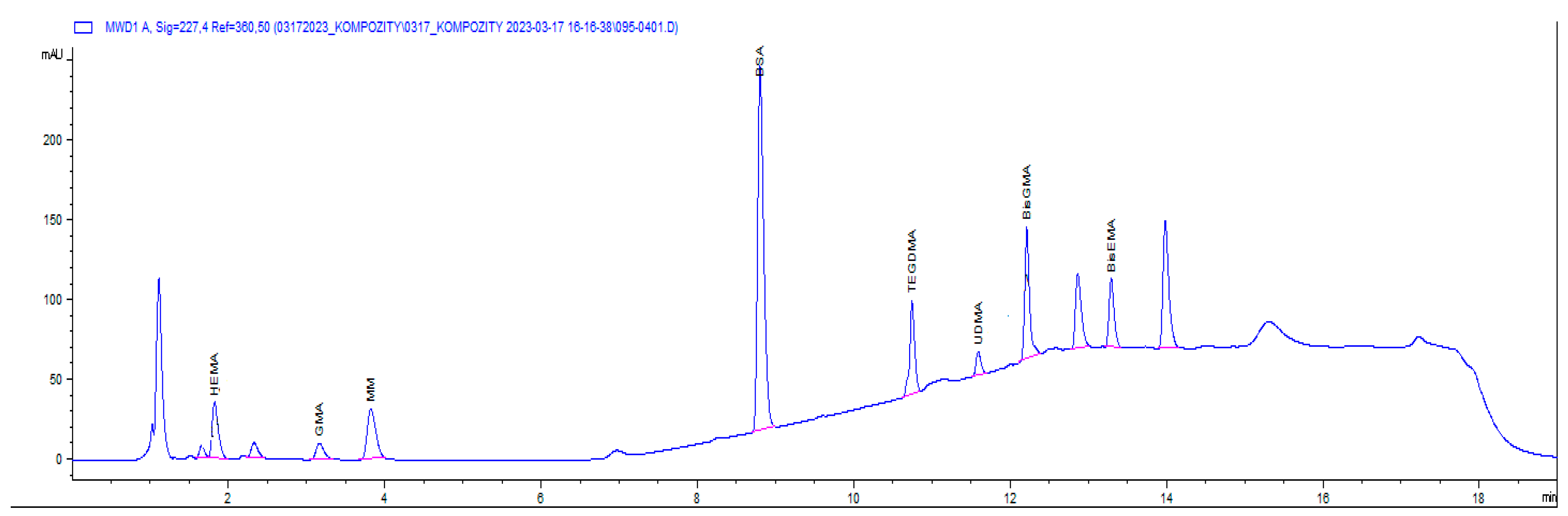
HPLC Conditions
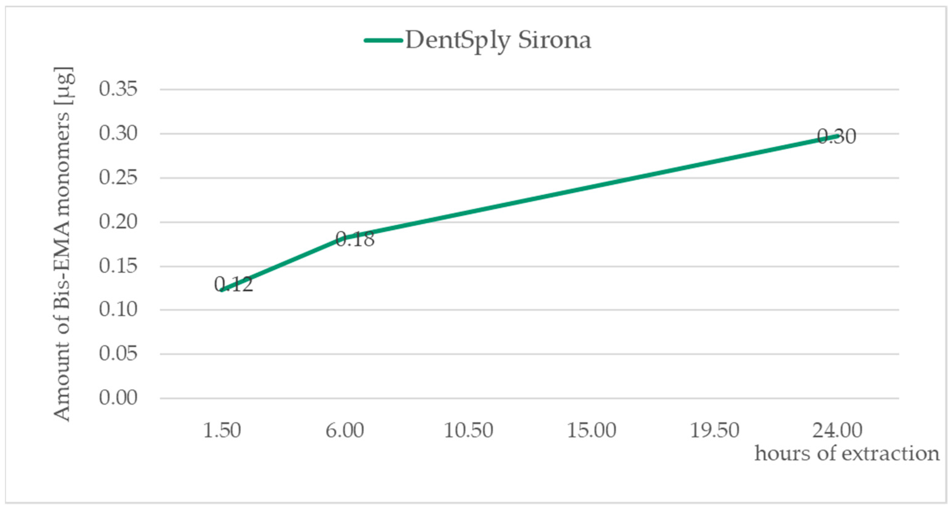
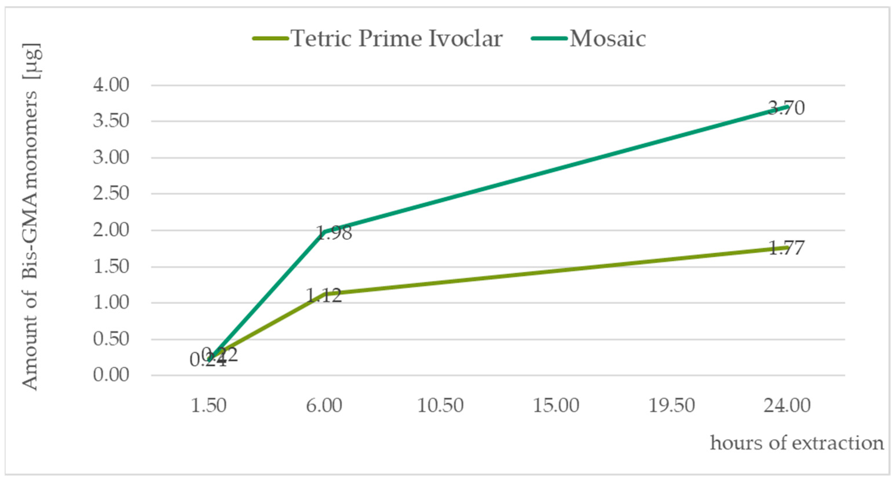
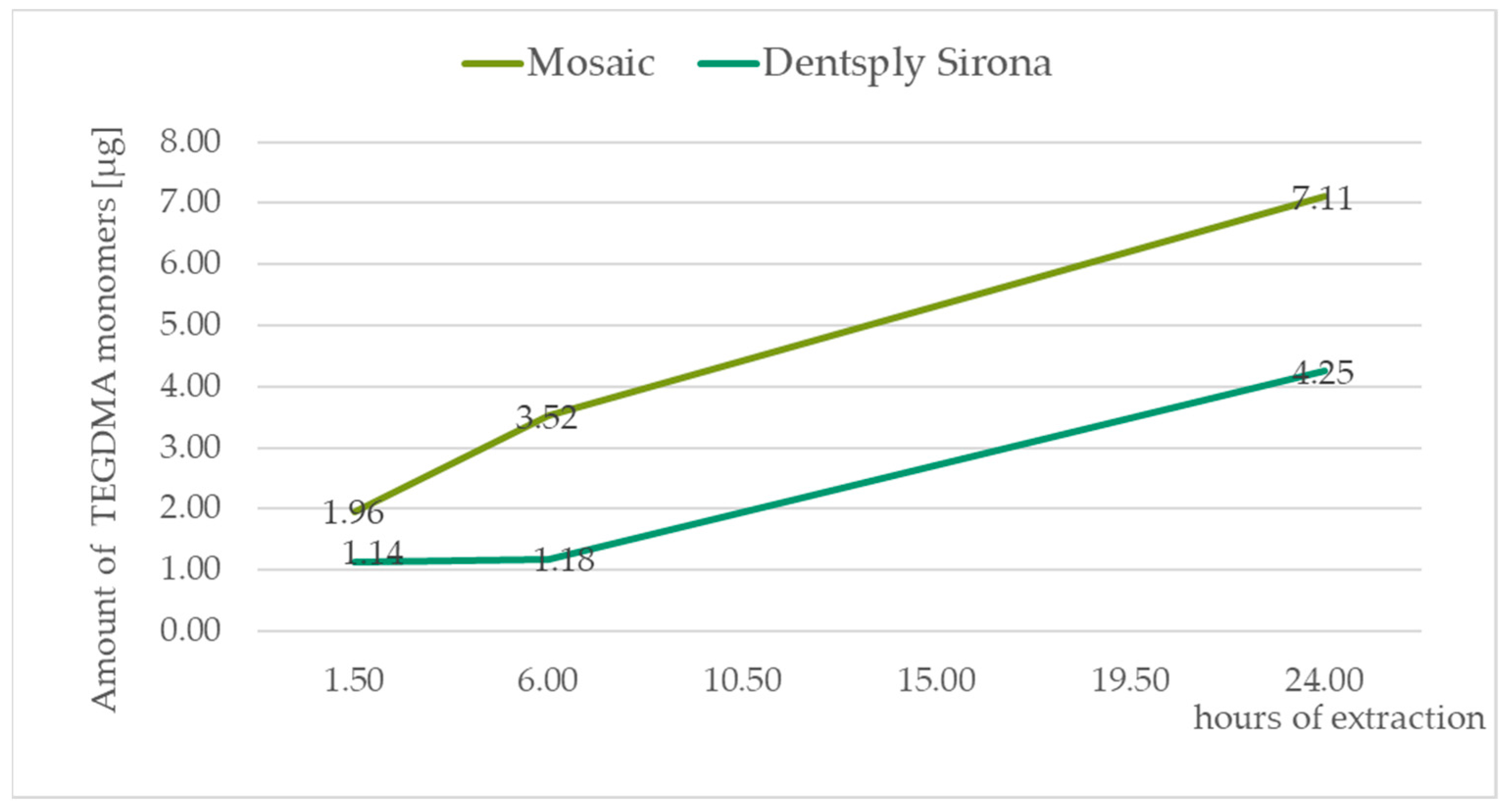
3. Results
4. Discussion
5. Conclusions
Author Contributions
Funding
Institutional Review Board Statement
Informed Consent Statement
Data Availability Statement
Acknowledgments
Conflicts of Interest
References
- Lempel, E.; Czibulya, Z.; Kunsági-Máté, S.; Szalma, J.; Sümegi, B.; Böddi, K. Quantification of Conversion Degree and Monomer Elution from Dental Composite Using HPLC and Micro-Raman Spectroscopy. Chromatographia 2014, 7, 1137–1144. [Google Scholar] [CrossRef]
- Lempel, E.; Czibulya, Z.; Kovács, B.; Szalma, J.; Tóth, Á.; Kunsági-Máté, S.; Varga, Z.; Böddi, K. Degree of Conversion and Bis-GMA, TEGDMA, UDMA Elution from Flowable Bulk Fill Composites. Int. J. Mol. Sci. 2016, 17, 732. [Google Scholar] [CrossRef] [PubMed] [Green Version]
- Gupta, S.K.; Saxena, P.; Pant, A.V.; Pant, A.B. Release and toxicity of dental resin composite. Toxicol. Int. 2012, 19, 225–234. [Google Scholar] [PubMed] [Green Version]
- Alizadehgharib, S.; Östberg, A.K.; Dahlstrand Rudin, A.; Dahlgren, U.; Christenson, K. The effects of the dental methacrylates TEGDMA, Bis-GMA, and UDMA on neutrophils in vitro. Clin. Exp. Dent. Res. 2020, 6, 439–447. [Google Scholar] [CrossRef] [PubMed]
- Reichl, F.X.; Esters, M.; Simon, S.; Seiss, M.; Kehe, K.; Kleinsasser, N. Cell death effects of resin-based dental material compounds and mercurials in human gingival fibroblasts. Arch. Toxicol. 2006, 80, 370–377. [Google Scholar] [CrossRef] [PubMed]
- Manojlovic, D.; Dramićanin, M.D.; Miletic, V.; Mitić-Ćulafić, D.; Jovanović, B.; Nikolić, B. Cytotoxicity and genotoxicity of a low-shrinkage monomer and monoacylphosphine oxide photoinitiator: Comparative analyses of individual toxicity and combination effects in mixtures. Dent. Mater. 2017, 33, 454–466. [Google Scholar] [CrossRef] [PubMed]
- Al-Hiyasat, A.S.; Darmani, H.; Milhem, M.M. Cytotoxicity evaluation of dental resin composites and their flowable derivatives. Clin. Oral. Invest. 2005, 9, 21–25. [Google Scholar] [CrossRef]
- Darmani, H.; Al-Hiyasat, A.S. The effect of Bis-GMA and TEG-DMA on female mouse fertility. Dent. Mat. 2006, 22, 353–358. [Google Scholar] [CrossRef]
- Geurtsen, W.; Leyhausen, G. Concise Review Biomaterials & Bioengineering: Chemical-Biological Interactions of the Resin Monomer Triethyleneglycol-dimethacrylate (TEGDMA). J. Dent. Res. 2001, 80, 2046–2050. [Google Scholar]
- Cornelio, R.B.; Wikant, A.; Mjøsund, H.; Kopperud, H.M.; Haasum, J.; Gedde, U.W.; Örtengren, U.T. The influence of bis-EMA vs bis GMA on the degree of conversion and water susceptibility of experimental composite materials. Acta Odontol. Scand. 2014, 72, 440–447. [Google Scholar] [CrossRef]
- Cebe, M.A.; Cebe, F.; Cengiz, M.F.; Cetin, A.R.; Arpag, O.F.; Ozturk, B. Elution of monomer from different bulk fill dental composite resins. Dent. Mater. 2015, 31, 141–149. [Google Scholar] [CrossRef]
- Ak, A.T.; Alpoz, A.R.; Bayraktar, O.; Ertugrul, F. Monomer Release from Resin Based Dental Materials Cured With LED and Halogen Lights. Eur. J. Dent. 2010, 4, 34–40. [Google Scholar] [CrossRef] [Green Version]
- Gerzinaab, T.M.; Humeab, W.R. Diffusion of monomers from bonding resin-resin composite combinations through dentine in vitro. J. Dent. 1996, 24, 125–128. [Google Scholar] [CrossRef]
- Ciucchi, B.; Bouillaguet, S.; Holz, J.; Pashley, D. Dentinal fluid dynamics in human teeth, in vivo. J. Endod. 1995, 21, 191–194. [Google Scholar] [CrossRef]
- Van Landuyt, K.L.; Nawrot, T.; Geebelen, B.; De Munck, J.; Snauwaert, J.; Yoshihara, K.; Scheers, H.; Godderis, L.; Hoet, P.; Van Meerbeek, B. How much do resin-based dental materials release? A meta-analytical approach. Dent. Mater. 2011, 27, 723–747. [Google Scholar] [CrossRef]
- The Swedish National Board of Health and Welfare: The National Register of Side-Effects of Dental Materials—Annual Report for 2001, [2002-125-6]; Socialstyrelsen: Stockholm, Sweden, 2002. (In Swedish)
- Tillberg, A.; Stenberg, B.; Berglund, A. Reactions to resin-based dental materials in patients-type, time to onset, duration, and consequence of the reaction. Contact Dermat. 2009, 61, 313–319. [Google Scholar] [CrossRef]
- Scully, C.; Beyli, M.; Ferreiro, M.C.; Ficarra, G.; Gill, Y.; Griffiths, M.; Holmstrup, P.; Mutlu, S.; Porter, S.; Wray, D. Update on oral lichen planus: Etiopathogenesis and management. Crit. Rev. Oral. Biol. Med. 1998, 9, 86–122. [Google Scholar] [CrossRef]
- Sorriento, D.; Iaccarino, G. Inflammation and Cardiovascular Diseases: The Most Recent Findings. Int. J. Mol. Sci. 2019, 20, 3879. [Google Scholar] [CrossRef] [Green Version]
- Sasaki, N.; Okuda, K.; Kato, T.; Kakishima, H.; Okuma, H.; Abe, K.; Tachino, H.; Tuchida, K.; Kubono, K. Salivary bisphenol-A levels detected by ELISA after restoration with composite resin. J. Mater. Sci. Mater. Med. 2005, 16, 297–300. [Google Scholar] [CrossRef]
- Rueggeberg, F.A.; Dlugokinski, M.; Ergle, J.W. Minimizing patients’ exposure to uncured components in a dental sealant. J. Am. Dent. Assoc. 1999, 130, 1751–1757. [Google Scholar] [CrossRef]
- Komurcuoglu, E.; Olmez, S.; Vural, N. Evaluation of residual monomer elimination methods in three different fissure sealants in vitro. J. OralRehabil 2005, 32, 116–121. [Google Scholar] [CrossRef] [PubMed]
- Koin, P.J.; Kilislioglu, A.; Zhou, M.; Drummond, J.L.; Hanley, L. Analysis of the degradation of a model dental composite. J. Dent. Res. 2008, 87, 661–665. [Google Scholar] [CrossRef] [PubMed] [Green Version]
- Shajii, L.; Santerre, J.P. Effect of filler content on the profile of released biodegradation products in micro-filled bis-GMA/TEGDMA dental composite resins. Biomaterials 1999, 20, 1897–1908. [Google Scholar] [CrossRef] [PubMed]
- Güzel, K.G.U.; Sönmez, I. Assessment of monomer release from 3 different fissure sealants. J. Appl. Biomater. Funct. Mater. 2018, 16, 90–96. [Google Scholar]


| Sample Number | Product Identifier | LOT Number | Manufacturer | Monitored Monomers |
|---|---|---|---|---|
| 1, 2, 3, 4 | Tetric Prime Ivoclar | Z00RW7 | Ivoclar Vivadent AG, Schaan, Liechtenstein | UDMA Bis-GMA |
| 5, 6, 7, 8 | Mosaic | BKMWT | Ultradent Products, Inc. South Jordan, UT, USA | TEGDMA |
| Bis-GMA | ||||
| 9, 10, 11,12 | DentSply Sirona | 2109000860 | DentSply Bensheim, Germany DeTrey GmbH Konstanz, Germany | TEGDMA Bis-EMA |
| 13,14,15,16 | 3M Filtek | NA52898 | 3M, | UDMA |
| Maplewood, MN, USA |
| time [min] | 0 | 1.5 | 10 | 15 | 15.5 | 20 |
| % MP B | 0 | 0 | 80 | 80 | 0 | 0 |
| Bis-GMA | Bis-EMA | TEGDMA | UDMA | |
|---|---|---|---|---|
| LOD [µg·mL−1] | 0.003 | 0.037 | 0.005 | 0.0134 |
| LOQ [µg·mL−1] | 0.0104 | 0.123 | 0.0166 | 0.044 |
| Sample | TEGDMA [µg] | SD [µg] | UDMA [µg] | SD [µg] | Bis-GMA [µg] | SD [µg] | Bis-EMA [µg] | SD [µg] |
|---|---|---|---|---|---|---|---|---|
| 1-1 | 9.087 | 0.282 | 0.244 | 0.006 | ||||
| 2-1 | 7.704 | 0.332 | 0.241 | 0.005 | ||||
| 3-1 | 8.991 | 0.371 | 0.243 | 0.005 | ||||
| 4-1 | 9.111 | 0.409 | 0.242 | 0.011 | ||||
| 5-1 | 1.684 | 0.004 | 0.195 | 0.007 | ||||
| 6-1 | 1.957 | 0.183 | 0.216 | 0.008 | ||||
| 7-1 | 1.200 | 0.024 | 0.148 | 0.012 | ||||
| 8-1 | 0.961 | 0.029 | 0.150 | 0.013 | ||||
| 9-1 | 1.297 | 0.026 | 0.109 | 0.005 | ||||
| 10-1 | 1.136 | 0.16 | 0.123 | 0.014 | ||||
| 11-1 | 1.497 | 0.058 | 0.063 | 0.002 | ||||
| 12-1 | 0.845 | 0.025 | 0.065 | 0.003 | ||||
| 13-1 | 2.965 | 0.118 | ||||||
| 14-1 | 2.606 | 0.071 | ||||||
| 15-1 | 2.229 | 0.124 | ||||||
| 16-1 | 1.859 | 0.065 | ||||||
| 1-2 | 9.067 | 0.618 | 0.885 | 0.008 | ||||
| 2-2 | 8.580 | 0.118 | 0.881 | 0.034 | ||||
| 3-2 | 8.514 | 0.011 | 0.884 | 0.017 | ||||
| 4-2 | 6.205 | 0.077 | 0.669 | 0.014 | ||||
| 5-2 | 1.916 | 0.012 | 1.777 | 0.003 | ||||
| 6-2 | 1.566 | 0.030 | 1.762 | 0.128 | ||||
| 7-2 | 2.610 | 0.064 | 1.706 | 0.029 | ||||
| 8-2 | 1.588 | 0.073 | 1.321 | 0.004 | ||||
| 9-2 | 0.061 | 0.001 | 0.068 | 0.020 | ||||
| 10-2 | 0.044 | 0.003 | 0.059 | 0.022 | ||||
| 11-2 | 0.024 | 0.003 | 0.058 | 0.017 | ||||
| 12-2 | 0.022 | 0.007 | 0.051 | 0.015 | ||||
| 13-2 | 2.439 | 0.041 | ||||||
| 14-2 | 2.372 | 0.004 | ||||||
| 15-2 | 1.881 | 0.078 | ||||||
| 16-2 | 1.562 | 0.047 | ||||||
| 1-3 | 7.120 | 0.697 | 0.661 | 0.012 | ||||
| 2-3 | 7.001 | 0.610 | 0.646 | 0.014 | ||||
| 3-3 | 6.087 | 0.544 | 0.624 | 0.077 | ||||
| 4-3 | 4.810 | 0.417 | 0.631 | 0.001 | ||||
| 5-3 | 3.312 | 0.342 | 1.533 | 0.115 | ||||
| 6-3 | 3.582 | 0.352 | 1.723 | 0.037 | ||||
| 7-3 | 4.022 | 0.303 | 1.210 | 0.132 | ||||
| 8-3 | 2.822 | 0.096 | 1.190 | 0.111 | ||||
| 9-3 | 2.048 | 0.301 | 0.144 | 0.024 | ||||
| 10-3 | 3.070 | 0.418 | 0.116 | 0.010 | ||||
| 11-3 | 1.219 | 0.058 | 0.075 | 0.000 | ||||
| 12-3 | 1.159 | 0.034 | 0.045 | 0.002 | ||||
| 13-3 | 1.234 | 0.002 | ||||||
| 14-3 | 0.976 | 0.030 | ||||||
| 15-3 | 0.870 | 0.060 | ||||||
| 16-3 | 0.746 | 0.006 |
Disclaimer/Publisher’s Note: The statements, opinions and data contained in all publications are solely those of the individual author(s) and contributor(s) and not of MDPI and/or the editor(s). MDPI and/or the editor(s) disclaim responsibility for any injury to people or property resulting from any ideas, methods, instructions or products referred to in the content. |
© 2023 by the authors. Licensee MDPI, Basel, Switzerland. This article is an open access article distributed under the terms and conditions of the Creative Commons Attribution (CC BY) license (https://creativecommons.org/licenses/by/4.0/).
Share and Cite
Tkáčiková, S.; Sabo, J. Release of Monomers from Dental Composite Materials into Saliva and the Possibility of Reducing the Toxic Risk for the Patient. Medicina 2023, 59, 1204. https://doi.org/10.3390/medicina59071204
Tkáčiková S, Sabo J. Release of Monomers from Dental Composite Materials into Saliva and the Possibility of Reducing the Toxic Risk for the Patient. Medicina. 2023; 59(7):1204. https://doi.org/10.3390/medicina59071204
Chicago/Turabian StyleTkáčiková, Soňa, and Ján Sabo. 2023. "Release of Monomers from Dental Composite Materials into Saliva and the Possibility of Reducing the Toxic Risk for the Patient" Medicina 59, no. 7: 1204. https://doi.org/10.3390/medicina59071204