

Evaluation of Clinical and Oral Findings in Patients with Epidermolysis bullosa
Abstract
:1. Introduction
2. Materials and Methods
3. Results
4. Discussion
5. Conclusions
- (1)
- The presence of extensive caries in individuals with DEB may result from delayed oral and dental rehabilitation due to physical disabilities, limitations and more pressing medical problems.
- (2)
- Microstomy, pain from mucosal lesions, and restricted access to the mouth can be caused by poor oral hygiene.
Author Contributions
Funding
Institutional Review Board Statement
Informed Consent Statement
Conflicts of Interest
References
- Fine, J.D. Inherited epidermolysis bullosa. Orphanet J. Rare Dis. 2010, 5, 12. [Google Scholar] [CrossRef] [PubMed] [Green Version]
- Bruckner, A.L.; Bedocs, L.A.; Keiser, E.; Tang, J.Y.; Doernbrack, C.; Arbuckle, H.A.; Berman, S.; Kent, K.; Bachrach, L.K. Correlates of low bone mass in children with generalized forms of epidermolysis bullosa. J. Am. Acad. Dermatol. 2011, 65, 1001–1009. [Google Scholar] [CrossRef]
- Kulali, F.; Özbakir, H.; Kundak, S.; Kalkanli, O.H.; Dilem, E.R.İ.Ş.; Çolak, R.; Çalkavur, Ş. Epidermolizis bülloza: Olgu serisi. Jinekoloji-Obstet. Ve Neonatoloji Tıp Derg. 2019, 16, 69–73. [Google Scholar]
- Bardhan, A.; Bruckner-Tuderman, L.; Chapple, I.L.; Fine, J.D.; Harper, N.; Has, C.; Heagerty, A.H. Epidermolysis bullosa. Nat. Rev. Dis. Primers. 2020, 6, 78. [Google Scholar] [CrossRef] [PubMed]
- Krämer, S.; Lucas, J.; Gamboa, F.; Diago, M.P.; Oltra, D.P.; Guzmán-Letelier, M.; Paul, S.; Molina, G.; Sepúlveda, L.; Araya, I.; et al. Clinical practice guidelines: Oral health care for children and adults living with epidermolysis bullosa. Spéc. Care Dent. 2020, 40, 3–81. [Google Scholar] [CrossRef]
- Bruckner, A.L.; Losow, M.; Wisk, J.; Patel, N.; Reha, A.; Lagast, H.; Gault, J.; Gershkowitz, J.; Kopelan, B.; Hund, M.; et al. The challenges of living with and managing epidermolysis bullosa: Insights from patients and caregivers. Orphanet J. Rare Dis. 2020, 15, 1. [Google Scholar] [CrossRef] [Green Version]
- Joseph, C.; Marty, M.; Dridi, S.M.; Verhaeghe, V.; Bailleul-Forestier, I.; Chiaverini, C.; Kémoun, P. Oral health status in patients with inherited epidermolysis bullosa: A comparative multicenter study. Quintessence Int. 2023, 54, 34–43. [Google Scholar]
- Wright, J.T. Oral Manifestations in the epidermolysis bullosa spectrum. Dermatol. Clin. 2010, 28, 159–164. [Google Scholar] [CrossRef] [Green Version]
- Simmer, J.P.; Hu, J.C.-C.; Hu, Y.; Zhang, S.; Liang, T.; Wang, S.-K.; Kim, J.-W.; Yamakoshi, Y.; Chun, Y.-H.; Bartlett, J.D.; et al. A genetic model for the secretory stage of dental enamel formation. J. Struct. Biol. 2021, 213, 107805. [Google Scholar] [CrossRef]
- Vahidnezhad, H.; Youssefian, L.; Zeinali, S.; Saeidian, A.H.; Sotoudeh, S.; Mozafari, N.; Abiri, M.; Kajbafzadeh, A.-M.; Barzegar, M.; Ertel, A.; et al. Dystrophic Epidermolysis Bullosa: COL7A1 Mutation Landscape in a Multi-Ethnic Cohort of 152 Extended Families with High Degree of Customary Consanguineous Marriages. J. Investig. Dermatol. 2017, 137, 660–669. [Google Scholar] [CrossRef] [Green Version]
- Gülşen, E.; Yavuz, İ. Epidermolizis Bülloza. HRU IJDOR 2021, 1, 19–30. [Google Scholar]
- Horn, H.; Tidman, M. The clinical spectrum of dystrophic epidermolysis bullosa. Br. J. Dermatol. 2002, 146, 267–274. [Google Scholar] [CrossRef] [PubMed]
- Manjunath, S.; Mahajan, R.; De, D.; Handa, S.; Attri, S.; Behera, B.N.; Bhasin, S.L.; Bolia, R. The severity of malnutrition in children with epidermolysis bullosa correlates with disease severity. Sci. Rep. 2021, 11, 16827. [Google Scholar] [CrossRef]
- Colomb, V.; Bourdon-Lannoy, E.; Lambe, C.; Sauvat, F.; Rabia, S.H.; Teillac, D.; De Prost, Y.; Bodemer, C. Nutritional outcome in children with severe generalized recessive dystrophic epidermolysis bullosa: A short- and long-term evaluation of gastrostomy and enteral feeding. Br. J. Dermatol. 2012, 166, 354–361. [Google Scholar] [CrossRef] [PubMed]
- Freeman, E.; Köglmeier, J.; Martinez, A.; Mellerio, J.; Haynes, L.; Sebire, N.; Lindley, K.; Shah, N. Gastrointestinal complications of epidermolysis bullosa in children. Br. J. Dermatol. 2008, 158, 1308–1314. [Google Scholar] [CrossRef]
- Liy-Wong, C.; Tarango, C.; Pope, E.; Coates, T.; Bruckner, A.L.; Feinstein, J.A.; Schwieger-Briel, A.; Hubbard, L.D.; Jane, C.; Torres-Pradilla, M.; et al. Consensus guidelines for diagnosis and management of anemia in epidermolysis bullosa. Orphanet J. Rare Dis. 2023, 18, 38. [Google Scholar] [CrossRef]
- Laimer, M.; Prodinger, C.; Bauer, J.W. Hereditary epidermolysis bullosa. J. Dtsch. Dermatol. Ges. 2015, 13, 1125–1133. [Google Scholar] [CrossRef] [Green Version]
- Chen, V.M.; Mehta, N.; Robbins, C.C.; Noh, E.; Pramil, V.; Duker, J.S.; Waheed, N.K. Anterior-segment spectral domain optical coherence tomography in epidermolysis bullosa. Ocul. Surf. 2020, 18, 912–919. [Google Scholar] [CrossRef]
- Figueira, E.C.; Murrell, D.F.; Coroneo, M.T. Ophthalmic Involvement in Inherited Epidermolysis Bullosa. Dermatol. Clin. 2010, 28, 143–152. [Google Scholar] [CrossRef]
- Bernardis, C.; Box, R. Surgery of the Hand in Recessive Dystrophic Epidermolysis Bullosa. Dermatol. Clin. 2010, 28, 335–341. [Google Scholar] [CrossRef]
- Tosti, A.; de Farias, D.C.; Murrell, D.F. Nail Involvement in Epidermolysis Bullosa. Dermatol. Clin. 2010, 28, 153–157. [Google Scholar] [CrossRef] [PubMed]
- Marinkovich, M.P.; Tang, J.Y. Gene Therapy for Epidermolysis Bullosa. J. Investig. Dermatol. 2019, 139, 1221–1226. [Google Scholar] [CrossRef] [Green Version]
- Niti, A.; Koliakos, G.; Michopoulou, A. Stem Cell Therapies for Epidermolysis Bullosa Treatment. Bioengineering 2023, 10, 422. [Google Scholar] [CrossRef]
- Arpag, O.F.; Arslanoglu, Z.; Altan, H.; Kale, E.; Bilgic, F. Epidermolysis bullosa in dentistry: Report of three cases and review of the literature. J. Int. Dent. Med. Res. 2015, 8, 133–139. [Google Scholar]
- Volovikov, O.; Velichko, E.; Razumova, S.; Said, O.B. The First Case Report about Noninvasive Impression Taking in Orthodontic Patient with Epidermolysis Bullosa. J. Int. Dent. Med. Res. 2021, 14, 1587–1591. [Google Scholar]
- Filho, G.A.N.; Caputo, B.V.; de Carvalhosa, A.A.; Costa, C.; Giovani, E.M. Dentistry approach of epidermolysis bullosa: Two case reports. J. Int. Dent. Med. Res. 2013, 6, 109–112. [Google Scholar]
- De Azevedo, B.L.R.; Roni, G.M.; Dettogni, R.S.; Torrelio, R.M.F.; Leal, L.F.; da Gama-de-Souza, L.N. Epidermolysis bullosa in oral health: Clinical manifestations and salivary alterations. Clin. Oral Investig. 2023, 27, 3117–3124. [Google Scholar] [CrossRef] [PubMed]
- Wright, J.T.; Fine, J.D.; Johnson, L. Dental caries risk in hereditary epidermolysis bullosa. Pediatr. Dent. 1994, 16, 427. [Google Scholar]
- Wagner, J.E.; Ishida-Yamamoto, A.; McGrath, J.A.; Hordinsky, M.; Keene, D.R.; Woodley, D.T.; Chen, M.; Riddle, M.J.; Osborn, M.J.; Lund, T.; et al. Bone marrow transplantation for recessive dystrophic epidermolysis bullosa. N. Engl. J. Med. 2010, 363, 629–639. [Google Scholar] [CrossRef] [Green Version]
- Harris, J.A.; Bryan, R.; Lucas, V.S.; Roberts, G.J. Dental disease and caries related microflora in children with dystrophic epidermolysis bullosa. Pediatr. Dent. 2001, 23, 438. [Google Scholar]
- Prevalence, H.D.C. Higher Dental Caries Prevalence and Its Association with Dietary Habits and Physical Limitation in Epidermolysis Bullosa Patients: A Case Control Study. J. Contemp. Dent. Pract. 2016, 17, 211–216. [Google Scholar]
- Wright, J.; Childens, N.K.; Evans, K.L.; Johnson, L.B.; Fine, J.-D. Salivary function of persons with hereditary epidermolysis bullosa. Oral Surg. Oral Med. Oral Pathol. 1991, 71, 553–559. [Google Scholar] [CrossRef] [PubMed]
- Polizzi, A.; Santonocito, S.; Patini, R.; Quinzi, V.; Mummolo, S.; Leonardi, R.; Bianchi, A.; Isola, G. Oral Alterations in Heritable Epidermolysis Bullosa: A Clinical Study and Literature Review. BioMed Res. Int. 2022, 2022, 6493156. [Google Scholar] [CrossRef] [PubMed]
- Merlya, R.; Darmawan, S.; Taufan, B. The Precede-proceed Model Implementation in Preventive Oral Health Programs for School-aged Children: A Scoping Review. J. Int. Dent. Med. Res. 2023, 16, 423–428. [Google Scholar]
- Hendiani, I.; Prasetyo, B.C.; Evangelina, I.A.; Rizqita, P.A. The Effects of Using Conventional and Self-Ligating Brackets on Oral Hygiene and Periodontal Health Status: A Rapid Review. J. Int. Dent. Med. Res. 2023, 16, 384–393. [Google Scholar]
- Komara, I.; Sopiatin, S.; Hafizh, F.R.; Miranda, A.; Metta, P. Oral Health Management through Plaque Control in Gingivitis with Autism Spectrum Disorder (ASD): A Rapid Review. J. Int. Dent. Med. Res. 2023, 16, 340–347. [Google Scholar]
- Hernawati, S.; Sulistiyani, S. Management of Varicella Zoster and Ulcer Manifestation in the Oral Cavity of a 5-years-old Patient. J. Int. Dent. Med. Res. 2019, 12, 1610–1612. [Google Scholar]
- Reza, M.; Subita, G.P.; Pradono, S.A. Oral Health Status and Subjective Complaint of Oral Dryness of Methadone User at Jakarta Drug Dependence Hospital—Indonesia. J. Int. Dent. Med. Res. 2019, 12, 1577–1584. [Google Scholar]
- Jafar, Z.J. Oral Health and Nutritional Status in Relation to Intelligence Quotient (IQ) of Children in Baghdad. J. Int. Dent. Med. Res. 2019, 12, 1487–1491. [Google Scholar]


| Epidermolysis bullosa Simplex (n:2) | Junctional Epidermolysis bullosa (n:1) | Dystrophic Epidermolysis bullosa (n:23) | Number of Cases (n) | ||
|---|---|---|---|---|---|
| Gender | Male | 1 | 14 | 15 | |
| Female | 1 | 1 | 9 | 11 | |
| Age (year) | 2 and 5 | 21 | 0.2–30 | 26 | |
| Parental consanguinity | 2 | 1 | 23 | 26 | |
| Number of affected siblings | 0 | 0 | 13 | 26 | |
| Malnutrition | 0 | 0 | 21 | 26 | |
| Anemia | 0 | 0 | 12 | 26 | |
| Growth retardation | 0 | 0 | 16 | 26 | |
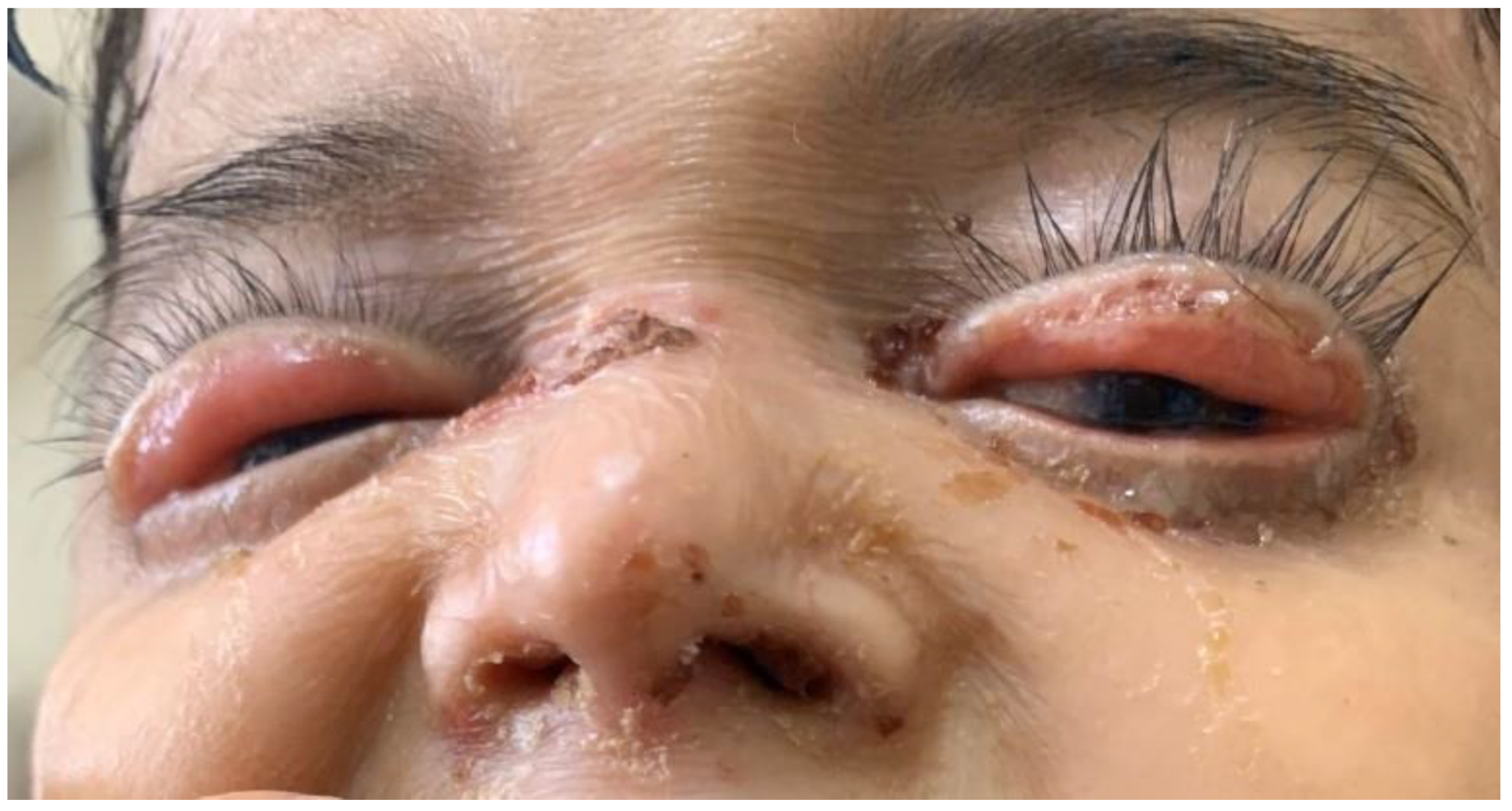
| Ocular involvement | 0 | 0 | 11 | 26 | |
| Auricular involvement | 0 | 0 | 0 | 26 | |
| Renal involvement | 0 | 0 | 1 | 26 | |
| Gastrointestinal system involvement | 1 | 1 | 20 | 26 | |
| Hair involvement | 0 | 1 | 9 | 26 | |
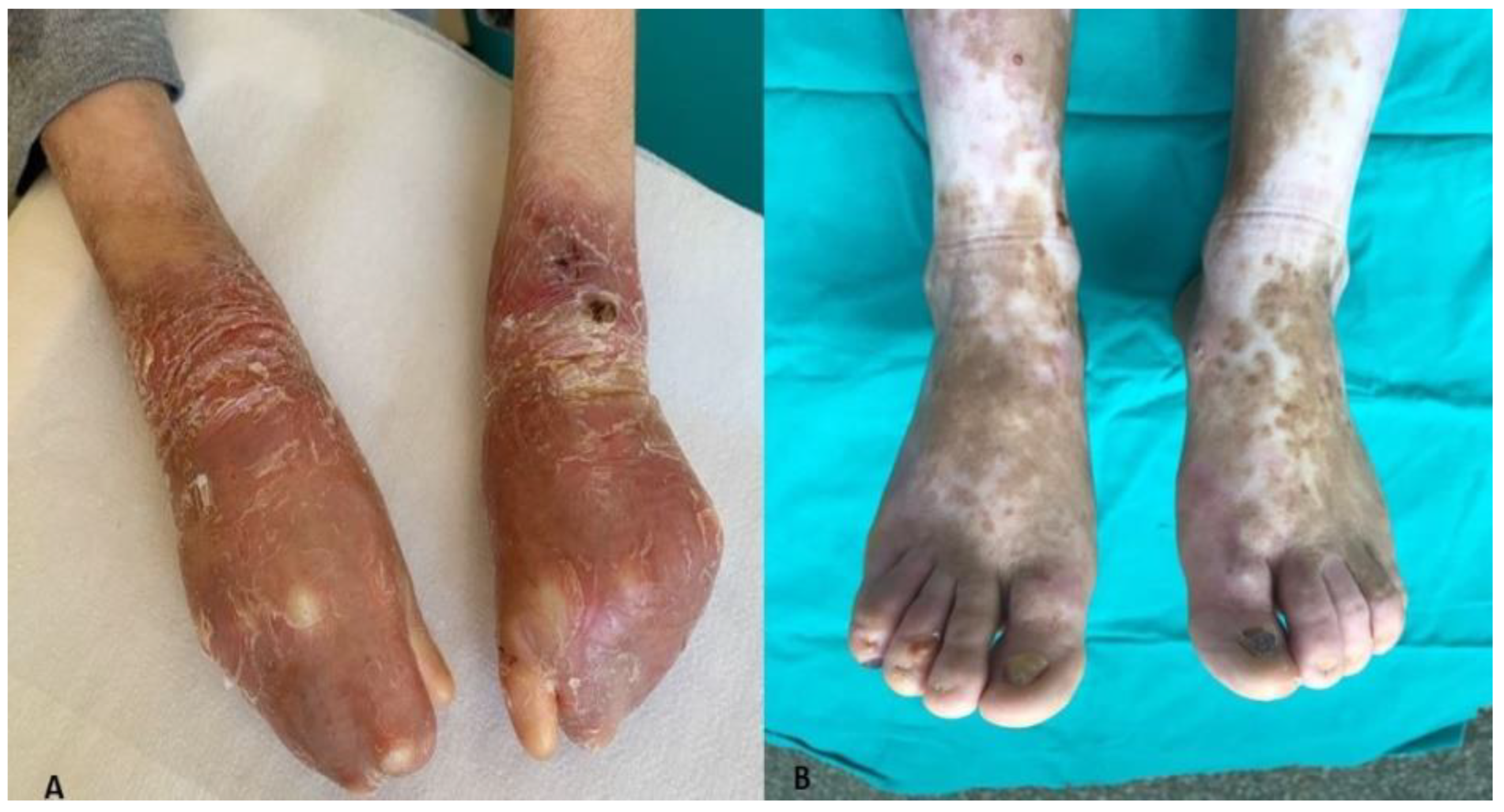
| Hand and nail deformity | 0 | 1 | 22 | 26 | |
| Oral Findings | Epidermolysis bullosa Simplex | Junctional Epidermolysis bullosa | Dystrophic Epidermolysis bullosa | Number of Cases (n) | |
|---|---|---|---|---|---|
| Gender | Male | 1 | 14 | 15 | |
| Female | 1 | 1 | 9 | 11 | |
| Age (year) | 21 | 0.2–30 | 26 | ||
| Tooth decay | 12 | 10 | 280 | 26 | |
| Enamel hypoplasia | 0 | 1 | 0 | 26 | |
| Extracted tooth | 0 | 1 | 17 | 26 | |
| Tooth to be extracted | 0 | 0 | 141 | 26 | |
| Periodontal disease | 0 | 1 | 17 | 26 | |
| Intraoral bulla and erosion | 1 | 1 | 23 | 26 | |
| Ankyglossia | 0 | 0 | 19 | 26 | |
| Microstomy | 0 | 0 | 18 | 26 | |
| Maxillary atrophy | 0 | 0 | 18 | 26 | |
| Vestibular sulcus insufficiency | 0 | 0 | 19 | 26 | |
| Restorative treatment history | 0 | 1 | 2 | 26 | |
| History of prosthetic rehabilitation | 0 | 1 | 0 | 26 | |
Disclaimer/Publisher’s Note: The statements, opinions and data contained in all publications are solely those of the individual author(s) and contributor(s) and not of MDPI and/or the editor(s). MDPI and/or the editor(s) disclaim responsibility for any injury to people or property resulting from any ideas, methods, instructions or products referred to in the content. |
© 2023 by the authors. Licensee MDPI, Basel, Switzerland. This article is an open access article distributed under the terms and conditions of the Creative Commons Attribution (CC BY) license (https://creativecommons.org/licenses/by/4.0/).
Share and Cite
Yavuz, Y.; An, I.; Yazmaci, B.; Akkus, Z.; Ortac, H. Evaluation of Clinical and Oral Findings in Patients with Epidermolysis bullosa. Medicina 2023, 59, 1185. https://doi.org/10.3390/medicina59071185
Yavuz Y, An I, Yazmaci B, Akkus Z, Ortac H. Evaluation of Clinical and Oral Findings in Patients with Epidermolysis bullosa. Medicina. 2023; 59(7):1185. https://doi.org/10.3390/medicina59071185
Chicago/Turabian StyleYavuz, Yasemin, Isa An, Betul Yazmaci, Zeki Akkus, and Hatice Ortac. 2023. "Evaluation of Clinical and Oral Findings in Patients with Epidermolysis bullosa" Medicina 59, no. 7: 1185. https://doi.org/10.3390/medicina59071185