
Association between Endodontic Infection, Its Treatment and Systemic Health: A Narrative Review


Abstract
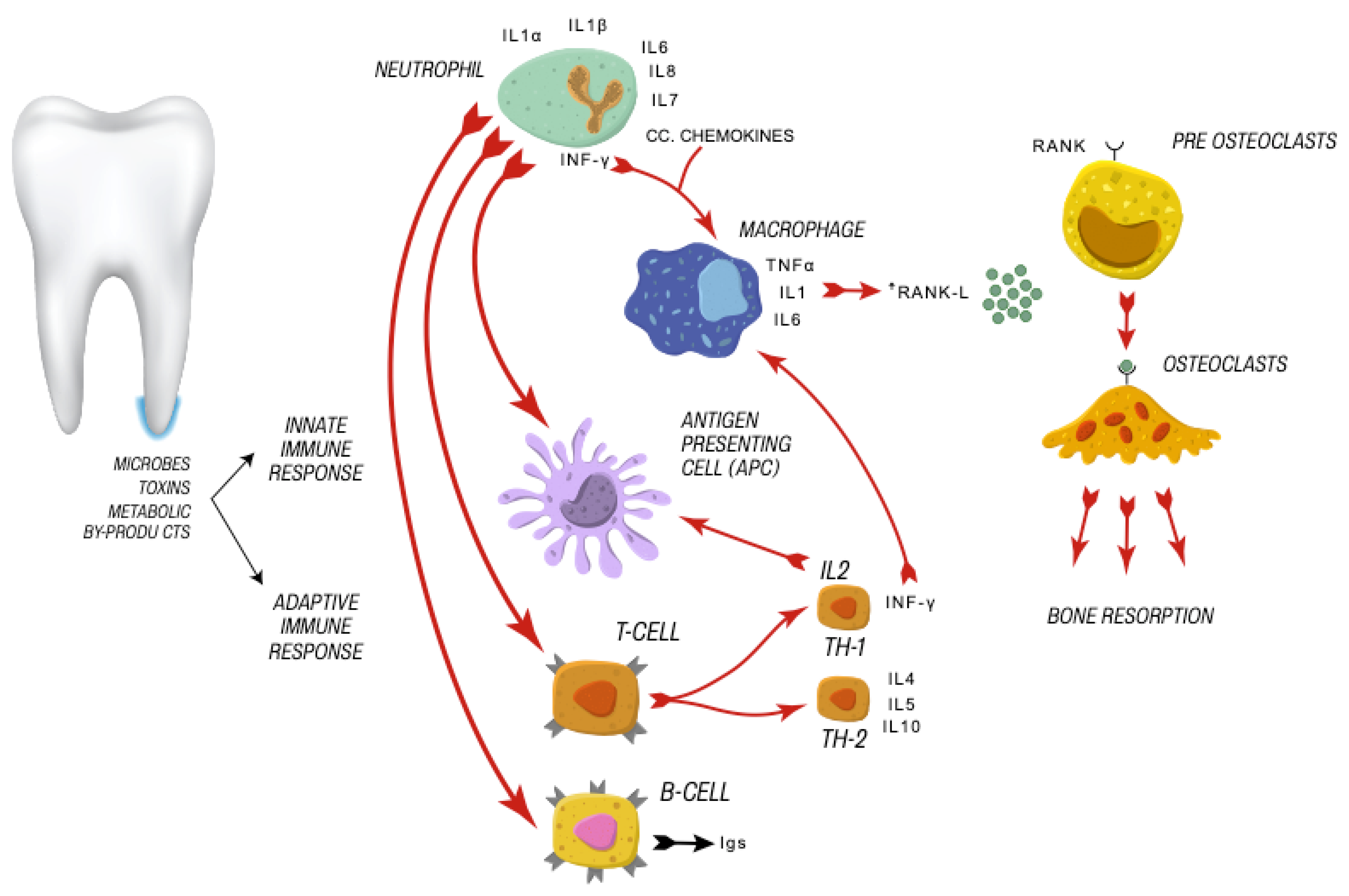
:1. Apical Periodontitis Aetiology
2. Apical Periodontitis-A Global Burden
3. Focal Infection
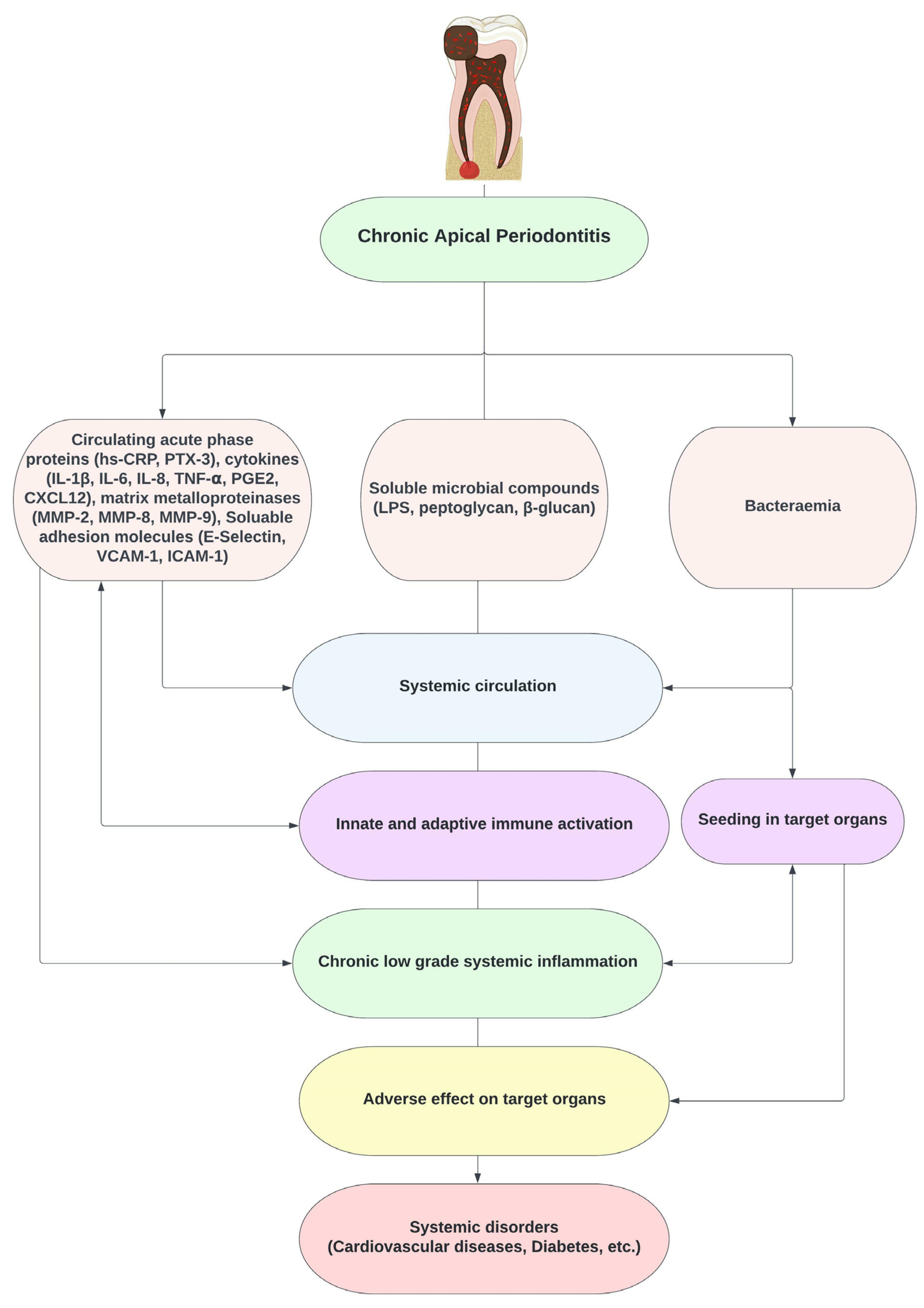
4. Endodontic Disease and Systemic Impact
5. Similarities between Periodontal and Endodontic Disease Impacting Systemic Health
6. Endodontic Bacteraemia
7. Systemic Inflammatory Mediators
8. Apical Periodontitis and Cardiovascular Diseases
9. Atherosclerosis
10. Inflammatory Mediators of Apical Periodontitis and Their Role in the Development of CVDs
10.1. C-Reactive Protein (CRP)
10.2. Pentraxin-3 (PTX-3)
10.3. Asymmetric Dimethylarginine (ADMA)
10.4. Fibroblast Growth Factor-23 (FGF-23)
10.5. Matrix Metalloproteinases (MMPs)
10.6. Human Complement C3
10.7. Statins and Apical Periodontitis
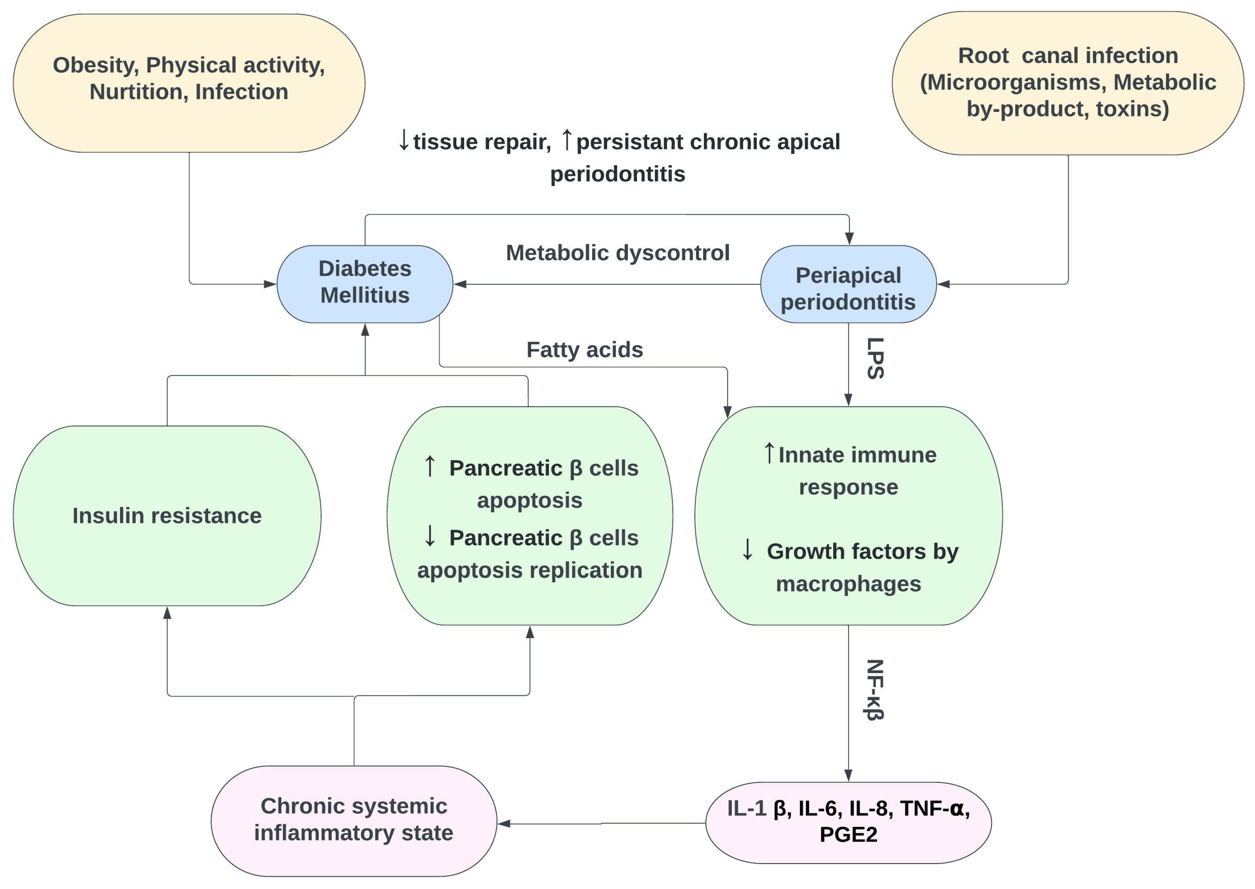
11. Apical Periodontitis and Diabetes Mellitus
12. Apical Periodontitis and Pregnancy
13. Apical Periodontitis and Autoimmune Disorder
14. Apical Periodontitis and Other Systemic Conditions
15. Conclusions
Author Contributions
Funding
Informed Consent Statement
Data Availability Statement
Conflicts of Interest
References
- Baumgartner, J.C. Microbiological and molecular analysis of endodontic infections. Endod. Top. 2004, 7, 35–51. [Google Scholar] [CrossRef]
- Nair, P.N. Pathogenesis of apical periodontitis and the causes of endodontic failures. Crit. Rev. Oral Biol. Med. 2004, 15, 348–381. [Google Scholar] [CrossRef] [PubMed] [Green Version]
- Nair, P.N. On the causes of persistent apical periodontitis: A review. Int. Endod. J. 2006, 39, 249–281. [Google Scholar] [CrossRef] [PubMed]
- Marton, I.J.; Kiss, C. Overlapping protective and destructive regulatory pathways in apical periodontitis. J. Endod. 2014, 40, 155–163. [Google Scholar] [CrossRef]
- Lucarotti, P.S.; Lessani, M.; Lumley, P.J.; Burke, F.J. Influence of root canal fillings on longevity of direct and indirect restorations placed within the General Dental Services in England and Wales. Br. Dent. J. 2014, 216, E14. [Google Scholar] [CrossRef] [PubMed]
- Chew, T.; Brennan, D.; Rossi-Fedele, G. Comparative Longitudinal Study on the Impact Root Canal Treatment and Other Dental Services Have on Oral Health-Related Quality of Life Using Self-reported Health Measures (Oral Health Impact Profile-14 and Global Health Measures). J. Endod. 2019, 45, 985–993.e1. [Google Scholar] [CrossRef]
- Cordis EU Research Results. Increasing the Success Rate of Root Canals. Available online: https://cordis.europa.eu/article/id/413194-increasing-the-success-rate-of-root-canals (accessed on 16 June 2022).
- Tiburcio-Machado, C.S.; Michelon, C.; Zanatta, F.B.; Gomes, M.S.; Marin, J.A.; Bier, C.A. The global prevalence of apical periodontitis: A systematic review and meta-analysis. Int. Endod. J. 2021, 54, 712–735. [Google Scholar] [CrossRef]
- Easlick, K.A. Evaluation of the action of focal dental infections on health. Med. Hyg. 1952, 10, 35. [Google Scholar]
- Billings, F. Focal Infection as the Cause of General Disease. Bull. N. Y. Acad. Med. 1930, 6, 759–773. [Google Scholar]
- Murray, C.A.; Saunders, W.P. Root canal treatment and general health: A review of the literature. Int. Endod. J. 2000, 33, 1–18. [Google Scholar] [CrossRef]
- Pallasch, T.J.; Wahl, M.J. The focal infection theory: Appraisal and reappraisal. J. Calif. Dent. Assoc. 2000, 28, 194–200. [Google Scholar] [PubMed]
- Miller, W.D. Diseases of the Human Body Which Have Been Traced to the Action of Mouth-Bacteria. Am. J. Dent. Sci. 1891, 25, 311–319. [Google Scholar] [PubMed]
- Hunter, W. Oral Sepsis as a Cause of Disease. Br. Med. J. 1900, 2, 215–216. [Google Scholar] [CrossRef] [PubMed] [Green Version]
- Hunter, W. The Coming of Age of Oral Sepsis. Br. Med. J. 1921, 1, 859. [Google Scholar] [CrossRef] [PubMed]
- Newman, H.N. Focal infection. J. Dent. Res. 1996, 75, 1912–1919. [Google Scholar] [CrossRef]
- Rautemaa, R.; Lauhio, A.; Cullinan, M.P.; Seymour, G.J. Oral infections and systemic disease—An emerging problem in medicine. Clin. Microbiol. Infect. 2007, 13, 1041–1047. [Google Scholar] [CrossRef]
- Rosen, J.; Sancheti, P.; Fierlinger, A.; Niazi, F.; Johal, H.; Bedi, A. Response to: Important Considerations When Determining the Cost-effectiveness of Viscosupplements in the Treatment of Knee Osteoarthritis. Adv. Ther. 2017, 33, 2273–2276. [Google Scholar] [CrossRef] [Green Version]
- Segura-Egea, J.J.; Jimenez-Moreno, E.; Calvo-Monroy, C.; Rios-Santos, J.V.; Velasco-Ortega, E.; Sanchez-Dominguez, B.; Castellanos-Cosano, L.; Llamas-Carreras, J.M. Hypertension and dental periapical condition. J. Endod. 2010, 36, 1800–1804. [Google Scholar] [CrossRef]
- Wang, C.H.; Chueh, L.H.; Chen, S.C.; Feng, Y.C.; Hsiao, C.K.; Chiang, C.P. Impact of diabetes mellitus, hypertension, and coronary artery disease on tooth extraction after nonsurgical endodontic treatment. J. Endod. 2011, 37, 1–5. [Google Scholar] [CrossRef]
- Joe Editorial Board. Relationship between systemic diseases and endodontics: An online study guide. J. Endod. 2008, 34 (Suppl. 5), e195–e200. [Google Scholar] [CrossRef]
- Caplan, D.J.; Chasen, J.B.; Krall, E.A.; Cai, J.; Kang, S.; Garcia, R.I.; Offenbacher, S.; Beck, J.D. Lesions of endodontic origin and risk of coronary heart disease. J. Dent. Res. 2006, 85, 996–1000. [Google Scholar] [CrossRef] [PubMed]
- Pasqualini, D.; Bergandi, L.; Palumbo, L.; Borraccino, A.; Dambra, V.; Alovisi, M.; Migliaretti, G.; Ferraro, G.; Ghigo, D.; Bergerone, S.; et al. Association among oral health, apical periodontitis, CD14 polymorphisms, and coronary heart disease in middle-aged adults. J. Endod. 2012, 38, 1570–1577. [Google Scholar] [CrossRef] [PubMed]
- Liljestrand, J.M.; Mantyla, P.; Paju, S.; Buhlin, K.; Kopra, K.A.; Persson, G.R.; Hernandez, M.; Nieminen, M.S.; Sinisalo, J.; Tjaderhane, L.; et al. Association of Endodontic Lesions with Coronary Artery Disease. J. Dent. Res. 2016, 95, 1358–1365. [Google Scholar] [CrossRef] [PubMed] [Green Version]
- Cotti, E.; Mercuro, G. Apical periodontitis and cardiovascular diseases: Previous findings and ongoing research. Int. Endod. J. 2015, 48, 926–932. [Google Scholar] [CrossRef] [PubMed]
- Segura-Egea, J.J.; Martín-González, J.; Castellanos-Cosano, L. Endodontic medicine: Connections between apical periodontitis and systemic diseases. Int. Endod. J. 2015, 48, 933–951. [Google Scholar] [CrossRef] [PubMed]
- Khalighinejad, N.; Aminoshariae, M.R.; Aminoshariae, A.; Kulild, J.C.; Mickel, A.; Fouad, A.F. Association between Systemic Diseases and Apical Periodontitis. J. Endod. 2016, 42, 1427–1434. [Google Scholar] [CrossRef]
- Aminoshariae, A.; Kulild, J.C.; Mickel, A.; Fouad, A.F. Association between Systemic Diseases and Endodontic Outcome: A Systematic Review. J. Endod. 2017, 43, 514–519. [Google Scholar] [CrossRef]
- Aminoshariae, A.; Kulild, J.C.; Fouad, A.F. The Impact of Endodontic Infections on the Pathogenesis of Cardiovascular Disease(s): A Systematic Review with Meta-analysis Using GRADE. J. Endod. 2018, 44, 1361–1366.e3. [Google Scholar] [CrossRef]
- Berlin-Broner, Y.; Febbraio, M.; Levin, L. Association between apical periodontitis and cardiovascular diseases: A systematic review of the literature. Int. Endod. J. 2017, 50, 847–859. [Google Scholar] [CrossRef] [Green Version]
- González Navarro, B.; Pintó Sala, X.; Jané Salas, E. Relationship between cardiovascular disease and dental pathology. Systematic review. Med. Clin. 2017, 149, 211–216. [Google Scholar] [CrossRef]
- Cintra, L.T.A.; Estrela, C.; Azuma, M.M.; Queiroz, I.O.A.; Kawai, T.; Gomes-Filho, J.E. Endodontic medicine: Interrelationships among apical periodontitis, systemic disorders, and tissue responses of dental materials. Braz. Oral Res. 2018, 32, e68. [Google Scholar] [CrossRef] [Green Version]
- Jimenez-Sanchez, M.C.; Cabanillas-Balsera, D.; Areal-Quecuty, V.; Velasco-Ortega, E.; Martin-Gonzalez, J.; Segura-Egea, J.J. Cardiovascular diseases and apical periodontitis: Association not always implies causality. Med. Oral Patol. Oral Cir. Bucal. 2020, 25, e652–e659. [Google Scholar] [CrossRef] [PubMed]
- Nagendrababu, V.; Segura-Egea, J.J.; Fouad, A.; Pulikkotil, S.J.; Dummer, P. Association between diabetes and the outcome of root canal treatment in adults: An umbrella review. Int. Endod. J. 2020, 53, 455–466. [Google Scholar] [CrossRef] [PubMed] [Green Version]
- Perez-Losada, F.L.; Estrugo-Devesa, A.; Castellanos-Cosano, L.; Segura-Egea, J.J.; Lopez-Lopez, J.; Velasco-Ortega, E. Apical periodontitis and diabetes mellitus type 2: A systematic review and meta-analysis. J. Clin. Med. 2020, 9, 540. [Google Scholar] [CrossRef] [PubMed] [Green Version]
- Bender, I.B.; Seltzer, S.; Yermish, M. The incidence of bacteremia in endodontic manipulation: Preliminary report. 1960. J. Endod. 2003, 29, 697–700; discussion 696. [Google Scholar] [CrossRef] [PubMed]
- Debelian, G.J.; Olsen, I.; Tronstad, L. Anaerobic bacteremia and fungemia in patients undergoing endodontic therapy: An overview. Ann. Periodontol. 1998, 3, 281–287. [Google Scholar] [CrossRef]
- Savarrio, L.; Mackenzie, D.; Riggio, M.; Saunders, W.P.; Bagg, J. Detection of bacteraemias during non-surgicalroot canal treatment. J. Dent. 2005, 33, 293–303. [Google Scholar] [CrossRef]
- Debelian, G.J.; Olsen, I.; Tronstad, L. Bacteremia in conjunction with endodontic therapy. Endod. Dent. Traumatol. 1995, 11, 142–149. [Google Scholar] [CrossRef]
- Gendron, R.; Grenier, D.; Maheu-Robert, L. The oral cavity as a reservoir of bacterial pathogens for focal infections. Microbes Infect. 2000, 2, 897–906. [Google Scholar] [CrossRef]
- Henderson, B.; Wilson, M. Commensal communism and the oral cavity. J. Dent. Res. 1998, 77, 1674–1683. [Google Scholar] [CrossRef]
- Li, X.; Kolltveit, K.M.; Tronstad, L.; Olsen, I. Systemic diseases caused by oral infection. Clin. Microbiol. Rev. 2000, 13, 547–558. [Google Scholar] [CrossRef] [PubMed]
- Oe, Y.; Soejima, H.; Nakayama, H.; Fukunaga, T.; Sugamura, K.; Kawano, H.; Sugiyama, S.; Matsuo, K.; Shinohara, M.; Izumi, Y.; et al. Significant association between score of periodontal disease and coronary artery disease. Heart Vessels 2009, 24, 103–107. [Google Scholar] [CrossRef] [PubMed]
- Byon, M.-J.; Kim, S.-Y.; Kim, J.-S.; Kim, H.-N.; Kim, J.-B. Association of Periodontitis with Atherosclerotic Cardiovascular Diseases: A Nationwide Population-based Retrospective Matched Cohort Study. Int. J. Environ. Res. Public Health 2020, 17, 7261. [Google Scholar] [CrossRef]
- Gita, B.; Sajja, C.; Padmanabhan, P. Are lipid profiles true surrogate biomarkers of coronary heart disease in periodontitis patients?: A case-control study in a south Indian population. J. Indian Soc. Periodontol. 2012, 16, 32. [Google Scholar] [CrossRef] [PubMed]
- Ameen, M.; Attia, A.M.; Felimban, A.; Al-Dweghri, T.; Fattni, A.; Azab, E.; Baz, S.; Dardir, A. Evaluation of cardiac biomarkers in smokers and non-smokers with chronic periodontitis. Int. J. Health Sci. 2020, 14, 26. [Google Scholar]
- Boyapati, R.; Vudathaneni, V.; Nadella, S.B.; Ramachandran, R.; Dhulipalla, R.; Adurty, C. Mapping the link between cardiac biomarkers and chronic periodontitis: A clinico-biochemical study. J. Indian Soc. Periodontol. 2020, 24, 309. [Google Scholar]
- Beukers, N.G.; van der Heijden, G.J.; van Wijk, A.J.; Loos, B.G. Periodontitis is an independent risk indicator for atherosclerotic cardiovascular diseases among 60 174 participants in a large dental school in the Netherlands. J. Epidemiol. Community Health 2017, 71, 37–42. [Google Scholar] [CrossRef] [Green Version]
- Dietrich, T.; Sharma, P.; Walter, C.; Weston, P.; Beck, J. The epidemiological evidence behind the association between periodontitis and incident atherosclerotic cardiovascular disease. J. Periodontol. 2013, 84, S70–S84. [Google Scholar] [CrossRef] [Green Version]
- Reyes, L.; Herrera, D.; Kozarov, E.; Roldan, S.; Progulske-Fox, A. Periodontal bacterial invasion and infection: Contribution to atherosclerotic pathology. J. Clin. Periodontol. 2013, 40 (Suppl. 14), S30–S50. [Google Scholar] [CrossRef]
- Schenkein, H.A.; Loos, B.G. Inflammatory mechanisms linking periodontal diseases to cardiovascular diseases. J. Clin. Periodontol. 2013, 40 (Suppl. 14), S51–S69. [Google Scholar] [CrossRef]
- D’Aiuto, F.; Orlandi, M.; Gunsolley, J.C. Evidence that periodontal treatment improves biomarkers and CVD outcomes. J. Clin. Periodontol. 2013, 40 (Suppl. 14), S85–S105. [Google Scholar] [CrossRef] [PubMed]
- Tonetti, M.S.; Van Dyke, T.E.; Working Group 1 of the Joint EFP/AAP Workshop. Periodontitis and atherosclerotic cardiovascular disease: Consensus report of the Joint EFP/AAP Workshop on Periodontitis and Systemic Diseases. J. Clin. Periodontol. 2013, 40 (Suppl. 14), S24–S29. [Google Scholar] [CrossRef] [PubMed]
- Sanz, M.; Marco Del Castillo, A.; Jepsen, S.; Gonzalez-Juanatey, J.R.; D’Aiuto, F.; Bouchard, P.; Chapple, I.; Dietrich, T.; Gotsman, I.; Graziani, F.; et al. Periodontitis and cardiovascular diseases: Consensus report. J. Clin. Periodontol. 2020, 47, 268–288. [Google Scholar] [CrossRef]
- Kristiansen, O.P.; Mandrup-Poulsen, T. Interleukin-6 and diabetes: The good, the bad, or the indifferent? Diabetes 2005, 54 (Suppl. 2), S114–S124. [Google Scholar] [CrossRef] [PubMed] [Green Version]
- Dubinski, A.; Zdrojewicz, Z. The role of interleukin-6 in development and progression of atherosclerosis. Pol. Merkur. Lek. 2007, 22, 291–294. [Google Scholar]
- Dowlati, Y.; Herrmann, N.; Swardfager, W.; Liu, H.; Sham, L.; Reim, E.K.; Lanctot, K.L. A meta-analysis of cytokines in major depression. Biol. Psychiatry 2010, 67, 446–457. [Google Scholar] [CrossRef]
- Swardfager, W.; Lanctot, K.; Rothenburg, L.; Wong, A.; Cappell, J.; Herrmann, N. A meta-analysis of cytokines in Alzheimer’s disease. Biol. Psychiatry 2010, 68, 930–941. [Google Scholar] [CrossRef]
- Gong, J.; Dong, H.; Xia, S.Q.; Huang, Y.Z.; Wang, D.; Zhao, Y.; Liu, W.; Tu, S.; Zhang, M.; Wang, Q.; et al. Correlation Analysis Between Disease Severity and Inflammation-related Parameters in Patients with COVID-19 Pneumonia. medRxiv 2020. [Google Scholar] [CrossRef] [Green Version]
- Wojtkowska, A.; Zapolski, T.; Wysokinska-Miszczuk, J.; Wysokinski, A.P. The inflammation link between periodontal disease and coronary atherosclerosis in patients with acute coronary syndromes: Case-control study. BMC Oral Health 2021, 21, 5. [Google Scholar] [CrossRef]
- Marouf, N.; Cai, W.; Said, K.N.; Daas, H.; Diab, H.; Chinta, V.R.; Hssain, A.A.; Nicolau, B.; Sanz, M.; Tamimi, F. Association between periodontitis and severity of COVID-19 infection: A case-control study. J. Clin. Periodontol. 2021, 48, 483–491. [Google Scholar] [CrossRef]
- Kane, S.F. The effects of oral health on systemic health. Gen. Dent. 2017, 65, 30–34. [Google Scholar]
- Sabharwal, A.; Gomes-Filho, I.S.; Stellrecht, E.; Scannapieco, F.A. Role of periodontal therapy in management of common complex systemic diseases and conditions: An update. Periodontol. 2000 2018, 78, 212–226. [Google Scholar] [CrossRef]
- Chen, C.K.; Wu, Y.T.; Chang, Y.C. Periodontal inflammatory disease is associated with the risk of Parkinson’s disease: A population-based retrospective matched-cohort study. PeerJ 2017, 5, e3647. [Google Scholar] [CrossRef] [Green Version]
- Dibello, V.; Lozupone, M.; Manfredini, D.; Dibello, A.; Zupo, R.; Sardone, R.; Daniele, A.; Lobbezoo, F.; Panza, F. Oral frailty and neurodegeneration in Alzheimer’s disease. Neural Regener. Res. 2021, 16, 2149–2153. [Google Scholar] [CrossRef]
- Borsa, L.; Dubois, M.; Sacco, G.; Lupi, L. Analysis the link between periodontal diseases and Alzheimer’s disease: A systematic review. Int. J. Environ. Res. Public Health 2021, 18, 9312. [Google Scholar] [CrossRef]
- Dibello, V.; Zupo, R.; Sardone, R.; Lozupone, M.; Castellana, F.; Dibello, A.; Daniele, A.; De Pergola, G.; Bortone, I.; Lampignano, L. Oral frailty and its determinants in older age: A systematic review. Lancet Healthy Longev. 2021, 2, e507–e520. [Google Scholar] [CrossRef]
- Costa, T.H.; de Figueiredo Neto, J.A.; de Oliveira, A.E.; Lopes e Maia Mde, F.; de Almeida, A.L. Association between chronic apical periodontitis and coronary artery disease. J. Endod. 2014, 40, 164–167. [Google Scholar] [CrossRef]
- Rechenberg, D.K.; Galicia, J.C.; Peters, O.A. Biological Markers for Pulpal Inflammation: A Systematic Review. PLoS ONE 2016, 11, e0167289. [Google Scholar] [CrossRef] [Green Version]
- Stashenko, P.; Teles, R.; D’Souza, R. Periapical inflammatory responses and their modulation. Crit. Rev. Oral Biol. Med. 1998, 9, 498–521. [Google Scholar] [CrossRef]
- Reis, L.C.; Rocas, I.N.; Siqueira, J.F., Jr.; de Uzeda, M.; Lacerda, V.S.; Domingues, R.M.; Moraes, S.R.; Saraiva, R.M. Bacteremia after Endodontic Procedures in Patients with Heart Disease: Culture and Molecular Analyses. J. Endod. 2016, 42, 1181–1185. [Google Scholar] [CrossRef]
- Berlin-Broner, Y.; Febbraio, M.; Levin, L. Apical periodontitis and atherosclerosis: Is there a link? Review of the literature and potential mechanism of linkage. Quintessence Int. 2017, 48, 527–534. [Google Scholar] [CrossRef] [PubMed]
- Jakovljevic, A.; Duncan, H.F.; Nagendrababu, V.; Jacimovic, J.; Milasin, J.; Dummer, P.M.H. Association between cardiovascular diseases and apical periodontitis: An umbrella review. Int. Endod. J. 2020, 53, 1374–1386. [Google Scholar] [CrossRef] [PubMed]
- Jakovljevic, A.; Sljivancanin Jakovljevic, T.; Duncan, H.; Nagendrababu, V.; Jacimovic, J.; Aminoshariae, A.; Milasin, J.; Dummer, P. The association between apical periodontitis and adverse pregnancy outcomes: A systematic review. Int. Endod. J. 2021, 54, 1527–1537. [Google Scholar] [CrossRef] [PubMed]
- Bender, I.B.; Seltzer, S.; Yermish, M. The incidence of bacteremia in endodontic manipulation: Preliminary report. Oral Surg. Oral Med. Oral Pathol. 1960, 13, 353–360. [Google Scholar] [CrossRef]
- Klotz, M.D.; Gerstein, H.; Bahn, A.N. Bacteremia after topical use of prednisolone in infected pulps. J. Am. Dent. Assoc. 1965, 71, 871–875. [Google Scholar] [CrossRef] [PubMed]
- Baumgartner, J.C.; Heggers, J.P.; Harrison, J.W. The incidence of bacteremias related to endodontic procedures. I. Nonsurgical endodontics. J. Endod. 1976, 2, 135–140. [Google Scholar] [CrossRef]
- Heimdahl, A.; Hall, G.; Hedberg, M.; Sandberg, H.; Soder, P.O.; Tuner, K.; Nord, C.E. Detection and quantitation by lysis-filtration of bacteremia after different oral surgical procedures. J. Clin. Microbiol. 1990, 28, 2205–2209. [Google Scholar] [CrossRef] [PubMed] [Green Version]
- Debelian, G.J.; Olsen, I.; Tronstad, L. Profiling of Propionibacterium acnes recovered from root canal and blood during and after endodontic treatment. Endod. Dent. Traumatol. 1992, 8, 248–254. [Google Scholar] [CrossRef]
- Debelian, G.J.; Olsen, I.; Tronstad, L. Electrophoresis of whole-cell soluble proteins of microorganisms isolated from bacteremias in endodontic therapy. Eur. J. Oral Sci. 1996, 104, 540–546. [Google Scholar] [CrossRef]
- Debelian, G.J.; Eribe, E.R.; Olsen, I.; Tronstad, L. Ribotyping of bacteria from root canal and blood of patients receiving endodontic therapy. Anaerobe 1997, 3, 237–243. [Google Scholar] [CrossRef]
- Ide, M.; Jagdev, D.; Coward, P.Y.; Crook, M.; Barclay, G.R.; Wilson, R.F. The short-term effects of treatment of chronic periodontitis on circulating levels of endotoxin, C-reactive protein, tumor necrosis factor-alpha, and interleukin-6. J. Periodontol. 2004, 75, 420–428. [Google Scholar] [CrossRef] [PubMed]
- Thornhill, M.H.; Jones, S.; Prendergast, B.; Baddour, L.M.; Chambers, J.B.; Lockhart, P.B.; Dayer, M.J. Quantifying infective endocarditis risk in patients with predisposing cardiac conditions. Eur. Heart J. 2018, 39, 586–595. [Google Scholar] [CrossRef] [PubMed] [Green Version]
- DeSimone, D.C.; Tleyjeh, I.M.; Correa de Sa, D.D.; Anavekar, N.S.; Lahr, B.D.; Sohail, M.R.; Steckelberg, J.M.; Wilson, W.R.; Baddour, L.M. Temporal trends in infective endocarditis epidemiology from 2007 to 2013 in Olmsted County, MN. Am. Heart J. 2015, 170, 830–836. [Google Scholar] [CrossRef] [Green Version]
- Selton-Suty, C.; Celard, M.; Le Moing, V.; Doco-Lecompte, T.; Chirouze, C.; Iung, B.; Strady, C.; Revest, M.; Vandenesch, F.; Bouvet, A.; et al. Preeminence of Staphylococcus aureus in infective endocarditis: A 1-year population-based survey. Clin. Infect. Dis. 2012, 54, 1230–1239. [Google Scholar] [CrossRef] [Green Version]
- Gomes, M.S.; Blattner, T.C.; Sant’Ana Filho, M.; Grecca, F.S.; Hugo, F.N.; Fouad, A.F.; Reynolds, M.A. Can apical periodontitis modify systemic levels of inflammatory markers? A systematic review and meta-analysis. J. Endod. 2013, 39, 1205–1217. [Google Scholar] [CrossRef] [PubMed]
- Georgiou, A.C.; Crielaard, W.; Armenis, I.; de Vries, R.; van der Waal, S.V. Apical Periodontitis Is Associated with Elevated Concentrations of Inflammatory Mediators in Peripheral Blood: A Systematic Review and Meta-analysis. J. Endod. 2019, 45, 1279–1295 e1273. [Google Scholar] [CrossRef] [PubMed]
- Torabinejad, M.; Theofilopoulos, A.N.; Ketering, J.D.; Bakland, L.K. Quantitation of circulating immune complexes, immunoglobulins G and M, and C3 complement component in patients with large periapical lesions. Oral Surg. Oral Med. Oral Pathol. 1983, 55, 186–190. [Google Scholar] [CrossRef]
- Sirin, D.A.; Ozcelik, F.; Uzun, C.; Ersahan, S.; Yesilbas, S. Association between C-reactive protein, neutrophil to lymphocyte ratio and the burden of apical periodontitis: A case-control study. Acta Odontol. Scand. 2019, 77, 142–149. [Google Scholar] [CrossRef]
- Ren, Y.F.; Malmstrom, H.S. Rapid quantitative determination of C-reactive protein at chair side in dental emergency patients. Oral Surg. Oral Med. Oral Pathol. Oral Radiol. Endod. 2007, 104, 49–55. [Google Scholar] [CrossRef]
- Matsushita, K.; Tajima, T.; Tomita, K.; Abeyama, K.; Maruyama, I.; Takada, H.; Nagaoka, S. Inflammatory cytokine production and specific antibody responses against possible causative bacteria in patients with multilesional periapical periodontitis. J. Endod. 1998, 24, 817–821. [Google Scholar] [CrossRef]
- Kimak, A.; Strycharz-Dudziak, M.; Bachanek, T.; Kimak, E. Lipids and lipoproteins and inflammatory markers in patients with chronic apical periodontitis. Lipids Health Dis. 2015, 14, 162. [Google Scholar] [CrossRef] [PubMed] [Green Version]
- Inchingolo, F.; Marrelli, M.; Annibali, S.; Cristalli, M.P.; Dipalma, G.; Inchingolo, A.D.; Palladino, A.; Inchingolo, A.M.; Gargari, M.; Tatullo, M. Influence of endodontic treatment on systemic oxidative stress. Int. J. Med. Sci. 2014, 11, 1–6. [Google Scholar] [CrossRef] [PubMed] [Green Version]
- Harjunmaa, U.; Doyle, R.; Jarnstedt, J.; Kamiza, S.; Jorgensen, J.M.; Stewart, C.P.; Shaw, L.; Hallamaa, L.; Ashorn, U.; Klein, N.; et al. Periapical infection may affect birth outcomes via systemic inflammation. Oral Dis. 2018, 24, 847–855. [Google Scholar] [CrossRef] [PubMed] [Green Version]
- Cotti, E.; Zedda, A.; Deidda, M.; Piras, A.; Flore, G.; Ideo, F.; Madeddu, C.; Pau, V.M.; Mercuro, G. Endodontic infection and endothelial dysfunction are associated with different mechanisms in men and women. J. Endod. 2015, 41, 594–600. [Google Scholar] [CrossRef]
- Garrido, M.; Cardenas, A.M.; Astorga, J.; Quinlan, F.; Valdes, M.; Chaparro, A.; Carvajal, P.; Pussinen, P.; Huaman-Chipana, P.; Jalil, J.E.; et al. Elevated Systemic Inflammatory Burden and Cardiovascular Risk in Young Adults with Endodontic Apical Lesions. J. Endod. 2019, 45, 111–115. [Google Scholar] [CrossRef]
- Gomes, C.; Martinho, F.C.; Barbosa, D.S.; Antunes, L.S.; Povoa, H.C.C.; Baltus, T.H.L.; Morelli, N.R.; Vargas, H.O.; Nunes, S.O.V.; Anderson, G.; et al. Increased Root Canal Endotoxin Levels are Associated with Chronic Apical Periodontitis, Increased Oxidative and Nitrosative Stress, Major Depression, Severity of Depression, and a Lowered Quality of Life. Mol. Neurobiol. 2018, 55, 2814–2827. [Google Scholar] [CrossRef]
- Anil, S.; Shanavas, K.R.; Beena, V.T.; Remani, P.; Vijayakumar, T. Quantitation of circulating immune complexes in patients with chronic periapical lesions. J. Nihon Univ. Sch. Dent. 1993, 35, 175–178. [Google Scholar] [CrossRef] [Green Version]
- Abdolsamadi, H.R.; Vahedi, M.; Esmaeili, F.; Nazari, S.; Abdollahzadeh, S. Serum Interleukin-6 as a Serologic Marker of Chronic Periapical Lesions: A Case-control Study. J. Dent. Res. Dent. Clin. Dent. Prospects 2008, 2, 43–47. [Google Scholar] [CrossRef]
- Cotti, E.; Dessi, C.; Piras, A.; Flore, G.; Deidda, M.; Madeddu, C.; Zedda, A.; Longu, G.; Mercuro, G. Association of endodontic infection with detection of an initial lesion to the cardiovascular system. J. Endod. 2011, 37, 1624–1629. [Google Scholar] [CrossRef]
- Kettering, J.D.; Torabinejad, M. Concentrations of immune complexes, IgG, IgM, IgE, and C3 in patients with acute apical abscesses. J. Endod. 1984, 10, 417–421. [Google Scholar] [CrossRef]
- Márion, I.; Kiss, C.; Balla, G.; Szabó, T.; Karmazsin, L. Acute phase proteins in patients with chronic periapical granuloma before and after surgical treatment. Oral Microbiol. Immunol. 1988, 3, 95–96. [Google Scholar] [CrossRef] [PubMed]
- Marton, I.J.; Kiss, C. Influence of surgical treatment of periapical lesions on serum and blood levels of inflammatory mediators. Int. Endod. J. 1992, 25, 229–233. [Google Scholar] [CrossRef]
- Poornima, L.; Ravishankar, P.; Abbott, P.V.; Subbiya, A.; PradeepKumar, A.R. Impact of root canal treatment on high-sensitivity C-reactive protein levels in systemically healthy adults with apical periodontitis—A preliminary prospective, longitudinal interventional study. Int. Endod. J. 2021, 54, 501–508. [Google Scholar] [CrossRef] [PubMed]
- Bakhsh, A.; Moyes, D.; Proctor, G.; Mannocci, F.; Niazi, S.A. The Impact of Apical Periodontitis, Non-surgical Root Canal Retreatment and Periapical Surgery on Serum Inflammatory Biomarkers. Int. Endod. J. 2022; Early View. [Google Scholar] [CrossRef]
- Thomas, H.; Diamond, J.; Vieco, A.; Chaudhuri, S.; Shinnar, E.; Cromer, S.; Perel, P.; Mensah, G.A.; Narula, J.; Johnson, C.O.; et al. Global Atlas of Cardiovascular Disease 2000-2016: The Path to Prevention and Control. Glob. Heart 2018, 13, 143–163. [Google Scholar] [CrossRef] [PubMed]
- World Health Organization. Cardiovascular Diseases (CVDs); World Health Organization: Geneva, Switzerland, 2017.
- Bhatnagar, P.; Wickramasinghe, K.; Williams, J.; Rayner, M.; Townsend, N. The epidemiology of cardiovascular disease in the UK 2014. Heart 2015, 101, 1182–1189. [Google Scholar] [CrossRef] [Green Version]
- Vasan, R.S.; Benjamin, E.J. The Future of Cardiovascular Epidemiology. Circulation 2016, 133, 2626–2633. [Google Scholar] [CrossRef] [Green Version]
- An, G.K.; Morse, D.E.; Kunin, M.; Goldberger, R.S.; Psoter, W.J. Association of Radiographically Diagnosed Apical Periodontitis and Cardiovascular Disease: A Hospital Records-based Study. J. Endod. 2016, 42, 916–920. [Google Scholar] [CrossRef]
- Virtanen, E.; Nurmi, T.; Soder, P.O.; Airila-Mansson, S.; Soder, B.; Meurman, J.H. Apical periodontitis associates with cardiovascular diseases: A cross-sectional study from Sweden. BMC Oral Health 2017, 17, 107. [Google Scholar] [CrossRef]
- Minassian, C.; D’Aiuto, F.; Hingorani, A.D.; Smeeth, L. Invasive dental treatment and risk for vascular events: A self-controlled case series. Ann. Intern. Med. 2010, 153, 499–506. [Google Scholar] [CrossRef]
- Graziani, F.; D’Aiuto, F.; Gennai, S.; Petrini, M.; Nisi, M.; Cirigliano, N.; Landini, L.; Bruno, R.M.; Taddei, S.; Ghiadoni, L. Systemic Inflammation after Third Molar Removal: A Case-Control Study. J. Dent. Res. 2017, 96, 1505–1512. [Google Scholar] [CrossRef]
- Chen, C.; Hemme, C.; Beleno, J.; Shi, Z.J.; Ning, D.; Qin, Y.; Tu, Q.; Jorgensen, M.; He, Z.; Wu, L.; et al. Oral microbiota of periodontal health and disease and their changes after nonsurgical periodontal therapy. ISME J. 2018, 12, 1210–1224. [Google Scholar] [CrossRef] [PubMed]
- Bergandi, L.; Giuggia, B.; Alovisi, M.; Comba, A.; Silvagno, F.; Maule, M.; Aldieri, E.; Scotti, N.; Scacciatella, P.; Conrotto, F.; et al. Endothelial Dysfunction Marker Variation in Young Adults with Chronic Apical Periodontitis before and after Endodontic Treatment. J. Endod. 2019, 45, 500–506. [Google Scholar] [CrossRef] [PubMed]
- Petersen, J.; Glassl, E.M.; Nasseri, P.; Crismani, A.; Luger, A.K.; Schoenherr, E.; Bertl, K.; Glodny, B. The association of chronic apical periodontitis and endodontic therapy with atherosclerosis. Clin. Oral Investig. 2014, 18, 1813–1823. [Google Scholar] [CrossRef] [PubMed] [Green Version]
- Aminoshariae, A.; Kulild, J.; Fouad, A.F. The impact of cardiovascular disease and endodontic outcome: A systematic review of longitudinal studies. Clin. Oral Investig. 2020, 24, 3813–3819. [Google Scholar] [CrossRef]
- Aloutaibi, Y.A.; Alkarim, A.S.; Qumri, E.M.; Almansour, L.A.; Alghamdi, F.T. Chronic Endodontic Infections and Cardiovascular Diseases: Does the Evidence Support an Independent Association? Cureus 2021, 13, e19864. [Google Scholar] [CrossRef]
- Hansson, G.K.; Libby, P. The immune response in atherosclerosis: A double-edged sword. Nat. Rev. Immunol. 2006, 6, 508–519. [Google Scholar] [CrossRef]
- Hansson, G.K.; Robertson, A.K.; Söderberg-Nauclér, C. Inflammation and atherosclerosis. Annu. Rev. Pathol. 2006, 1, 297–329. [Google Scholar] [CrossRef]
- Wolf, D.; Ley, K. Immunity and Inflammation in Atherosclerosis. Circ. Res. 2019, 124, 315–327. [Google Scholar] [CrossRef]
- Milutinović, A.; Šuput, D.; Zorc-Pleskovič, R. Pathogenesis of atherosclerosis in the tunica intima, media, and adventitia of coronary arteries: An updated review. Bosn. J. Basic Med. Sci. 2020, 20, 21–30. [Google Scholar] [CrossRef]
- Ross, R. Atherosclerosis--an inflammatory disease. N. Engl. J. Med. 1999, 340, 115–126. [Google Scholar] [CrossRef]
- Libby, P.; Ridker, P.M.; Maseri, A. Inflammation and atherosclerosis. Circulation 2002, 105, 1135–1143. [Google Scholar] [CrossRef] [PubMed]
- Cleland, S.J.; Sattar, N.; Petrie, J.R.; Forouhi, N.G.; Elliott, H.L.; Connell, J.M. Endothelial dysfunction as a possible link between C-reactive protein levels and cardiovascular disease. Clin. Sci. 2000, 98, 531–535. [Google Scholar] [CrossRef]
- Moore, K.J.; Sheedy, F.J.; Fisher, E.A. Macrophages in atherosclerosis: A dynamic balance. Nat. Revi. Immunol. 2013, 13, 709–721. [Google Scholar] [CrossRef]
- Lawson, J.S. Multiple Infectious Agents and the Origins of Atherosclerotic Coronary Artery Disease. Front. Cardiovasc. Med. 2016, 3, 30. [Google Scholar] [CrossRef] [Green Version]
- Di Pietro, M.; Filardo, S.; Falasca, F.; Turriziani, O.; Sessa, R. Infectious Agents in Atherosclerotic Cardiovascular Diseases through Oxidative Stress. Int. J. Mol. Sci. 2017, 18, 2459. [Google Scholar] [CrossRef] [Green Version]
- Shindo, A.; Tanemura, H.; Yata, K.; Hamada, K.; Shibata, M.; Umeda, Y.; Asakura, F.; Toma, N.; Sakaida, H.; Fujisawa, T.; et al. Inflammatory biomarkers in atherosclerosis: Pentraxin 3 can become a novel marker of plaque vulnerability. PLoS ONE 2014, 9, e100045. [Google Scholar] [CrossRef]
- van Lammeren, G.W.; Moll, F.L.; De Borst, G.J.; de Kleijn, D.P.V.; de Vries, J.P.P.M.; Pasterkamp, G. Atherosclerotic plaque biomarkers: Beyond the horizon of the vulnerable plaque. Curr. Cardiol. Rev. 2011, 7, 22–27. [Google Scholar] [CrossRef] [Green Version]
- Kazanci, E.; Oguz, K.K.; Gurgey, A.; Topçu, M. Streptococcus oralis as a risk factor for middle cerebral artery thrombosis. J. Child Neurol. 2005, 20, 611–613. [Google Scholar] [CrossRef]
- Wu, Y.; Potempa, L.A.; El Kebir, D.; Filep, J.G. C-reactive protein and inflammation: Conformational changes affect function. Biol. Chem. 2015, 396, 1181–1197. [Google Scholar] [CrossRef]
- Pepys, M.B.; Hirschfield, G.M. C-reactive protein: A critical update. J. Clin. Investig. 2003, 111, 1805–1812. [Google Scholar] [CrossRef]
- Koenig, W.; Sund, M.; Fröhlich, M.; Fischer, H.G.; Löwel, H.; Döring, A.; Hutchinson, W.L.; Pepys, M.B. C-Reactive protein, a sensitive marker of inflammation, predicts future risk of coronary heart disease in initially healthy middle-aged men: Results from the MONICA (Monitoring Trends and Determinants in Cardiovascular Disease) Augsburg Cohort Study, 1984 to 1992. Circulation 1999, 99, 237–242. [Google Scholar] [CrossRef] [PubMed] [Green Version]
- Devaraj, S.; Yun, J.M.; Adamson, G.; Galvez, J.; Jialal, I. C-reactive protein impairs the endothelial glycocalyx resulting in endothelial dysfunction. Cardiovasc. Res. 2009, 84, 479–484. [Google Scholar] [CrossRef] [PubMed] [Green Version]
- Singh, U.; Devaraj, S.; Jialal, I. C-reactive protein stimulates myeloperoxidase release from polymorphonuclear cells and monocytes: Implications for acute coronary syndromes. Clin. Chem. 2009, 55, 361–364. [Google Scholar] [CrossRef] [PubMed] [Green Version]
- Luan, Y.Y.; Yao, Y.M. The Clinical Significance and Potential Role of C-Reactive Protein in Chronic Inflammatory and Neurodegenerative Diseases. Front. Immunol. 2018, 9, 1302. [Google Scholar] [CrossRef] [Green Version]
- Ding, S.; Yang, N.; Lu, Q.; Gong, Z.; Jiang, J.; Chen, Z.; Li, Z.; Wang, R. Clinical Analysis of Risk Factors and Lesion Characteristics of Coronary Artery in Patients with Coronary Heart Disease. Biomed. Res. 2013, 24, 104–109. [Google Scholar]
- Ridker, P.M. Clinical Application of C-Reactive Protein for Cardiovascular Disease Detection and Prevention. Circulation 2003, 107, 363–369. [Google Scholar] [CrossRef] [PubMed]
- Pradhan, A.D.; Manson, J.E.; Rifai, N.; Buring, J.E.; Ridker, P.M. C-reactive protein, interleukin 6, and risk of developing type 2 diabetes mellitus. JAMA 2001, 286, 327–334. [Google Scholar] [CrossRef]
- Dehghan, A.; Kardys, I.; de Maat, M.P.; Uitterlinden, A.G.; Sijbrands, E.J.; Bootsma, A.H.; Stijnen, T.; Hofman, A.; Schram, M.T.; Witteman, J.C. Genetic variation, C-reactive protein levels, and incidence of diabetes. Diabetes 2007, 56, 872–878. [Google Scholar] [CrossRef] [Green Version]
- Sproston, N.R.; Ashworth, J.J. Role of C-Reactive Protein at Sites of Inflammation and Infection. Front. Immunol. 2018, 9, 754. [Google Scholar] [CrossRef]
- Kotronia, E.; Wannamethee, S.G.; Papacosta, A.O.; Whincup, P.H.; Lennon, L.T.; Visser, M.; Kapila, Y.L.; Weyant, R.J.; Ramsay, S.E. Poor oral health and inflammatory, haemostatic and cardiac biomarkers in older age: Results from two studies in the UK and USA. J. Gerontol. Ser. A Biol. Sci. Med. Sci. 2020, 76, 346–351. [Google Scholar] [CrossRef]
- Garrido, M.; Dezerega, A.; Bordagaray, M.J.; Reyes, M.; Vernal, R.; Melgar-Rodríguez, S.; Ciuchi, P.; Paredes, R.; García-Sesnich, J.; Ahumada-Montalva, P.; et al. C-reactive protein expression is up-regulated in apical lesions of endodontic origin in association with interleukin-6. J. Endod. 2015, 41, 464–469. [Google Scholar] [CrossRef] [PubMed]
- Salazar, J.; Martinez, M.S.; Chavez-Castillo, M.; Nunez, V.; Anez, R.; Torres, Y.; Toledo, A.; Chacin, M.; Silva, C.; Pacheco, E.; et al. C-Reactive Protein: An In-Depth Look into Structure, Function, and Regulation. Int. Sch. Res. Not. 2014, 2014, 653045. [Google Scholar] [CrossRef] [PubMed] [Green Version]
- Zhang, J.; Huang, X.; Lu, B.; Zhang, C.; Cai, Z. Can apical periodontitis affect serum levels of CRP, IL-2, and IL-6 as well as induce pathological changes in remote organs? Clin. Oral Investig. 2016, 20, 1617–1624. [Google Scholar] [CrossRef] [PubMed]
- Vidal, F.; Fontes, T.V.; Marques, T.V.; Goncalves, L.S. Association between apical periodontitis lesions and plasmatic levels of C-reactive protein, interleukin 6 and fibrinogen in hypertensive patients. Int. Endod. J. 2016, 49, 1107–1115. [Google Scholar] [CrossRef] [PubMed]
- Jenny, N.S.; Blumenthal, R.S.; Kronmal, R.A.; Rotter, J.I.; Siscovick, D.S.; Psaty, B.M. Associations of pentraxin 3 with cardiovascular disease: The Multi-Ethnic Study of Atherosclerosis. J. Thromb. Haemost. 2014, 12, 999–1005. [Google Scholar] [CrossRef] [PubMed] [Green Version]
- Ristagno, G.; Fumagalli, F.; Bottazzi, B.; Mantovani, A.; Olivari, D.; Novelli, D.; Latini, R. Pentraxin 3 in Cardiovascular Disease. Front. Immunol. 2019, 10, 823. [Google Scholar] [CrossRef]
- Inoue, K.; Kodama, T.; Daida, H. Pentraxin 3: A novel biomarker for inflammatory cardiovascular disease. Int. J. Vasc. Med. 2012, 2012, 657025. [Google Scholar] [CrossRef] [Green Version]
- Sjaarda, J.; Gerstein, H.; Chong, M.; Yusuf, S.; Meyre, D.; Anand, S.S.; Hess, S.; Pare, G. Blood CSF1 and CXCL12 as Causal Mediators of Coronary Artery Disease. J. Am. Coll. Cardiol. 2018, 72, 300–310. [Google Scholar] [CrossRef]
- Casula, M.; Montecucco, F.; Bonaventura, A.; Liberale, L.; Vecchie, A.; Dallegri, F.; Carbone, F. Update on the role of Pentraxin 3 in atherosclerosis and cardiovascular diseases. Vascul. Pharmacol. 2017, 99, 1–12. [Google Scholar] [CrossRef]
- Vallance, P.; Leone, A.; Calver, A.; Collier, J.; Moncada, S. Accumulation of an endogenous inhibitor of nitric oxide synthesis in chronic renal failure. Lancet 1992, 339, 572–575. [Google Scholar] [CrossRef]
- McDermott, J.R. Studies on the catabolism of Ng-methylarginine, Ng, Ng-dimethylarginine and Ng, Ng-dimethylarginine in the rabbit. Biochem. J. 1976, 154, 179–184. [Google Scholar] [CrossRef] [Green Version]
- Cooke, J.P. Does ADMA Cause Endothelial Dysfunction? Arterioscler. Thromb. Vasc. Biol. 2000, 20, 2032–2037. [Google Scholar] [CrossRef] [PubMed] [Green Version]
- Fukuchi, M.; Giaid, A. Endothelial expression of endothelial nitric oxide synthase and endothelin-1 in human coronary artery disease: Specific reference to underlying lesion. Lab. Investig. 1999, 79, 659–670. [Google Scholar] [PubMed]
- Kuvin, J.T.; Rämet, M.E.; Patel, A.R.; Pandian, N.G.; Mendelsohn, M.E.; Karas, R.H. A novel mechanism for the beneficial vascular effects of high-density lipoprotein cholesterol: Enhanced vasorelaxation and increased endothelial nitric oxide synthase expression. Am. Heart J. 2002, 144, 165–172. [Google Scholar] [CrossRef] [PubMed] [Green Version]
- van der Zwan, L.P.; Scheffer, P.G.; Dekker, J.M.; Stehouwer, C.D.A.; Heine, R.J.; Teerlink, T. Systemic inflammation is linked to low arginine and high ADMA plasma levels resulting in an unfavourable nos substrate-to-inhibitor ratio: The Hoorn study. Clin. Sci. 2011, 121, 71–78. [Google Scholar] [CrossRef] [Green Version]
- Schulze, F.; Lenzen, H.; Hanefeld, C.; Bartling, A.; Osterziel, K.J.; Goudeva, L.; Schmidt-Lucke, C.; Kusus, M.; Maas, R.; Schwedhelm, E.; et al. Asymmetric dimethylarginine is an independent risk factor for coronary heart disease: Results from the multicenter Coronary Artery Risk Determination investigating the Influence of ADMA Concentration (CARDIAC) study. Am. Heart J. 2006, 152, 493.e1–493.e8. [Google Scholar] [CrossRef]
- Liu, S.; Guo, R.; Simpson, L.G.; Xiao, Z.S.; Burnham, C.E.; Quarles, L.D. Regulation of fibroblastic growth factor 23 expression but not degradation by PHEX. J. Biol. Chem. 2003, 278, 37419–37426. [Google Scholar] [CrossRef] [Green Version]
- Stubbs, J.; Liu, S.; Quarles, L.D. Role of fibroblast growth factor 23 in phosphate homeostasis and pathogenesis of disordered mineral metabolism in chronic kidney disease. Semin. Dial. 2007, 20, 302–308. [Google Scholar] [CrossRef]
- Itoh, N. Hormone-like (endocrine) Fgfs: Their evolutionary history and roles in development, metabolism, and disease. Cell Tissue Res. 2010, 342, 1–11. [Google Scholar] [CrossRef] [Green Version]
- Batra, J.; Buttar, R.S.; Kaur, P.; Kreimerman, J.; Melamed, M.L. FGF-23 and cardiovascular disease: Review of literature. Current Opin. Endocrinol. Diabetes Obes. 2016, 23, 423–429. [Google Scholar] [CrossRef]
- Vázquez-Sánchez, S.; Poveda, J.; Navarro-García, J.A.; González-Lafuente, L.; Rodríguez-Sánchez, E.; Ruilope, L.M.; Ruiz-Hurtado, G. An Overview of FGF-23 as a Novel Candidate Biomarker of Cardiovascular Risk. Front. Physiol. 2021, 12, 632260. [Google Scholar] [CrossRef] [PubMed]
- Woessner, J.F., Jr. Matrix metalloproteinases and their inhibitors in connective tissue remodeling. FASEB J. 1991, 5, 2145–2154. [Google Scholar] [CrossRef] [Green Version]
- Birkedal-Hansen, H.; Moore, W.G.; Bodden, M.K.; Windsor, L.J.; Birkedal-Hansen, B.; DeCarlo, A.; Engler, J.A. Matrix metalloproteinases: A review. Crit. Rev. Oral Biol. Med. 1993, 4, 197–250. [Google Scholar] [CrossRef] [PubMed] [Green Version]
- Lin, S.K.; Kok, S.H.; Kuo, M.Y.; Wang, T.J.; Wang, J.T.; Yeh, F.T.; Hsiao, M.; Lan, W.H.; Hong, C.Y. Sequential expressions of MMP-1, TIMP-1, IL-6, and COX-2 genes in induced periapical lesions in rats. Eur. J. Oral Sci. 2002, 110, 246–253. [Google Scholar] [CrossRef] [PubMed]
- Hong, C.Y.; Lin, S.K.; Kok, S.H.; Cheng, S.J.; Lee, M.S.; Wang, T.M.; Chen, C.S.; Lin, L.D.; Wang, J.S. The role of lipopolysaccharide in infectious bone resorption of periapical lesion. J. Oral Pathol. Med. 2004, 33, 162–169. [Google Scholar] [CrossRef]
- Lin, S.K.; Chiang, C.P.; Hong, C.Y.; Lin, C.P.; Lan, W.H.; Hsieh, C.C.; Kuo, M.Y. Immunolocalization of interstitial collagenase (MMP-1) and tissue inhibitor of metalloproteinases-1 (TIMP-1) in radicular cysts. J. Oral Pathol. Med. 1997, 26, 458–463. [Google Scholar] [CrossRef]
- Shin, S.J.; Lee, J.I.; Baek, S.H.; Lim, S.S. Tissue levels of matrix metalloproteinases in pulps and periapical lesions. J. Endod. 2002, 28, 313–315. [Google Scholar] [CrossRef]
- Wahlgren, J.; Salo, T.; Teronen, O.; Luoto, H.; Sorsa, T.; Tjäderhane, L. Matrix metalloproteinase-8 (MMP-8) in pulpal and periapical inflammation and periapical root-canal exudates. Int. Endod. J. 2002, 35, 897–904. [Google Scholar] [CrossRef]
- Leonardi, R.; Caltabiano, R.; Loreto, C. Collagenase-3 (MMP-13) is expressed in periapical lesions: An immunohistochemical study. Int. Endod. J. 2005, 38, 297–301. [Google Scholar] [CrossRef]
- de Paula-Silva, F.W.; D’Silva, N.J.; da Silva, L.A.; Kapila, Y.L. High matrix metalloproteinase activity is a hallmark of periapical granulomas. J. Endod. 2009, 35, 1234–1242. [Google Scholar] [CrossRef] [Green Version]
- Hannas, A.R.; Pereira, J.C.; Granjeiro, J.M.; Tjaderhane, L. The role of matrix metalloproteinases in the oral environment. Acta Odontol. Scand. 2007, 65, 1–13. [Google Scholar] [CrossRef]
- Sousa, N.G.; Cardoso, C.R.; Silva, J.S.; Kuga, M.C.; Tanomaru-Filho, M.; Faria, G. Association of matrix metalloproteinase inducer (EMMPRIN) with the expression of matrix metalloproteinases-1, -2 and -9 during periapical lesion development. Arch. Oral Biol. 2014, 59, 944–953. [Google Scholar] [CrossRef]
- Brown, D.L.; Hibbs, M.S.; Kearney, M.; Loushin, C.; Isner, J.M. Identification of 92-kD gelatinase in human coronary atherosclerotic lesions. Association of active enzyme synthesis with unstable angina. Circulation 1995, 91, 2125–2131. [Google Scholar] [CrossRef] [PubMed]
- Kramsch, D.M.; Franzblau, C.; Hollander, W. The protein and lipid composition of arterial elastin and its relationship to lipid accumulation in the atherosclerotic plaque. J. Clin. Investig. 1971, 50, 1666–1677. [Google Scholar] [CrossRef] [PubMed]
- Newby, A.C. Dual role of matrix metalloproteinases (matrixins) in intimal thickening and atherosclerotic plaque rupture. Physiol. Rev. 2005, 85, 1–31. [Google Scholar] [CrossRef] [PubMed] [Green Version]
- Rekhter, M.D. Collagen synthesis in atherosclerosis: Too much and not enough. Cardiovasc. Res. 1999, 41, 376–384. [Google Scholar] [CrossRef]
- Horiba, N.; Maekawa, Y.; Abe, Y.; Ito, M.; Matsumoto, T.; Nakamura, H. Correlations between endotoxin and clinical symptoms or radiolucent areas in infected root canals. Oral Surg. Oral Med. Oral Pathol. 1991, 71, 492–495. [Google Scholar] [CrossRef]
- Pattamapun, K.; Handagoon, S.; Sastraruji, T.; Gutmann, J.L.; Pavasant, P.; Krisanaprakornkit, S. Decreased levels of matrix metalloproteinase-2 in root-canal exudates during root canal treatment. Arch. Oral Biol. 2017, 82, 27–32. [Google Scholar] [CrossRef]
- Hertle, E.; Stehouwer, C.D.A.; van Greevenbroek, M.M.J. The complement system in human cardiometabolic disease. Mol. Immunol. 2014, 61, 135–148. [Google Scholar] [CrossRef]
- Onat, A.; Hergenç, G.; Can, G.; Kaya, Z.; Yüksel, H. Serum complement C3: A determinant of cardiometabolic risk, additive to the metabolic syndrome, in middle-aged population. Metabolism 2010, 59, 628–634. [Google Scholar] [CrossRef]
- Tu, Y.K.; D’Aiuto, F.; Lin, H.J.; Chen, Y.W.; Chien, K.L. Relationship between metabolic syndrome and diagnoses of periodontal diseases among participants in a large Taiwanese cohort. J. Clin. Periodontol. 2013, 40, 994–1000. [Google Scholar] [CrossRef] [PubMed]
- Morita, T.; Yamazaki, Y.; Mita, A.; Takada, K.; Seto, M.; Nishinoue, N.; Sasaki, Y.; Motohashi, M.; Maeno, M. A cohort study on the association between periodontal disease and the development of metabolic syndrome. J. Periodontol. 2010, 81, 512–519. [Google Scholar] [CrossRef] [PubMed]
- Illingworth, D.R.; Tobert, J.A. HMG-CoA reductase inhibitors. Adv. Protein Chem. 2001, 56, 77–114. [Google Scholar] [CrossRef] [PubMed]
- Baek, K.H.; Lee, W.Y.; Oh, K.W.; Tae, H.J.; Lee, J.M.; Lee, E.J.; Han, J.H.; Kang, M.I.; Cha, B.Y.; Lee, K.W.; et al. The effect of simvastatin on the proliferation and differentiation of human bone marrow stromal cells. J. Korean Med. Sci. 2005, 20, 438–444. [Google Scholar] [CrossRef] [Green Version]
- Liu, M.; Wang, K.; Tang, T.; Dai, K.; Zhu, Z. The effect of simvastatin on the differentiation of marrow stromal cells from aging rats. Pharmazie 2009, 64, 43–48. [Google Scholar] [PubMed]
- Maeda, T.; Matsunuma, A.; Kawane, T.; Horiuchi, N. Simvastatin promotes osteoblast differentiation and mineralization in MC3T3-E1 cells. Biochem. Biophy.s Res. Commun. 2001, 280, 874–877. [Google Scholar] [CrossRef]
- Chuang, S.C.; Liao, H.J.; Li, C.J.; Wang, G.J.; Chang, J.K.; Ho, M.L. Simvastatin enhances human osteoblast proliferation involved in mitochondrial energy generation. Eur. J. Pharmacol. 2013, 714, 74–82. [Google Scholar] [CrossRef]
- Chen, P.Y.; Sun, J.S.; Tsuang, Y.H.; Chen, M.H.; Weng, P.W.; Lin, F.H. Simvastatin promotes osteoblast viability and differentiation via Ras/Smad/Erk/BMP-2 signaling pathway. Nutr. Res. 2010, 30, 191–199. [Google Scholar] [CrossRef]
- Hu, F.; Zhang, X.Y.; Wang, C.X.; Zhou, L. Effects of Simvastatin on osteoblast activity of human periodontal ligament cells. Hua Xi Kou Qiang Yi Xue Za Zhi 2009, 27, 313–316. [Google Scholar]
- Maeda, T.; Matsunuma, A.; Kurahashi, I.; Yanagawa, T.; Yoshida, H.; Horiuchi, N. Induction of osteoblast differentiation indices by statins in MC3T3-E1 cells. J. Cell. Biochem. 2004, 92, 458–471. [Google Scholar] [CrossRef]
- Montazerolghaem, M.; Ning, Y.; Engqvist, H.; Karlsson Ott, M.; Tenje, M.; Mestres, G. Simvastatin and zinc synergistically enhance osteoblasts activity and decrease the acute response of inflammatory cells. J. Mater. Sci. Mater. Med. 2016, 27, 23. [Google Scholar] [CrossRef] [PubMed]
- Rosselli, J.E.; Martins, D.M.; Martins, J.L.; Oliveira, C.R.; Fagundes, D.J.; Taha, M.O. The effect of simvastatin on the regeneration of surgical cavities in the femurs of rabbits. Acta Cir. Bras. 2014, 29, 87–92. [Google Scholar] [CrossRef] [PubMed] [Green Version]
- Alghofaily, M.; Tordik, P.; Romberg, E.; Martinho, F.; Fouad, A.F. Healing of Apical Periodontitis after Nonsurgical Root Canal Treatment: The Role of Statin Intake. J. Endod. 2018, 44, 1355–1360. [Google Scholar] [CrossRef] [PubMed]
- Ambrosio, L.M.B.; Rovai, E.S.; Sendyk, D.I.; Holzhausen, M.; Pannuti, C.M. Does the adjunctive use of statins provide additional benefits to nonsurgical periodontal treatment? A systematic review and meta-analysis. J. Periodontal Res. 2018, 53, 12–21. [Google Scholar] [CrossRef]
- Lin, S.-K.; Kok, S.-H.; Lee, Y.-L.; Hou, K.-L.; Lin, Y.-T.; Chen, M.-H.; Wang, C.-C.; Hong, C.-Y. Simvastatin as a novel strategy to alleviate periapical lesions. J. Endod. 2009, 35, 657–662. [Google Scholar] [CrossRef]
- Pereira, J.M.; Semenoff-Segundo, A.; Silva, N.F.d.; Borges, Á.H.; Semenoff, T.A.D.V. Effect of Simvastatin on induced apical periodontitis in rats: A tomographic and biochemical analysis. Rev. Odontol. UNESP 2016, 45, 189–194. [Google Scholar] [CrossRef]
- Vernillo, A.T. Dental considerations for the treatment of patients with diabetes mellitus. J. Am. Dent. Assoc. 2003, 134, 24S–33S. [Google Scholar] [CrossRef]
- Delamaire, M.; Maugendre, D.; Moreno, M.; Le Goff, M.C.; Allannic, H.; Genetet, B. Impaired leucocyte functions in diabetic patients. Diabet. Med. 1997, 14, 29–34. [Google Scholar] [CrossRef]
- Williams, R.C. Understanding and managing periodontal diseases: A notable past, a promising future. J. Periodontol. 2008, 79, 1552–1559. [Google Scholar] [CrossRef]
- American Diabetes Association. Diagnosis and classification of diabetes mellitus. Diabetes Care 2013, 36 (Suppl. 1), S67–S74. [Google Scholar] [CrossRef] [Green Version]
- Saeedi, P.; Salpea, P.; Karuranga, S.; Petersohn, I.; Malanda, B.; Gregg, E.W.; Unwin, N.; Wild, S.H.; Williams, R. Mortality attributable to diabetes in 20–79 years old adults, 2019 estimates: Results from the International Diabetes Federation Diabetes Atlas. Diabetes Res. Clin. Pract. 2020, 162, 108086. [Google Scholar] [CrossRef] [PubMed] [Green Version]
- Hussain, M.J.; Peakman, M.; Gallati, H.; Lo, S.S.; Hawa, M.; Viberti, G.C.; Watkins, P.J.; Leslie, R.D.; Vergani, D. Elevated serum levels of macrophage-derived cytokines precede and accompany the onset of IDDM. Diabetologia 1996, 39, 60–69. [Google Scholar] [CrossRef] [PubMed]
- Koenig, W.; Khuseyinova, N.; Baumert, J.; Thorand, B.; Loewel, H.; Chambless, L.; Meisinger, C.; Schneider, A.; Martin, S.; Kolb, H.; et al. Increased concentrations of C-reactive protein and IL-6 but not IL-18 are independently associated with incident coronary events in middle-aged men and women: Results from the MONICA/KORA Augsburg case-cohort study, 1984-2002. Arterioscler. Thromb. Vasc. Biol. 2006, 26, 2745–2751. [Google Scholar] [CrossRef] [Green Version]
- Sasaki, H.; Hirai, K.; Martins, C.M.; Furusho, H.; Battaglino, R.; Hashimoto, K. Interrelationship Between Periapical Lesion and Systemic Metabolic Disorders. Curr. Pharm. Des. 2016, 22, 2204–2215. [Google Scholar] [CrossRef] [Green Version]
- Lien, Y.H.; Stern, R.; Fu, J.C.; Siegel, R.C. Inhibition of collagen fibril formation in vitro and subsequent cross-linking by glucose. Science 1984, 225, 1489–1491. [Google Scholar] [CrossRef]
- Garber, S.E.; Shabahang, S.; Escher, A.P.; Torabinejad, M. The effect of hyperglycemia on pulpal healing in rats. J. Endod. 2009, 35, 60–62. [Google Scholar] [CrossRef] [PubMed]
- Segura-Egea, J.J.; Castellanos-Cosano, L.; Machuca, G.; Lopez-Lopez, J.; Martin-Gonzalez, J.; Velasco-Ortega, E.; Sanchez-Dominguez, B.; Lopez-Frias, F.J. Diabetes mellitus, periapical inflammation and endodontic treatment outcome. Med. Oral Patol. Oral Cir. Bucal. 2012, 17, e356–e361. [Google Scholar] [CrossRef] [Green Version]
- Britto, L.R.; Katz, J.; Guelmann, M.; Heft, M. Periradicular radiographic assessment in diabetic and control individuals. Oral Surg. Oral Med. Oral Pathol. Oral Radiol. Endod. 2003, 96, 449–452. [Google Scholar] [CrossRef]
- Segura-Egea, J.J.; Jimenez-Pinzon, A.; Rios-Santos, J.V.; Velasco-Ortega, E.; Cisneros-Cabello, R.; Poyato-Ferrera, M. High prevalence of apical periodontitis amongst type 2 diabetic patients. Int. Endod. J. 2005, 38, 564–569. [Google Scholar] [CrossRef]
- Fouad, A.F.; Burleson, J. The effect of diabetes mellitus on endodontic treatment outcome: Data from an electronic patient record. J. Am. Dent. Assoc. 2003, 134, 43–51; quiz 117–118. [Google Scholar] [CrossRef] [Green Version]
- Yip, N.; Liu, C.; Wu, D.; Fouad, A.F. The association of apical periodontitis and type 2 diabetes mellitus: A large hospital network cross-sectional case-controlled study. J. Am. Dent. Assoc. 2021, 152, 434–443. [Google Scholar] [CrossRef] [PubMed]
- Arya, S.; Duhan, J.; Tewari, S.; Sangwan, P.; Ghalaut, V.; Aggarwal, S. Healing of Apical Periodontitis after Nonsurgical Treatment in Patients with Type 2 Diabetes. J. Endod. 2017, 43, 1623–1627. [Google Scholar] [CrossRef] [PubMed]
- Bender, I.B.; Seltzer, S.; Freedland, J. The Relationship of Systemic Diseases to Endodontic Failures and Treatment Procedures. Oral Surg. Oral Med. Oral Pathol. 1963, 16, 1102–1115. [Google Scholar] [CrossRef]
- Ng, Y.L.; Mann, V.; Gulabivala, K. A prospective study of the factors affecting outcomes of non-surgical root canal treatment: Part 2: Tooth survival. Int. Endod. J. 2011, 44, 610–625. [Google Scholar] [CrossRef] [PubMed]
- Mindiola, M.J.; Mickel, A.K.; Sami, C.; Jones, J.J.; Lalumandier, J.A.; Nelson, S.S. Endodontic treatment in an American Indian population: A 10-year retrospective study. J. Endod. 2006, 32, 828–832. [Google Scholar] [CrossRef] [PubMed]
- Pattanashetti, J.I.; Nagathan, V.M.; Rao, S.M. Evaluation of Periodontitis as a Risk for Preterm Birth among Preeclamptic and Non-Preeclamptic Pregnant Women—A Case Control Study. J. Clin. Diagn. Res. 2013, 7, 1776–1778. [Google Scholar] [CrossRef] [PubMed]
- Contreras, A.; Herrera, J.A.; Soto, J.E.; Arce, R.M.; Jaramillo, A.; Botero, J.E. Periodontitis is associated with preeclampsia in pregnant women. J. Periodontol. 2006, 77, 182–188. [Google Scholar] [CrossRef] [Green Version]
- Offenbacher, S.; Boggess, K.A.; Murtha, A.P.; Jared, H.L.; Lieff, S.; McKaig, R.G.; Mauriello, S.M.; Moss, K.L.; Beck, J.D. Progressive periodontal disease and risk of very preterm delivery. Obstet. Gynecol. 2006, 107, 29–36. [Google Scholar] [CrossRef] [Green Version]
- Xiong, X.; Buekens, P.; Fraser, W.D.; Beck, J.; Offenbacher, S. Periodontal disease and adverse pregnancy outcomes: A systematic review. BJOG 2006, 113, 135–143. [Google Scholar] [CrossRef]
- Harjunmaa, U.; Jarnstedt, J.; Alho, L.; Dewey, K.G.; Cheung, Y.B.; Deitchler, M.; Ashorn, U.; Maleta, K.; Klein, N.J.; Ashorn, P. Association between maternal dental periapical infections and pregnancy outcomes: Results from a cross-sectional study in Malawi. Trop. Med. Int. Health 2015, 20, 1549–1558. [Google Scholar] [CrossRef]
- Leal, A.S.; de Oliveira, A.E.; Brito, L.M.; Lopes, F.F.; Rodrigues, V.P.; Lima, K.F.; de Araujo Martins, I.C. Association between chronic apical periodontitis and low-birth-weight preterm births. J. Endod. 2015, 41, 353–357. [Google Scholar] [CrossRef] [PubMed]
- Khalighinejad, N.; Aminoshariae, A.; Kulild, J.C.; Mickel, A. Apical Periodontitis, a Predictor Variable for Preeclampsia: A Case-control Study. J. Endod. 2017, 43, 1611–1614. [Google Scholar] [CrossRef] [PubMed]
- Mammaro, A.; Carrara, S.; Cavaliere, A.; Ermito, S.; Dinatale, A.; Pappalardo, E.M.; Militello, M.; Pedata, R. Hypertensive disorders of pregnancy. J. Prenat. Med. 2009, 3, 1–5. [Google Scholar] [PubMed]
- Backes, C.H.; Markham, K.; Moorehead, P.; Cordero, L.; Nankervis, C.A.; Giannone, P.J. Maternal preeclampsia and neonatal outcomes. J. Pregnancy 2011, 2011, 214365. [Google Scholar] [CrossRef] [PubMed]
- Wang, L.; Wang, F.S.; Gershwin, M.E. Human autoimmune diseases: A comprehensive update. J. Intern. Med. 2015, 278, 369–395. [Google Scholar] [CrossRef]
- Baumgart, D.C.; Carding, S.R. Inflammatory bowel disease: Cause and immunobiology. Lancet 2007, 369, 1627–1640. [Google Scholar] [CrossRef]
- Piras, V.; Usai, P.; Mezzena, S.; Susnik, M.; Ideo, F.; Schirru, E.; Cotti, E. Prevalence of Apical Periodontitis in Patients with Inflammatory Bowel Diseases: A Retrospective Clinical Study. J. Endod. 2017, 43, 389–394. [Google Scholar] [CrossRef]
- Karataş, E.; Kul, A.; Tepecik, E. Association between Rheumatoid Arthritis and Apical Periodontitis: A Cross-sectional Study. Eur. Endod. J. 2020, 5, 155–158. [Google Scholar] [CrossRef]
- Poyato-Borrego, M.; Segura-Sampedro, J.J.; Martín-González, J.; Torres-Domínguez, Y.; Velasco-Ortega, E.; Segura-Egea, J.J. High Prevalence of Apical Periodontitis in Patients With Inflammatory Bowel Disease: An Age- and Gender- matched Case-control Study. Inflamm. Bowel Dis. 2020, 26, 273–279. [Google Scholar] [CrossRef] [Green Version]
- Ideo, F.; Niazi, S.; Mezzena, S.; Mannocci, F.; Cotti, E. Prevalence of Apical Periodontitis in Patients with Autoimmune Diseases under Immunomodulators: A Retrospective Cohort Study. J. Endod. 2022, 48, 722–729. [Google Scholar] [CrossRef]
- Lai, Y.; Dong, C. Therapeutic antibodies that target inflammatory cytokines in autoimmune diseases. Int.l Immunol. 2015, 28, 181–188. [Google Scholar] [CrossRef] [Green Version]
- Cotti, E.; Schirru, E.; Acquas, E.; Usai, P. An overview on biologic medications and their possible role in apical periodontitis. J. Endod. 2014, 40, 1902–1911. [Google Scholar] [CrossRef]
- McInnes, I.B.; Buckley, C.D.; Isaacs, J.D. Cytokines in rheumatoid arthritis—Shaping the immunological landscape. Nat. Rev. Rheumatol. 2016, 12, 63–68. [Google Scholar] [CrossRef] [PubMed]
- NICE. Rheumatoid Arthritis in Adults: Management; NICE Guideline [NG100]; NICE: London, UK, 2018; Available online: https://www.nice.org.uk/guidance/ng100 (accessed on 10 August 2021).
- Benjamin, O.; Bansal, P.; Goyal, A.; Lappin, S.L. Disease Modifying Anti-Rheumatic Drugs (DMARD). In StatPearls; StatPearls Publishing: Treasure Island, FL, USA, 2021. [Google Scholar]
- Smolen, J.S.; Landewé, R.B.M.; Bijlsma, J.W.J.; Burmester, G.R.; Dougados, M.; Kerschbaumer, A.; McInnes, I.B.; Sepriano, A.; van Vollenhoven, R.F.; de Wit, M.; et al. EULAR recommendations for the management of rheumatoid arthritis with synthetic and biological disease-modifying antirheumatic drugs: 2019 update. Ann. Rheum. Dis. 2020, 79, 685–699. [Google Scholar] [CrossRef] [PubMed] [Green Version]
- Wilsdon, T.D.; Hill, C.L. Managing the drug treatment of rheumatoid arthritis. Aust. Prescr. 2017, 40, 51. [Google Scholar] [CrossRef] [PubMed] [Green Version]
- Nam, J.L.; Takase-Minegishi, K.; Ramiro, S.; Chatzidionysiou, K.; Smolen, J.S.; Van Der Heijde, D.; Bijlsma, J.W.; Burmester, G.R.; Dougados, M.; Scholte-Voshaar, M. Efficacy of biological disease-modifying antirheumatic drugs: A systematic literature review informing the 2016 update of the EULAR recommendations for the management of rheumatoid arthritis. Ann. Rheum. Dis. 2017, 76, 1113–1136. [Google Scholar] [CrossRef]
- Sepriano, A.; Kerschbaumer, A.; Smolen, J.S.; Van Der Heijde, D.; Dougados, M.; Van Vollenhoven, R.; McInnes, I.B.; Bijlsma, J.W.; Burmester, G.R.; De Wit, M. Safety of synthetic and biological DMARDs: A systematic literature review informing the 2019 update of the EULAR recommendations for the management of rheumatoid arthritis. Ann. Rheum. Dis. 2020, 79, 760–770. [Google Scholar] [CrossRef] [Green Version]
- Willrich, M.A.; Murray, D.L.; Snyder, M.R. Tumor necrosis factor inhibitors: Clinical utility in autoimmune diseases. Transl. Res. 2015, 165, 270–282. [Google Scholar] [CrossRef]
- Diamond, B.; Lipsky, P.E. Autoimmunity and autoimmune diseases. In Harrison’s Principles of Internal Medicine; Kasper, D.L., Braunwald, E., Fauci, A.S., Hauser, S.L., Longo, D.L., Jameson, J.L., Eds.; McGraw-Hill Companies, Inc.: New York, NY, USA, 2011; Volume 2. [Google Scholar]
- Cotti, E.; Mezzena, S.; Schirru, E.; Ottonello, O.; Mura, M.; Ideo, F.; Susnik, M.; Usai, P. Healing of Apical Periodontitis in Patients with Inflammatory Bowel Diseases and under Anti-tumor Necrosis Factor Alpha Therapy. J. Endod. 2018, 44, 1777–1782. [Google Scholar] [CrossRef]
- Cotti, E.; Careddu, R.; Schirru, E.; Marongiu, S.; Barca, M.P.; Manconi, P.E.; Mercuro, G. A Case of SAPHO Syndrome with Endodontic Implications and Treatment with Biologic Drugs. J. Endod. 2015, 41, 1565–1570. [Google Scholar] [CrossRef]
- Castellanos-Cosano, L.; Machuca-Portillo, G.; Segura-Sampedro, J.J.; Torres-Lagares, D.; López-López, J.; Velasco-Ortega, E.; Segura-Egea, J.J. Prevalence of apical periodontitis and frequency of root canal treatments in liver transplant candidates. Med. Oral Patol. Oral Cir. Bucal. 2013, 18, e773. [Google Scholar] [CrossRef] [PubMed]
- Castellanos-Cosano, L.; Machuca-Portillo, G.; Sánchez-Domínguez, B.; Torrés-Lagares, D.; López-López, J.; Segura-Egea, J.J. High prevalence of radiolucent periapical lesions amongst patients with inherited coagulation disorders. Haemophilia 2013, 19, e110–e115. [Google Scholar] [CrossRef] [PubMed]


| Study | Method Used | Findings |
|---|---|---|
| [77] | Culture-based approach |
|
| [78] | Culture-based approach |
|
| [79] | Culture-based approach |
|
| [39] | Culture-based approach |
|
| [80] | Culture-based approach using sodium dodecyl sulfate-polyacrylamide gel electrophoresis |
|
| [81] | DNA-Hybridization |
|
| [37] | Phenotypic and genotypic approach |
|
| [38] | Culture-based approach |
|
| [71] | Molecular approach (qPCR) |
|
Publisher’s Note: MDPI stays neutral with regard to jurisdictional claims in published maps and institutional affiliations. |
© 2022 by the authors. Licensee MDPI, Basel, Switzerland. This article is an open access article distributed under the terms and conditions of the Creative Commons Attribution (CC BY) license (https://creativecommons.org/licenses/by/4.0/).
Share and Cite
Niazi, S.A.; Bakhsh, A. Association between Endodontic Infection, Its Treatment and Systemic Health: A Narrative Review. Medicina 2022, 58, 931. https://doi.org/10.3390/medicina58070931
Niazi SA, Bakhsh A. Association between Endodontic Infection, Its Treatment and Systemic Health: A Narrative Review. Medicina. 2022; 58(7):931. https://doi.org/10.3390/medicina58070931
Chicago/Turabian StyleNiazi, Sadia Ambreen, and Abdulaziz Bakhsh. 2022. "Association between Endodontic Infection, Its Treatment and Systemic Health: A Narrative Review" Medicina 58, no. 7: 931. https://doi.org/10.3390/medicina58070931