Article

Clinical Significance of Ventricular Premature Contraction Provoked by the Treadmill Test

Samsung Medical Center, Division of Cardiology, Heart Vascular and Stroke Institute, Sungkyunkwan University School of Medicine, Seoul 06351, Korea
*
Author to whom correspondence should be addressed.
Medicina 2022, 58(4), 556; https://doi.org/10.3390/medicina58040556
Submission received: 6 March 2022 / Revised: 9 April 2022 / Accepted: 15 April 2022 / Published: 18 April 2022
(This article belongs to the Section Cardiology)

Abstract

:
Background and Objectives: The treadmill test (TMT) is a predictive tool for myocardial ischemia. Recently, exercise-provoked ventricular premature contracture (VPC) during TMT was shown to have a relation with coronary artery disease and cardiovascular mortality. Therefore, we evaluate clinical correlates of exercise-provoked VPC and compare the predictive power for myocardial ischemia and cardiovascular events. Method: Data of 408 patients (≥18 years of age) who underwent TMT for work up of angina, palpitation, dyspnea, syncope, or arrhythmia between February 2015, and January 2016, were collected with consent at Samsung Medical Center, Seoul, Republic of Korea. Among total of 408 patients, 208 were excluded according to the previous history of PCI or CABG, previous MI, decreased left ventricular ejection fraction lower than 50%, arrhythmia that could affect ST-segment change on ECG. Results: Among 200 patients, 32 (16.0%) developed exercise-provoked VPC (21 patients in the exercise phase, 20 patients in the recovery phase). Of them, 20 patients (10.0%) showed positive TMT, and 4 patients (2.0%) underwent revascularization after TMT. Among 21 patients showing exercise phase VPC, 5 (23.8%) showed positive TMT results. In patients younger than 65 years, exercise phase VPC was associated with positive TMT (odds ratio 6.879, 1.458–32.453) considering demographics and previous medical history in multivariable analysis. Among the 20 patients showing recovery phase VPC, 2 (10.0%) underwent revascularization after TMT. In multivariable analysis, recovery phase VPC was associated with revascularization (odds ratio 9.381, 1.144–76.948) considering age, sex, BMI, and TMT result. Conclusion: VPC during the treadmill test was a useful predictor of myocardial ischemia in this study.

1. Introduction

Change in ST-segment in the treadmill test (TMT) [1,2,3,4] is traditionally used to predict myocardial ischemia. In addition to the ST depression sign during TMT, studies have been conducted on whether exercise-provoked arrhythmia, a finding was frequently seen during TMT, could predict myocardial ischemia or mortality, VPC can occur by ectopic nodal automaticity, re-entrance signaling, or triggered beat. Myocardial ischemia is known to reduce the arrhythmic threshold and ectopic pacemaker cells can carry a sub-threshold potential for firing [5,6]. Some studies have suggested exercise-induced ventricular arrhythmia to be associated with exercise-induced ischemia [7,8], and other studies have reported opposing results [9]. There are reports [10,11,12,13] that patients with ventricular activity noted during exercise have higher mortality compared to those with no such activity. However, other reports conclude no such relationship with mortality [14,15] or coronary artery disease [16,17,18]. As such, there have been many reports that have studied the relationship between exercise-provoked VPC and cardiovascular events. However, most of those studies were conducted in Europe or the USA, and there is a paucity of data on Asian populations. Therefore, we evaluate clinical correlates of exercise-provoked VPC and compare the predictive power for myocardial ischemia and cardiovascular events in the Asian population.

2. Materials and Methods

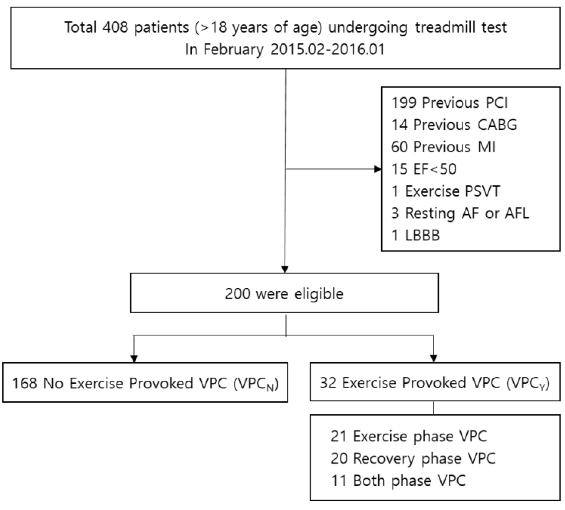
2.1. Study Population

Data of 408 patients (≥18 years of age) who underwent TMT for work up of angina, palpitation, dyspnea, syncope, or arrhythmia between February 2015, and January 2016, were collected with consent at Samsung Medical Center, Seoul, Republic of Korea. This study complied with the Declaration of Helsinki, and the research protocol was approved by the Ethics committee of Samsung Medical Center. Of these patients, 51% were referred for testing because of angina pectoris; 39% for non-ischemic chest pain, resting ECG abnormality, or elevated risk factors; 5% to evaluate arrhythmias; 3% for exercise capacity assessment; less than 2% for dyspnea on exertion; and the rest for miscellaneous reasons.

2.2. Data Collection

All exercise tests were supervised, and all results were read by two of the investigators (MJP and JHC). A thorough clinical history including medications and risk factors was recorded prospectively at the time of the exercise test using computerized forms.

2.3. Exercise Test

The treadmill test was conducted according to a symptom-limited Bruce protocol that divides step one to increase exercise capacity slowly. Exercise capacity was estimated in metabolic equivalents based on treadmill speed and grade, and 12-lead ECG data were recorded with Quinton Q-stress. An abnormal ST-segment response was defined as 1 mm or more of horizontal or down-sloping depression measured visually at the J junction. Underlying medical history, family history, follow-up data, and death was collected from hospital medical records. Exclusion criteria were previous history of coronary revascularization (percutaneous coronary intervention or coronary artery bypass graft), previous MI, left ventricular ejection fraction lower than 50%, arrhythmia during TMT, and any disease that could affect ST-segment change on ECG. A cardiovascular event was defined as a history of documented coronary artery disease on coronary angiography or computed tomography angiography, myocardial infarction (MI), stroke, percutaneous coronary intervention (PCI), or coronary artery bypass graft surgery (CABG).

2.4. Classification of VPC

Exercise-provoked VPC was noted if frequent VPCs (more than 10% of all VPCs during any 30-s ECG recording) or ventricular tachycardia (three or more consecutive VPCs) were visually detected during the exercise test or recovery. Resting VPC was recorded if a VPC was detected in the 10-s ECG prior to exercise. We divided the patients into the VPC group (VPCY) and the non-VPC group (VPCN) based on the results of the TMT. We defined VPCY as VPC that occurred during TMT (exercise phase or recovery phase) regardless of evidence of resting phase VPC. Further, we compared the clinical outcomes of patients with VPC according to phase, exercise (VPCE), or recovery (VPCR). Categorization of VPCE means that VPC occurred during the exercise phase of TMT, while VPCR is VPC that occurred during the recovery phase. There was an overlap between the VPCE and VPCR groups.

2.5. Statistics

Continuous variables were tested using an independent two-sample t-test and are presented as mean ± standard deviation. Categorical data were compared using the Chi-square test and are presented as numbers and relative frequency (%). The multivariable models comprised the covariates that were significant on univariate analysis (p < 0.1) or that were clinically relevant. All probability values were two-sided, and a p-value < 0.05 was considered statistically significant. Statistical analyses were performed using SPSS statistics 20 (SPSS Inc., Chicago, IL, USA).

3. Results

Among total of 408 patients, 208 were excluded according to the previous history of PCI or CABG, previous MI, decreased left ventricular ejection fraction lower than 50%, arrhythmia like paroxysmal supraventricular ventricular tachycardia during TMT, and any disease that could affect ST-segment change on ECG, such as atrial fibrillation/atrial flutter, bundle branch block, or Wolff-Parkinson-White syndrome. Finally, we enrolled 200 patients with a median follow-up of 32 months. Among these 200 patients, 32 patients (16.0%) developed VPC. Of these 32 patients, 21 were exercise VPC, 20 were recovery VPC and 11 were both exercise and recovery phase VPC (Figure 1).
In baseline characteristics of demographics, past medical history, clinical finding, and exercise test record, there was no significant deference between VPCY and VPCN; between VPCE and all others; or between VPCR group and all others. The VPCR group had greater family history of sudden death compared to the others (p = 0.014), while the VPCE group showed higher prevalence of positive TMT result (p = 0.042). (Table 1) Based on baseline characteristics, we analyzed clinical correlation with logistic regression in the VPCY, VPCE, and VPCR groups. The VPCY group (OR 5.786 (1.111–30.120)) and VPCR group (OR 10.875 (2.026–58.363)) showed significant correlation with family history of sudden cardiac death (Supplementary Table S1).
Among 200 enrolled patients, 20 (10%) showed positive TMT (TMT (+)). There was no significant difference in incidence of VPCY between TMT (+) and TMT (−) patients (25% vs. 15%, p = 0.329). In the study, 8 patients (4%) were diagnosed with significant coronary artery disease in coronary angiography. Further, 4 patients (2%) underwent revascularization. Among the TMT (+) patients, 5 (25%) were diagnosed with significant coronary artery disease in coronary angiography, a significantly higher percentage compared to the 1.7% in TMT (−) patients (p-value < 0.001). Among 5 patients, one (5.0%) underwent PCI and one (5.0%) underwent CABG during follow-up. The trend of revascularization rate was higher in patients with VPCY than with VPCN in TMT (+) patients (VPCY vs. VPCN: 20.0% vs. 6.7%, p = 0.447), but the difference was not significant.
To determine the relationship of VPC with TMT (+), we conducted univariable and multivariable logistic regression. In univariable logistic regression, VPCY and TMT (+) had no significant relation (OR = 1.889 (0.634–5.628)), p-value = 0.254). After dividing VPCY into specific phases, exercise phase VPC (OR = 3.417 (1.098–10.628), p-value = 0.034) showed significant relation with TMT (+), while recovery phase VPC (OR = 1.692 (0.450–6.367), p value = 0.437) did not. In multivariable analysis, VPCE had a relation with positive TMT in patients younger than 65 years (OR = 6.879 (1.458–32.453), p value = 0.015), after correcting for demographics and previous medical history (Table 2). In the prediction of coronary artery disease which is significant stenosis in more than 1 vessel confirmed by coronary angiography, age (OR = 1.157 (1.030–1.300), p value = 0.014) and positive TMT result (OR = 22.224 (3.267–151.154), p value = 0.002) was significantly related after correcting for sex, BMI and the presence of exercise phase VPC or recovery phase VPC. In the prediction of revascularization, VPCR had a relation with revascularization (OR = 9.381 (1.144–76.948), p-value = 0.037) after correcting for age, sex, BMI and TMT result(Table 3). When we compared baseline characteristics under the assumption that VPCE and VPCR groups were independent without overlap, there was no difference in demographics, previous medical history, or exercise test results between the two groups (Supplementary Table S2).

4. Discussion

With this study, we suggested that exercise-provoked VPC could be the sign of myocardial ischemia with respect to predicting revascularization events after TMT even in the Asian population.
With respect to revascularization, VPCR showed significant predictive value in our study. In the previous Framingham Heart Study [12], there was no relation between coronary heart disease (CHD) event and exercise-provoked VPC. However, another study [19] conducted on patients with suspected coronary artery disease showed a significant relation between CHD events and exercise-induced ventricular arrhythmia. Considering the results of those previous studies, the reason that exercise-provoked VPC and revascularization were found to be related in the present study seems to have been influenced by the analysis of symptomatic patients with suspected coronary artery disease, not the community population. Among exercise-induced VPCs, recovery phase VPCs are known to predict myocardial ischemia better than exercise-phase VPCs, which is also consistent with the results of our study [19,20]. This means that VPCR was closer to achieving actual clinical relevance than was VPCE. Another notable point in this study is that recovery phase VPC was associated with revascularization independently of TMT results. Therefore, even if the TMT result is negative, paying attention to the recovery phase VPC will be helpful in finding patients with significant epicardial coronary artery stenosis.
In our study, exercise phase VPC showed a relation with positive segments-segment change in TMT after adjusting for cardiovascular risk factors (age, sex, BMI, DM, HTN, dyslipidemia, history of CVA or PAD). Unlike the recovery phase VPC, the exercise phase VPC was not associated with epicardial coronary artery stenosis and clinical outcome but was correlated with ST depression. ST depression in ECG is also known to be associated with coronary microvascular dysfunction (CMD) in the absence of epicardial coronary artery stenosis [21]. However, in this study, since neither instantaneous flow ratio (IMR) nor coronary flow reserve (CFR) was measured during coronary angiography, the presence of CMD could not be confirmed. Based on these results, future studies are expected to verify the predictability of exercise phase VPC with coronary microvascular disease. The analysis of the relationship between exercise provoked VPC and TMT results was established in patients under 65 years of age in this study. Considering that TMT is mainly performed in ambulatory young patients, this restriction does not seem to have a significant impact on the interpretation of the results. Although there are reports that exercise-provoked VPC increased with age [9,10,12,22] and male sex [12], there was no such difference in incidence in the present study. Here, patients with resting phase VPC showed more frequent exercise-provoked VPC, in agreement with a previous study [22].
Exercise-provoked VPC patients with or without known coronary artery disease had a more frequent family history of sudden cardiac death, corresponding to a previous study [23]. This indicates that such patients could have unknown familial heart failure with preserved ejection fraction [24,25]. We excluded patients according to baseline ejection fraction. If we had excluded patients based on echocardiographic morphology, such as hypertrophy, valve abnormality, and regional wall motion abnormality, different results may have been observed.
There is no clear pathophysiology of exercise provoked VPC. Some studies have suggested that exercise-induced ventricular arrhythmia is associated with exercise-induced ischemia [7,8], though other studies with a myocardial infarction group or healthy population have reported opposing results [9,12]. It is possible that exercise-provoked VPC is caused by viable ventricular ischemic cells with altered electrical conductance.
The treadmill test is a useful modality in the outpatient clinic because it is simple, easy to interpret, shows the low inter-observer difference, and is inexpensive and noninvasive. Although the convenience and accessibility of TMT are good advantages, low negative predictive value and low positive predictive value are the limitations of TMT, especially compared with updated tools such as stress echocardiography or perfusion cardiovascular magnetic resonance imaging [3,26,27,28]. Exercise provoked VPC during TMT could compensate for this limitation of the TMT. There is much evidence that exercise-provoked VPC is related to cardiovascular mortality [10,11], but the predictive value combined with standard ST change criteria has not been established. There was no statistical difference in VPCY incidence based on TMT results in our study. If ST change and exercise-provoked VPC have different mechanisms, complementary effects would be expected for TMT.

Limitations

The present study population comprised patients visiting a hospital for specific symptoms. Because of this, they were relatively old and showed a higher percentage of VPC compared to another study of healthy asymptomatic patients [11]. Our results are more representative of those of a general ambulatory clinic. We defined exercise-induced VPC during TMT as any single VPC during the exercise or recovery phase with or without evidence of VPC on resting ECG. Thus, VPCY could overlap with resting phase VPC. However, based only on the 10-s baseline ECG, we cannot exclude resting phase VPC. Selection bias could have been produced by the required consent of the study population and by retrospective analysis of prospectively collected data. Although there also is a limitation as a single-center study, the residence of patients spread nationwide rather than being limited to Seoul because of center characteristics. We collected data from a large population over a short period (1 year), follow up period (32 months) was relatively short, and follow-up data were collected from in-hospital medical records. There were only three mortality cases without cardiovascular cause during follow-up, though there is a possibility of underestimated mortality cases. If we trace patients consistently and collect data based on in-hospital medical records and on the National Institute of Health, a reanalysis would produce a stronger conclusion.

5. Conclusions

Since the exercise phase, VPC is related to ST depression, and the recovery phase VPC could predict revascularization, exercise-provoked VPC during TMT can be used as a marker of myocardial ischemia.

6. Patents

This section is not mandatory but may be added if there are patents resulting from the work reported in this manuscript.

Supplementary Materials

The following supporting information can be downloaded at: https://www.mdpi.com/article/10.3390/medicina58040556/s1, Table S1: Clinical Correlation in exercise provoked VPC group; Table S2: Baseline characteristics of VPCE and VCPR.

Author Contributions

Conceptualization—K.-M.P.; Data curation—H.-J.K.; Investigation—S.-J.P.; Resources—J.-S.K.; Software—M.-J.B. and J.-H.C.; Writing—review and editing—Y.-K.O. and K.-M.P. All authors have read and agreed to the published version of the manuscript.

Funding

This research received no external funding.

Institutional Review Board Statement

The study was approved by the Ethics committee of Samsung Medical Center Approval Code: 2014-06-117 Approval Date: 11 July 2014.

Informed Consent Statement

Informed consent was obtained from all subjects involved in the study.

Data Availability Statement

Data sharing not applicable.

Conflicts of Interest

The authors declare no conflict of interest.

References

  1. Gianrossi, R.; Detrano, R.; Mulvihill, D.; Lehmann, K.; Dubach, P.; Colombo, A.; McArthur, D.; Froelicher, V. Exercise-induced ST depression in the diagnosis of coronary artery disease. A meta-analysis. Circulation 1989, 80, 87–98. [Google Scholar] [CrossRef] [PubMed] [Green Version]
  2. Pryor, D.B.; Harrell, F.E.; Lee, K.L.; Califf, R.M.; Rosati, R.A. Estimating the likelihood of significant coronary artery disease. Am. J. Med. 1983, 75, 771–780. [Google Scholar] [CrossRef]
  3. Gibbons, R.J.; Balady, G.J.; Bricker, J.T.; Chaitman, B.R.; Fletcher, G.F.; Froelicher, V.F.; Mark, D.B.; McCallister, B.D.; Mooss, A.N.; O’Reilly, M.G.; et al. ACC/AHA 2002 guideline update for exercise testing: Summary article. A report of the American college of cardiology/American Heart Association task force on practice guidelines (committee to update the 1997 exercise testing guidelines). J. Am. Coll. Cardiol. 2002, 40, 1531–1540. [Google Scholar] [CrossRef] [Green Version]
  4. Fletcher, G.F.; Ades, P.A.; Kligfield, P.; Arena, R.; Balady, G.J.; Bittner, V.; Coke, L.A.; Fleg, J.L.; Forman, D.E.; Gerber, T.C.; et al. Exercise standards for testing and training: A scientific statement from the American Heart Association. Circulation 2013, 128, 873–934. [Google Scholar] [CrossRef]
  5. Farzam, K.; Richards, J.R. Premature Ventricular Contraction (PVC); StatPearls: Treasure Island, San Francisco, FL, USA, 2020. [Google Scholar]
  6. Weiner, D.A.; Levine, S.R.; Klein, M.D.; Ryan, T.J. Ventricular arrhythmias during exercise testing: Mechanism, response to coronary bypass surgery and prognostic significance. Am. J. Cardiol. 1984, 53, 1553–1557. [Google Scholar] [CrossRef]
  7. Marieb, M.A.; Beller, G.A.; Gibson, R.S.; Lerman, B.B.; Kaul, S. Clinical relevance of exercise-induced ventricular arrhythmias in suspected coronary artery disease. Am. J. Cardiol. 1990, 66, 172–178. [Google Scholar] [CrossRef]
  8. Schweikert, R.A.; Pashkow, F.J.; Snader, C.E.; Marwick, T.H.; Lauer, M.S. Association of exercise-induced ventricular ectopic activity with thallium myocardial perfusion and angiographic coronary artery disease in stable, low-risk populations. Am. J. Cardiol. 1999, 83, 530–534. [Google Scholar] [CrossRef]
  9. Casella, G.; Pavesi, P.C.; Sangiorgio, P.; Rubboli, A.; Bracchetti, D. Exercise-induced ventricular arrhythmias in patients with healed myocardial infarction. Int. J. Cardiol. 1993, 40, 229–235. [Google Scholar] [CrossRef]
  10. Beckerman, J.; Mathur, A.; Stahr, S.; Myers, J.; Chun, S.; Froelicher, V. Exercise-induced ventricular arrhythmias and cardiovascular death. Ann. Noninvasive Electrocardiol. 2005, 10, 47–52. [Google Scholar] [CrossRef]
  11. Jouven, X.; Zureik, M.; Desnos, M.; Courbon, D.; Ducimetière, P. Long-term outcome in asymptomatic men with exercise-induced premature ventricular depolarizations. New Engl. J. Med. 2000, 343, 826–833. [Google Scholar] [CrossRef]
  12. Morshedi-Meibodi, A.; Evans, J.C.; Levy, D.; Larson, M.; Vasan, R.S. Clinical correlates and prognostic significance of exercise-induced ventricular premature beats in the community: The framingham heart study. Circulation 2004, 109, 2417–2422. [Google Scholar] [CrossRef] [Green Version]
  13. Partington, S.; Myers, J.; Cho, S.; Froelicher, V.; Chun, S. Prevalence and prognostic value of exercise-induced ventricular arrhythmias. Am. Hear. J. 2003, 145, 139–146. [Google Scholar] [CrossRef] [Green Version]
  14. Marine, J.E.; Shetty, V.; Chow, G.V.; Wright, J.G.; Gerstenblith, G.; Najjar, S.S.; Lakatta, E.G.; Fleg, J.L. Prevalence and prognostic significance of exercise-induced nonsustained ventricular tachycardia in asymptomatic volunteers: BLSA (Baltimore Longitudinal Study of Aging). J. Am. Coll. Cardiol. 2013, 62, 595–600. [Google Scholar] [CrossRef] [Green Version]
  15. Sami, M.; Chaitman, B.; Fisher, L.; Holmes, D.; Fray, D.; Alderman, E. Significance of exercise-induced ventricular arrhythmia in stable coronary artery disease: A coronary artery surgery study project. Am. J. Cardiol. 1984, 54, 1182–1188. [Google Scholar] [CrossRef]
  16. McHenry, P.L.; Morris, S.N.; Kavalier, M.; Jordan, J.W. Comparative study of exercise-induced ventricular arrhythmias in normal subjects and patients with documented coronary artery disease. Am. J. Cardiol. 1976, 37, 609–616. [Google Scholar] [CrossRef]
  17. Bruce, R.A.; DeRouen, T.A.; Hossack, K.F.; Blake, B.; Hofer, V.R. Value of maximal exercise tests in risk assessment of primary coronary heart disease events in healthy men: Five years’ experience of the Seattle heart watch study. Am. J. Cardiol. 1980, 46, 371–378. [Google Scholar] [CrossRef]
  18. Drory, Y.; Pines, A.; Fisman, E.Z.; Kellermann, J.J. Persistence of arrhythmia exercise response in healthy young men. Am. J. Cardiol. 1990, 66, 1092–1094. [Google Scholar] [CrossRef]
  19. Elhendy, A.; Chandrasekaran, K.; Gersh, B.J.; Mahoney, D.; Burger, K.N.; Pellikka, P.A. Functional and prognostic significance of exercise-induced ventricular arrhythmias in patients with suspected coronary artery disease. Am. J. Cardiol. 2002, 90, 95–100. [Google Scholar] [CrossRef]
  20. Meine, T.J.; Patel, M.R.; Shaw, L.K.; Borges-Neto, S. Relation of ventricular premature complexes during recovery from a myocardial perfusion exercise stress test to myocardial ischemia. Am. J. Cardiol. 2006, 97, 1570–1572. [Google Scholar] [CrossRef]
  21. Lanza, G.A.; De Vita, A.; Kaski, J.-C. ‘Primary’ microvascular angina: Clinical characteristics, pathogenesis and management. Interv. Cardiol. Rev. 2018, 13, 108–111. [Google Scholar] [CrossRef] [PubMed]
  22. Dewey, F.E.; Kapoor, J.R.; Williams, R.S.; Lipinski, M.J.; Ashley, E.A.; Hadley, D.; Myers, J.; Froelicher, V.F. Ventricular arrhythmias during clinical treadmill testing and prognosis. Arch. Intern. Med. 2008, 168, 225–234. [Google Scholar] [CrossRef] [Green Version]
  23. Abdalla, I.S.; Prineas, R.J.; Neaton, J.D.; Jacobs, D.R.J.; Crow, R.S. Crow RS: Relation between ventricular premature complexes and sudden cardiac death in apparently healthy men. Am. J. Cardiol. 1987, 60, 1036. [Google Scholar] [CrossRef]
  24. Spirito, P.; Chiarella, F.; Carratino, L.; Berisso, M.Z.; Bellotti, P.; Vecchio, C. Clinical course and prognosis of hypertrophic cardiomyopathy in an outpatient population. N. Engl. J. Med. 1989, 320, 749–755. [Google Scholar] [CrossRef]
  25. Fontaine, G.; Fontaliran, F.; Frank, R. Arrhythmogenic right ventricular cardiomyopathies: Clinical forms and main differential diagnoses. Circulation 1998, 97, 1532–1535. [Google Scholar] [CrossRef] [PubMed] [Green Version]
  26. Bourque, J.M.; Beller, G.A. Value of exercise ECG for risk stratification in suspected or known CAD in the era of advanced imaging technologies. JACC Cardiovasc. Imaging 2015, 8, 1309–1321. [Google Scholar] [CrossRef] [Green Version]
  27. Klem, I.; Heitner, J.F.; Shah, D.J.; Sketch, M.H.; Behar, V.; Weinsaft, J.; Cawley, P.; Parker, M.; Elliott, M.; Judd, R.M.; et al. Improved detection of coronary artery disease by stress perfusion cardiovascular magnetic resonance with the use of delayed enhancement infarction imaging. J. Am. Coll. Cardiol. 2006, 47, 1630–1638. [Google Scholar] [CrossRef] [Green Version]
  28. Dang, G.; Shahreyar, M.; Oktay, A.A.; Sulaiman, S.; Husein, H.; Inashvili, A.; Elhangouche, N.; Subramanian, P.; Bashir, M.W.; Pathak, A.; et al. Positive predictive value of noninvasive stress testing is poor in the real world. Crit. Pathw. Cardiol. 2018, 17, 38–42. [Google Scholar] [CrossRef]
Figure 1. Study flow chart with number of patients.
Medicina 58 00556 g001
Table 1. Baseline characteristics (n = 200).
VPCY
(n = 32)
VPCN
(n = 168)
p Value
(VPCY vs. VPCN)
VPCE+
(n = 21)
p Value
(VPCE vs. Others)
VPCR
(n = 20)
p Value
(VPCR vs. Others)
Demographics
Age56.4 ± 16.652.7 ± 14.00.18057.7 ± 16.10.13953.8 ± 17.90.869
Sex (male)27 (84.4%)122 (72.6%)0.1623 (14.3%)0.2135 (25.0%)0.957
Height167.0 ± 7.7167.0 ± 8.70.988166.3 ± 8.00.696165.7 ± 7.20.495
Weight68.4 ± 9.468.7 ± 11.90.88869.8 ± 10.20.64967.3 ± 10.10.586
BMI24.5 ± 2.824.6 ± 3.10.95125.2 ± 2.80.31924.5 ± 2.70.907
Family Hx
FHxCAD4 (12.5%)14 (8.3%)0.4984 (19.0%)0.1033 (15.0%)0.399
FHxSCD3 (9.4%)3 (1.8%)0.0531 (4.8%)0.4913 (15.0%)0.014
Pre. Medical Hx
DM5 (15.6%)23 (13.7%)0.7823 (14.3%)1.0003 (15.0%)1.000
HTN12 (37.5%)71 (42.3%)0.6169 (42.9%)0.8949 (45.0%)0.738
Dyslipidemia7 (21.9%)55 (32.7%)0.2233 (14.3%)0.0806 (30.0%)0.919
CVA3 (9.4%)7 (4.2%)0.2022 (9.5%)0.2832 (10.0%)0.263
PAD2 (6.2%)14 (8.3%)1.0002 (9.5%)0.6782 (10.0%)0.665
Clinical finding
Resting HR79.7 ± 11.577.0 ± 14.20.29979.3 ± 11.50.49778.5 ± 11.80.719
Resting SBP127.8 ± 18.2124.5 ± 19.00.379131.3 ± 18.50.110126.0 ± 17.50.814
ECG finding
QRS duration95.5 ± 21.395.3 ± 15.80.95397.4 ± 25.80.55193.8 ± 13.80.673
QTc422.6 ± 34.6424.3 ± 27.80.754426.5 ± 35.20.683425.9 ± 38.10.762
VPC at rest7 (21.9%)3 (1.8%)0.0006 (28.6%)0.0005 (25.0%)0.001
Exercise test
Maximal HR157.0 ± 23.2156.8 ± 25.10.954154.1 ± 20.70.604158.1 ± 24.70.805
Maximal SBP190.1 ± 32.4180.3 ± 27.30.074190.8 ± 35.00.130185.9 ± 33.50.507
Exercise capacity11.3 ± 2.311.6 ± 2.10.46811.4 ± 2.00.80911.2 ± 2.70.402
TMT positive5 (15.6%)15 (8.9%)0.3295 (23.8%)0.0423 (15.0%)0.430
Data are presented as mean ± standard deviation or n (%). Abbreviations: BMI, body mass index; FHx, family history; CAD, coronary artery disease; SCD, sudden cardiac death, DM, diabetes mellitus; HTN, hypertension; CVA, cerebro-vascular accident; PAD, peripheral artery disease; HR, heart rate; SBP, systolic blood pressure; VPC, ventricular prem-ature contracture; TMT, treadmill.
Table 2. Risk for TMT positive in exercise provoked VPC group (n = 153, age ≤ 65).
TMT Positive Result
UnivariableMultivariable
Age1.105 (1.030–1.186)1.118 (1.036–1.207)
Sex 0.182 (0.023–1.426)
BMI1.012 (0.855–1.199)
DM1.892 (0.483–7.410)
HTN2.571 (0.901–7.343)
Dyslipidemia2.538 (0.890–7.237)
CVA 6.429 (0.989–41.790)
PAD1.248 (0.144–10.843)
Exercise phase VPC4.778 (1.279–17.849)6.879 (1.458–32.453)
Recovery phase VPC1.500 (0.304–7.396)
Data are presented as odds ratios (95% confidence interval).
Table 3. Risk for CAD and revascularization in exercise provoked VPC group (n = 200).
Coronary Artery DiseaseRevascularization
UnivariableMultivariableUnivariableMultivariable
Age1.095 (1.028–1.165)1.108 (1.026–1.196)1.083 (0.984–1.192)
Sex0.000 (0.000–0.000) 0.000 (0.000–0.000)
BMI1.045 (0.857–1.275) 0.967 (0.697–1.342)
TMT positive result1.930 (0.566–6.581)7.547 (1.812–31.434)2.267 (0.312–16.472)8.797 (1.073–72.127)
Exercise phase VPC1.988 (0.400–9.886) 2.933 (0.291–29.550)
Recovery phase VPC2.111 (0.423–10.533) 9.889 (1.313–74.473)9.381 (1.144–76.948)
Data are presented as odds ratios (95% confidence interval). Included variable: age, sex, BMI, TMT positive result, exercise phase VPC, recovery phase VPC.
Publisher’s Note: MDPI stays neutral with regard to jurisdictional claims in published maps and institutional affiliations.

