Communication

The Ethyl Acetate Extract of Caulerpa microphysa Promotes Collagen Homeostasis and Inhibits Inflammation in the Skin

1
Department of Nursing, Division of Basic Medical Sciences, Chang-Gung University of Science and Technology, Taoyuan 333, Taiwan
2
Department of General Surgery, Chang Gung Memorial Hospital at Linkou, Taoyuan 333, Taiwan
3
Graduate Institute of Oral Biology, School of Dentistry, National Taiwan University, Taipei 100, Taiwan
4
Institute of Environmental and Occupational Health Sciences, College of Public Health, National Taiwan University, Taipei 100, Taiwan
5
Department of Dermatology, Drug Hypersensitivity Clinical and Research Center, Chang Gung Memorial Hospital, Taoyuan 333, Taiwan
6
Graduate Institute of Clinical Dentistry, School of Dentistry, National Taiwan University, Taipei 100, Taiwan
7
Department of Dentistry, National Taiwan University Hospital, Taipei 100, Taiwan
*
Author to whom correspondence should be addressed.
These authors contributed equally to this work.
Curr. Issues Mol. Biol. 2024, 46(3), 2701-2712; https://doi.org/10.3390/cimb46030170
Submission received: 2 March 2024 / Revised: 18 March 2024 / Accepted: 18 March 2024 / Published: 21 March 2024
(This article belongs to the Special Issue The Role of Bioactives in Inflammation)

Abstract

:
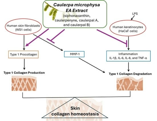
Inflammation and collagen-degrading enzymes’ overexpression promote collagen decomposition, which affects the structural integrity of the extracellular matrix. The polysaccharide and peptide extracts of the green alga Caulerpa microphysa (C. microphysa) have been proven to have anti-inflammatory, wound healing, and antioxidant effects in vivo and in vitro. However, the biological properties of the non-water-soluble components of C. microphysa are still unknown. In the present study, we demonstrated the higher effective anti-inflammatory functions of C. microphysa ethyl acetate (EA) extract than water extract up to 16–30% in LPS-induced HaCaT cells, including reducing the production of interleukin (IL)-1β, IL-6, IL-8, and tumor necrosis factor-α (TNF-α). Furthermore, the excellent collagen homeostasis effects from C. microphysa were proven by suppressing the matrix metalloproteinase-1 (MMP-1) secretion, enhancing type 1 procollagen and collagen expressions dose-dependently in WS1 cells. Moreover, using UHPLC-QTOF-MS analysis, four terpenoids, siphonaxanthin, caulerpenyne, caulerpal A, and caulerpal B, were identified and may be involved in the superior collagen homeostasis and anti-inflammatory effects of the C. microphysa EA extract.

Graphical Abstract

1. Introduction

Seaweed, also known as marine macroalgae, mainly grows in coastal intertidal zones and most types of seaweed are common edible sea vegetables for coastal residents. The intertidal habitat is harsh, especially in the high- and mid-tidal areas. Green algae, which comprise the main seaweed growing in the high- and mid-tidal zones must adapt to daily tidal changes and resist strong sunlight and high temperature. The green algae produce many adaptive bioactive compounds and metabolites to protect against environmental damage [1,2,3], including phenolic compounds [4,5], pigments [6,7], polysaccharides [8,9,10], terpenoids [11,12,13], etc.
In recent years, research has shown that these active metabolites of algae also exhibit the potential for a variety of pharmacological applications. For example, water-soluble polysaccharides from some green algae have been discovered to have antioxidant and immunomodulatory activities [14,15]. A variety of water-insoluble terpenoids from seaweed also have been identified to have diverse physiological activities, such as carotenoids, tetraterpene pigments, which are the most widely distributed pigments in nature and are also found in large amounts in photosynthetic seaweeds. Their conjugated double-bond structures are considered extremely effective natural antioxidants because of their ability to scavenge reactive oxygen species [16,17]. Siphonaxanthin is a marine carotenoid that exists in green algae. It is an oxidative metabolite of lutein. Its structure is similar to lutein but with one keto group located at C-8 and an extra hydroxyl group at C-19 [17]. Siphonaxanthin has been shown to have angiogenesis-inhibiting, anti-inflammatory, and lipid metabolism-regulating activities [12,18,19,20,21]. Caulerpenyne, a sesquiterpene metabolite of Caulerpa, has shown antioxidant, antiproliferative, antibiotic, and antiviral activities [13,22,23,24]. Caulerpal A and B are two sesquiterpenes isolated from Caulerpa taxifolia with an unusual aromatic pentene carbon skeleton and caulerpin. These two metabolites exhibit a broad spectrum of antifungal activity [25].
The green alga Caulerpa microphysa (C. microphysa) is an edible green alga native to the intertidal zones of Taiwan, China, and the Philippines. C. microphysa belongs to the green algae genus Caulerpa. The thallus is composed of a horizontal stolon connected by rhizoids and erect stems connected by rounded vesicular fronds. Its appearance is similar to C. lentillifera but with shorter erect stems and smaller bulbous fronds. C. microphysa grows all year round but grows slowly in summer due to the hot sun and high temperatures. Previous studies have found that its polysaccharide extract has anti-inflammatory and wound healing effects in 3T3-L1 fibroblasts [26], as well as antioxidant and anticoagulant effects in an ex vivo study [27]. Peptides extracted from C. microphysa also exhibit antitumor properties in human promyelocytic leukemia cells (HL-60) [28]. However, there is a lack of literature mentioning the effect of C. microphysa on dermatology and discussing the physiological activities of the non-water-soluble components of this edible alga.
Type I collagen is one of the most abundant molecules in the body, especially in the skin, bones, and connective tissues [29]. In the skin, it accounts for three-quarters of the dry weight of skin and is highly expressed in fibroblasts [30,31]. When fibroblasts complete triple-helical procollagen synthesis, the procollagen is secreted into the extracellular matrix (ECM) and cleaves the carboxy terminals and the amino terminals through proteinases. The terminal-cutting procollagen chains, tropocollagen, combine with each other to form collagen fibers [32,33,34]. Collagen homeostasis means the stability of collagen content in ECM, which is determined by the balance of synthesis and degradation. Previous studies have indicated that collagen is degraded during various normal physiological processes, such as wound healing and skin aging. In addition, pathological conditions such as inflammation, atherosclerotic cardiovascular disease, and sunburn accelerate collagen degradation [35,36,37]. Members of the MMP family are involved in collagen degradation [38,39], especially MMP-1, known as interstitial collagenase, which is primarily responsible for the degradation of dermal type I collagen. MMP-1 is expressed in unstimulated fibroblasts and upregulated by inflammation and photoaging [40,41,42].
The aim of this study was to clarify whether the non-water-soluble components of C. microphysa have physiological effects on the inflammation and collagen homeostasis of skin cells and to further analyze which potential secondary metabolites are present in the water-insoluble extract of C. microphysa. In a comparison of the anti-inflammatory effects between the water extract and EA extract of C. microphysa on keratinocytes, surprisingly, the EA extract is more effective than the water extract in inhibiting LPS-induced IL-1β, IL-6, IL-8, and TNF-α expressions. For human fibroblasts, the EA extract of C. microphysa also exhibits superior collagen homeostasis effects by increasing type I procollagen and collagen expressions and suppressing MMP-1, the interstitial collagen-degrading enzyme, secretion. By UHPLC-QTOF-MS analysis, we observed the presence of four terpenoid compounds, which are siphonaxanthin, caulerpenyne, caulerpal A, and caulerpal B in the C. microphysa EA extract. Whether these four terpenoid compounds are involved in the anti-inflammatory and collagen-homeostasis effects of C. microphysa EA extract remains to be further clarified. This experiment was the first to verify the superior anti-inflammatory and collagen-homeostasis effects of the water-insoluble extract of C. microphysa on dermatology and to analyze its potential secondary metabolites.

2. Materials and Methods

2.1. Cells

A human epidermal keratinocyte cell line, HaCaT (300493, Cell Lines Service GmbH, Eppelheim, Deutschland), was cultured in DMEM (with 10% FBS, 2 mM L-glutamine, 4.5 g/L glucose, and 1% Penicillin–Streptomycin–Neomycin Solution) in a 37 °C oven with 5% CO2. Human skin fibroblast, WS1 cells (60300, BCRC), was cultured in MEM (with 10% FBS and 1% Penicillin–Streptomycin–Neomycin Solution) in a 37 °C oven with 5% CO2. The split ratio in both cells was 1:3. Finally, 0–200 ng/mL LPS was applied to trigger cell inflammation.

2.2. Preparation of Caulerpa microphysa Extracts

The Caulerpa microphysa (C. microphysa), after morphological identification, was cultivated in Penghu waters, Taiwan, and harvested in autumn. After washing with reverse osmosis water, it was homogenized 3 times at 12,000 rpm for 5 min. The homogenate was filtrated and centrifuged with 6000× g at 4 °C for 10 min; then, we collected the supernatant (the supernatant/fresh specimens was about 55%). The supernatant was filtered using a 0.22 μm filter; then, we added EA to the filtrate at a ratio of 1:1 by weight and used a separatory funnel. After violently shaking to mix, we let it stand to separate the lower water layer (WL) and upper EA layer (EAL). Each layer was collected using five repeats for separation. Then, the EAL was concentrated with a reduced pressure vacuum concentrator (BUCHI/ROTAVAPOR R-300, Taipei, Taiwan) in 80 mbar and 45 °C conditions. The yield was 0.015%. The WL was dried using a freeze dryer (Yihua/FDS-5B-0.5-LT, New Taipei City, Taiwan).

2.3. Ultra-Performance Liquid Chromatography (UPLC) and Quadrupole Time-of-Flight Mass Spectrometry (QTOF)

We took the 17-fold concentrated EAL of C. microphysa for UHPLC-QTOF MS analysis. The analysis was performed using an Agilent 1290 Infinity II LC System (Agilent Technologies, Santa Clara, CA, USA) coupled with an Agilent 6545 LC/QTOF MS. The extracts were separated on a Waters BEH C18 column (100 × 2.1 mm, 1.7 µm). The mobile phases consisted of (A) 5 mM ammonium acetate(aq) and (B) methanol. The liquid chromatography gradient started at 5% of (B) for 30 s and increased to 60% of (B) at 7 min; then, it turned to 100% of (B) at 14 min, which was held for 3 min; then, it was decreased to the initial proportion and re-equilibrated for 2.5 min. The flow rate was 0.3 mL/min, the injection volume was 6 μL, and the column temperature was 40 °C. The QTOF MS was conducted on an Agilent Jet Stream ESI positive mode, with a mass range of m/z 100 to 1500 at a scan rate of 3 spectra per second. Accurate mass measurements by the instrument were ensured using an automated calibrant delivery system that continuously introduced a reference solution with a mass mix of m/z 121.050873 (purine) and m/z 922.009798 (HP-0921) in the ESI-positive mode. The parameters set for ESI-MS included the following: drying gas temperature: 325 °C; drying gas flow rate: 9 L/min; nebulizer gas pressure: 45 psi; sheath gas temperature: 350 °C; sheath gas flow: 11 L/min; capillary voltage: 4500 V; fragmentor voltage: 175 V; radiofrequency voltage in the octupole: 750 V; and fixed collision energies of 0 eV, 20 eV, and 40 eV. Data acquisition was performed on Mass Hunter Workstation Software Data Acquisition for Q-TOF, version B.06.01.

2.4. WST-1

The cellular number was evaluated by WST-1 assay (Roche, Basel, Switzerland) based on the manufacturer’s instruction. At specified time intervals, the cultured cells underwent a pulsing regimen with the oxidation–reduction indicator WST-1 (10% vol/vol) for a duration of 4 h. The color development (A450 nm–A600 nm) was then quantified to assess the activity of mitochondrial dehydrogenases. This activity was deemed proportional to the quantity of viable cultured cells and expressed as the cell proliferating index [43]. The outcome following pre-incubation (on 0 h) served to adjust the results as a ratio.

2.5. Enzyme-Linked Immunosorbent Assay (ELISA)

Cellular media from the cells were analyzed by IL-1b (DLB50), IL-6 (D6050B), IL-8 (D8000C), TNF-α (DTA00D), MMP-1 (DY901B), and type-1 collagen (DY6220-05) kits (R & D system, Minneapolis, MN, USA) according to the manufacturer’s instructions. The limits of detection of the above kits were 3.9, 3.1, 31.2, 15.6, 62.5, and 31.2 pg/mL, respectively.

2.6. Western Blot

Cells were lysed using RIPA lysis buffer (Genestar, Taipei, Taiwan) containing 1% NP-40, 0.1% SDS, 150 mM sodium chloride, 0.5% sodium deoxycholate, and 50 mM Tris. The cell lysates were centrifuged at 12,000 rpm for 5 min before the supernatant was collected. The extracted protein was quantified by protein assay. Equal amounts of protein were separated using 10% SDS-polyacrylamide gel electrophoresis and transferred to Immobilon-P membranes (Millipore, Burlington, MA, USA). After blocking with 5% skimmed milk, the membranes were incubated with Collagen I pAb (A16891) (ABclonal, Woburn, MA, USA) (1:1000) and then incubated with the Goat anti-Rabbit IgG (H + L)-HRP (C04003, Croyez, Taipei, Taiwan) (1:5000). The protein bands were detected using an Immobilon Western Chemiluminescent HRP Substrate (Millipore, Burlington, MA, USA) and quantified by the ImageQuant 5.2 software (GE Healthcare Bio-Sciences, Pittsburgh, PA, USA).

2.7. Statistical Analyses

We used the two-tailed Student’s t-test to compare the difference between two groups with different treatments. For the comparisons among data from more than two experimental conditions, we first used ANOVA to check the existence of differences; then, post hoc Tukey’s test was used to identify significant differences between two specific groups. A p value less than 0.05 was considered statistically significant.

3. Results

The cellular viability of HaCaT was tested under various doses of LPS, EAL, or WL extracts of C. microphysa. The results of WST-1 indicated that 200 ng/mL LPS and a high dose of C. microphysa’s EAL and WL (200 mg/mL) affected HaCaT cell viability (Figure 1). Therefore, we chose 100 mg/mL as the maximum treating concentration for both C. microphysa extracts and 100 ng/mL for the LPS treatment, which had triggered HaCaT cells to secrete inflammatory markers such as IL-1 β, IL-6, IL-8, and TNF-α (Figure 2).
To compare the anti-inflammatory effects between the EAL and WL extracts of C. microphysa in LPS-induced keratinocyte inflammation, we pretreated HaCaT cells with 100 mg/mL of EAL or WL extraction of C. microphysa for 2 h; then, we treated with 100 ng/mL LPS (L). The results of the ELISA assay showed that both the EAL and WL extracts of C. microphysa could attenuate LPS-increased IL-1 β, IL-6, IL-8, and TNF-α secretion (Figure 3). Notably, the C. microphysa EA extract significantly inhibited LPS-induced IL-1 β, IL-6, IL-8, and TNF-α more than the water extract by up to 16–30% in HaCaT cells. This result implies that the anti-inflammatory components of C. microphysa should be abundant in the water-insoluble layer.
In addition to the anti-inflammatory effects of the C. microphysa EA extract in keratinocytes, we also designed a collagen homeostasis experiment to detect the physiological activity of the EAL extract of C. microphysa on the human fibroblasts, WS1. In the extracellular matrix, collagen homeostasis mainly depends on the balance between collagen production and breakdown. Therefore, we investigated the effects of the C. microphysa EA extract on the production of type I collagen and the secretion of type I procollagen and MMP-1 from WS1 cells, respectively. WS1 cells were treated with a 0–100 mg/mL EA extract of C. microphysa for 24 h; then, we collected the cellular medium. The data of the ELISA indicated that the C. microphysa EA extract dose-dependently elevated the type I procollagen secretion but decreased the MMP-1 level in the WS1 cell medium (Figure 4). Furthermore, according to the result of the Western blot, the C. microphysa EA extract dose-dependently triggered type I collagen expression up to 3.8-fold in WS1 cells (Figure 5). The above results show that the non-water-soluble compounds of C. microphysa contribute to collagen homeostasis in the extracellular matrix. They may have great potential for dermatological applications.
In order to further understand which potential secondary metabolites contained in C. microphysa EA extract may contribute to the abovementioned anti-inflammation and collagen homeostasis, we applied UHPLC-QTOF-MS analysis in positive ionization mode to identify the C. microphysa EA extract. The total ion chromatogram of the EA extract of C. microphysa showed several peaks, suggesting the presence of several components in the extract (Figure 6A). The data were processed with Agilent MassHunter Qualitative Analysis Software (Version B.07.00), using the Find by Formula algorithm to view the output data after instrumental analysis. After the suspect screening, four terpenoid metabolites of the C. microphysa EA extract were identified, including siphonaxanthin (C40H56O4), caulerpenyne (C21H26O6), caulerpal A (C17H20O4), and caulerpal B (C16H20O3) (Figure 6C–F).

4. Discussion

This is the first study that demonstrated that C. microphysa EA extract effectively inhibited the LPS-induced inflammatory response in human skin keratinocytes and promoted collagen homeostasis by human fibroblasts.
Collagen is the main protein of the ECM. Collagen homeostasis, the balance between synthesis and degradation, is tightly regulated for the structural integrity of the ECM and is critical for tissue physiological homeostasis and repair [44]. Type I collagen is the most abundant ECM collagen in the skin, bones, and connective tissues [29], which is degraded during various normal physiological processes, such as wound healing and skin aging, and pathological conditions such as inflammation, atherosclerotic cardiovascular disease, and sunburn [35,36,37]. MMP-1, also known as interstitial collagenase, is primarily responsible for the degradation of dermal type I collagen. It is secreted by unstimulated fibroblasts involved in collagen homeostasis and upregulated by inflammation and photoaging [40,41,42].
Inflammation is a series of protective responses for the host defense system of the skin, which is exposed to various external stimuli throughout life. Even in physiological cellular aging, the pro-inflammatory cytokine, IL-1α, was upregulated in senescent keratinocytes [45]. The chronic inflammatory condition in senescent skin accelerates collagen loss and skin thinning. Further, immune cells activated by inflammation or other stimulators produce proteases (e.g., MMPs), cytokines (e.g., IL-1β, IFN-α), growth factors (e.g., TGF-β), and ECM components (e.g., fibronectin) and then participate in the remodeling of ECM components [46,47]. On the other hand, collagen peptides were found to suppress the LPS-induced inflammation in fibroblasts and keratinocytes [47]. The above experiments seem to imply that there is dynamic crosstalk between anti-inflammation and collagen stability in skin. Most anti-inflammatory studies on green algae have focused on the activity of its polysaccharides [26,48], but there has been a lack of research on the non-water-soluble components of green algae. In this present study, we demonstrated that the water-insoluble extract of C. microphysa not only has superior anti-inflammatory effects in keratinocytes but also has great potential to stimulate fibroblasts to express type I collagen and to decrease the collagen-degrading enzyme, MMP-1, secretion. Therefore, regardless of the causal relationship between anti-inflammation and collagen stabilization, the EA extract of C. microphysa is clearly beneficial for collagen homeostasis. And the skin-protective effects of C. microphysa EA extract imply the pharmacological and cosmetic application potential of this edible green alga.
In addition, in order to explore the possible secondary metabolites responsible for the beneficial effects on anti-LPS-induced inflammation and collagen production in skin cells, we analyzed the C. microphysa EA extract by UPLC with QTof and identified four terpenoids. They are siphonaxanthin, caulerpenyne, caulerpal A, and caulerpal B, respectively. Siphonaxanthin is a specific keto-carotenoid, which was isolated from green algae and is found to suppress advanced glycation end product-induced inflammatory responses in RAW264 macrophages [19]. It also has been shown to have angiogenesis-inhibiting and lipid metabolism-regulating activities [18,20,21]. Caulerpenyne is a sesquiterpene metabolite of Caulerpa and was extracted from Caulerpa taxifolia, which exhibits antiproliferative effects on the SK-N-SH tumor cell line and alters the structure of the microtubule network [49]. It also shows antioxidant, antiproliferative, antibiotic, and antiviral activities [13,22,23,24]. Caulerpal A and B are the sesquiterpenes with a unique aromatic valerenane-type carbon structure, and they exhibit a broad spectrum of antifungal activity [25]. However, standards for these four terpenoids are not readily available; so, we have no further insight into their content in C. microphysa EA extracts. Furthermore, the contributions of these four terpenoid compounds in the anti-inflammatory and collagen-homeostasis effects of C. microphysa EA extract remain to be further clarified.
For a long time, research on the physiological activities of green algae has mostly focused on water-soluble components. However, compared to the water-soluble extract, the non-water-soluble extract not only avoids the interference of salt on osmolarity but usually contains a variety of antioxidants, pigments, and fragrance components. Through this study, we also proved the excellent anti-inflammatory and anti-wrinkle effects of C. microphysa EA extract. Therefore, for the cosmetics industry, the non-water-soluble extract of C. microphysa is a potential raw material with multiple functions.
In summary, the present study was the first to verify the superior anti-inflammatory activities in keratinocytes and type I collagen-homeostasis enhancing effects in fibroblasts of the water-insoluble extract of C. microphysa, which contains several interpenoids, including siphonaxanthin, caulerpenyne, caulerpal A, and caulerpal B. This is the first time the potential of the non-water-soluble extract of C. microphysa for dermatological and cosmetic applications has been suggested.

Author Contributions

Conceptualization, K.-Y.L. and H.-H.H.; Validation, Z.-C.H.; Investigation, Z.-C.H. and Z.-Y.C.; Writing—original draft, K.-Y.L. and H.-H.H.; Writing—review and editing, L.-C.C. and C.-W.W. All authors have read and agreed to the published version of the manuscript.

Funding

This research received a grant from the College of Medicine, National Taiwan University (111M116).

Data Availability Statement

The data presented in this study are available on request from the corresponding author.

Acknowledgments

In addition, thanks to Thalassa Mystery Biotech Co., Ltd. for providing C. microphysa for this study.

Conflicts of Interest

The authors declare no conflicts of interest.

References

  1. Domozych, D.S.; LoRicco, J.G. The extracellular matrix of green algae. Plant Physiol. 2023, 194, 15–32. [Google Scholar] [CrossRef] [PubMed]
  2. Popper, Z.A.; Michel, G.; Hervé, C.; Domozych, D.S.; Willats, W.G.; Tuohy, M.G.; Kloareg, B.; Stengel, D.B. Evolution and diversity of plant cell walls: From algae to flowering plants. Annu. Rev. Plant Biol. 2011, 62, 567–590. [Google Scholar] [CrossRef] [PubMed]
  3. de Vries, J.; Stanton, A.; Archibald, J.M.; Gould, S.B. Streptophyte Terrestrialization in Light of Plastid Evolution. Trends Plant Sci. 2016, 21, 467–476. [Google Scholar] [CrossRef] [PubMed]
  4. Kumar, S.; Abedin, M.M.; Singh, A.K.; Das, S. Role of phenolic compounds in plant-defensive mechanisms. In Plant Phenolics in Sustainable Agriculture; Lone, R., Shuab, R., Kamili, A., Eds.; Springer: Singapore, 2020; pp. 517–533. [Google Scholar]
  5. Abu-Ghannam, N.; Rajauria, G. Antimicrobial activity of compounds isolated from algae. In Functional Ingredients from Algae for Foods and Nutraceuticals; Domínguez, H., Ed.; Woodhead Publishing: Cambridge, UK, 2013; pp. 287–306. [Google Scholar]
  6. Pangestuti, R.; Kim, S.K. Biological activities and health benefit effects of natural pigments derived from marine algae. J. Funct. Foods 2011, 3, 255–2667. [Google Scholar] [CrossRef]
  7. Hegazi, M.M.; Perez-Ruzafa, A.; Almela, L.; María-Emilia, C. Separation and identification of chlorophylls and carotenoids from Caulerpa prolifera, Jania rubens and Padina pavonica by reversed-phase high-performance liquid chromatography. J. Chromatogr. A 1998, 829, 153–159. [Google Scholar] [CrossRef]
  8. Wahlström, N.; Nylander, F.; Malmhäll-Bah, E.; Sjövold, K.; Edlund, U.; Westman, G.; Albers, E. Composition and structure of cell wall ulvans recovered from Ulva spp. along the Swedish west coast. Carbohydr. Polym. 2020, 233, 115852. [Google Scholar] [CrossRef]
  9. Domozych, D.S.; Bagdan, K. The cell biology of charophytes: Exploring the past and models for the future. Plant Physiol. 2022, 190, 1588–1608. [Google Scholar] [CrossRef]
  10. Kloareg, B.; Badis, Y.; Cock, J.M.; Michel, G. Role and Evolution of the Extracellular Matrix in the Acquisition of Complex Multicellularity in Eukaryotes: A Macroalgal Perspective. Genes 2021, 12, 1059. [Google Scholar] [CrossRef]
  11. Takaichi, S. Carotenoids in algae: Distributions, biosyntheses and functions. Mar. Drugs 2011, 9, 1101–1118. [Google Scholar] [CrossRef]
  12. Sugawara, T.; Ganesan, P.; Li, Z.; Manabe, Y.; Hirata, T. Siphonaxanthin, a green algal carotenoid, as a novel functional compound. Mar. Drugs 2014, 12, 3660–3668. [Google Scholar] [CrossRef]
  13. Sureda, A.; Box, A.; Deudero, S.; Pons, A. Reciprocal effects of caulerpenyne and intense herbivorism on the antioxidant response of Bittium reticulatum and Caulerpa taxifolia. Ecotoxicol. Environ. Saf. 2009, 72, 795–801. [Google Scholar] [CrossRef] [PubMed]
  14. Shao, P.; Pei, Y.; Fang, Z.; Sun, P. Effects of partial desulfation on antioxidant and inhibition of DLD cancer cell of Ulva fasciata polysaccharide. Int. J. Biol. Macromol. 2014, 65, 307–313. [Google Scholar] [CrossRef] [PubMed]
  15. Peasura, N.; Laohakunjit, N.; Kerdchoechuen, O.; Vongsawasdi, P.; Chao, L.K. Assessment of biochemical and immunomodulatory activity of sulphated polysaccharides from Ulva intestinalis. Int. J. Biol. Macromol. 2016, 91, 269–277. [Google Scholar] [CrossRef] [PubMed]
  16. Foote, C.S.; Chang, Y.C.; Denny, R.W. Chemistry of singlet oxygen. XI. Cis-trans isomerization of carotenoids by singlet oxygen and a probable quenching mechanism. J. Am. Chem. Soc. 1970, 92, 5218–5219. [Google Scholar] [CrossRef]
  17. Martin, H.D.; Ruck, C.; Schmidt, M.; Sell, S.; Beutner, S.; Mayer, B.; Walsh, R. Chemistry of carotenoid oxidation and free radical reactions. Pure Appl. Chem. 1999, 71, 2253–2262. [Google Scholar] [CrossRef]
  18. Ganesan, P.; Matsubara, K.; Sugawara, T.; Hirata, T. Marine algal carotenoids inhibit angiogenesis by down-regulating FGF-2-mediated intracellular signals in vascular endothelial cells. Mol. Cell Biochem. 2013, 380, 1–9. [Google Scholar] [CrossRef]
  19. Manabe, Y.; Takii, Y.; Sugawara, T. Siphonaxanthin, a carotenoid from green algae, suppresses advanced glycation end product-induced inflammatory responses. J. Nat. Med. 2020, 74, 127–134. [Google Scholar] [CrossRef]
  20. Li, Z.S.; Noda, K.; Fujita, E.; Manabe, Y.; Hirata, T.; Sugawara, T. The green algal carotenoid siphonaxanthin inhibits adipogenesis in 3T3-L1 preadipocytes and the accumulation of lipids in white adipose tissue of KK-Ay mice. J. Nutr. 2015, 145, 490–498. [Google Scholar] [CrossRef]
  21. Li, Z.S.; Zheng, J.W.; Manabe, Y.; Hirata, T.; Sugawara, T. Anti-Obesity Properties of the Dietary Green Alga, Codium cylindricum, in High-Fat Diet-Induced Obese Mice. J. Nutr. Sci. Vitaminol. 2018, 64, 347–356. [Google Scholar] [CrossRef] [PubMed]
  22. Fischel, J.L.; Lemee, R.; Formento, P.; Caldani, C.; Moll, J.L.; Pesando, D.; Meinesz, A.; Grelier, P.; Pietra, P.; Guerriero, A.; et al. Cell growth inhibitory effects of caulerpenyne, a sesquiterpenoid from the marine algae Caulerpa taxifolia. Anticancer Res. 1995, 15, 2155–2160. [Google Scholar] [PubMed]
  23. Cavas, L.; Baskin, Y.; Yurdakoc, K.; Olgun, N. Antiproliferative and newly attributed apoptotic activities from an invasive marine alga: Caulerpa racemosa var. cylindracea. J. Exp. Mar. Biol. Ecol. 2006, 339, 111–119. [Google Scholar] [CrossRef]
  24. Nicoletti, E.; Della Pietà, F.; Calderone, V.; Bandecchi, P.; Pistello, M.; Morelli, I.; Cinelli, F. Antiviral properties of a crude extract from a green alga Caulerpa taxifolia (Vahl) C. Agardh. Phytother. Res. 1999, 13, 245–247. [Google Scholar] [CrossRef]
  25. Liu, A.H.; Liu, D.Q.; Liang, T.J.; Yu, X.Q.; Feng, M.T.; Yao, L.G.; Fang, Y.; Wang, B.; Feng, L.H.; Zhang, M.X.; et al. Caulerprenylols A and B, two rare antifungal prenylated para-xylenes from the green alga Caulerpa racemosa. Bioorg. Med. Chem. Lett. 2013, 23, 2491–2494. [Google Scholar] [CrossRef] [PubMed]
  26. Lee, M.C.; Yeh, H.Y.; Shih, W.L. Extraction Procedure, Characteristics, and Feasibility of Caulerpa microphysa (Chlorophyta) Polysaccharide Extract as a Cosmetic Ingredient. Mar. Drugs 2021, 19, 524. [Google Scholar] [CrossRef] [PubMed]
  27. Costa, M.S.S.P.; Costa, L.S.; Cordeiro, S.L.; Almeida-Lima, J.; Dantas-Santos, N.; Magalhães, K.D.; Sabry, D.A.; Albuquerque, I.R.L.; Pereira, M.R.; Leite, E.L.; et al. Evaluating the possible anticoagulant and antioxidant effects of sulfated polysaccharides from the tropical green alga Caulerpa cupressoides var. flabellata. J. Appl. Phycol. 2012, 24, 1159–1167. [Google Scholar] [CrossRef]
  28. Lin, H.C.; Chou, S.T.; Chuang, M.Y.; Liao, T.Y.; Tsai, W.S.; Chiu, T.H. The effects of Caulerpa microphysa enzyme-digested extracts on ACE-inhibitory activity and in vitro anti-tumour properties. Food Chem. 2012, 134, 2235–2241. [Google Scholar] [CrossRef]
  29. Henriksen, K.; Karsdal, M.A. Type I Collagen. In Biochemistry of Collagens, Laminins and Elastin: Structure, Function and Biomarkers; Elsevier Inc.: Amsterdam, The Netherlands, 2016; pp. 1–11. [Google Scholar]
  30. Osman, O.S.; Selway, J.L.; Harikumar, P.E.; Stocker, C.J.; Wargent, E.T.; Cawthorne, M.A.; Jassim, S.; Langlands, K. A novel method to assess collagen architecture in skin. BMC Bioinform. 2013, 14, 260. [Google Scholar] [CrossRef]
  31. Ivarsson, M.; McWhirter, A.; Borg, T.K.; Rubin, K. Type I collagen synthesis in cultured human fibroblasts: Regulation by cell spreading, platelet-derived growth factor and interactions with collagen fibers. Matrix Biol. 1998, 16, 409–425. [Google Scholar] [CrossRef]
  32. Risteli, J.; Risteli, L. Analysing connective tissue metabolites in human serum. Biochemical, physiological and methodological aspects. J. Hepatol. 1995, 22 (Suppl. 2), 77–81. [Google Scholar]
  33. Risteli, L.; Risteli, J. Non-Invasive Methods for Detection of Organ Fibrosis; CRC Press: Boca Raton, FL, USA, 1990. [Google Scholar]
  34. Shoulders, M.D.; Raines, R.T. Collagen structure and stability. Annu. Rev. Biochem. 2009, 78, 929–958. [Google Scholar] [CrossRef]
  35. Chang, S.W.; Buehler, M.J. Molecular biomechanics of collagen molecules. Mater. Today 2014, 17, 70–76. [Google Scholar] [CrossRef]
  36. Ricard-Blum, S. The collagen family. Cold Spring Harb. Perspect. Biol. 2011, 3, a004978. [Google Scholar] [CrossRef] [PubMed]
  37. Quan, T.; Little, E.; Quan, H.; Qin, Z.; Voorhees, J.J.; Fisher, G.J. Elevated matrix metalloproteinases and collagen fragmentation in photodamaged human skin: Impact of altered extracellular matrix microenvironment on dermal fibroblast function. J. Investig. Dermatol. 2013, 133, 1362–1366. [Google Scholar] [CrossRef] [PubMed]
  38. Lauer-Fields, J.L.; Juska, D.; Fields, G.B. Matrix metalloproteinases and collagen catabolism. Biopolymers 2002, 66, 19–32. [Google Scholar] [CrossRef] [PubMed]
  39. Nagase, H.; Visse, R.; Murphy, G. Structure and function of matrix metalloproteinases and TIMPs. Cardiovasc. Res. 2006, 69, 562–573. [Google Scholar] [CrossRef] [PubMed]
  40. Brennan, M.; Bhatti, H.; Nerusu, K.C.; Bhagavathula, N.; Kang, S.; Fisher, G.J.; Varani, J.; Voorhees, J.J. Matrix metalloproteinase-1 is the major collagenolytic enzyme responsible for collagen damage in UV-irradiated human skin. Photochem. Photobiol. 2003, 78, 43–48. [Google Scholar] [CrossRef]
  41. Lindner, D.; Zietsch, C.; Becher, P.M.; Schulze, K.; Schultheiss, H.P.; Tschöpe, C.; Westermann, D. Differential expression of matrix metalloproteases in human fibroblasts with different origins. Biochem. Res. Int. 2012, 2012, 875742. [Google Scholar] [CrossRef]
  42. Du, G.; Liu, C.; Li, X.; Chen, W.; He, R.; Wang, X.; Feng, P.; Lan, W. Induction of matrix metalloproteinase-1 by tumor necrosis factor-α is mediated by interleukin-6 in cultured fibroblasts of keratoconus. Exp. Biol. Med. 2016, 241, 2033–2041. [Google Scholar] [CrossRef]
  43. Sakamoto, T.; Ojima, N.; Yamashita, M. Induction of Mrnas in Response to Acclimation of Trout Cells to Different Osmolalities. Fish Physiol. Biochem. 2000, 22, 255–262. [Google Scholar] [CrossRef]
  44. Okazaki, M.; Yoshimura, K.; Uchida, G.; Harii, K. Correlation between age and the secretions of melanocyte-stimulating cytokines in cultured keratinocytes and fibroblasts. Br. J. Dermatol. 2005, 153 (Suppl. 2), 23–29. [Google Scholar] [CrossRef]
  45. Watt, F.M.; Fujiwara, H. Cell-extracellular matrix interactions in normal and diseased skin. Cold Spring Harb. Perspect. Biol. 2011, 3, a005124. [Google Scholar] [CrossRef] [PubMed]
  46. Bhattacharjee, O.; Ayyangar, U.; Kurbet, A.S.; Ashok, D.; Raghavan, S. Unraveling the ECM-Immune Cell Crosstalk in Skin Diseases. Front. Cell Dev. Biol. 2019, 7, 68. [Google Scholar] [CrossRef] [PubMed]
  47. Brandao-Rangel, M.A.R.; Oliveira, C.R.; da Silva Olímpio, F.R.; Aimbire, F.; Mateus-Silva, J.R.; Chaluppe, F.A.; Vieira, R.P. Hydrolyzed Collagen Induces an Anti-Inflammatory Response That Induces Proliferation of Skin Fibroblast and Keratinocytes. Nutrients 2022, 14, 4975. [Google Scholar] [CrossRef] [PubMed]
  48. Sun, Y.; Liu, Z.; Song, S.; Zhu, B.; Zhao, L.; Jiang, J.; Liu, N.; Wang, J.; Chen, X. Anti-inflammatory activity and structural identification of a sulfated polysaccharide CLGP4 from Caulerpa lentillifera. Int. J. Biol. Macromol. 2020, 146, 931–938. [Google Scholar] [CrossRef]
  49. Barbier, P.; Guise, S.; Huitorel, P.; Amade, P.; Pesando, D.; Briand, C.; Peyrot, V. Caulerpenyne from Caulerpa taxifolia has an antiproliferative activity on tumor cell line SK-N-SH and modifies the microtubule network. Life Sci. 2001, 70, 415–429. [Google Scholar] [CrossRef]
Figure 1. Cytotoxicity assay for C. microphysa’s ethyl acetate layer (EAL) and water layer (WL) extraction and lipopolysaccharide (LPS) treatments. After pretreating HaCaT cells with various doses of the (A) EAL, WL, and (B) LPS for 24 h, the cellular lysates were collected for the WST-1 assay. (* p < 0.05 vs. corresponding zero control).
Cimb 46 00170 g001
Figure 2. LPS triggers IL-1β, IL-6, IL-8, and TNF-α secretion. HaCaT cells were treated with various doses of LPS (ng/mL) for 24 h. The collected cellular medium was applied to (A) IL-1β, (B) IL-6, (C) IL-8, and (D) TNF-α ELISA assays. (* p < 0.05 vs. corresponding zero control).
Cimb 46 00170 g002
Figure 3. EAL extract of C. microphysa inhibits LPS-induced IL-1β, IL-6, IL-8, and TNF-α secretion more significantly than the WL extract. HaCaT cells were pretreated with 100 mg/mL of an EAL or WL extract of C. microphysa for 2 h; then, they were treated with 100 ng/mL LPS (L) for 24 h. The collected cellular media were applied to (A) IL-1β, (B) IL-6, (C) IL-8, and (D) TNF-α ELISA assays. (* p < 0.05 vs. L group, # p < 0.05 vs. L + EAL group).
Cimb 46 00170 g003
Figure 4. EAL extract of C. microphysa dose-dependently decreases MMP-1 and promotes type I procollagen secretion. WS1 cells were treated with various doses of the EAL extract of C. microphysa for 24 h. The collected cellular medium was applied to (A) MMP-1 and (B) type I pro-collagen specific ELISA. (* p < 0.05 vs. corresponding zero control).
Cimb 46 00170 g004
Figure 5. EAL extract of C. microphysa promotes type I collagen expression. WS1 cells were treated with various doses of the EAL extract of C. microphysa for 24 h. The collected cellular lysate was applied to a Western blot assay with type I collagen and β-actin antibodies. (* p < 0.05 vs. zero control).
Cimb 46 00170 g005
Figure 6. Four terpenoid compounds are present in C. microphysa EA extract. The UHPLC-ESI-QTOF-MS analysis in the positive electrospray ionization mode shows the chromatogram intensity against the acquisition time; (A) EA extract of C. microphysa, (B) blank (EA), (C) siphonaxanthin, (D) caulerpenyne, (E) caulerpal A, and (F) caulerpal B. The four marker ingredients are indicated by red arrows, and the structures are shown in the upper left.
Cimb 46 00170 g006
Disclaimer/Publisher’s Note: The statements, opinions and data contained in all publications are solely those of the individual author(s) and contributor(s) and not of MDPI and/or the editor(s). MDPI and/or the editor(s) disclaim responsibility for any injury to people or property resulting from any ideas, methods, instructions or products referred to in the content.