Article

Genome-Wide Identification and Expression Analysis of Calmodulin-Like Gene Family in Paspalums vaginatium Revealed Their Role in Response to Salt and Cold Stress

1
Guangdong Engineering Research Center for Grassland Science, State Key Laboratory for Conservation and Utilization of Subtropical Agro-Bioresources, College of Life Sciences, South China Agricultural University, Guangzhou 510642, China
2
Key Laboratory of Plant Resources Conservation and Sustainable Utilization, South China Botanical Garden, Chinese Academy of Sciences, Guangzhou 510650, China
*
Author to whom correspondence should be addressed.
Curr. Issues Mol. Biol. 2023, 45(2), 1693-1711; https://doi.org/10.3390/cimb45020109
Submission received: 28 December 2022 / Revised: 5 February 2023 / Accepted: 9 February 2023 / Published: 16 February 2023
(This article belongs to the Special Issue Functional Genomics and Comparative Genomics Analysis in Plants)

Abstract

:
The calmodulin-like (CML) family is an important calcium (Ca2+) sensor in plants and plays a pivotal role in the response to abiotic and biotic stresses. As one of the most salt-tolerant grass species, Paspalums vaginatum is resistant to multiple abiotic stresses, such as salt, cold, and drought. However, investigations of PvCML proteins in P. vaginatum have been limited. Based on the recently published P. vaginatum genome, we identified forty-nine PvCMLs and performed a comprehensive bioinformatics analysis of PvCMLs. The main results showed that the PvCMLs were unevenly distributed on all chromosomes and that the expansion of PvCMLs was shaped by tandem and segmental duplications. In addition, cis-acting element analysis, expression profiles, and qRT–PCR analysis revealed that PvCMLs were involved in the response to salt and cold stress. Most interestingly, we found evidence of a tandem gene cluster that independently evolved in P. vaginatum and may participate in cold resistance. In summary, our work provides important insight into how grass species are resistant to abiotic stresses such as salt and cold and could be the basis of further gene function research on CMLs in P. vaginatum.

1. Introduction

Plants are subjected to various biotic and abiotic stresses, such as pathogens, low temperature, salt, and drought stresses [1,2]. As an important second messenger in organisms, calcium (Ca2+) plays a crucial role in various signal transduction pathways [3,4]. Increasing evidence shows that a wide range of external stimuli, such as gravity, light, cold, heat, drought, hypoxia, salt, wind, touch, mechanical damage, and pathogen attack, can rapidly induce an increase in the intracellular Ca2+ concentration [5,6]. Interestingly, the transient changes in the intracellular Ca2+ concentration caused by various stresses, so-called calcium signals [2,7,8], are different in amplitude, duration, frequency, and spatial distribution in cells. Our current knowledge has confirmed that calcium signals are sensed by unique Ca2+ sensors or Ca2+-binding proteins [9,10]. Ca2+-binding proteins binding to Ca2+ trigger conformational changes, and the modulation of activity subsequently regulates downstream targets, thereby transmitting Ca2+ signals [10,11].
There are three types of Ca2+ sensor proteins in plants, including calmodulin (CaM) and calmodulin-like protein (CML), Ca2+-dependent protein kinases (CDPK/CPKs), and calcineurin-B-like protein (CBL) [12,13,14]. Among them, CaMs and CMLs do not contain any functional domains except for EF-hand motifs [15]. Previous studies have shown that CaM proteins are highly conserved in all eukaryotes, while CML proteins are only present in plants. The identification of CMLs has been accomplished in several plants, such as Arabidopsis thaliana [13], rice [16,17], tomato [18], soybean [19,20], and other species [21,22,23,24,25]. These studies demonstrated that the members of the CML family are quite different in sequence, length, and number of EF-hand motifs [13,26,27]. Moreover, gene expression profiles in multiple species show that the expression patterns of CML genes vary greatly in response to external stimuli and hormones [28]. This evidence highly suggests that the divergent functions are likely to have evolved in CMLs [27]. In model species, the functions of CMLs were reported to participate in the plant developmental process and response to various stresses, such as the accumulation of JA [29], resistance to pathogens [30,31], salinity stress [32,33,34], cold stress [33,35,36], and drought stress [33,34]. These works suggest that CMLs are critical to plant survival. However, until now, the mechanisms of how CMLs regulate different pathways to respond to environmental stimuli have not been fully explored, especially in nonmodel species.
Paspalums vaginatum Sw. (seashore paspalum) is a perennial grass species of Paspalum L. (Poaceae), and as a warm-season turfgrass, has been widely planted in tropical and subtropical areas [37,38,39,40]. P. vaginatum is native to high-salinity regions such as saltwater beaches and shows great salt tolerance to saline environments [37,41,42,43]. Other studies have reported that P. vaginatum is also advantageous compared with other grasses due to its excellent tolerance to drought, cold, low light, waterlogging, barren land, and wear [37,41]. All these characteristics make P. vaginatum a good model for learning how plants respond to abiotic stimuli, like salt and cold stress. Understanding the molecular mechanisms of how P. vaginatum is resistant to environmental stresses could not only help to better utilize the germplasm resource of P. vaginatum but also contribute to crop breeding in other crop species, such as rice, Zea mays, and Sorghum bicolor [44].
Here, we investigated the CML gene family in the salt-tolerant species P. vaginatum. We identified 49 PvCML genes in the P. vaginatum genome and performed comprehensive bioinformatics analyses, including molecular characterization, phylogenetic classification, and analyses of motif and gene structure, chromosome location, and cis-elements. Our results clearly show that the functions of PvCMLs have largely diverged and that the evolutionary history of PvCMLs was shaped by both tandem and segmental duplications. Moreover, we identified fourteen PvCMLs that respond to salt and cold stresses by integrating the analysis of expression profiles, putative cis-element identification, and qRT–PCR. These results will provide valuable insights into further studies of PvCML genes at the physiological and molecular levels.

2. Materials and Methods

2.1. Identification of CML Genes in the Paspalum vaginatum Genome

To perform CML gene identification in P. vaginatum, we downloaded the P. vaginatum genome and nonredundant protein sequences from Phytozome 13 (https://phytozome-next.jgi.doe.gov/info/Pvaginatumv3_1/, accessed on 1 April 2022) [44]. Using the HMM search tool embedded in HMMER v3.3.2 and a hidden Markov model (HMM) profile of EF-hand (PF00036) obtained from the Pfam database (http://pfam.xfam.org/, accessed on 2 April 2022) [45], we searched for potential CML proteins in P. vaginatum (e-value < 1 × 10⁻⁵). For all retrieved potential sequences, we checked the presence of the EF-hand domain using SMART (http://smart.embl-heidelberg.de/, accessed on 20 April 2022) [46] and Pfam. Only those proteins containing EF-hand motifs and not containing other functional motifs were retained for downstream analyses. Additionally, we downloaded 50 CML protein sequences of A. thaliana from TAIR (https://www.arabidopsis.org/, accessed on 1 April 2022) and 54 nonredundant CML protein sequences identified by Zhu et al., 2015 [15] and Boonburapong and Buaboocha 2007 [17] for downstream phylogenetic and collinearity analyses.

2.2. Sequence Alignment and Phylogenetic Analysis

We performed multiple sequence alignments in MUSCLE v3.8.31 [47] with the default settings for CML protein sequences of P. vaginatum, rice, and A. thaliana and then manually adjusted the incorrect alignments. For the phylogenetic analysis, we performed neighborhood joining (NJ) analysis in MEGA 11 [48] with the parameter settings of pairwise deletion and 1000 bootstrap replicates. Visualization of the NJ tree was conducted using the online tool Evoview (http://www.evolgenius.info/evolview/, accessed on 15 June 2022) [49].

2.3. Characterization and Assessments of Gene Structure and Conserved Domains of PvCML Genes

We predicted the physical and chemical characteristics of molecular weight (MW), theoretical isoelectric point (pI), and grand average of hydropathicity (GRAVY) for the proteins in the ExpasyProtParam server (http://web.expasy.org/protparam/, accessed on 6 June 2022). For gene structure analysis, we extracted exon–intron information from the P. vaginatum genome annotation file downloaded from Phytozome 13 and visualized it using in-house R scripts. For conserved domain detection, we performed analyses using both MEME tools [50] and Pfam. The parameters of MEME analysis were set to the minimum width of motifs of 10 and the maximum width of motifs of 40. Visualization of the conserved domains was conducted using TBtools [51].

2.4. Collinearity and Chromosomal Locations of PvCML Genes

We retrieved the chromosomal locations of CML genes in P. vaginatum, rice, and A. thaliana from each genome annotation file (gff3 format). Using MCScanX [52], we performed collinearity analyses to identify the collinear relationship between P. vaginatum and rice and between P. vaginatum and A. thaliana and assessed gene duplication events. Synteny relationships and chromosomal locations of CML genes were visualized using Circos v0.69-8 [53] and JCVI [54]. In addition, we calculated the Ka/Ks for each gene pair using the function yn00 embedded in PAML v4.9 [55].

2.5. Analysis of Putative Cis-Acting Elements

We extracted upstream 2-kb genomic sequences of PvCMLs to conduct this analysis. Retrieved nucleic acid sequences were used to identify putative cis-acting elements in the PlantCARE database (http://bioinformatics.psb.ugent.be/webtools/plantcare/html/, accessed on 8 July 2022) [56].

2.6. Transcription Expression Analysis of PvCML Genes under the Stress and Cold Treatments

To explore the expression changes of PvCMLs under salt stress conditions, we obtained Illumina RNA-seq data from the NCBI Sequence Read Archive (SRA) with accession number PRJNA395934 [38]. After filtering low-quality bases of reads using Trimmomatic v0.38 [57], we generated a de novo transcription assembly using Trinity v2.14.0 [58]. Using TransDecoder v5.5.0 [59], we identified 92,967 ORFs (47.8% of all trinity transcripts) among 220,886 trinity transcript sequences. After clustering the potential coding genes with a sequence identity ≥95%, we retained 77,735 unigenes for downstream analyses. To identify the differential expression between the two treatments (control and salt-treated), we aligned the trimmed reads to 77,735 unigenes and estimated the FPKM counts for each unigene in each sample with RSEM software [60]. The differential expression analysis was then conducted using the R package DESeq2 [61]. The differentially expressed genes were defined with a threshold of adjusted p value ≤ 0.05 and |log2(fold change)| > 1.5. Then, we searched PvCMLs in the unigenes using Blastp [62] with a default parameter, and only unigenes with coverage higher than 95% and identity scores higher than 95% to PvCMLs were recognized as the same gene.
Moreover, we downloaded RNA-seq data from the NCBI Sequence Read Archive (SRA) under accession number PRJNA343268 [63] to explore the expression profile of PvCMLs under 6 °C cold stress conditions. Here, we performed reference-based transcriptome analysis because the length of paired-end reads was too short to ensure the accuracy of de novo assembly (paired-end 50 bp). To perform this analysis, we first trimmed low-quality bases of reads using Trimmomatic, aligned clean reads to the P. vaginatum genome using HISAT2 [64], and then evaluated the expression level of PvCMLs using StringTie v2.1.6 [65]. The differential expression analysis was conducted using the R package DESeq2. The differentially expressed genes were determined using the same thresholds as mentioned above.

2.7. Plant Materials and Treatments

We generated seashore paspalum cultivars (sea spray) from 2.5 cm diameter plugs and grew the plants in pots containing a mixture of sand and peat. After four weeks of growth, the plants were carefully removed from the pot, washed briefly, and then incubated with Hoagland’s nutrient solution for one week. For salt treatment, the plants were immersed in nutrient solutions containing 200 mM NaCl, and three leaves of paspalum were collected at 0 h, 2 h, and 24 h following treatment. For cold treatment, the plants were immersed in nutrient solutions, and three leaves of paspalum were collected at 0 h, 2 h, and 24 h following 4 °C treatment. Both treatments were performed with three biological replicates, and all harvested samples were frozen in liquid nitrogen immediately after sampling and stored at −80 °C for RNA extraction.

2.8. Gene Expression Analysis Using Real-Time PCR

TRIZOL reagent (AG21102, Accurate, Guangzhou, China) was used to extract the total RNA of collected samples, and the PrimeScriptTM RT reagent kit (RR047A, TaKaRa, Dalian, China) was used to synthesize first-strand cDNA according to the manufacturer’s protocol. The gene-specific primers were designed using the online software Primer-3Plus (https://primer3plus.com/, accessed on 8 July 2022). Quantitative real-time PCR (qRT–PCR) was conducted using gene-specific primers (Table S5) in a 20 μL reaction with TB Green® Premix Ex TaqTM Supermix (RR420L, TaKaRa, Dalian, China). The qRT–PCR program was carried out as follows: 40 cycles of denaturation at 95 °C for 30 s, annealing at 55 °C for 10 s, and extension at 72 °C for 34 s. The 2−ΔΔCt method was used to analyze the data. The Pvactin gene was used as the internal control [66]. All experiments were performed with three technical replicates and three biological replicates.

3. Results

3.1. Identification and Characterization of CML Proteins in the P. vaginatum Genome

Based on the EF-hand motif sequence (PF00036) and the HMM search method, we searched for potential calmodulin-like genes in P. vaginatum and identified 49 nonredundant CML proteins (Table 1 and Table S1). These proteins were further confirmed using Pfam and SMART to ensure that they did not possess identifiable functional domains other than Ca2+-binding motifs. We then designated these PvCMLs as PvCML01 to PvCML49 according to their relative location in the reference genome (Table 1). Of these PvCML proteins, the amino acid length ranged from 83 aa (PvCML34) to 313 aa (PvCML24) with an average of 180 aa, the molecular weights ranged from 8.93 kDa (PvCML34) to 32.46 kDa (PvCML24), and the isoelectric points ranged from 3.87 (PvCML46) to 9.73 (PvCML12) (Table 1). The results indicated a great variety of PvCMLs in seashore paspalum.

3.2. Phylogenetic Alignments of CMLs among P. vaginatum, Rice and Aribidopsis

We inferred the phylogenetic relationships of CMLs among P. vaginatum, rice, and A. thaliana using amino acid sequences of 50 AtCMLs, 54 OsCMLs, and 49 PvCMLs and classified 49 PvCMLs into eight cluster groups (Figure 1). These groups were designated as I to VIII, containing 7, 3, 7, 11, 3, 6, 4, and 8 PvCML genes, respectively. Most PvCMLs had similar proteins in rice (with BS > 70), indicating the potential conserved molecular functions of CMLs between seashore paspalum and rice. However, some gene clusters, such as PvCML09, PvCML11, PvCML12, and PvCML49 in group III and the tandem repeats PvCML29, PvCML30, PvCML31, and PvCML32 in group IV, formed an independent clade, demonstrating that P. vaginatum has evolved some novel CML proteins. Considering that seashore paspalum is more tolerant to salt, drought, and cold environments than rice, these novel PvCML proteins may contribute to resistance to abiotic and biotic stresses.

3.3. Conserved Motifs and Gene Structure of the PvCMLs

The variety of structural components can provide insight into the corresponding functions and evolutionary relationships of this gene family. The conserved motif analysis revealed that motifs 1 and 2 were present in all PvCML members, demonstrating functional conserved motifs. Of all PvCMLs, nearly half (23 out of 49) contained two pairs of EF-hand motifs, nine contained three EF-hand motifs, and the remaining 16 only contained a pair of EF-hand motifs (Figure 2A). The motif composition of PvCMLs was shown to correspond to their phylogenetic relationships. For example, CML members in group IV mainly contained two conserved motifs, whereas CMLs in groups I, II, V, VI, and VII mainly contained four conserved motifs (Figure 2B). Furthermore, we counted the number of introns among the PvCMLs (Figure 2C). The results showed that the number of introns ranged widely among PvCMLs (from 0 to 17). For most PvCMLs (34/49) without any introns, four PvCMLs contained only one intron, and ten PvCMLs contained 2–6 introns (Figure 2C). PvCMLs with multiple introns mainly belonged to groups I, II, and III, which indicated that the PvCMLs with closer evolutionary relationships have similar gene structures.

3.4. Chromosomal Location and Collinearity of PvCMLs

We inferred the chromosomal location of PvCMLs based on the genome sequence and genome annotation of P. vaginatum. The results clearly showed that the 49 PvCMLs were unevenly distributed on all 10 chromosomes (Figure 3A). For example, chromosomes 1, 7, and 9 contained seven CML genes, whereas chromosome 8 contained only one CML gene (PvCML35). To explore how PvCMLs evolved, we examined the collinearity of PvCMLs within P. vaginatum, between P. vaginatum and rice and between P. vaginatum and A. thaliana (Figure 3B,C). The results showed that most PvCML genes included more than one paralog in P. vaginatum (Figure 3A, Table S6), indicating that the expansion of PvCMLs is mainly driven by segmental duplications. Twelve CML genes did not have any paralogs, suggesting that these genes may not have experienced duplications or that their duplicated genes were lost during evolution (Table S6). Moreover, we identified two tandem gene arrays (PvCML11-PvCML12 in group III and PvCML29-PvCML32 in group VI) in PvCMLs, demonstrating that the evolutionary processes of PvCMLs were also shaped by tandem duplication. Interestingly, there were no genes in rice and A. thaliana that were homologous to these tandem-arrayed genes (Table S7), implying that the tandem genes independently evolved in P. vaginatum. Homology prediction and Ka/Ks analysis indicated that PvCMLs were under purifying selection after divergence from rice (Figure 4, Table S7), suggesting that the gene functions of CMLs may be conserved between rice and P. vaginatum.

3.5. Putative Cis-Element Analysis in Promoter Regions of PvCML Genes

Cis-elements and trans-regulatory factors play critical roles in gene expression. Various hormone response elements and stress response elements have been identified in the promoter regions of the CaM and CML genes in plants [67]. To obtain a better understanding of the functional and transcriptional regulation of PvCML genes, we extracted the upstream 2000 bp sequences of all PvCML genes from the reference genome to identify cis-elements and trans-regulatory factors. The results showed that the hormone response element, stress response element, and other regulatory elements were enriched in the upstream promoter region of PvCMLs (Figure 5, Table S4). The cis-regulatory elements were involved in hormone responses, including auxin, gibberellin, ethylene, salicylic acid, and MeJA. Stress response elements were mainly composed of drought response elements, dehydration low temperature and salt stress response elements, low-temperature response elements, wound responsive elements, and defense and stress response elements. Almost all PvCMLs contained ABA response elements in their promoter regions, with a maximum of 31 in PvCML38 and a minimum of 1 in PvCML3, PvCML22, and PvCML34. In addition, dehydration low temperature and salt stress response elements were widely present in PvCMLs. PvCML38, PvCML21, and PvCML41 were shown to contain 19, 18, and 14 dehydration low-temperature and salt stress response elements, respectively. Most PvCMLs contained MeJA-responsive elements, with PvCML38 and PvCML21 containing 19 and 18, respectively. The flavonoid synthesis gene regulatory elements were found only in PvCML45 and PvCML37. These results indicated that PvCMLs were involved in various abiotic and biotic stresses.

3.6. Transcriptome of PvCMLs under Salt and Cold Treatment Conditions

To gain insights into the putative molecular functions under abiotic stresses, we analyzed the expression pattern of PvCMLs using publicly accessible RNA-seq data. The RNA-Seq data were generated from two different experimental treatments, 400 mM NaCl treatment and 4 °C cold treatment, which reflected the response of PvCMLs to different abiotic stresses. Our results showed that the expression of one PvCML gene was significantly increased under salt treatment (Figure 6A,B, Table S2), and eight PvCMLs were significantly increased under cold treatment (Figure 6C,D, Table S3). Among all differentially expressed genes, PvCML18 increased the gene expression in both salt and cold treatments; PvCML06, PvCML14, PvCML40, PvCML25, and PvCML31 demonstrated a more than two-fold expression level increase under cold treatment, while the expression of PvCML18, PvCML27, and PvCML03 was increased only one-fold.

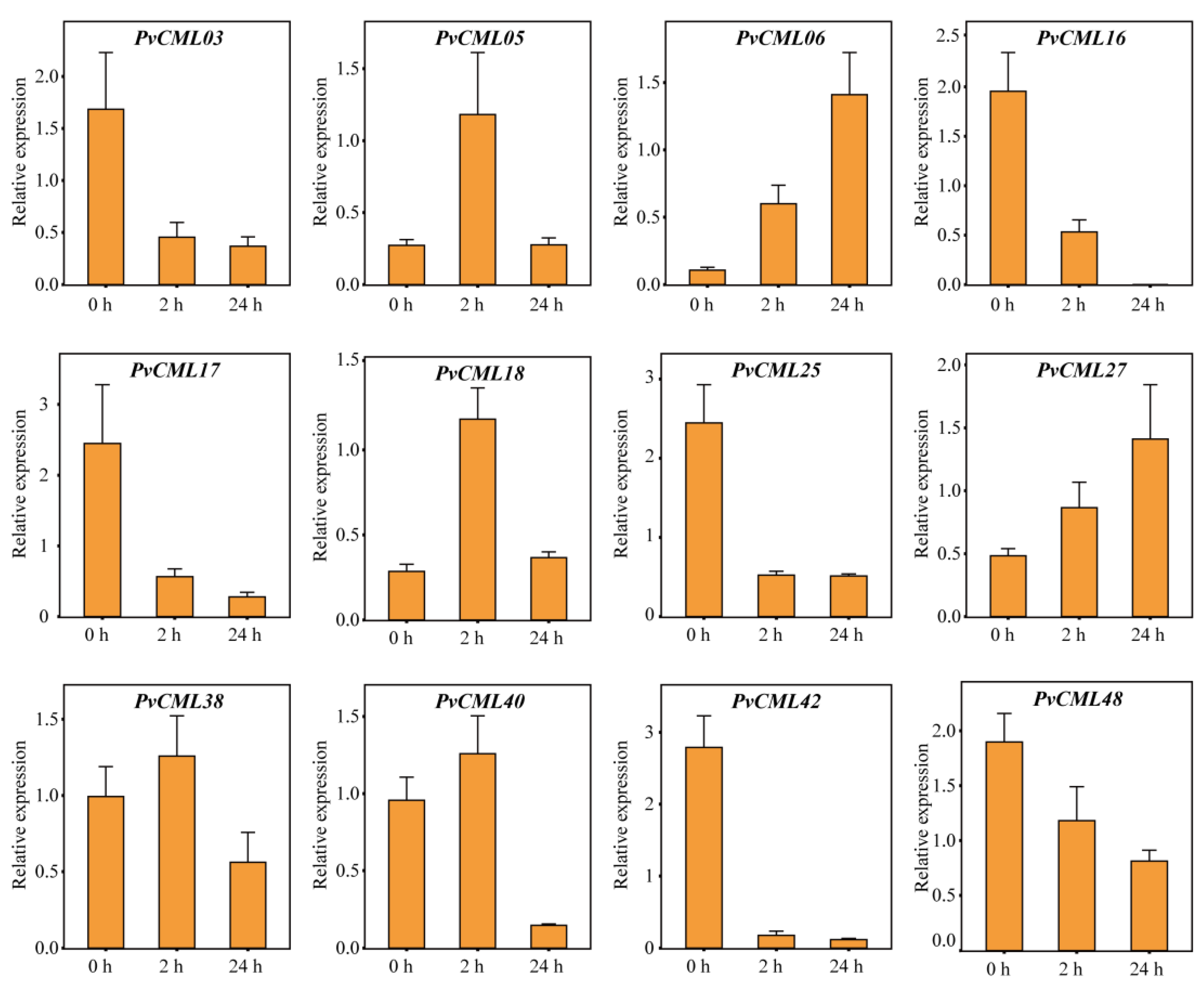
3.7. qRT–PCR of PvCMLs under Salt and Cold Treatment Conditions

To further investigate the response of PvCMLs to salt and cold stresses, we estimated the expression levels of PvCMLs using qRT–PCR analysis. Genes with excess “dehydration low temperature and salt stress response” elements (more than six elements) or excess “dehydration low temperature and salt stress response” elements (more than three elements) or genes with differential expression in expression profiles were selected for this analysis. Under salt treatment conditions, the expression of PvCML18 increased at 2 h, identical to the results of the expression profile, while at 24 h, PvCML18 gene expression recovered to the control level (Figure 7). In addition, we found that PvCML06 and PvCML27 expression progressively increased over time, whereas PvCML03, PvCML17, PvCML25, PvCML42, and PvCML48 expression progressively decreased over time (Figure 7 and Figure S2). Under cold treatment conditions, seven differentially expressed genes (PvCML06, PvCML14, PvCML18. PvCML25, PvCML31, PvCML40, and PvCML27) were found to be upregulated in the qRT–PCR analysis, validating the results of the expression profile (Figure 8 and Figure S3). Overall, the qRT–PCR analysis validated the expression profile results and provided new insight into how PvCMLs respond to salt and cold stresses.

4. Discussion

Plants have evolved a series of physiological and biochemical mechanisms to cope with environmental and developmental stimuli. One of the most important ways is to recruit Ca2+ as a second messenger in response to a given stimulus [68]. During this process, Ca2+ sensor proteins play a crucial role in transducing the increase in cytosolic Ca2+ concentrations by associating with and altering downstream target proteins [28]. As a member of the Ca2+ sensors, the CML family is specific in green plants and crucial in both developmental processes and adaptation to environmental stimuli [15]. In the past decade, the publication of an increasing number of reference genomes has enhanced our ability to identify the members and functions of CMLs in different species. The identification of CML proteins has been accomplished in several plant species, such as rice [15,17], A. thaliana [13], maize [22], tomato [18], and other species [20,24,25,69]. However, most of these are model or well-studied species, and the functions and evolutionary processes of the CML family in nonmodel species have not been fully studied. In this study, 49 CML family proteins were identified in one of the most salt-tolerant turfgrass species, P. vaginatum. The number of PvCMLs differs from those in rice (54 OsCMLs), maize (46 ZmCMLs), A. thaliana (50 AtCMLs), soybean (144 GmCMLs), and grapevine (62 VviCMLs), indicating that the evolutionary processes of PvCMLs are not completely shared with those of other species. Moreover, a comprehensive analysis of PvCMLs, including construction of a phylogenetic tree and analyses of conserved motifs, exon-intron structure, chromosome location, synteny, cis-regulatory elements, interaction network, and expression patterns under salt stress and cold stress conditions was completed. These results provide novel and valuable information that could help us to explore the functions and regulatory mechanisms of CML genes in P. vaginatum, especially in salt and cold resistance.
Our results demonstrated that the gene length, theoretical molecular weight (Mw), and isoelectric point (pI) of PvCMLs largely varied (Table 1), which is identical to other species [16,70], indicating that divergent functions are likely to have evolved. Moreover, PvCMLs have less homology with AtCMLs, indicating that CML proteins have highly diverged between monocotyledons and dicotyledons, which is also consistent with the results of other studies [21,71]. The gene location results demonstrated that the PvCMLs were unevenly distributed on all 10 chromosomes, indicating that the evolutionary processes of PvCMLs were shaped by segmental duplication. Several tandem gene arrays, such as PvCMLs29-PvCML32 on chromosome 7 and PvCML11-PvCML12 on chromosome 2, were detected in P. vaginatum, suggesting that the evolutionary processes of PvCMLs were also affected by tandem duplications. Additionally, we predicted complex interaction networks among PvCMLs, which indicates the function of PvCMLs associated with each other (Figure S1).
Phylogenetic analysis and collinearity relationships showed that most PvCMLs had homologous genes in rice, suggesting the potential conserved molecular function of CML genes between the two species. All homologous gene pairs were under purifying selection, which further confirmed our inference. In addition, eight PvCMLs were identified to have independently evolved in P. vaginatum, and these genes may contribute to adaptation to harsh environments. Four of them (PvCML29-PvCML32) were adjacent to each other, forming a tandem gene array. The expression profile and qRT–PCR analysis demonstrated that the tandem gene array participated in the regulatory process of cold stress (Figure S4), which indicates that the function of this tandem gene array may be associated with cold stimulus. The impact of tandem duplication on plant abiotic and biotic resistance has been reported in multiple taxa [72,73,74,75,76]. Here, we provide another case of the potential role of tandem gene array contributions to resistance to abiotic stress.
The specific expression of genes is determined by the cis-elements in the promoter region. Previous studies have shown that certain CML genes with special cis-acting elements in the promoter region participate in hormonal or abiotic stress responses [67]. For example, low-temperature responsive elements (LTRs) were enriched in the promoter regions of MtCML16 and MtCML33, and the expression of MtCML16 and MtCML33 was significantly induced under cold stress conditions [24]. The promoter activities of AtCML37, AtCML38, and AtCML39 were altered under conditions of drought, salt, and oxidative stresses and mechanical damage, and their transcript expression levels were also significantly increased [77]. In different tissues at different developmental stages of rice, the expression of OsMSR2 was induced by different stresses, such as low temperature, drought, and high temperature [33]. In P. vaginatum, PvCMLs were shown to contain various cis-elements related to plant growth and development, abiotic stress, and plant hormones. Almost all PvCML genes were found to contain cis-elements related to abscisic acid responsiveness, indicating that PvCML proteins extensively participate in ABA and drought regulation. Low-temperature and salt-responsive elements were present in most promoters of PvCMLs, suggesting that PvCML proteins are also associated with high salt tolerance. More than half of the PvCMLs contained anaerobic responsive elements (AREs) and MeJA responsive elements, which might be related to the adaptability to the aquatic environment and involvement in the jasmonic acid pathway. The PvCML38 and PvCML21 promoter regions contained almost all cis-acting elements, indicating that PvCML38 and PvCML21 may participate in multiple developmental and stress responses.
Previous studies have demonstrated that CML genes are key regulators involved in salt stress and cold stress. For example, AtCML24 was induced after cold treatment in A. thaliana [78]. AtCML9 was associated with salt and low-temperature treatments, and the AtCML9 mutants enhanced resistance to salt stress [34]. Soybean GsCML27 was induced by salt stress, and heterologous expression of GsCML27 enhanced tolerance to salt stress in A. thaliana [79]. MtCML40 was induced by salt and low-temperature stress and negatively regulated salt tolerance in M. truncatula [32]. MtCML42 and MtCML10 were induced by cold treatment, and overexpression in transgenic plants improved cold tolerance [35,36]. Tomato ShCML44 was induced by a variety of abiotic stresses, and overexpression of ShCML44 could improve tolerance to cold and salt stress [80]. All these works in model species clearly show that CMLs can enhance resistance to salt and cold stresses. In P. vaginatum, we analyzed the expression patterns of PvCMLs through expression profiles and qRT–PCR and found evidence of PvCMLs in response to salt and cold stresses. Under salt stress conditions, the expression of PvCML06, PvCML18, PvCML27, PvCML38, and PvCML40 increased over time, indicating that these genes played an important role in regulating the salt tolerance of P. vaginatum, while PvCML16, PvCML42, and PvCML48 were downregulated under salt stress conditions, indicating that these genes participated in the negative regulation of salt stress. Furthermore, we found the expression of six CML genes (PvCML06, PvCML14, PvCML18. PvCML25, PvCML31, PvCML40, and PvCML27) were significantly induced under cold stress conditions, indicating that PvCMLs also played important roles in the cold tolerance of P. vaginatum. In rice, as a homologous gene of PvCML14 (one of the cold-inducing genes), OsCML16 has been proven to interact with OsPi304 to enhance cold tolerance [81], which provides additional evidence for our results. Overall, our work provides important insights into the role of PvCMLs in salt and cold resistance and could provide a good foundation for further molecular function investigation of PvCMLs in the future.

5. Conclusions

As one of the most salt tolerance grass species and famous warm-season turfgrass, the mechanism of excellent abiotic stress resistance of P. vaginatum is still largely unknown. In this study, we investigated CML gene family in P. vaginatum to evaluate the role of CML genes played in response to salt and cold stress. A total of 49 CML proteins were identified in P. vaginatum. Among them, the gene length, molecular weight, theoretical isoelectric point, composition of the conserved domain, and gene structure varied greatly, which demonstrated that the functions of PvCMLs have largely diverged. Synteny analysis indicated that the evolutionary history of PvCMLs was largely affected by tandem and segmental duplications. Moreover, the expression profile and qRT–PCR results suggested that PvCMLs participated in regulating the salt and cold tolerance of P. vaginatum. Most notably, we found that evidence of a tandem gene array (PvCML29-PvCML32) indicated that evolution of novel CMLs in P. vaginatum may be associated with cold stress. Altogether, our work contributes to insights into a deep understanding of the evolution and expression characteristics of PvCMLs, and the findings of a potential stress resistance gene will guide further molecular function investigation of PvCMLs and germplasm resource utilization of P. vaginatum in the future. Moreover, understanding how grass species resist abiotic and biotic stresses could pave the road to crop breeding in other closely related crop species, such as rice, Z. may, and S. bicolor.

Supplementary Materials

The following supporting information can be downloaded at: https://www.mdpi.com/article/10.3390/cimb45020109/s1. Figure S1: predicted protein-protein interaction network (PPI) of PvCMLs; Figure S2: qRT-PCR results of 14 PvCMLs under salt stress; Figure S3: qRT-PCR results of 14 PvCMLs under cold stress; Figure S4: RT-qPCR results of PvCML29–32 under cold stress; Table S1: Protein sequence of PvCMLs; Table S2: Expression profile under salt stress; Table S3: Expression profile under cold stress; Table S4: The promoter information of PvCMLs genes; Table S5: Sequences of primers used in RT-qPCR; Table S6: Pairs of paralogous PvCML proteins; Table S7: The Ka/Ks value of pairs of homologous proteins between P. vaginatium and rice. All code associated with this project is available at GitHub (https://github.com/zzbbf123/PvCMLs, accessed on 7 December 2022).

Author Contributions

Conceptualization, B.-F.Z.; methodology, B.-F.Z. and M.Y.; software, M.Y.; validation, M.Y., J.C. and T.L.; formal analysis, M.Y., J.C., T.L. and L.X.; investigation, M.Y., J.C., T.L. and L.X.; resources, B.-F.Z.; data curation, B.-F.Z. and M.Y.; writing—original draft preparation, M.Y.; writing—review and editing, B.-F.Z.; visualization, M.Y.; supervision, B.-F.Z.; project administration, B.-F.Z.; funding acquisition, B.-F.Z. All authors have read and agreed to the published version of the manuscript.

Funding

This research was funded by the China Postdoctoral Science Foundation (grant number: 2022M713197).

Institutional Review Board Statement

No applicable.

Informed Consent Statement

Not applicable.

Data Availability Statement

The transcriptome data that support the findings of this study are available from National Center for Biotechnology Information (https://www.ncbi.nlm.nih.gov/) (accessed on 17 February 2022) with the access number of SRR5876943, SRR5876944, SRR4280403, SRR4280409, SRR4280414, SRR4280420, SRR4280426 and SRR4280427.

Conflicts of Interest

The authors declare no conflict of interest.

References

  1. Zhu, J.-K. Abiotic Stress Signaling and Responses in Plants. Cell 2016, 167, 313–324. [Google Scholar] [CrossRef] [PubMed] [Green Version]
  2. Tian, W.; Wang, C.; Gao, Q.; Li, L.; Luan, S. Calcium spikes, waves and oscillations in plant development and biotic interactions. Nat. Plants 2020, 6, 750–759. [Google Scholar] [CrossRef] [PubMed]
  3. Kudla, J.; Batistič, O.; Hashimoto, K. Calcium Signals: The Lead Currency of Plant Information Processing. Plant Cell 2010, 22, 541–563. [Google Scholar] [CrossRef] [PubMed]
  4. La Verde, V.; Dominici, P.; Astegno, A. Towards Understanding Plant Calcium Signaling through Calmodulin-Like Proteins: A Biochemical and Structural Perspective. Int. J. Mol. Sci. 2018, 19, 1331. [Google Scholar] [CrossRef] [Green Version]
  5. Hashimoto, K.; Kudla, J. Calcium decoding mechanisms in plants. Biochimie 2011, 93, 2054–2059. [Google Scholar] [CrossRef]
  6. Perochon, A.; Aldon, D.; Galaud, J.-P.; Ranty, B. Calmodulin and calmodulin-like proteins in plant calcium signaling. Biochimie 2011, 93, 2048–2053. [Google Scholar] [CrossRef]
  7. Webb, A.A.; McAinsh, M.R.; Taylor, J.E.; Hetherington, A.M. Calcium Ions as Intracellular Second Messengers in Higher Plants. Adv. Bot. Res. 1996, 22, 45–96. [Google Scholar] [CrossRef]
  8. Hetherington, A.M.; Brownlee, C. The generation of Ca2+ signals in plants. Annu. Rev. Plant Biol. 2004, 55, 401–427. [Google Scholar] [CrossRef] [Green Version]
  9. Yang, T.; Poovaiah, B. Calcium/calmodulin-mediated signal network in plants. Trends Plant Sci. 2003, 8, 505–512. [Google Scholar] [CrossRef]
  10. Edel, K.H.; Marchadier, E.; Brownlee, C.; Kudla, J.; Hetherington, A.M. The Evolution of Calcium-Based Signalling in Plants. Curr. Biol. 2017, 27, R667–R679. [Google Scholar] [CrossRef]
  11. Luan, S.; Kudla, J.; Rodriguez-Concepcion, M.; Yalovsky, S.; Gruissem, W. Calmodulins and Calcineurin B–like Proteins: Cal-cium sensors for specific signal response coupling in plants. Plant Cell 2002, 14, S389–S400. [Google Scholar] [CrossRef] [PubMed] [Green Version]
  12. Mohanta, T.K.; Yadav, D.; Khan, A.L.; Hashem, A.; Abd_Allah, E.; Al-Harrasi, A. Molecular Players of EF-hand Containing Calcium Signaling Event in Plants. Int. J. Mol. Sci. 2019, 20, 1476. [Google Scholar] [CrossRef] [PubMed] [Green Version]
  13. McCormack, E.; Braam, J. Calmodulins and related potential calcium sensors of Arabidopsis. New Phytol. 2003, 159, 585–598. [Google Scholar] [CrossRef] [PubMed] [Green Version]
  14. DeFalco, T.A.; Bender, K.W.; Snedden, W.A. Breaking the code: Ca2+ sensors in plant signalling. Biochem. J. 2010, 425, 27–40. [Google Scholar] [CrossRef] [Green Version]
  15. Zhu, X.; Dunand, C.; Snedden, W.; Galaud, J.-P. CaM and CML emergence in the green lineage. Trends Plant Sci. 2015, 20, 483–489. [Google Scholar] [CrossRef]
  16. Zhu, X.; Perez, M.; Aldon, D.; Galaud, J.-P. Respective contribution of CML8 and CML9, two arabidopsis calmodulin-like proteins, to plant stress responses. Plant Signal. Behav. 2017, 12, e1322246. [Google Scholar] [CrossRef] [Green Version]
  17. Boonburapong, B.; Buaboocha, T. Genome-wide identification and analyses of the rice calmodulin and related potential calcium sensor proteins. BMC Plant Biol. 2007, 7, 4. [Google Scholar] [CrossRef] [Green Version]
  18. Munir, S.; Khan, M.R.G.; Song, J.; Munir, S.; Zhang, Y.; Ye, Z.; Wang, T. Genome-wide identification, characterization and expression analysis of calmodulin-like (CML) proteins in tomato (Solanum lycopersicum). Plant Physiol. Biochem. 2016, 102, 167–179. [Google Scholar] [CrossRef]
  19. Yadav, M.; Pandey, J.; Chakraborty, A.; Hassan, I.; Kundu, J.K.; Roy, A.; Singh, I.K.; Singh, A. A Comprehensive Analysis of Calmodulin-Like Proteins of Glycine max Indicates Their Role in Calcium Signaling and Plant Defense Against Insect Attack. Front. Plant Sci. 2022, 13, 817950. [Google Scholar] [CrossRef]
  20. Zeng, H.; Zhang, Y.; Zhang, X.; Pi, E.; Zhu, Y. Analysis of EF-Hand Proteins in Soybean Genome Suggests Their Potential Roles in Environmental and Nutritional Stress Signaling. Front. Plant Sci. 2017, 8, 877. [Google Scholar] [CrossRef] [Green Version]
  21. Zhang, X.; Tian, J.; Li, S.; Liu, Y.; Feng, T.; Wang, Y.; Li, Y.; Huang, X.; Li, D. Characterization of the Calmodulin/Calmodulin-like Protein (CAM/CML) Family in Ginkgo biloba, and the Influence of an Ectopically Expressed GbCML Gene (Gb_30819) on Seedling and Fruit Development of Transgenic Arabidopsis. Plants 2022, 11, 1506. [Google Scholar] [CrossRef] [PubMed]
  22. Wang, Z.; Wang, L.; Li, J.; Yang, W.; Ci, J.; Ren, X.; Wang, W.; Wang, Y.; Jiang, L.; Yang, W. Identification and expression analysis revealed drought stress-responsive Calmodulin and Calmodulin-like genes in maize. J. Plant Interact. 2022, 17, 450–461. [Google Scholar] [CrossRef]
  23. Cai, K.; Kuang, L.; Yue, W.; Xie, S.; Xia, X.; Zhang, G.; Wang, J. Calmodulin and calmodulin-like gene family in barley: Identification, characterization and expression analyses. Front. Plant Sci. 2022, 13, 964888. [Google Scholar] [CrossRef] [PubMed]
  24. Sun, Q.; Yu, S.; Guo, Z. Calmodulin-Like (CML) Gene Family in Medicago truncatula: Genome-Wide Identification, Characterization and Expression Analysis. Int. J. Mol. Sci. 2020, 21, 7142. [Google Scholar] [CrossRef] [PubMed]
  25. Guo, N.; Wang, G.; Zong, M.; Han, S.; Liu, F. Genome-wide identification, and phylogenetic and expression profiling analyses of CaM and CML genes in Brassica rapa and Brassica oleracea. Gene 2018, 677, 232–244. [Google Scholar] [CrossRef] [PubMed]
  26. Bender, K.W.; Snedden, W.A. Calmodulin-Related Proteins Step Out from the Shadow of Their Namesake. Plant Physiol. 2013, 163, 486–495. [Google Scholar] [CrossRef] [PubMed] [Green Version]
  27. McCormack, E.; Tsai, Y.-C.; Braam, J. Handling calcium signaling: Arabidopsis CaMs and CMLs. Trends Plant Sci. 2005, 10, 383–389. [Google Scholar] [CrossRef] [PubMed]
  28. Chinpongpanich, A.; Limruengroj, K.; Phean-O-Pas, S.; Limpaseni, T.; Buaboocha, T. Expression analysis of calmodulin and calmodulin-like genes from rice, Oryza sativa L. BMC Res. Notes 2012, 5, 625. [Google Scholar] [CrossRef] [Green Version]
  29. Scholz, S.S.; Vadassery, J.; Heyer, M.; Reichelt, M.; Bender, K.W.; Snedden, W.A.; Boland, W.; Mithöfer, A. Mutation of the Arabidopsis Calmodulin-Like Protein CML37 Deregulates the Jasmonate Pathway and Enhances Susceptibility to Herbivory. Mol. Plant 2014, 7, 1712–1726. [Google Scholar] [CrossRef] [Green Version]
  30. Cheng, H.-Q.; Han, L.-B.; Yang, C.-L.; Wu, X.-M.; Zhong, N.-Q.; Wu, J.-H.; Wang, F.-X.; Wang, H.-Y.; Xia, G.-X. The cotton MYB108 forms a positive feedback regulation loop with CML11 and participates in the defense response against Verticillium dahlia infection. J. Exp. Bot. 2016, 67, 1935–1950. [Google Scholar] [CrossRef] [Green Version]
  31. Xu, B.; Cheval, C.; Laohavisit, A.; Hocking, B.; Chiasson, D.; Olsson, T.S.G.; Shirasu, K.; Faulkner, C.; Gilliham, M. A calmodulin-like protein regulates plasmodesmal closure during bacterial immune responses. New Phytol. 2017, 215, 77–84. [Google Scholar] [CrossRef] [PubMed] [Green Version]
  32. Zhang, X.; Wang, T.; Liu, M.; Sun, W.; Zhang, W.-H. Calmodulin-like gene MtCML40 is involved in salt tolerance by regulating MtHKTs transporters in Medicago truncatula. Environ. Exp. Bot. 2019, 157, 79–90. [Google Scholar] [CrossRef]
  33. Xu, G.-Y.; Rocha, P.S.C.F.; Wang, M.-L.; Xu, M.-L.; Cui, Y.-C.; Li, L.-Y.; Zhu, Y.-X.; Xia, X. A novel rice calmodulin-like gene, OsMSR2, enhances drought and salt tolerance and increases ABA sensitivity in Arabidopsis. Planta 2011, 234, 47–59. [Google Scholar] [CrossRef] [PubMed]
  34. Magnan, F.; Ranty, B.; Charpenteau, M.; Sotta, B.; Galaud, J.-P.; Aldon, D.; Ranty, B. Mutations in AtCML9, a calmodulin-like protein from Arabidopsis thaliana, alter plant responses to abiotic stress and abscisic acid. Plant J. 2008, 56, 575–589. [Google Scholar] [CrossRef]
  35. Sun, Q.; Huang, R.; Zhu, H.; Sun, Y.; Guo, Z. A novel Medicago truncatula calmodulin-like protein (MtCML42) regulates cold tolerance and flowering time. Plant J. 2021, 108, 1069–1082. [Google Scholar] [CrossRef]
  36. Yu, S.; Wu, J.; Sun, Y.; Zhu, H.; Sun, Q.; Zhao, P.; Huang, R.; Guo, Z. A calmodulin-like protein (CML10) interacts with cytosolic enzymes GSTU8 and FBA6 to regulate cold tolerance. Plant Physiol. 2022, 190, 1321–1333. [Google Scholar] [CrossRef]
  37. Lee, G.-J.; Carrow, R.N.; Duncan, R.R. Photosynthetic responses to salinity stress of halophytic seashore paspalum ecotypes. Plant Sci. 2004, 166, 1417–1425. [Google Scholar] [CrossRef]
  38. Wu, P.; Cogill, S.; Qiu, Y.; Li, Z.; Zhou, M.; Hu, Q.; Chang, Z.; Noorai, R.E.; Xia, X.; Saski, C.; et al. Comparative transcriptome profiling provides insights into plant salt tolerance in seashore paspalum (Paspalum vaginatum). BMC Genom. 2020, 21, 131. [Google Scholar] [CrossRef]
  39. Qi, P.; Eudy, D.; Schnable, J.C.; Schmutz, J.; Raymer, P.L.; Devos, K.M. High Density Genetic Maps of Seashore Paspalum Using Genotyping-By-Sequencing and Their Relationship to The Sorghum Bicolor Genome. Sci. Rep. 2019, 9, 12183. [Google Scholar] [CrossRef] [Green Version]
  40. Liao, L.; Hu, X.; Hao, J.; Tang, M.; Ren, L.; Pan, L.; Xie, S.; Raymer, P.; Qi, P.; Chen, Z.; et al. The chromosome-based genome of Paspalum vaginatum provides new insights into salt-stress adaptation. bioRxiv 2022. [Google Scholar] [CrossRef]
  41. Lee, G.-J.; Duncan, R.R.; Carrow, R.N. Salinity Tolerance of Seashore Paspalum Ecotypes: Shoot Growth Responses and Criteria. Hortscience 2004, 39, 1138–1142. [Google Scholar] [CrossRef] [Green Version]
  42. Goad, D.M.; Baxter, I.; Kellogg, E.A.; Olsen, K.M. Hybridization, polyploidy and clonality influence geographic patterns of diversity and salt tolerance in the model halophyte seashore paspalum (Paspalum vaginatum). Mol. Ecol. 2021, 30, 148–161. [Google Scholar] [CrossRef] [PubMed]
  43. Uddin, M.K.; Juraim, A.S.; Ismail, M.R.; Alam, M.A. The effect of salinity on growth and ion accumulation in six turfgrass species. Plant Omics. 2012, 5, 244–252. [Google Scholar]
  44. Sun, G.; Wase, N.; Shu, S.; Jenkins, J.; Zhou, B.; Torres-Rodríguez, J.V.; Chen, C.; Sandor, L.; Plott, C.; Yoshinga, Y.; et al. Genome of Paspalum vaginatum and the role of trehalose mediated autophagy in increasing maize biomass. Nat. Commun. 2022, 13, 7731. [Google Scholar] [CrossRef]
  45. Mistry, J.; Chuguransky, S.; Williams, L.; Qureshi, M.; Salazar, G.A.; Sonnhammer, E.L.L.; Tosatto, S.C.; Paladin, L.; Raj, S.; Richardson, L.J.; et al. Pfam: The protein families database in 2021. Nucleic Acids Res. 2021, 49, D412–D419. [Google Scholar] [CrossRef]
  46. Letunic, I.; Khedkar, S.; Bork, P. SMART: Recent updates, new developments and status in 2020. Nucleic Acids Res. 2021, 49, D458–D460. [Google Scholar] [CrossRef]
  47. Edgar, R.C. MUSCLE: Multiple sequence alignment with high accuracy and high throughput. Nucleic Acids Res. 2004, 32, 1792–1797. [Google Scholar] [CrossRef] [Green Version]
  48. Tamura, K.; Stecher, G.; Kumar, S. MEGA11: Molecular Evolutionary Genetics Analysis Version 11. Mol. Biol. Evol. 2021, 38, 3022–3027. [Google Scholar] [CrossRef]
  49. Subramanian, B.; Gao, S.; Lercher, M.J.; Hu, S.; Chen, W.-H. Evolview v3: A webserver for visualization, annotation, and management of phylogenetic trees. Nucleic Acids Res. 2019, 47, W270–W275. [Google Scholar] [CrossRef] [PubMed]
  50. Bailey, T.L.; Johnson, J.; Grant, C.E.; Noble, W.S. The MEME Suite. Nucleic Acids Res. 2015, 43, W39–W49. [Google Scholar] [CrossRef] [PubMed] [Green Version]
  51. Chen, C.J.; Chen, H.; Zhang, Y.; Thomas, H.R.; Frank, M.H.; He, Y.H.; Xia, R. TBtools: An Integrative Toolkit Developed for Interactive Analyses of Big Biological Data. Mol. Plant 2020, 13, 1194–1202. [Google Scholar] [CrossRef] [PubMed]
  52. Wang, Y.; Tang, H.; DeBarry, J.D.; Tan, X.; Li, J.; Wang, X.; Lee, T.-H.; Jin, H.; Marler, B.; Guo, H.; et al. MCScanX: A toolkit for detection and evolutionary analysis of gene synteny and collinearity. Nucleic Acids Res. 2012, 40, e49. [Google Scholar] [CrossRef] [PubMed] [Green Version]
  53. Krzywinski, M.; Schein, J.; Birol, I.; Connors, J.; Gascoyne, R.; Horsman, D.; Jones, S.J.; Marra, M.A. Circos: An information aesthetic for comparative genomics. Genome Res. 2009, 19, 1639–1645. [Google Scholar] [CrossRef] [PubMed] [Green Version]
  54. Tang, H.; Bowers, J.E.; Wang, X.; Ming, R.; Alam, M.; Paterson, A.H. Synteny and Collinearity in Plant Genomes. Science 2008, 320, 486–488. [Google Scholar] [CrossRef] [Green Version]
  55. Yang, Z. PAML 4: Phylogenetic Analysis by Maximum Likelihood. Mol. Biol. Evol. 2007, 24, 1586–1591. [Google Scholar] [CrossRef] [Green Version]
  56. Lescot, M.; Déhais, P.; Thijs, G.; Marchal, K.; Moreau, Y.; Van de Peer, Y.; Rouzé, P.; Rombauts, S. PlantCARE, a database of plant cis-acting regulatory elements and a portal to tools for in silico analysis of promoter sequences. Nucleic Acids Res. 2002, 30, 325–327. [Google Scholar] [CrossRef]
  57. Bolger, A.M.; Lohse, M.; Usadel, B. Trimmomatic: A flexible trimmer for Illumina sequence data. Bioinformatics 2014, 30, 2114–2120. [Google Scholar] [CrossRef] [Green Version]
  58. Grabherr, M.G.; Haas, B.J.; Yassour, M.; Levin, J.Z.; Thompson, D.A.; Amit, I.; Adiconis, X.; Fan, L.; Raychowdhury, R.; Zeng, Q.D.; et al. Full-length transcriptome assembly from RNA-Seq data without a reference genome. Nat. Biotechnol. 2011, 29, 644–652. [Google Scholar] [CrossRef] [Green Version]
  59. Haas, B.J.; Papanicolaou, A.; Yassour, M.; Grabherr, M.; Blood, P.D.; Bowden, J.; Couger, M.B.; Eccles, D.; Li, B.; Lieber, M.; et al. De novo transcript sequence reconstruction from RNA-seq using the Trinity platform for reference generation and analysis. Nat. Protoc. 2013, 8, 1494–1512. [Google Scholar] [CrossRef]
  60. Li, B.; Dewey, C.N. RSEM: Accurate transcript quantification from RNA-Seq data with or without a reference genome. BMC Bioinform. 2011, 12, 323. [Google Scholar] [CrossRef] [Green Version]
  61. Love, M.I.; Huber, W.; Anders, S. Moderated estimation of fold change and dispersion for RNA-seq data with DESeq2. Genome Biol. 2014, 15, 550. [Google Scholar] [CrossRef] [PubMed] [Green Version]
  62. Camacho, C.; Coulouris, G.; Avagyan, V.; Ma, N.; Papadopoulos, J.; Bealer, K.; Madden, T.L. BLAST+: Architecture and applications. BMC Bioinform. 2009, 10, 421. [Google Scholar] [CrossRef] [PubMed] [Green Version]
  63. Zhang, Y.; Ngu, D.W.; Carvalho, D.; Liang, Z.; Qiu, Y.; Roston, R.L.; Schnable, J.C. Differentially Regulated Orthologs in Sorghum and the Subgenomes of Maize. Plant Cell 2017, 29, 1938–1951. [Google Scholar] [CrossRef] [PubMed] [Green Version]
  64. Kim, D.; Paggi, J.M.; Park, C.; Bennett, C.; Salzberg, S.L. Graph-based genome alignment and genotyping with HISAT2 and HISAT-genotype. Nat. Biotechnol. 2019, 37, 907–915. [Google Scholar] [CrossRef] [PubMed]
  65. Pertea, M.; Pertea, G.M.; Antonescu, C.M.; Chang, T.-C.; Mendell, J.T.; Salzberg, S.L. StringTie enables improved reconstruction of a transcriptome from RNA-seq reads. Nat. Biotechnol. 2015, 33, 290–295. [Google Scholar] [CrossRef] [Green Version]
  66. Livak, K.J.; Schmittgen, T.D. Analysis of relative gene expression data using real-time quantitative PCR and the 2−ΔΔCT Method. Methods 2001, 25, 402–408. [Google Scholar] [CrossRef]
  67. He, X.; Liu, W.; Li, W.; Liu, Y.; Wang, W.; Xie, P.; Kang, Y.; Liao, L.; Qian, L.; Liu, Z.; et al. Genome-wide identification and expression analysis of CaM/CML genes in Brassica napus under abiotic stress. J. Plant Physiol. 2020, 255, 153251. [Google Scholar] [CrossRef]
  68. Dale, S.; Colin, B.; Jeffrey, F.H. Communicating with Calcium. Plant Cell 1999, 11, 691–706. [Google Scholar]
  69. Vandelle, E.; Vannozzi, A.; Wong, D.; Danzi, D.; Digby, A.-M.; Santo, S.D.; Astegno, A. Identification, characterization, and expression analysis of calmodulin and calmodulin-like genes in grapevine (Vitis vinifera) reveal likely roles in stress responses. Plant Physiol. Biochem. 2018, 129, 221–237. [Google Scholar] [CrossRef]
  70. Mohanta, T.K.; Kumar, P.; Bae, H. Genomics and evolutionary aspect of calcium signaling event in calmodulin and calmodulin-like proteins in plants. BMC Plant Biol. 2017, 17, 38. [Google Scholar] [CrossRef] [Green Version]
  71. Gao, L.; Damaris, R.N.; Yu, F.; Yang, P. Genome-wide Identification and Expression Analysis of CaM/CML Gene Family in Sacred Lotus (Nelumbo nucifera). Plant Mol. Biol. Rep. 2022, 40, 418–432. [Google Scholar] [CrossRef]
  72. Bellieny-Rabelo, D.; Oliveira, A.E.A.; Venancio, T.M. Impact of Whole-Genome and Tandem Duplications in the Expansion and Functional Diversification of the F-Box Family in Legumes (Fabaceae). PLoS ONE 2013, 8, e55127. [Google Scholar] [CrossRef] [PubMed] [Green Version]
  73. Hofberger, J.A.; Lyons, E.; Edger, P.P.; Pires, J.C.; Schranz, M.E. Whole Genome and Tandem Duplicate Retention Facilitated Glucosinolate Pathway Diversification in the Mustard Family. Genome Biol. Evol. 2013, 5, 2155–2173. [Google Scholar] [CrossRef] [PubMed] [Green Version]
  74. Hu, X.; Hao, J.; Pan, L.; Xu, T.; Ren, L.; Chen, Y.; Tang, M.; Liao, L.; Wang, Z. Genome-wide analysis of tandem duplicated genes and their expression under salt stress in seashore paspalum. Front. Plant Sci. 2022, 13, 971999. [Google Scholar] [CrossRef]
  75. Knight, J.C. Tandem and segmental gene duplication and recombination in the evolution of plant disease resistance genes. Trends Genet. 2004, 20, 113–116. [Google Scholar] [CrossRef]
  76. Rizzon, C.; Ponger, L.; Gaut, B.S. Striking Similarities in the Genomic Distribution of Tandemly Arrayed Genes in Arabidopsis and Rice. PLoS Comput. Biol. 2006, 2, e115. [Google Scholar] [CrossRef] [Green Version]
  77. Vanderbeld, B.; Snedden, W.A. Developmental and stimulus-induced expression patterns of Arabidopsis calmodulin-like genes CML37, CML38 and CML39. Plant Mol. Biol. 2007, 64, 683–697. [Google Scholar] [CrossRef]
  78. Delk, N.A.; Johnson, K.A.; Chowdhury, N.I.; Braam, J. CML24, Regulated in Expression by Diverse Stimuli, Encodes a Potential Ca2+ Sensor That Functions in Responses to Abscisic Acid, Daylength, and Ion Stress. Plant Physiol. 2005, 139, 240–253. [Google Scholar] [CrossRef] [Green Version]
  79. Chen, C.; Sun, X.; Duanmu, H.; Zhu, D.; Yu, Y.; Cao, L.; Liu, A.; Jia, B.; Xiao, J.; Zhu, Y. GsCML27, a Gene Encoding a Calcium-Binding Ef-Hand Protein from Glycine soja, Plays Differential Roles in Plant Responses to Bicarbonate, Salt and Osmotic Stresses. PLoS ONE 2015, 10, e0141888. [Google Scholar] [CrossRef] [Green Version]
  80. Munir, S.; Liu, H.; Xing, Y.; Hussain, S.; Ouyang, B.; Zhang, Y.; Li, H.; Ye, Z. Overexpression of calmodulin-like (ShCML44) stress-responsive gene from Solanum habrochaites enhances tolerance to multiple abiotic stresses. Sci. Rep. 2016, 6, 31772. [Google Scholar] [CrossRef]
  81. Yang, J.; Ji, L.; Zhu, B.; Yuan, X.; Jin, D.; Xie, G. OsCML16 interacts with a novel CC-NBS-LRR protein OsPi304 in the Ca2+/Mg2+ dependent and independent manner in rice. Biochem. Biophys. Res. Commun. 2018, 504, 346–351. [Google Scholar] [CrossRef] [PubMed]
Figure 1. Phylogenetic relationship of calmodulin-like (CML) proteins in P. vaginatum (green), A. thaliana (red), and rice (blue). An unrooted tree was built using the NJ method in MEGA with 1000 bootstrap replications. The bootstrap values of each node lower than 30 were not shown.
Cimb 45 00109 g001
Figure 2. The conserved motif composition and gene structure of CMLs proteins in P. vaginatum. (A) phylogenetic relationship of PvCMLs. (B) The conserved EF-hand motifs of PvCMLs. (C) Gene structure of PvCMLs. Pink boxes represent untranslated regions (UTR), green boxes represent coding regions (CDS), and black lines represent introns.
Cimb 45 00109 g002
Figure 3. Chromosomal distribution and synteny analysis in the genomes of P. vaginatum, Oryza sativa, and A. thaliana. (A) Paralogous genes of PvCMLs in P. vaginatum. (B) Orthologous CML genes between P. vaginatum and rice. (C) Orthologous CML genes between P. vaginatum and A. thaliana and between P. vaginatum and rice. Red lines indicate orthologous gene pairs.
Cimb 45 00109 g003
Figure 4. Bar plot of average Ka/Ks ratios for homologous CML gene pairs between rice and P. vaginatum. The different colors indicate eight subfamily groups identified in phylogenetic analysis. The error bar is shown in the figure.
Cimb 45 00109 g004
Figure 5. Prediction of cis-acting elements contained in PvCMLs. Analysis of cis-elements was carried out by the PlantCARE database. The analysis showed the presence of 14 cis-elements. The different colors represent the different numbers of cis-elements, with red indicating a high number of cis-elements, while white represents a low number of cis-elements. Values in the boxes represent the number of cis-regulatory elements.
Cimb 45 00109 g005
Figure 6. Expression pattern of PvCML genes under salt and cold stress conditions. (A). Heatmap of log2–transformed RPKM of PvCML genes after 400 nM NaCl treatment. The colors changing from blue to red indicate the expression level transition from low to high. (B). Volcano plot displaying the differentially expressed genes after salt treatment. The blue dots represent genes differentially expressed (adjusted p < 0.05) in the 400 nM NaCl treatment group vs. the normal control group. The red dots represent PvCMLs. (C). Heatmap of log2–transformed RPKM of PvCML genes after 6 °C cold treatment. The colors changing from blue to red indicate the expression level transition from low to high. (D). Volcano plot displaying the differentially expressed genes after 6 °C cold treatment. The blue dots represent genes differentially expressed (adjusted p < 0.05) in the 6 °C treatment group vs. the normal control group. The red dots represent PvCMLs.
Cimb 45 00109 g006
Figure 7. Gene expression analysis by RT–qPCR for the PvCMLs under salt stress conditions. The four-week-old seashore paspalum cultivars (sea spray) were treated with 200 mM NaCl, and three leaves were collected for each sample at 0 h, 2 h, and 24 h following treatment. Three biological replicates were performed for this analysis. The bar plot indicates the relative mRNA expression of each PvCML gene at each treatment time.
Cimb 45 00109 g007
Figure 8. Gene expression analysis by RT–qPCR for the PvCMLs under cold stress conditions. The four-week-old seashore paspalum cultivars (sea spray) were treated with 4 °C, and three leaves were collected for each sample at 0 h, 2 h, and 24 h following treatment. Three biological replicates were performed for this analysis. The bar plot indicates the relative mRNA expression of each PvCML gene at each treatment time.
Cimb 45 00109 g008
Table 1. Information of PvCMLs identified in the P. vaginatium.
Gene NameLocus IDLength (aa)MW (kDa)pIGRAVYEF-Hand Domain Number
PvCML01Pavag01G03620024926,549.834.93−0.2352
PvCML02Pavag01G06740017919,525.775.66−0.4442
PvCML03Pavag01G08500015517,270.083.98−0.5884
PvCML04Pavag01G23870017820,176.574.62−0.7494
PvCML05Pavag01G27480021622,865.164.54−0.2894
PvCML06Pavag01G36530018920,387.824.46−0.2973
PvCML07Pavag01G37490016717,550.554.16−0.1493
PvCML08Pavag02G20500018421,026.198.76−0.6493
PvCML09Pavag02G2051009811,265.65.45−0.7381
PvCML10Pavag02G35190017319,858.134.76−0.9234
PvCML11Pavag02G3521009510,825.188.18−0.6742
PvCML12Pavag02G35220012013,500.429.73−0.4792
PvCML13Pavag02G39240020021,075.54.47−0.2073
PvCML14Pavag03G07440018319,175.094.48−0.5384
PvCML15Pavag03G18700023924,717.864.95−0.1454
PvCML16Pavag03G28750021923,379.894.96−0.5482
PvCML17Pavag03G40110018819,570.964.88−0.2332
PvCML18Pavag03G40130020220,647.084.79−0.1254
PvCML19Pavag03G40590016717,384.444.420.0614
PvCML20Pavag03G40630015216,591.574.2−0.2964
PvCML21Pavag04G07490023026,143.374.59−0.5593
PvCML22Pavag04G26840018319,286.474.26−0.2454
PvCML23Pavag05G03170019622,180.574.48−0.6454
PvCML24Pavag05G03810031332,468.236.42−0.3722
PvCML25Pavag05G03870018919,421.474.8−0.2432
PvCML26Pavag05G19580019822,248.024.57−0.3662
PvCML27Pavag06G14470026327,960.34.78−0.4294
PvCML28Pavag07G02050014916,585.774.78−0.343
PvCML29Pavag07G18100014115,274.294.53−0.0252
PvCML30Pavag07G18110014115,343.274.48−0.0732
PvCML31Pavag07G18120014115,250.154.54−0.0632
PvCML32Pavag07G18130014115,341.344.53−0.0212
PvCML33Pavag07G22690024326,137.436.07−0.1932
PvCML34Pavag07G229200838931.054.29−0.152
PvCML35Pavag08G06550020622,885.434.62−0.74
PvCML36Pavag08G13740017218,391.34.28−0.3094
PvCML37Pavag09G04260023126,890.244.95−0.6853
PvCML38Pavag09G08840022723,777.515.61−0.4934
PvCML39Pavag09G12470019320,236.494.3−0.3144
PvCML40Pavag09G12660019219,711.924.7−0.1844
PvCML41Pavag09G18590015416,812.84.51−0.3552
PvCML42Pavag09G24670018619,477.014.73−0.034
PvCML43Pavag10G05920025127,068.034.51−0.5244
PvCML44Pavag10G0857009911,520.989.38−0.5132
PvCML45Pavag10G18850022525,934.324.75−0.484
PvCML46Pavag10G24160016016,085.053.870.3163
PvCML47PavagK0544009711,196.586.41−0.6352
PvCML48PavagK12310019822,203.984.57−0.3682
PvCML49PavagK3175008910,140.336.41−0.7412
Note: Abbreviations: MW, molecular weight; pI, isoelectric point; GRAVY, grand average of hydropathicity.
Disclaimer/Publisher’s Note: The statements, opinions and data contained in all publications are solely those of the individual author(s) and contributor(s) and not of MDPI and/or the editor(s). MDPI and/or the editor(s) disclaim responsibility for any injury to people or property resulting from any ideas, methods, instructions or products referred to in the content.

