Article

Matrix Metalloproteinases in Human Decidualized Endometrial Stromal Cells

1
Department of Obstetrics and Gynecology, Kansai Medical University, Osaka 573-1010, Japan
2
Department of Anatomy, Kansai Medical University, Osaka 573-1010, Japan
*
Authors to whom correspondence should be addressed.
Curr. Issues Mol. Biol. 2021, 43(3), 2111-2123; https://doi.org/10.3390/cimb43030146
Submission received: 10 November 2021 / Accepted: 25 November 2021 / Published: 26 November 2021
(This article belongs to the Collection Feature Papers in Current Issues in Molecular Biology)

Abstract

:
Cyclic changes, such as growth, decidualization, shedding, and regeneration, in the human endometrium are regulated by the reciprocal action of female hormones, such as estradiol (E2), and progesterone (P4). Matrix metalloproteases (MMPs) and tissue inhibitors of MMPs (TIMPs) control the invasion of extravillous trophoblast cells after implantation. Several MMPs and TIMPs function in the decidua and endometrial stromal cells (ESCs). Here, we aimed to systematically investigate the changes in MMPs and TIMPs associated with ESC decidualization. We evaluated the expression of 23 MMPs, four TIMPs, and four anti-sense non-coding RNAs from MMP loci. Primary ESC cultures treated with E2 + medroxyprogesterone acetate (MPA), a potent P4 receptor agonist, showed significant down-regulation of MMP3, MMP10, MMP11, MMP12, MMP20, and MMP27 in decidualized ESCs, as assessed by quantitative reverse transcription PCR. Further, MMP15 and MMP19 were significantly upregulated in decidualized ESCs. siRNA-mediated silencing of Heart and Neural Crest Derivatives Expressed 2 (HAND2), a master transcriptional regulator in ESC decidualization, significantly increased MMP15 expression in untreated human ESCs. These results collectively indicate the importance of MMP15 and MMP19 in ESC decidualization and highlight the role of HAND2 in repressing MMP15 transcription, thereby regulating decidualization.

1. Introduction

In the human endometrium, growth, decidualization, shedding, and regeneration occur repeatedly with each menstrual cycle, even without embryo implantation [1,2]. These events are controlled by the reciprocal action of estradiol (E2) and progesterone (P4), the major ovarian steroid hormones [3]. Human endometrial stromal cells (ESCs) are decidualized spontaneously by endometrial changes during the secretory phase [4]. ESC decidualization is essential to provide a healthy environment for the embryo before placentation, protecting the embryo from maternal immune rejection [5,6].
Upon implantation, placental extravillous trophoblast (EVT) cells have been shown to invade through the decidua and migrate towards the spiral arteries of the placenta [7]. EVT cells first invade the uterine epithelium, then the basement membrane, followed by the extracellular matrix (ECM), and finally the decidual stroma [8]. Matrix metalloproteases (MMPs), controlled by tissue inhibitors of MMPs (TIMPs), play a notable role in regulating the invasion potential of EVTs by degrading ECM components. Several MMPs and TIMPs are known to be expressed in the decidua and ESCs [9]. ESCs and decidualized ESCs undergo a cycle of endometrial breakdown and regeneration and decidua remodeling by secreting various MMPs that degrade ECM components and activate growth factors [10,11]. MMPs belong to a group of structurally related zinc-containing endopeptidases. MMPs degrade and remodel specific components of the ECM. In total, 23 types of human MMP gene families with various vital functions have been identified to date [12]. Further, TIMPS regulate MMP activity by processing pro-zymogens and inhibiting active enzymes [13].
Increasing studies have highlighted the role of MMPs in the endometrium; however, a systematic investigation of the changes in MMPs and TIMPs associated with ESC decidualization is lacking. In this study, we examined the expression of 23 MMPs, four non-coding RNAs from MMP loci (MMP2-AS1, MMP23A, MMP24OS, and MMP25-AS1), and four TIMPs in control and decidualized human ESCs induced by E2 + medroxyprogesterone acetate (MPA), a synthetic progestin that acts as a potent P4 receptor agonist [14]. Our findings indicate that many MMPs are significantly downregulated, whereas MMP15 and MMP19 are significantly upregulated during the decidualization of ESCs.

2. Materials and Methods

2.1. Ethics Statement

Explanations were given orally and in written form, and informed consent was obtained in written form from each patient. This study was performed with approval from the review board of Kansai Medical University (protocol number 2006101). This study also adheres to the principles of the Declaration of Helsinki.

2.2. ESCs Collection

Human endometrial tissues from 11 patients aged 42–50 years with regular menstrual cycles were obtained (Table 1). These patients underwent hysterectomies without preoperative hormonal therapy. Uterine histological examinations revealed myomas, which are benign tumors. All endometrial specimens were confirmed to be histologically normal. ESCs were then immediately purified from endometrial tissues as described below.

2.3. Human ESC Culture and Treatment with Steroid Hormones

To purify human ESCs from endometrial tissues, a standard enzyme digestion method was used as described previously [15]. Purified ESCs were cultured in phenol red-free DMEM/F12 medium (Thermo Fisher Scientific, Waltham, MA, USA) with 2 mmol/L Glutamax (Thermo Fisher Scientific), 10% dextran-coated charcoal-stripped fetal calf serum (Thermo Fisher Scientific), 100 μg/mL streptomycin, and 100 IU/mL penicillin at 37 °C under a humidified atmosphere of 5% CO2 in air. Phenol red-free DMEM/F12 medium and dextran-coated charcoal-stripped fetal calf serum were used to remove the effects of endogenous steroid hormones. The culture media were exchanged every three days. ESCs were used for the experiments when they were nearly confluent. Immunohistochemical analysis for vimentin was performed to confirm ESC purity, and >99% vimentin-positive cells were detected in confluent ESCs as described previously [15]. For experiments, ESCs were seeded in a 6-well plate and allowed to reach confluence before treatment. Confluent cultures of ESCs were treated with E2 (10−8 mol/L) and medroxyprogesterone acetate (MPA) (10−7 mol/L) (E2 + MPA treatment) for up to 12 days to induce decidualization, as per a previously established method [14,16]. Untreated cells were used as controls.

2.4. siRNA-Meditated HAND2 Silencing

HAND2 silencing was performed by reusing the samples prepared in a previous study [14,17]. Briefly, ESCs were transfected with 10 nmol/L of two different siRNAs targeting the human HAND2 gene (HAND2-1; catalog no. HSS145155 and HAND2-2; HSS190355) or a non-silencing RNA (catalog no. HSS12935–112) using Lipofectamine RNAiMAX reagent (Thermo Fisher Scientific) on day 0 and were then cultured for 5 days. Silencing of HAND2 was verified by RT-qPCR and Western blotting in a previous study [17]. Experiments were repeated at least four times using cell preparations drove from different patients.

2.5. Quantitative Reverse Transcription PCR

Total RNA was extracted from cultured ESCs with/without E2+MPA treatment for twelve days using the RNeasy Mini kit (Qiagen, Venlo, The Netherlands). cDNA was reverse-transcribed with a ReverTra Ace™ qPCR RT master mix with gDNA remover (TOYOBO, Osaka, Japan). Quantitative PCR was then performed with a Rotor-Gene platform (Qiagen) and the Thunderbird qPCR Mix (Toyobo). Primer sequences corresponding to each gene used for PCR are shown in Table 2. The 2-ΔΔCt method was used to calculate relative target gene expression levels [18] using Elongation factor-1α (EF1A) as a housekeeping gene [19]. PCR with all cDNA samples was performed in duplicate.

2.6. Immunoblot Analysis

Total soluble protein was purified from human ESCs that were cultured with/without E2 + MPA treatment for twelve days using Mammalian Protein Extraction Reagent (Thermo Fisher Scientific) and a protease inhibitor cocktail (Nacalai Tesque, Osaka, Japan). Immunoblotting was performed to analyze MMP15 protein levels. Soluble proteins were loaded onto 10% sodium dodecyl sulfate-polyacrylamide gel electrophoresis (SDS-PAGE gel), electrotransferred to a polyvinylidene diflouride membrane, and blocked with Blocking One (Nacalai Tesque). The membrane was incubated overnight at 4 °C with Rabbit Anti-MMP-15 antibody (1:1000; Sigma-Aldrich, Tokyo, Japan, Cat# SAB4501903, RRID: AB_10746129) or β-actin mouse antibody (1:5000; Sigma-Aldrich, Cat# A5316, RRID: AB_476743) in 5% Blocking One in TBS containing 0.1% Tween-20. For the secondary antibody, goat anti-rabbit peroxidase-labeled IgG (H+L) (1:5000; VECTOR laboratories, Cat# PI-1000, RRID: AB_2336198) or sheep peroxidase-linked anti-mouse IgG antibody (1:10,000; GE Healthcare Life Science, Cat# NA931, RRID: AB_772210) was used. ECL Prime Western Blotting Detection Reagent (GE Healthcare Life Science) was used to visualize the resulting complexes. LAS 4000 (GE Healthcare Life Science) was used to detect the bands, protein band intensities were analyzed using ImageJ [20], and MMP15 mounts were normalized to β-actin levels.

2.7. Statistical Analyses

The Shapiro–Wilk normality test showed normal distribution in each group. Comparisons of MMP and TIMP levels between the control and E2 + MPA groups were performed using Welch’s t-test. siRNA effects were compared using Welch’s t-test with/without Bonferroni correction. All values were two-sided with statistical significance set at a p value < 0.05. Statistical analyses were performed using IBM SPSS Statistics version 21.0 (IBM Corp., Armonk, NY, USA).

3. Results

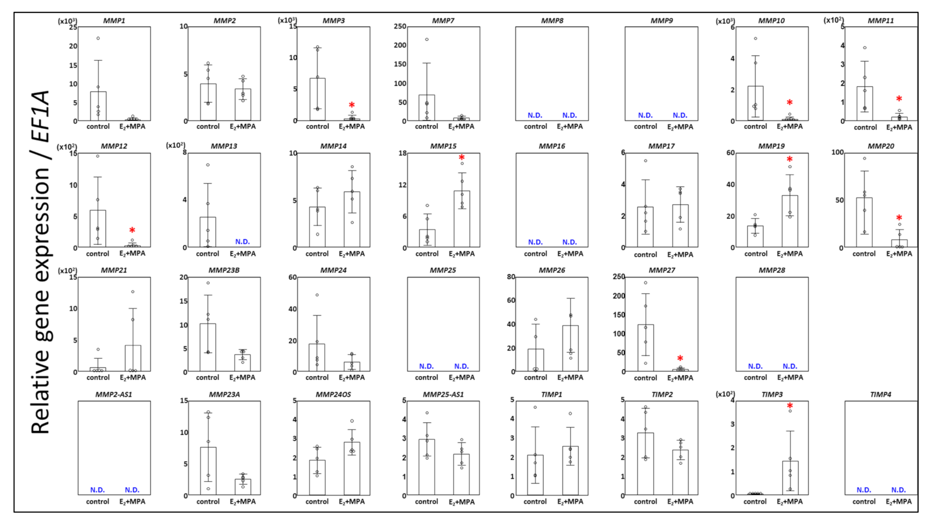
3.1. Expression Changes in MMPs

First, we investigated the gene expression profile of control ESCs or ESCs treated with E2 + MPA for 12 days to investigate the involvement of MMPs in the decidualization process. Our analysis revealed that MMP3, MMP10, MMP11, MMP12, MMP20, and MMP27 were significantly downregulated in decidualized ESCs compared with those in the control (Figure 1). MMP1, MMP7, MMP13, and MMP23B tend to have lower expression in decidualized ESCs than in the control but did not show significant down-regulation due to variability among samples. Moreover, MMP15 and MMP19 were significantly upregulated in decidualized ESCs compared to those in the control (Figure 1), with no significant differences in MMP2, MMP7, MMP14, MMP17, MMP21, MMP24, and MMP26 between the two groups. MMP15 protein expression in ESCs was also confirmed (Figure 2). Furthermore, as with gene expression, E2 + MPA treatment significantly increased the amount of MMP15 protein (Figure 2).
Although all genes were amplified from the endometrial cDNA pool from cells in the proliferative to secretory phase, the CT values in the RT-qPCR analysis of MMP8, MMP9, MMP16, MMP25, and MMP28 in both decidualized and control ESCs were about the same levels as the CT value of the no-template control (with only distilled water, negative control). Therefore, we hypothesized that MMP8, MMP9, MMP16, MMP25, and MMP28 are not expressed in ESCs (Figure 1).
It is well known that antisense long non-coding RNAs are involved in the expression of protein-coding genes at their loci [21]. Thus, we examined the expression of MMP2-AS1, MMP23A, MMP24OS, and MMP25AS-1 to find a novel mechanism regulating MMP expression in ESCs by antisense long non-coding RNAs. However, we did not detect MMP2-AS1 expression in ESCs or find any involvement of MMP23A, MMP24OS, and MMP25AS-1 in decidualization (Figure 1).

3.2. Expression Chages in TIMPs

TIMP3 was significantly increased in decidualized ESCs (Figure 1) as in our previous studies [19]. However, no significant differences in TIMP1, TIMP2, and TIMP4 expression were detected between E2 + MPA and the control group.

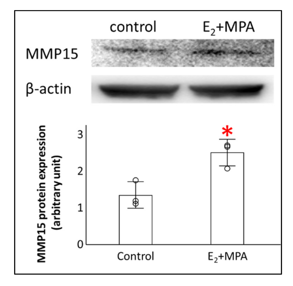
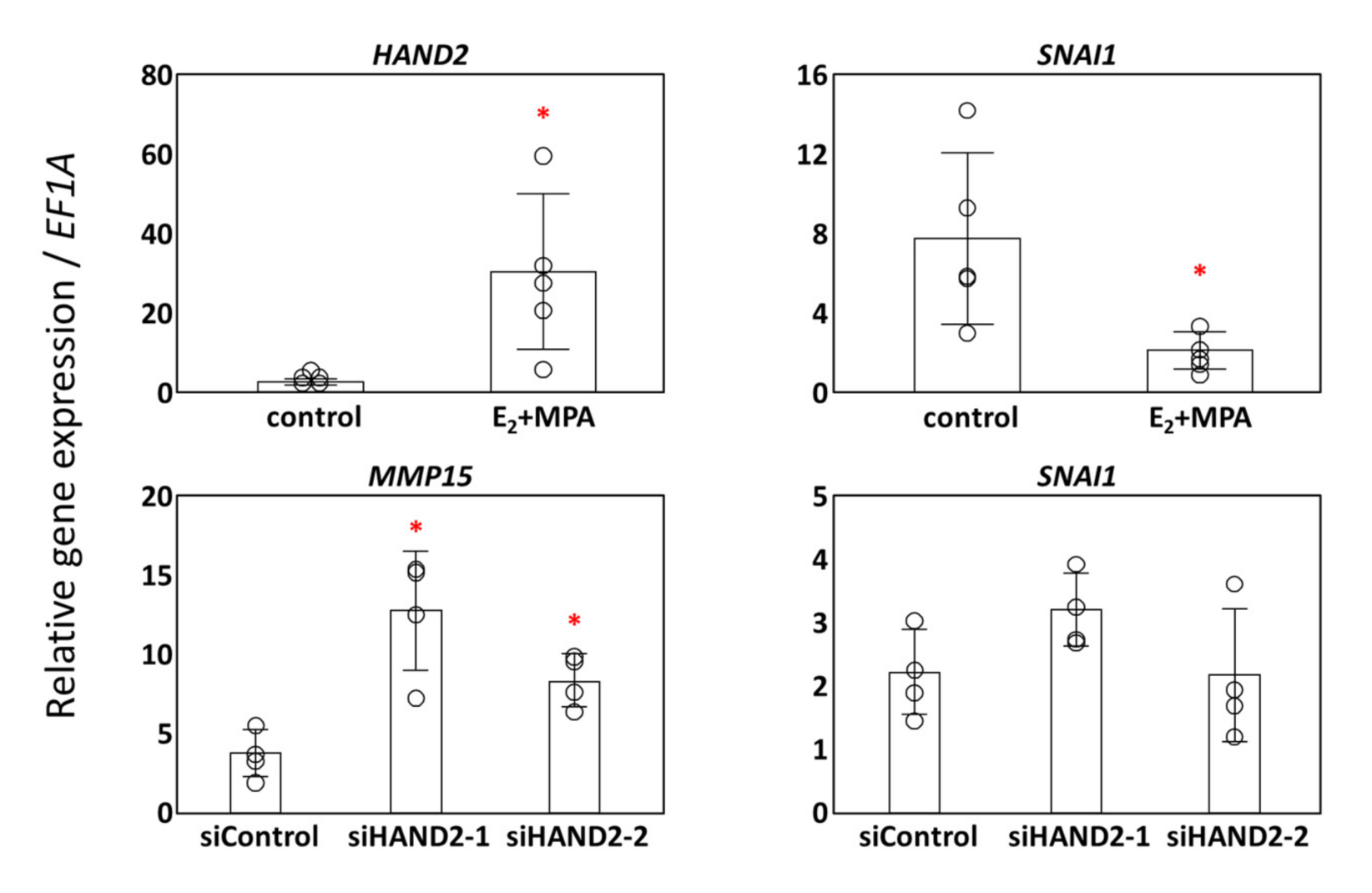
3.3. Potential Transcription Factors for MMP15

Progesterone receptor (PGR) is known to bind to the MMP19 genomic locus and to regulate MMP19 expression during decidualization [22], whereas the transcriptional regulation of MMP15 during decidualization is not well understood. Therefore, we searched the ChIP-Atlas database (https://chip-atlas.org/, accessed on 23 April 2021) for transcription factors that may regulate the transcription of MMPs [23]. We found that HAND2, a master transcriptional regulator [24,25], which is upregulated in decidualization of ESCs, may bind to the upstream region of MMP2 and MMP15 genes. There are no ChIP-based studies that report the binding of HAND2 to the upstream region of other MMPs or to that of any of the TIMPs in the ChIP-Atlas database. Moreover, MMP15 is regulated by SNAI1, a transcriptional repressor, and SNAI1 is regulated by HAND2 in the heart [26,27]. Therefore, we examined the gene expression changes of HAND2 and SNAI1 during the decidualization of ESCs to understand their involvement in the regulation of MMP15 expression. As reported in our previous study [25], HAND2 expression was significantly upregulated in ESCs with E2 + MPA treatment compared with that in the control group (Figure 3). In contrast, SNAI1 expression was significantly suppressed in ESCs with E2 + MPA treatment (Figure 3). Therefore, we examined the expression of SNAI1 and MMP15 in siRNA-mediated HAND2-silenced samples from our previous study [14]. We found upregulation of MMP15 expression upon silencing of HAND2 (Figure 3), suggesting a crucial role of HAND2 in regulating the baseline of MMP15 expression in ESCs. However, SNAI1 expression was not affected by the decrease in the HAND2 expression (Figure 3), thereby suggesting that SNAI1 expression in ESCs was independent of HAND2.

4. Discussion

In this study, we surprisingly found significant downregulation of many MMPs and significant upregulation of MMP15 and MMP19 during ESC decidualization. Moreover, consistent with findings from our previous study [19], only TIMP3 expression was significantly increased among TIMP family members. Such observations collectively led us to conclude that the downregulation of many MMPs and up-regulation of MMP15, MMP19, and TIMP3 must be necessary for ESC decidualization.
To check the purity of the patient-derived ESCs after isolation, we confirmed vimentin expression on IHC staining and elevation of IGFBP1 and PRL, which are known as decidualized-ESCs markers. Furthermore, proof that other endometrial mesenchymal cells were eliminated in this study derives from the following: (1) MMP9 has been reported to be upregulated in umbilical cord-derived mesenchymal stem cells in uterine scars [28], whereas MMP9 was not expressed in our study; (2) MMP16 is known to be expressed in endometrial mesenchymal stem cells [29] but was not found in our study; and (3) MMP19 is known to show lower expression in endometrial mesenchymal stem cells than in glandular cells [29], but MMP19 expression was found to be elevated in our study. Therefore, the influence of mesenchymal stem cells is considered negligible in this study.
MMP15 is the only membrane type (MT)-MMP expressed in the human placenta during early pregnancy [30,31,32]. MT-MMPs including MMP15, are specifically expressed in the front line of invading cells and can degrade ECM in a site-specific manner during cell invasion in cancer [33]. Moreover, cytotrophoblasts (CTB) from human decidual tissue show significant MMP15 expression [9]. Majali-Martinez A et al. also showed that stromal CTBs in the decidua basalis of the first trimester are the primary source of MMP15 [34]. Therefore, we hypothesized that decidualized ESCs might use MMP15, together with stromal CTBs, to break MMP15-specific substrates, including several decidual ECM proteins such as collagen I and IV, laminin, and vitronectin [35,36] for CTB invasion.
We found a possibility that HAND2, a master transcriptional regulator [24,25] upregulated in ESC decidualization, may bind to the upstream regions of MMP2 and MMP15 genes with analysis of the ChIP-Atlas database (https://chip-atlas.org/, accessed on 23 April 2021) [23]. There are no ChIP reports on HAND2 binding to the upstream regions of MMPs without MMP2 and MMP15 and TIMPs in the ChIP-Atlas database. Similarly, the transcriptional regulator FOXO1, which is upregulated in decidualization [17,37,38,39], was found to not bind to the upstream region of any MMP, including MMP2 and MMP15 or TIMPs from the Chip-Atlas search. The consensus motif for HAND2 binding (CAGATG) [40] is found in two locations (−1253/−1248, −1523/−1518) of the 1.5-kilobase region upstream of the human MMP15 gene. These sequences appear to be vital because they are conserved even in the proximal upstream region of the mouse MMP15 gene (−1472/−1467, −1636/−1631). However, the HAND2 consensus motif (CAGATG) was absent even in the 5-kilobase upstream region of the human MMP2 gene. In contrast, the results of siRNA-mediated HAND2 silencing showed HAND2 repressed MMP15 transcription, suggesting that a group of other transcription factors increased in ESCs under the influence of P4 upregulate MMP15 transcription. Subsequently, the increased amount of MMP15 protein might promote menstruation by cleaving proMMP2 to the active form, MMP2, which is compatible with many substrates [41,42]. Mmp15 is known to be regulated by the P4 receptor and the CCAAT/enhancer-binding protein β (Cebpb) in medaka follicles [43]. CEBPBs are known regulators affecting the proliferation and differentiation of ESCs [44]. We applied TFBIND [45] used in a study of the medaka genome [43] to the 2 kb upstream region of the human MMP15 gene; however, unlike in the medaka, a putative P4 receptor motif was not found in the human counterpart. Moreover, multiple putative CEBPBs motifs were found in the 2k upstream region of the human MMP15 gene (−194/183, −164/−155, −151/−140), suggesting that CEBPB might be implicated in the transcriptional regulation of human MMP15. Interestingly, we found putative estrogen receptor 1 binding motifs in the 2k upstream region of the human MMP15 gene (−85/−67). The involvement of these factors in regulating MMP15 expression in ESCs needs further investigation to clearly delineate the mechanistic basis of the regulation.
MMP15 expression is also known to be driven by SNAI1, an epithelial-mesenchymal transition (EMT)-related transcription factor [46] in endocardial cushion development [27]. SNAI1 is also known to be transcriptionally regulated by HAND2 [26]. E-box sequence-mediated regulation by SNAI1 is conserved between humans and mice [27]. In contrast to endocardial development, SNAI1 might suppress the expression of MMP15 in ESCs, according to many reports showing how the same DNA binding factors regulate the transcription of several mRNAs differentially during the developmental stages [47]. A similar phenomenon depending on cell type is supposed to occur between MMP15 expression and binding of SNAI1 to the motif in the promoter region of MMP15 gene. Our observations from siRNA-mediated HAND2 silencing experiments also support that there is no direct relationship between SNAI1 transcription and HAND2 in ESCs. Therefore, we propose SNAI1-mediated MMP15 regulation to be independent of HAND2 in ESCs. Interestingly, in the process of decidualization, MMP15 itself is a regulator of EMT [48,49]. Furthermore, as MMP15 is upregulated in preeclampsia [49], proper regulation of MMP15 in decidualized ESCs could be necessary for normal pregnancy.
It is known that progesterone receptor (PGR) binds to the MMP19 genomic locus and regulates MMP19 expression during decidualization [22]. Angiogenesis is the process by which endothelial cells migrate and invade through the ECM by inducing angiogenesis-promoting secretory factors, and new blood vessels are formed from existing ones. MMP19 localizes in vascular endothelial and smooth muscle cells [50] and exerts anti-angiogenic effects on endothelial cells by generating angiostatin-like fragments [51]. These angiostatin-like fragments stabilize blood vessels by inhibiting the proliferation of human microvascular endothelial cells and reducing the formation of capillary-like structures [51]. Similarly, PGR-induced MMP19, which decreases in low fertility animals, is considered necessary in maintaining vascular stability during decidualization, which may be critical for pregnancy [52,53].
In this study, we found downregulated expression of multiple MMPs in association with decidualization. Interestingly, a relationship between recurrent spontaneous abortion and MMP10 mutation is known [54]. Therefore, we hypothesized that MMP10 in ESCs might be essential for embryo receptivity.
The family of TIMPs has four non-redundant members in the human genome to physiologically inhibit MMPs. The balance between MMP and TIMP activity is vital for the normal function and stability of the ECM. TIMP3 is the only TIMP family member with a high affinity for ECM proteoglycans, and the widest range of substrates, including all MMPs, disintegrins, and metalloproteinases (ADAMs), with thrombospondin motifs [55,56,57,58,59,60,61]. Therefore, we hypothesized that TIMP3 has more diverse functions beyond MMP inhibition, such as regulating MMPs during decidualization. TIMP3 has been reported to have MMP-independent functions with a variety of interactors [62,63,64,65,66,67,68,69]. For example, TIMP3 is known to inhibit the binding of VEGF to VEGFR2 [62], thereby increasing vascular stability, as TIMP3 inhibits vascular endothelial cell proliferation and migration, and increases vascular permeability caused by VEGF–VEGFR2 interaction.

5. Conclusions

In conclusion, from the results of our study, we established an association between MMP15 expression and decidualization. However, as we could not prove whether MMP15 elevation is a result of decidualization or whether it is involved in decidualization itself, further studies of MMP15 knockdown in ESCs are necessary to understand the mechanistic details of MMP15 function.

Author Contributions

Y.H. (Yoji Hisamatsu), H.M. and S.T. designed the study; Y.H. (Yoji Hisamatsu), H.M., S.T., H.T.; and Y.H. (Yoshiko Hashimoto) performed the experiments and analyzed data; Y.H. (Yoji Hisamatsu), H.M., S.T., M.K.; and H.O. wrote the paper. All authors have read and agreed to the published version of the manuscript.

Funding

This research was funded by JSPS KAKENHI [grant numbers. 21K09480 (H.O.), 21K09529 (H.M.), and 19K06891 (S.T.), the Yamaguchi Endocrine Research Foundation (H.M.), and the Takeda Science Foundation (S.T.). The funding agency had no role in study design, data collection, analysis, decision to publish, or manuscript preparation.

Institutional Review Board Statement

This study was performed with approval from the review board of Kansai Medical University (protocol number 2006101). This study also adheres to the principles of the Declaration of Helsinki.

Informed Consent Statement

We obtained informed consent from each patient. Explanations were given orally and in written form, and informed consent was obtained in written form.

Data Availability Statement

The data that support the findings of this study are available from the corresponding author, (S.T.), upon reasonable request.

Acknowledgments

We thank our colleagues from the Department of Obstetrics and Gynecology, Kansai Medical University, for patient recruitment, and those from the Biomedical Science Central Research Center of Kansai Medical University for providing technical support.

Conflicts of Interest

The authors declare no conflict of interest in connection with this article.

References

  1. Cha, J.; Sun, X.; Dey, S.K. Mechanisms of implantation: Strategies for successful pregnancy. Nat. Med. 2012, 18, 1754–1767. [Google Scholar] [CrossRef] [PubMed]
  2. Evans, J.; Salamonsen, L.A.; Winship, A.; Menkhorst, E.; Nie, G.; Gargett, C.E.; Dimitriadis, E. Fertile ground: Human endometrial programming and lessons in health and disease. Nat. Rev. Endocrinol. 2016, 12, 654–667. [Google Scholar] [CrossRef] [PubMed]
  3. Fox, C.; Morin, S.; Jeong, J.W.; Scott, R.T., Jr.; Lessey, B.A. Local and systemic factors and implantation: What is the evidence? Fertil. Steril. 2016, 105, 873–884. [Google Scholar] [CrossRef] [Green Version]
  4. Patel, B.; Elguero, S.; Thakore, S.; Dahoud, W.; Bedaiwy, M.; Mesiano, S. Role of nuclear progesterone receptor isoforms in uterine pathophysiology. Hum. Reprod. Update 2015, 21, 155–173. [Google Scholar] [CrossRef] [PubMed] [Green Version]
  5. Vinketova, K.; Mourdjeva, M.; Oreshkova, T. Human Decidual Stromal Cells as a Component of the Implantation Niche and a Modulator of Maternal Immunity. J. Pregnancy 2016, 2016, 8689436. [Google Scholar] [CrossRef] [Green Version]
  6. Mori, M.; Bogdan, A.; Balassa, T.; Csabai, T.; Szekeres-Bartho, J. The decidua-the maternal bed embracing the embryo-maintains the pregnancy. Semin. Immunopathol. 2016, 38, 635–649. [Google Scholar] [CrossRef] [PubMed] [Green Version]
  7. Burton, G.J.; Woods, A.W.; Jauniaux, E.; Kingdom, J.C. Rheological and physiological consequences of conversion of the maternal spiral arteries for uteroplacental blood flow during human pregnancy. Placenta 2009, 30, 473–482. [Google Scholar] [CrossRef] [Green Version]
  8. Fitzgerald, J.S.; Germeyer, A.; Huppertz, B.; Jeschke, U.; Knofler, M.; Moser, G.; Scholz, C.; Sonderegger, S.; Toth, B.; Markert, U.R. Governing the invasive trophoblast: Current aspects on intra- and extracellular regulation. Am. J. Reprod. Immunol. 2010, 63, 492–505. [Google Scholar] [CrossRef] [PubMed]
  9. Anacker, J.; Segerer, S.E.; Hagemann, C.; Feix, S.; Kapp, M.; Bausch, R.; Kammerer, U. Human decidua and invasive trophoblasts are rich sources of nearly all human matrix metalloproteinases. Mol. Hum. Reprod. 2011, 17, 637–652. [Google Scholar] [CrossRef] [Green Version]
  10. Dong, J.C.; Dong, H.; Campana, A.; Bischof, P. Matrix metalloproteinases and their specific tissue inhibitors in menstruation. Reproduction 2002, 123, 621–631. [Google Scholar] [CrossRef]
  11. Anacker, J.; Feix, S.; Kapp, M.; Bausch, R.; Kammerer, U. Expression pattern of matrix metalloproteinases (MMPs) in human decidua during pregnancy. J. Reprod. Immunol. 2010, 86, 79. [Google Scholar] [CrossRef]
  12. Murphy, G.; Nagase, H. Progress in matrix metalloproteinase research. Mol. Asp. Med. 2008, 29, 290–308. [Google Scholar] [CrossRef] [PubMed] [Green Version]
  13. Kessenbrock, K.; Plaks, V.; Werb, Z. Matrix metalloproteinases: Regulators of the tumor microenvironment. Cell 2010, 141, 52–67. [Google Scholar] [CrossRef] [Green Version]
  14. Murata, H.; Tsuzuki, T.; Kido, T.; Kakita-Kobayashi, M.; Kida, N.; Hisamatsu, Y.; Okada, H. Progestin-induced heart and neural crest derivatives-expressed transcript 2 inhibits angiopoietin 2 via fibroblast growth factor 9 in human endometrial stromal cells. Reprod. Biol. 2019, 19, 14–21. [Google Scholar] [CrossRef]
  15. Okada, H.; Nakajima, T.; Yoshimura, T.; Yasuda, K.; Kanzaki, H. The inhibitory effect of dienogest, a synthetic steroid, on the growth of human endometrial stromal cells in vitro. Mol. Hum. Reprod. 2001, 7, 341–347. [Google Scholar] [CrossRef] [PubMed] [Green Version]
  16. Zhu, H.H.; Huang, J.R.; Mazella, J.; Rosenberg, M.; Tseng, L. Differential effects of progestin and relaxin on the synthesis and secretion of immunoreactive prolactin in long term culture of human endometrial stromal cells. J. Clin. Endocrinol. Metab. 1990, 71, 889–899. [Google Scholar] [CrossRef] [PubMed]
  17. Shindoh, H.; Okada, H.; Tsuzuki, T.; Nishigaki, A.; Kanzaki, H. Requirement of heart and neural crest derivatives-expressed transcript 2 during decidualization of human endometrial stromal cells in vitro. Fertil. Steril. 2014, 101, 1781–1790. [Google Scholar] [CrossRef]
  18. Livak, K.J.; Schmittgen, T.D. Analysis of relative gene expression data using real-time quantitative PCR and the 2(-Delta Delta C(T)) Method. Methods 2001, 25, 402–408. [Google Scholar] [CrossRef]
  19. Cho, H.; Okada, H.; Tsuzuki, T.; Nishigaki, A.; Yasuda, K.; Kanzaki, H. Progestin-induced heart and neural crest derivatives expressed transcript 2 is associated with fibulin-1 expression in human endometrial stromal cells. Fertil. Steril. 2013, 99, 248–255. [Google Scholar] [CrossRef]
  20. Schneider, C.A.; Rasband, W.S.; Eliceiri, K.W. NIH Image to ImageJ: 25 years of image analysis. Nat. Methods 2012, 9, 671–675. [Google Scholar] [CrossRef]
  21. Wahlestedt, C. Targeting long non-coding RNA to therapeutically upregulate gene expression. Nat. Rev. Drug Discov. 2013, 12, 433–446. [Google Scholar] [CrossRef]
  22. Mazur, E.C.; Vasquez, Y.M.; Li, X.; Kommagani, R.; Jiang, L.; Chen, R.; Lanz, R.B.; Kovanci, E.; Gibbons, W.E.; DeMayo, F.J. Progesterone receptor transcriptome and cistrome in decidualized human endometrial stromal cells. Endocrinology 2015, 156, 2239–2253. [Google Scholar] [CrossRef]
  23. Oki, S.; Ohta, T.; Shioi, G.; Hatanaka, H.; Ogasawara, O.; Okuda, Y.; Kawaji, H.; Nakaki, R.; Sese, J.; Meno, C. ChIP-Atlas: A data-mining suite powered by full integration of public ChIP-seq data. EMBO Rep. 2018, 19, e46255. [Google Scholar] [CrossRef]
  24. Okada, H.; Tsuzuki, T.; Murata, H. Decidualization of the human endometrium. Reprod. Med. Biol. 2018, 17, 220–227. [Google Scholar] [CrossRef] [PubMed] [Green Version]
  25. Murata, H.; Tanaka, S.; Tsuzuki-Nakao, T.; Kido, T.; Kakita-Kobayashi, M.; Kida, N.; Hisamatsu, Y.; Tsubokura, H.; Hashimoto, Y.; Kitada, M.; et al. The transcription factor HAND2 up-regulates transcription of the IL15 gene in human endometrial stromal cells. J. Biol. Chem. 2020, 295, 9596–9605. [Google Scholar] [CrossRef]
  26. Laurent, F.; Girdziusaite, A.; Gamart, J.; Barozzi, I.; Osterwalder, M.; Akiyama, J.A.; Lincoln, J.; Lopez-Rios, J.; Visel, A.; Zuniga, A.; et al. HAND2 Target Gene Regulatory Networks Control Atrioventricular Canal and Cardiac Valve Development. Cell Rep. 2017, 19, 1602–1613. [Google Scholar] [CrossRef] [PubMed] [Green Version]
  27. Tao, G.; Levay, A.K.; Gridley, T.; Lincoln, J. Mmp15 is a direct target of Snai1 during endothelial to mesenchymal transformation and endocardial cushion development. Dev. Biol. 2011, 359, 209–221. [Google Scholar] [CrossRef] [PubMed] [Green Version]
  28. Xu, L.; Ding, L.; Wang, L.; Cao, Y.; Zhu, H.; Lu, J.; Li, X.; Song, T.; Hu, Y.; Dai, J. Umbilical cord-derived mesenchymal stem cells on scaffolds facilitate collagen degradation via upregulation of MMP-9 in rat uterine scars. Stem Cell Res. Ther. 2017, 8, 84. [Google Scholar] [CrossRef] [Green Version]
  29. Spitzer, T.L.; Rojas, A.; Zelenko, Z.; Aghajanova, L.; Erikson, D.W.; Barragan, F.; Meyer, M.; Tamaresis, J.S.; Hamilton, A.E.; Irwin, J.C.; et al. Perivascular human endometrial mesenchymal stem cells express pathways relevant to self-renewal, lineage specification, and functional phenotype. Biol. Reprod. 2012, 86, 58. [Google Scholar] [CrossRef]
  30. Majali-Martinez, A.; Hiden, U.; Ghaffari-Tabrizi-Wizsy, N.; Lang, U.; Desoye, G.; Dieber-Rotheneder, M. Placental membrane-type metalloproteinases (MT-MMPs): Key players in pregnancy. Cell Adhes. Migr. 2016, 10, 136–146. [Google Scholar] [CrossRef] [Green Version]
  31. Pollheimer, J.; Fock, V.; Knofler, M. Review: The ADAM metalloproteinases—Novel regulators of trophoblast invasion? Placenta 2014, 35, S57–S63. [Google Scholar] [CrossRef] [PubMed]
  32. Takino, T.; Sato, H.; Shinagawa, A.; Seiki, M. Identification of the second membrane-type matrix metalloproteinase (MT-MMP-2) gene from a human placenta cDNA library. MT-MMPs form a unique membrane-type subclass in the MMP family. J. Biol. Chem. 1995, 270, 23013–23020. [Google Scholar] [CrossRef] [PubMed] [Green Version]
  33. Hernandez-Barrantes, S.; Bernardo, M.; Toth, M.; Fridman, R. Regulation of membrane type-matrix metalloproteinases. Semin. Cancer Biol. 2002, 12, 131–138. [Google Scholar] [CrossRef] [PubMed]
  34. Majali-Martinez, A.; Hoch, D.; Tam-Amersdorfer, C.; Pollheimer, J.; Glasner, A.; Ghaffari-Tabrizi-Wizsy, N.; Beristain, A.G.; Hiden, U.; Dieber-Rotheneder, M.; Desoye, G. Matrix metalloproteinase 15 plays a pivotal role in human first trimester cytotrophoblast invasion and is not altered by maternal obesity. FASEB J. 2020, 34, 10720–10730. [Google Scholar] [CrossRef]
  35. Smith, S.D.; Choudhury, R.H.; Matos, P.; Horn, J.A.; Lye, S.J.; Dunk, C.E.; Aplin, J.D.; Jones, R.L.; Harris, L.K. Changes in vascular extracellular matrix composition during decidual spiral arteriole remodeling in early human pregnancy. Histol. Histopathol. 2016, 31, 557–571. [Google Scholar] [CrossRef]
  36. Huppertz, B.; Kertschanska, S.; Demir, A.Y.; Frank, H.G.; Kaufmann, P. Immunohistochemistry of matrix metalloproteinases (MMP), their substrates, and their inhibitors (TIMP) during trophoblast invasion in the human placenta. Cell Tissue Res. 1998, 291, 133–148. [Google Scholar] [CrossRef] [PubMed]
  37. Halari, C.D.; Nandi, P.; Jeyarajah, M.J.; Renaud, S.J.; Lala, P.K. Decorin production by the human decidua: Role in decidual cell maturation. Mol. Hum. Reprod. 2020, 26, 784–796. [Google Scholar] [CrossRef] [PubMed]
  38. Huyen, D.V.; Bany, B.M. Evidence for a conserved function of heart and neural crest derivatives expressed transcript 2 in mouse and human decidualization. Reproduction 2011, 142, 353–368. [Google Scholar] [CrossRef]
  39. Murata, H.; Tanaka, S.; Hisamatsu, Y.; Tsubokura, H.; Hashimoto, Y.; Kitada, M.; Okada, H. Transcriptional regulation of LGALS9 by HAND2 and FOXO1 in human endometrial stromal cells in women with regular cycles. Mol. Hum. Reprod. 2021, 27, gaab063. [Google Scholar] [CrossRef]
  40. Stormo, G.D. Modeling the specificity of protein-DNA interactions. Quant. Biol. 2013, 1, 115–130. [Google Scholar] [CrossRef] [Green Version]
  41. Morrison, C.J.; Butler, G.S.; Bigg, H.F.; Roberts, C.R.; Soloway, P.D.; Overall, C.M. Cellular activation of MMP-2 (gelatinase A) by MT2-MMP occurs via a TIMP-2-independent pathway. J. Biol. Chem. 2001, 276, 47402–47410. [Google Scholar] [CrossRef] [Green Version]
  42. Morrison, C.J.; Overall, C.M. TIMP independence of matrix metalloproteinase (MMP)-2 activation by membrane type 2 (MT2)-MMP is determined by contributions of both the MT2-MMP catalytic and hemopexin C domains. J. Biol. Chem. 2006, 281, 26528–26539. [Google Scholar] [CrossRef] [PubMed] [Green Version]
  43. Ogiwara, K.; Takahashi, T. Involvement of the nuclear progestin receptor in LH-induced expression of membrane type 2-matrix metalloproteinase required for follicle rupture during ovulation in the medaka, Oryzias latipes. Mol. Cell. Endocrinol. 2017, 450, 54–63. [Google Scholar] [CrossRef] [PubMed]
  44. Wang, W.; Taylor, R.N.; Bagchi, I.C.; Bagchi, M.K. Regulation of human endometrial stromal proliferation and differentiation by C/EBPbeta involves cyclin E-cdk2 and STAT3. Mol. Endocrinol. 2012, 26, 2016–2030. [Google Scholar] [CrossRef] [PubMed] [Green Version]
  45. Tsunoda, T.; Takagi, T. Estimating transcription factor bindability on DNA. Bioinformatics 1999, 15, 622–630. [Google Scholar] [CrossRef] [PubMed]
  46. Carver, E.A.; Jiang, R.; Lan, Y.; Oram, K.F.; Gridley, T. The mouse snail gene encodes a key regulator of the epithelial-mesenchymal transition. Mol. Cell. Biol. 2001, 21, 8184–8188. [Google Scholar] [CrossRef] [PubMed] [Green Version]
  47. Schroer, U.; Volk, G.F.; Liedtke, T.; Thanos, S. Translin-associated factor-X (Trax) is a molecular switch of growth-associated protein (GAP)-43 that controls axonal regeneration. Eur. J. Neurosci. 2007, 26, 2169–2178. [Google Scholar] [CrossRef]
  48. Liu, Y.; Sun, X.; Feng, J.; Deng, L.L.; Li, B.; Zhu, M.; Lu, C.; Zhou, L. MT2-MMP induces proteolysis and leads to EMT in carcinomas. Oncotarget 2016, 7, 48193–48205. [Google Scholar] [CrossRef] [PubMed] [Green Version]
  49. Kaitu’u-Lino, T.J.; Palmer, K.; Tuohey, L.; Ye, L.; Tong, S. MMP-15 is upregulated in preeclampsia, but does not cleave endoglin to produce soluble endoglin. PLoS ONE 2012, 7, e39864. [Google Scholar] [CrossRef] [Green Version]
  50. Kolb, C.; Mauch, S.; Krawinkel, U.; Sedlacek, R. Matrix metalloproteinase-19 in capillary endothelial cells: Expression in acutely, but not in chronically, inflamed synovium. Exp. Cell Res. 1999, 250, 122–130. [Google Scholar] [CrossRef]
  51. Brauer, R.; Beck, I.M.; Roderfeld, M.; Roeb, E.; Sedlacek, R. Matrix metalloproteinase-19 inhibits growth of endothelial cells by generating angiostatin-like fragments from plasminogen. BMC Biochem. 2011, 12, 38. [Google Scholar] [CrossRef] [Green Version]
  52. Mattos, R.; Staples, C.R.; Thatcher, W.W. Effects of dietary fatty acids on reproduction in ruminants. Rev. Reprod. 2000, 5, 38–45. [Google Scholar] [CrossRef]
  53. Killeen, A.P.; Morris, D.G.; Kenny, D.A.; Mullen, M.P.; Diskin, M.G.; Waters, S.M. Global gene expression in endometrium of high and low fertility heifers during the mid-luteal phase of the estrous cycle. BMC Genom. 2014, 15, 234. [Google Scholar] [CrossRef] [Green Version]
  54. Quintero-Ronderos, P.; Mercier, E.; Fukuda, M.; Gonzalez, R.; Suarez, C.F.; Patarroyo, M.A.; Vaiman, D.; Gris, J.C.; Laissue, P. Novel genes and mutations in patients affected by recurrent pregnancy loss. PLoS ONE 2017, 12, e0186149. [Google Scholar] [CrossRef]
  55. Staskus, P.W.; Masiarz, F.R.; Pallanck, L.J.; Hawkes, S.P. The 21-kDa protein is a transformation-sensitive metalloproteinase inhibitor of chicken fibroblasts. J. Biol. Chem. 1991, 266, 449–454. [Google Scholar] [CrossRef]
  56. Pavloff, N.; Staskus, P.W.; Kishnani, N.S.; Hawkes, S.P. A new inhibitor of metalloproteinases from chicken: ChIMP-3. A third member of the TIMP family. J. Biol. Chem. 1992, 267, 17321–17326. [Google Scholar] [CrossRef]
  57. Leco, K.J.; Khokha, R.; Pavloff, N.; Hawkes, S.P.; Edwards, D.R. Tissue inhibitor of metalloproteinases-3 (TIMP-3) is an extracellular matrix-associated protein with a distinctive pattern of expression in mouse cells and tissues. J. Biol. Chem. 1994, 269, 9352–9360. [Google Scholar] [CrossRef]
  58. Apte, S.S.; Olsen, B.R.; Murphy, G. The gene structure of tissue inhibitor of metalloproteinases (TIMP)-3 and its inhibitory activities define the distinct TIMP gene family. J. Biol. Chem. 1995, 270, 14313–14318. [Google Scholar] [CrossRef] [PubMed] [Green Version]
  59. Brew, K.; Nagase, H. The tissue inhibitors of metalloproteinases (TIMPs): An ancient family with structural and functional diversity. Biochim. Biophys. Acta 2010, 1803, 55–71. [Google Scholar] [CrossRef] [PubMed] [Green Version]
  60. Moore, L.; Fan, D.; Basu, R.; Kandalam, V.; Kassiri, Z. Tissue inhibitor of metalloproteinases (TIMPs) in heart failure. Heart Fail. Rev. 2012, 17, 693–706. [Google Scholar] [CrossRef]
  61. Jackson, H.W.; Defamie, V.; Waterhouse, P.; Khokha, R. TIMPs: Versatile extracellular regulators in cancer. Nat. Rev. Cancer 2017, 17, 38–53. [Google Scholar] [CrossRef] [PubMed]
  62. Qi, J.H.; Ebrahem, Q.; Moore, N.; Murphy, G.; Claesson-Welsh, L.; Bond, M.; Baker, A.; Anand-Apte, B. A novel function for tissue inhibitor of metalloproteinases-3 (TIMP3): Inhibition of angiogenesis by blockage of VEGF binding to VEGF receptor-2. Nat. Med. 2003, 9, 407–415. [Google Scholar] [CrossRef] [PubMed]
  63. Klenotic, P.A.; Munier, F.L.; Marmorstein, L.Y.; Anand-Apte, B. Tissue inhibitor of metalloproteinases-3 (TIMP-3) is a binding partner of epithelial growth factor-containing fibulin-like extracellular matrix protein 1 (EFEMP1). Implications for macular degenerations. J. Biol. Chem. 2004, 279, 30469–30473. [Google Scholar] [CrossRef] [PubMed] [Green Version]
  64. Nour, N.; Mayer, G.; Mort, J.S.; Salvas, A.; Mbikay, M.; Morrison, C.J.; Overall, C.M.; Seidah, N.G. The cysteine-rich domain of the secreted proprotein convertases PC5A and PACE4 functions as a cell surface anchor and interacts with tissue inhibitors of metalloproteinases. Mol. Biol. Cell 2005, 16, 5215–5226. [Google Scholar] [CrossRef] [Green Version]
  65. Kang, K.H.; Park, S.Y.; Rho, S.B.; Lee, J.H. Tissue inhibitor of metalloproteinases-3 interacts with angiotensin II type 2 receptor and additively inhibits angiogenesis. Cardiovasc. Res. 2008, 79, 150–160. [Google Scholar] [CrossRef] [PubMed] [Green Version]
  66. Scilabra, S.D.; Troeberg, L.; Yamamoto, K.; Emonard, H.; Thogersen, I.; Enghild, J.J.; Strickland, D.K.; Nagase, H. Differential regulation of extracellular tissue inhibitor of metalloproteinases-3 levels by cell membrane-bound and shed low density lipoprotein receptor-related protein 1. J. Biol. Chem. 2013, 288, 332–342. [Google Scholar] [CrossRef] [Green Version]
  67. Huttlin, E.L.; Ting, L.; Bruckner, R.J.; Gebreab, F.; Gygi, M.P.; Szpyt, J.; Tam, S.; Zarraga, G.; Colby, G.; Baltier, K.; et al. The BioPlex Network: A Systematic Exploration of the Human Interactome. Cell 2015, 162, 425–440. [Google Scholar] [CrossRef] [PubMed] [Green Version]
  68. Huttlin, E.L.; Bruckner, R.J.; Paulo, J.A.; Cannon, J.R.; Ting, L.; Baltier, K.; Colby, G.; Gebreab, F.; Gygi, M.P.; Parzen, H.; et al. Architecture of the human interactome defines protein communities and disease networks. Nature 2017, 545, 505–509. [Google Scholar] [CrossRef]
  69. Kwasna, D.; Abdul Rehman, S.A.; Natarajan, J.; Matthews, S.; Madden, R.; De Cesare, V.; Weidlich, S.; Virdee, S.; Ahel, I.; Gibbs-Seymour, I.; et al. Discovery and Characterization of ZUFSP/ZUP1, a Distinct Deubiquitinase Class Important for Genome Stability. Mol. Cell 2018, 70, 150–164, e156. [Google Scholar] [CrossRef] [Green Version]
Figure 1. Effect of E2 + MPA treatment on MMPs or TIMPs in human endometrial stromal cells. Human endometrial stromal cells were treated with E2 (10−8 mol/L) + MPA (10−7 mol/L) (E2 + MPA) or without hormonal treatment (control) for 12 days. The expression levels of all protein-coding MMPs, TIMPs, and long non-coding antisense RNAs from MMP loci were analyzed by quantitative RT-PCR. MMP and TIMP RNA levels were normalized to those of EF1A. Values are shown as mean fold change ± SD of independent experiments using cells derived from different patients (n = 5). Each dot indicates the value for different cell preparations. An asterisk indicates statistically significant differences from the control group, * p < 0.05.
Cimb 43 00146 g001
Figure 2. Effect of E2 + MPA treatment on MMP15 proteins in human endometrial stromal cells. Human endometrial stromal cells were treated with E2 (10−8 mol/L) + MPA (10−7 mol/L) (E2 + MPA) or without hormonal treatment (control) for 12 days. The protein expression levels of MMP15 and β-actin were analyzed by immunoblot analysis. MMP15 were normalized to those of β-actin. Values are shown as mean fold change ± SD of independent experiments using cells derived from different patients (n = 3). Each dot indicates the value for different cell preparations. An asterisk indicates statistically significant differences from the control group, * p < 0.05.
Cimb 43 00146 g002
Figure 3. Effect of siRNA-mediated HAND2 silencing on the expression of MMP15 or SNAI1 in human endometrial stromal cells. Human endometrial stromal cells (ESCs) were treated with E2 (10−8 mol/L) + MPA (10−7 mol/L) or without hormonal treatment (control) for 12 days. HAND2 and SNAI1 were analyzed by quantitative RT-PCR (n = 5, upper panels). ESCs were transfected with human HAND2 siRNA (HAND2-1 and HAND2-2) or a non-silencing RNA on day 0 and then cultured for 5 days without hormonal treatment (lower panels). MMP15 and SNAI1 were quantified using quantitative RT-PCR and corrected for EF1A (lower panels). The bars show the mean of relative gene expression ± SD of independent experiments using cells derived from different patients (n = 4). Each dot indicates the value for cells derived from different patients. An asterisk indicates statistically significant differences from the control group, * p < 0.05.
Cimb 43 00146 g003
Table 1. Sample Information.
Sample No.MaterialsMethodsAgeMenstrual Cycle Phase at the Time of Collection
1Primary culture ESCsTreated with E2 + MPA for 12 days RT-qPCR50Proliferative
2Primary culture ESCsTreated with E2 + MPA for 12 days RT-qPCR45Mid-secretory
3Primary culture ESCsTreated with E2 + MPA for 12 days RT-qPCR48Late-secretory
4Primary culture ESCsTreated with E2 + MPA for 12 days RT-qPCR50Mid-secretory
5Primary culture ESCsTreated with E2 + MPA for 12 days RT-qPCR44Late-secretory
6Primary culture ESCsTransfection of HAND2-siRNART-qPCR49Mid-secretory
7Primary culture ESCsTransfection of HAND2-siRNART-qPCR46Late-secretory
8Primary culture ESCsTransfection of HAND2-siRNART-qPCR42Proliferative
9Primary culture ESCsTransfection of HAND2-siRNART-qPCR47Late-secretory
10Primary culture ESCsTreated with E2 + MPA for 12 daysImmunoblot analysis45Mid-Secretory
11Primary culture ESCsTreated with E2 + MPA for 12 daysImmunoblot analysis42Proliferative
12Primary culture ESCsTreated with E2 + MPA for 12 daysImmunoblot analysis49Proliferative
Table 2. Oligonucleotides.
MMP1MMP1_884F5′-ACAAACCCCAAAAGCGTGTG-3′
MMP1_993R5′-AGAAGGGATTTGTGCGCATG-3′
MMP2MMP2_697F5′-ACAGTGGATGATGCCTTTGC-3
MMP2_808R5′-AGCGGCCAAAGTTGATCATG-3′
MMP3MMP3_1578F5′-TTCGTTTTCTCCTGCCTGTG-3′
MMP3_1693R5′-AGCAGCAGCCCATTTGAATG-3′
MMP7MMP7_362F5′-TTCCAAAGTGGTCACCTACAGG-3′
MMP7_459R5′-TGCCCCACATGTTTAAAGCC-3′
MMP8MMP8_355F5′-ATGAAAAAGCCTCGCTGTGG-3′
MMP8_436R5′-AGTTAGTGCGTTCCCACTTG-3′
MMP9MMP9_1560F5′-ATGCCTGCAACGTGAACATC-3′
MMP9_1646R5′-AGAATCGCCAGTACTTCCCATC-3′
MMP10MMP10_373F5′-CCTTACATACAGGATTGTGAATTATACACC-3′
MMP10_519R5′-GAGATCATTATATCAGCCTCTCCTTCATAC-3′
MMP11MMP11_558F5′-ATGCCTTCTTCCCCAAGACTC-3′
MMP11_684R5′-AGCACGTGGCCAAATTCATG-3′
MMP12MMP12_440F5′-ACGCAATCCGGAAAGCTTTC-3′
MMP12_527R5′-ACCAAAATGTCAGCCATGCC-3′
MMP13MMP13_864F5′-AACGCCAGACAAATGTGACC-3′
MMP13_991R5′-AAAACAGCTCCGCATCAACC-3′
MMP14MMP14_461F5′-AACAGGCAAAGCTGATGCAG-3′
MMP14_567R5′-AGCGCTTCCTTCGAACATTG-3′
MMP15MMP15_1187F5′-TCATGGTACTCTTTGCCTCTGG-3′
MMP15_1310R5′-TCTGCGTCAAAATGGGTGTC-3′
MMP16MMP16_654F5′-AATGGCAGCACAAGCACATC-3′
MMP16_748R5′-ATCAAAGGCACGGCGAATAG-3′
MMP17MMP17_1095F5′-ATGCAGCACTCACTTTGACG-3′
MMP17_1169R5′-CGCCAGAAGTACTTGCCTTTG-3′
MMP19MMP19_1606F5′-AGCCACAGAAACCACGTTTG-3′
MMP19_1698R5′-AAATGAAAGGGTGGGTGGTG-3′
MMP20MMP20_1053F5′-TGGATGCAGCTTACGAAGTG-3′
MMP20_1195R5′-TATTTGCTGCACGTGCCTTG-3′
MMP21MMP21_889F5′-ACGGGATCCATAATGCAACC-3′
MMP21_1025R5′-TTGCGAATCCAGTCAAACGC-3′
MMP23BMMP23B_1021F5′-CCTCCACAAGAAAGGGAAAGTG-3′
MMP23B_1151R5′-TGTAGGTGCCCTCATTGACG-3′
MMP24MMP24_847F5′-AGGAAATGCCAACCATGACG-3′
MMP24_990R5′-AGCTTGAAGTTGTGCGTCTC-3′
MMP25MMP25_3278F5′-TGGCTGTTTCGTGGCATTTC-3′
MMP25_3407R5’-TGGACAGCAACTTAGGAAGTGG-3′
MMP26MMP26_1127F5′-AAAGCACTAGAGCAGCCTTG-3′
MMP26_1215R5′-AGCGTTTTGAGTGTCGGTTC-3′
MMP27MMP27_853F5′-ACCTGCTAAGCCAAAGGAAC-3′
MMP27_932R5′-TGCGGAAAGTTGTGATAGCG-3′
MMP28MMP28_1214F5′-AAACGCAGGGCCCTAAATAC-3′
MMP28_1283R5′-TACAGTTGCTGTTGCCTGTC-3′
TIMP1TIMP1_490F5′-AGGAATGCACAGTGTTTCCC-3′
TIMP1_592R5′-AAGCCCTTTTCAGAGCCTTG-3′
TIMP2TIMP2_2430F5-ACACGCAATGAAACCGAAGC-3′
TIMP2_2503R5′-AACAGGCTAAGGTGGCTTTG-3′
TIMP3TIMP3_1700F5′-TGCCTGCCTTGTACAAAAGC-3′
TIMP3_1805R5′-TGGCCAAATCTACCAAAGCG-3′
TIMP4TIMP4_611F5′-ACTGGCTGTTGGAACGAAAG-3′
TIMP4_681R5′-GCCGTCAACATGCTTCATACAG-3′
MMP2-AS1MMP2-AS1_541F5′-ATGTTGTGAGCAGCCCAATG-3′
MMP2-AS1_682R5′-TTGCCACTCAGCATCATCAC-3′
MMP23AMMP23A_682F5′-CCTCCACAAGAAAGGGAAAGTG-3′
MMP23A_812R5′-TGTAGGTGCCCTCATTGACG-3′
MMP24OSMMP24OS_1029F5′-ATCCCAGGGAAATGACACACTC-3′
MMP24OS_1144R5′-GGGACATCACAGCATTTCAGTG-3′
MMP25-AS1MMP25-AS1_2017F5′-AGTTCCGGAATGCAAAACCC-3′
MMP25-AS1_2086R5′-AGGACCTTGAAAGCATGTGG-3′
HAND2hHAND2-Forward5′-AGAGGAAGAAGGAGCTGAACGA-3′
hHAND2-Reverse5′-CGTCCGGCCTTTGGTTTT-3′
SNAI1SNAI1_1405F5′-TTTCAGCCTCCTGTTTGGTG-3′
SNAI1_1489R5′-TGACAGCCATTACTCACAGTCC-3′
EF1AhEF1A_F5′-TCTGGTTGGAATGGTGACAACATGC-3′
hEF1A_R5′-AGAGCTTCACTCAAAGCTTCATGG-3′
Publisher’s Note: MDPI stays neutral with regard to jurisdictional claims in published maps and institutional affiliations.

