
Andrographolide Relieves Post-Operative Wound Pain but Affects Local Angiogenesis

 , , , and
Abstract
:1. Introduction
2. Results
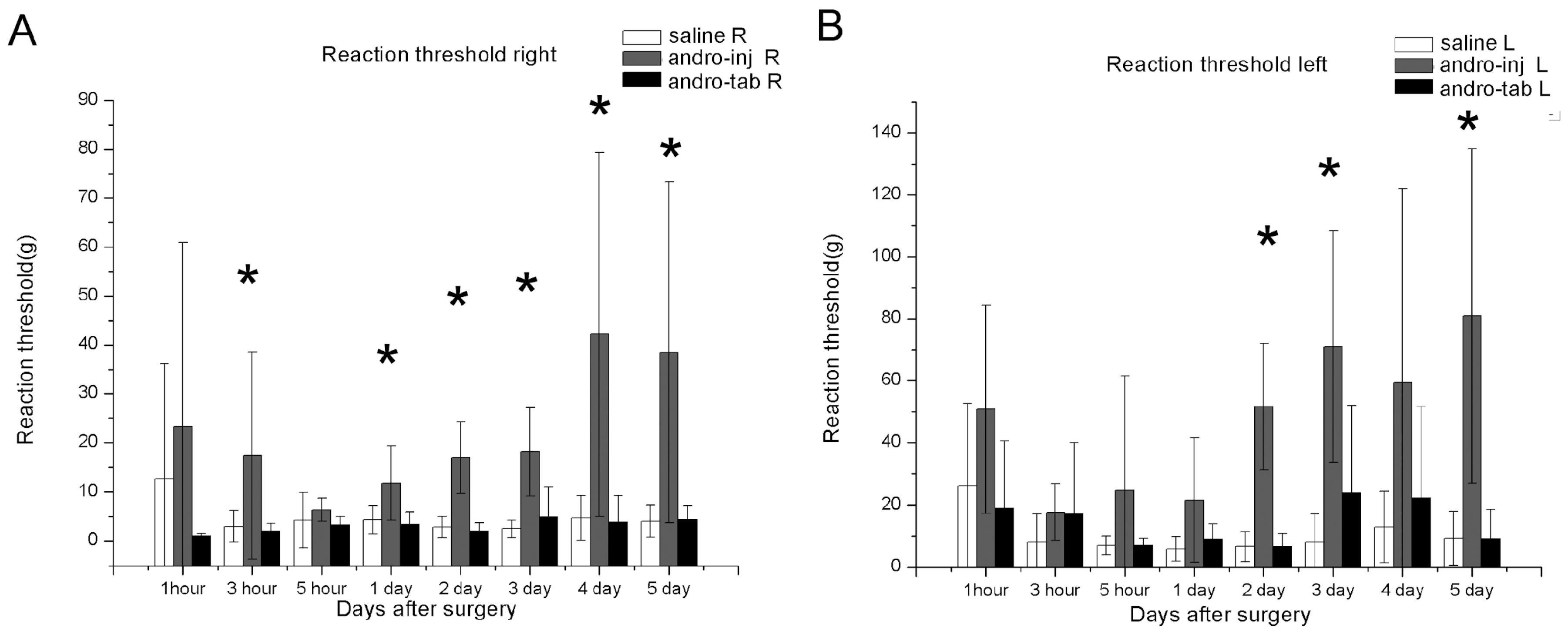
2.1. Von Frey Tests: Reaction Threshold (Left and Right)
2.1.1. The Reaction Thresholds of the Right Hind Paw in the Injection Group Were Significantly Increased Compared to the Saline Group
2.1.2. The Reaction Thresholds of the Left Paw in the Injection Group Were Significantly Increased Compared to Saline Group
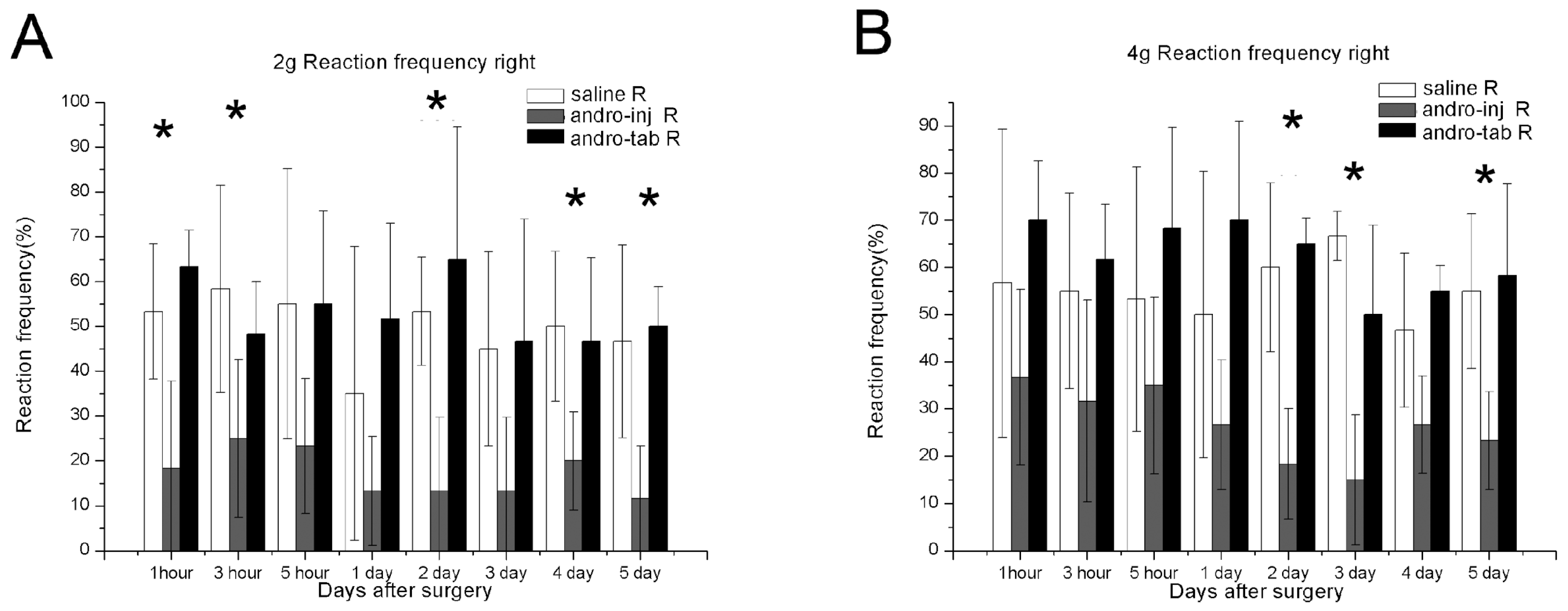
2.1.3. The Reaction Frequency of the Right Paw in the Injection Group Was Significantly Reduced Compared to the Saline Group
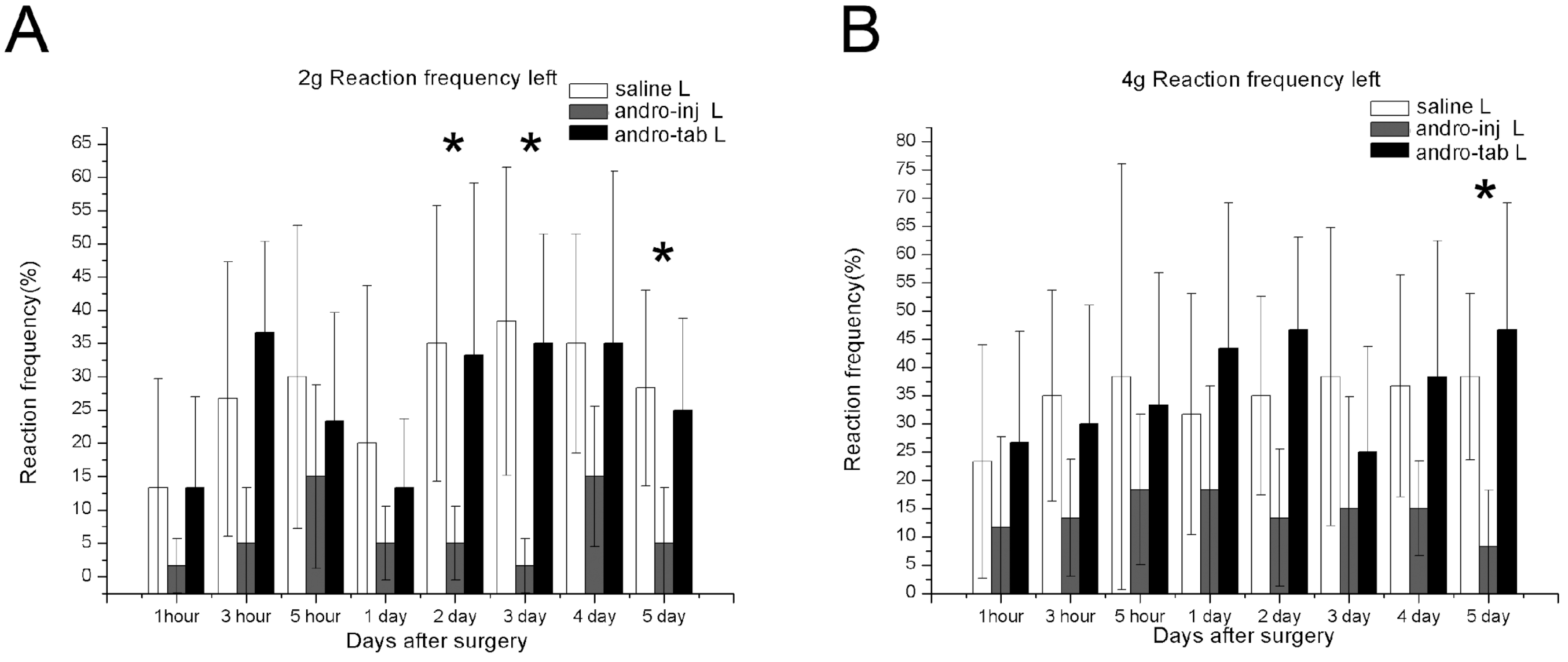
2.1.4. The Reaction Frequency of the Left Paw in the Injection Group Was Significantly Reduced Compared to the Saline Group
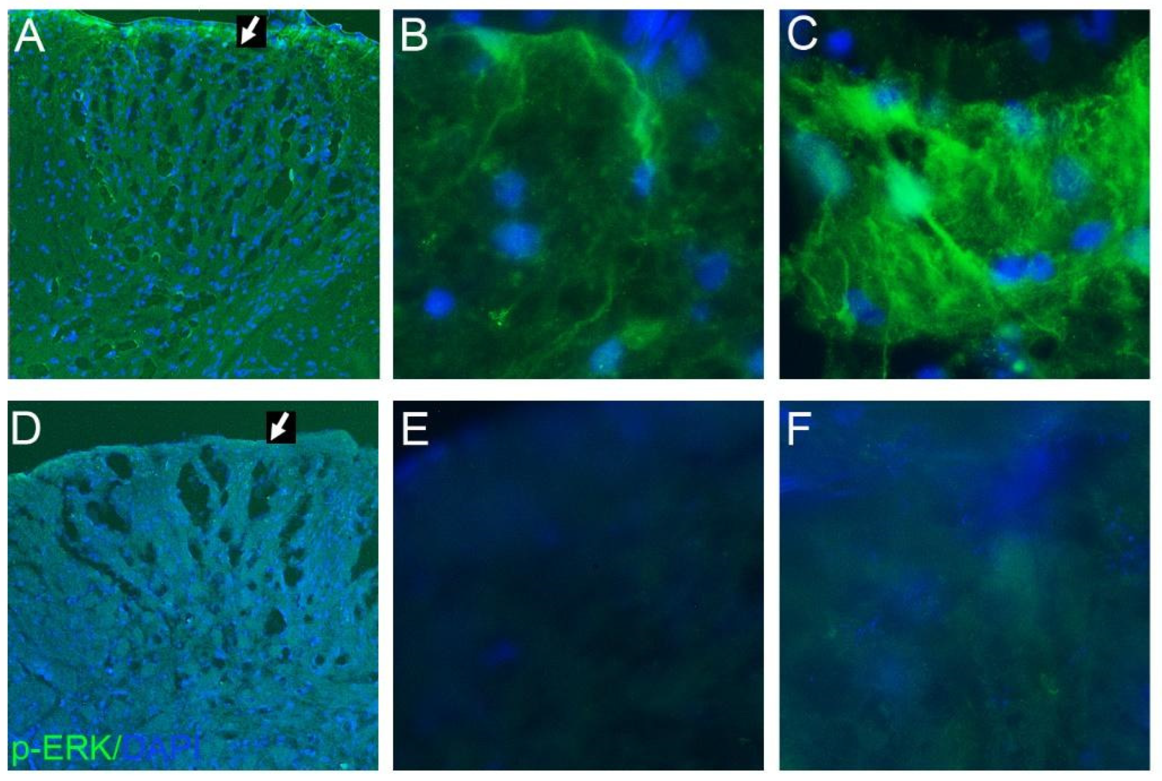
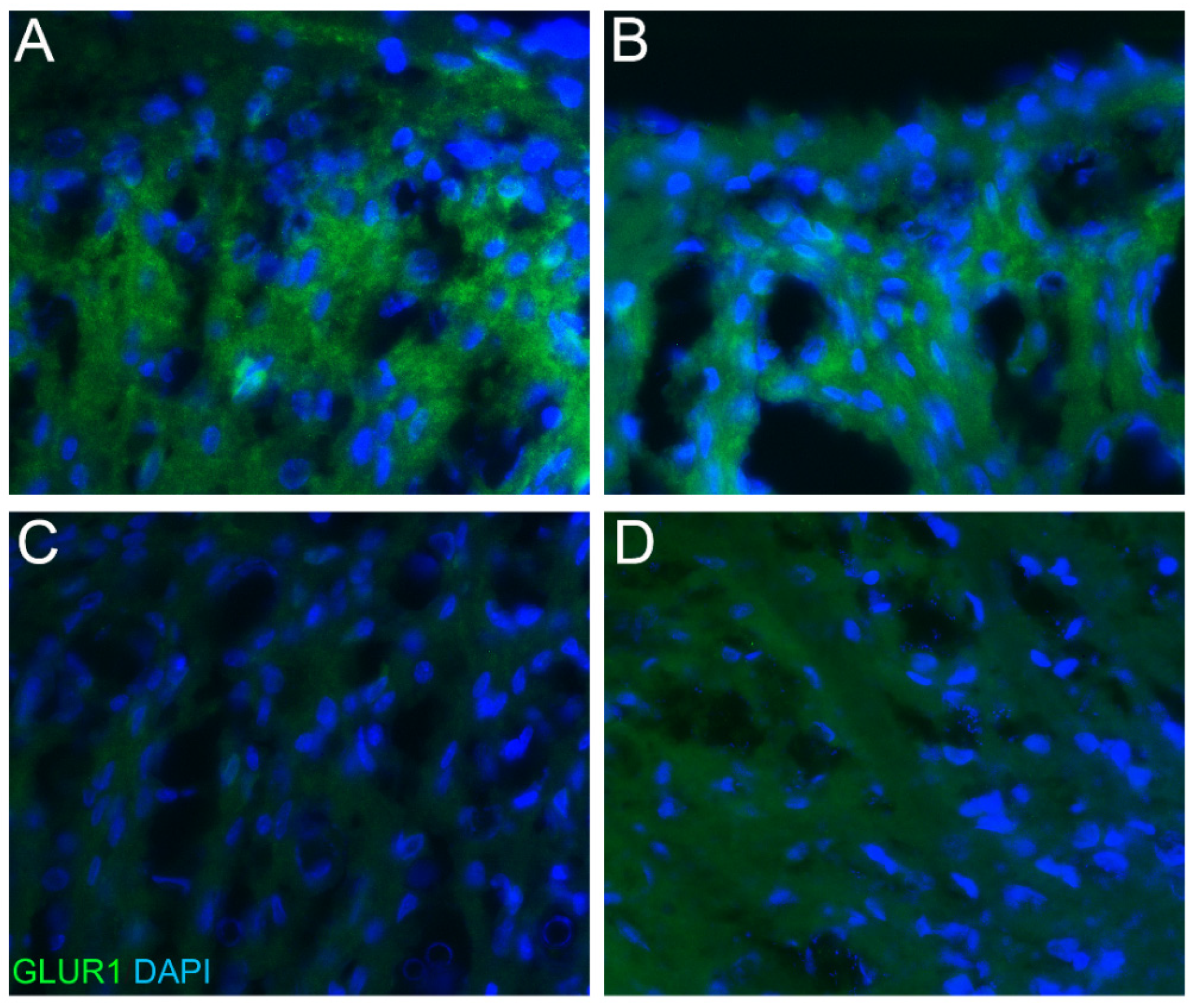
2.2. Phospho-ERK and GluR1 Staining
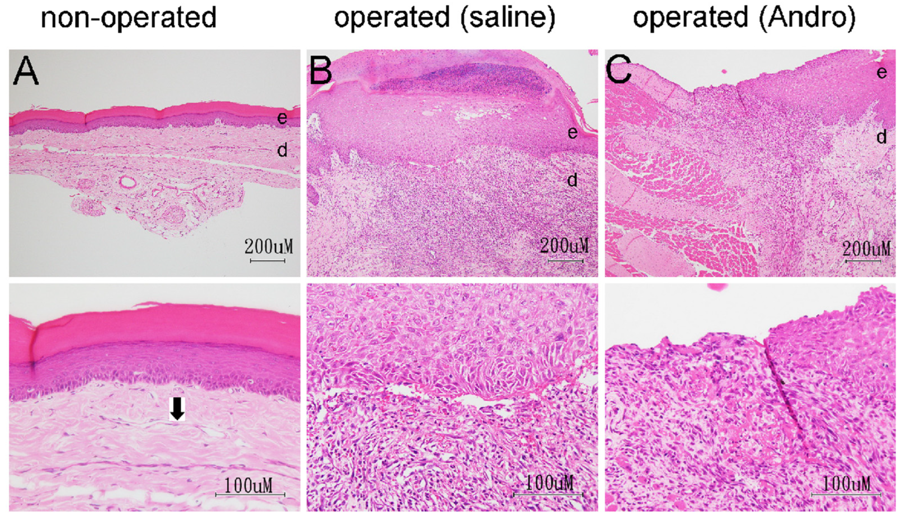
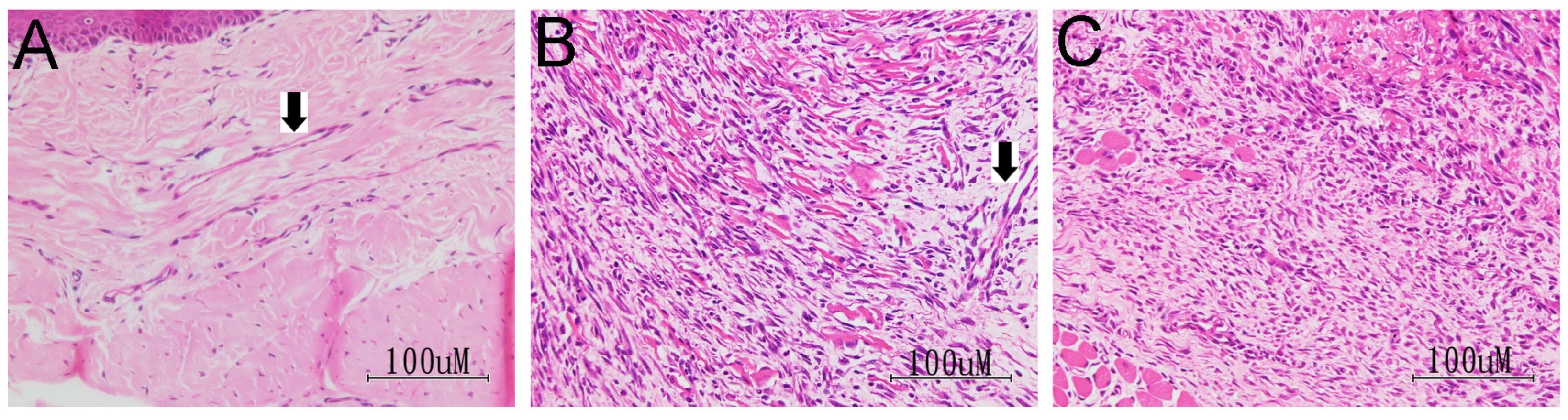
2.3. HE-Based Pathological Evaluation
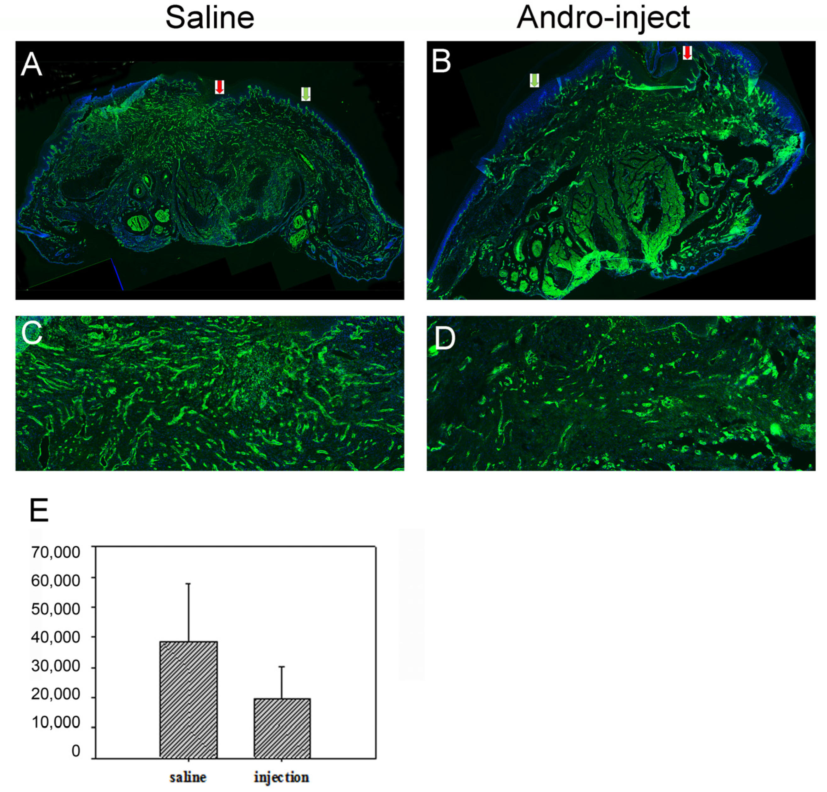
2.4. Laminin Staining for Pathological Evaluation
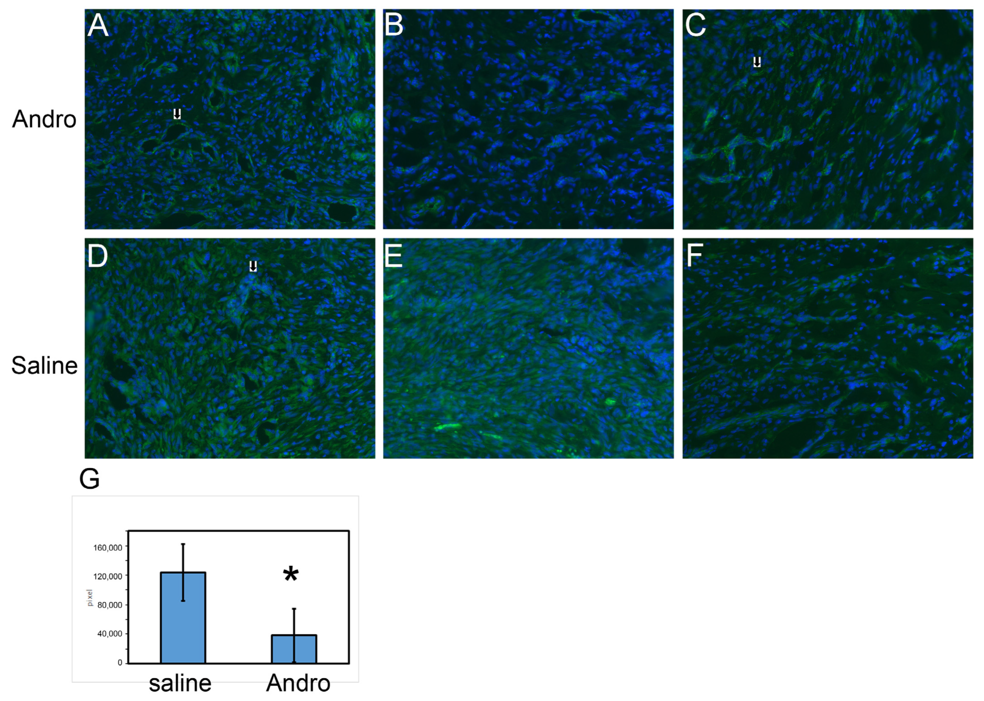
2.5. CD31 Staining for Blood Vessels
3. Discussion
3.1. Reduced Allodynia Due to pERK Downregulation in Andro-Injected Rats
3.2. Reduced Allodynia Due to GluR1 Downregulation in Andro-Injected Rats
3.3. Comparison between Andro and Cox Inhibitors for Incisional Pain: Andro Performs Better
3.4. Comparison between the Spared Sciatic Nerve Model and Postoperative Pain Model
3.5. Possible Modulation to the Wound Healing Process by Andro
4. Materials and Methods
4.1. Animals
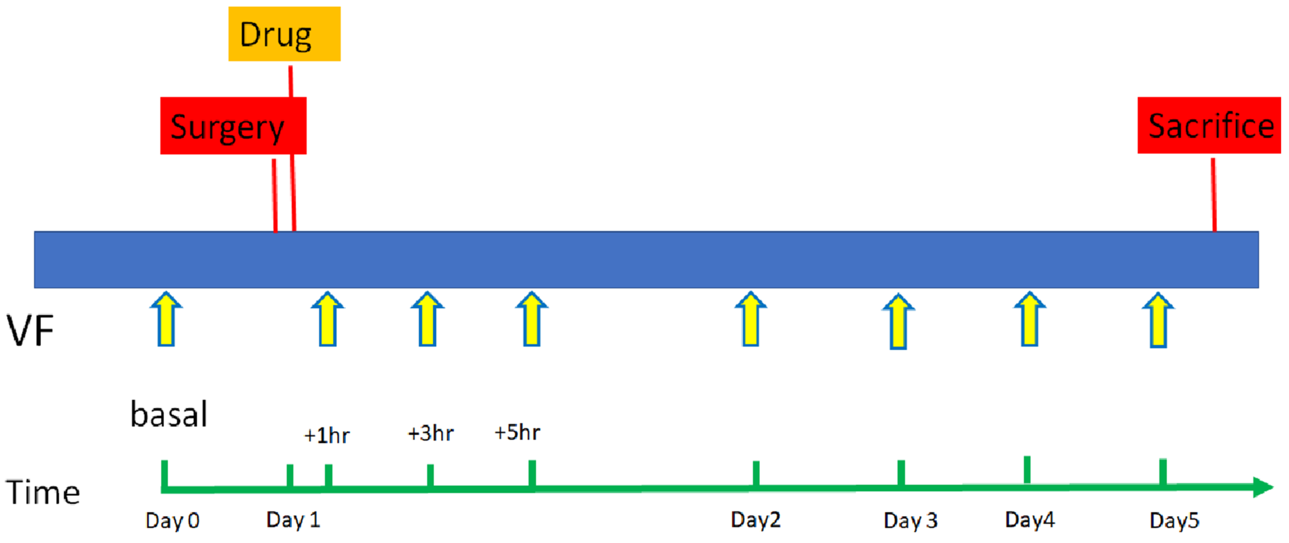
4.2. Surgery
4.3. Treatment Groups
4.4. Behaviour Test for Allodynia Thresholds: Von Frey Hair Test
4.5. Drug Preparation and Administration
4.6. Collection of Tissue Samples
4.7. Immunohistochemistry
4.8. Pathological Grading for Wound Area
4.9. Quantification of Immunoreactivity (IR) for Blood Vessels
4.10. Statistical Analysis
5. Conclusions
Author Contributions
Funding
Institutional Review Board Statement
Data Availability Statement
Acknowledgments
Conflicts of Interest
References
- Rao, Y.K.; Vimalamma, G.; Rao, C.V.; Tzeng, Y.-M. Flavonoids and andrographolides from Andrographis paniculata. Phytochemistry 2004, 65, 2317–2321. [Google Scholar] [CrossRef]
- Kumar, G.; Singh, D.; Tali, J.A.; Dheer, D.; Shankar, R. Andrographolide: Chemical modification and its effect on biological activities. Bioorg. Chem. 2020, 95, 103511. [Google Scholar] [CrossRef]
- Wang, T.; Liu, B.; Zhang, W.; Wilson, B.; Hong, J.-S. Andrographolide reduces inflammation-mediated dopaminergic neurodegeneration in mesencephalic neuron-glia cultures by inhibiting microglial activation. J. Pharmacol. Exp. Ther. 2004, 308, 975–983. [Google Scholar] [CrossRef] [PubMed] [Green Version]
- Tzeng, Y.-M.; Lee, Y.-C.; Cheng, W.-T.; Shih, H.-N.; Wang, H.-c.; Rao, Y.K.; Lee, M.-J. Effects of andrographolide and 14-deoxy-11, 12-didehydroandrographolide on cultured primary astrocytes and PC12 cells. Life Sci. 2012, 90, 257–266. [Google Scholar] [CrossRef] [PubMed]
- Wang, H.-C.; Tsay, H.-S.; Shih, H.-N.; Chen, Y.-A.; Chang, K.-M.; Agrawal, D.C.; Huang, S.; Lin, Y.-L.; Lee, M.-J. Andrographolide relieved pathological pain generated by spared nerve injury model in mice. Pharm. Biol. 2018, 56, 124–131. [Google Scholar] [CrossRef] [PubMed] [Green Version]
- Brennan, T.J. Postoperative models of nociception. ILAR J. 1999, 40, 129–136. [Google Scholar] [CrossRef] [Green Version]
- Zahn, P.K.; Umali, E.; Brennan, T.J. Intrathecal non-NMDA excitatory amino acid receptor antagonists inhibit pain behaviors in a rat model of postoperative pain. Pain 1998, 74, 213–223. [Google Scholar] [CrossRef]
- Jones, M.K.; Wang, H.; Peskar, B.M.; Levin, E.; Itani, R.M.; Sarfeh, I.J.; Tarnawski, A.S. Inhibition of angiogenesis by nonsteroidal anti-inflammatory drugs: Insight into mechanisms and implications for cancer growth and ulcer healing. Nat. Med. 1999, 5, 1418–1423. [Google Scholar] [CrossRef]
- Tonnesen, M.G.; Feng, X.; Clark, R.A. Angiogenesis in wound healing. J. Investig. Dermatol. Symp. Proc. 2020, 5, 40–46. [Google Scholar] [CrossRef] [Green Version]
- Gould, T.; Crosby, D.; Harmer, M.; Lloyd, S.; Lunn, J.; Rees, G.; Roberts, D.; Webster, J. Policy for controlling pain after surgery: Effect of sequential changes in management. Br. Med. J. 1992, 305, 1187–1193. [Google Scholar] [CrossRef]
- Segelcke, D.; Pogatzki-Zahn, E.M. 5.34-Pathophysiology of Postoperative Pain. In The Senses: A Comprehensive Reference, 2nd ed.; Fritzsch, B., Ed.; Elsevier: Oxford, UK, 2020; pp. 604–627. [Google Scholar] [CrossRef]
- Segelcke, D.; Pradier, B.; Pogatzki-Zahn, E. Advances in assessment of pain behaviors and mechanisms of post-operative pain models. Curr. Opin. Physiol. 2019, 11, 85–92. [Google Scholar] [CrossRef]
- Shi, X.D.; Fu, D.; Xu, J.M.; Zhang, Y.L.; Dai, R.P. Activation of spinal ERK1/2 contributes to mechanical allodynia in a rat model of postoperative pain. Mol. Med. Rep. 2013, 7, 1661–1665. [Google Scholar] [CrossRef] [PubMed] [Green Version]
- Lin, X.; Wang, M.; Zhang, J.; Xu, R. p38 MAPK: A potential target of chronic pain. Curr. Med. Chem. 2014, 21, 4405–4418. [Google Scholar] [CrossRef] [PubMed]
- Mai, L.; Zhu, X.; Huang, F.; He, H.; Fan, W. p38 mitogen-activated protein kinase and pain. Life Sci. 2020, 256, 117885. [Google Scholar] [CrossRef] [PubMed]
- Sridharan, B.; Lin, Y.-L.; Kung, Y.-J.; Lee, M.-J. Andrographolide regulates phosphorylated p38 mitogen-activated protein kinases and phosphorylated ERK expression in spinal cord and reduces postoperative pain in rats. Mater. Today Proc. 2022, 57, 829–833. [Google Scholar] [CrossRef]
- Skopelja-Gardner, S.; Saha, M.; Alvarado-Vazquez, P.A.; Liponis, B.S.; Martinez, E.; Romero-Sandoval, E.A. Mitogen-activated protein kinase phosphatase-3 (MKP-3) in the surgical wound is necessary for the resolution of postoperative pain in mice. J. Pain Res. 2017, 10, 763–774. [Google Scholar] [CrossRef] [Green Version]
- Park, J.-S.; Yaster, M.; Guan, X.; Xu, J.-T.; Shih, M.-H.; Guan, Y.; Raja, S.N.; Tao, Y.-X. Role of spinal cord alpha-amino-3-hydroxy-5-methyl-4-isoxazolepropionic acid receptors in complete Freund’s adjuvant-induced inflammatory pain. Mol. Pain 2008, 4, 67. [Google Scholar] [CrossRef] [Green Version]
- Pogatzki-Zahn, E.M.; Segelcke, D.; Schug, S.A. Postoperative pain—From mechanisms to treatment. Pain Rep. 2017, 2, e588. [Google Scholar] [CrossRef]
- Wang, Y.; Wu, J.; Guo, R.; Zhao, Y.; Wang, Y.; Zhang, M.; Chen, Z.; Wu, A.; Yue, Y. Surgical incision induces phosphorylation of AMPA receptor GluR1 subunits at Serine-831 sites and GluR1 trafficking in spinal cord dorsal horn via a protein kinase Cγ-dependent mechanism. Neuroscience 2013, 240, 361–370. [Google Scholar] [CrossRef]
- Zahn, P.K.; Pogatzki-Zahn, E.M.; Brennan, T.J. Spinal administration of MK-801 and NBQX demonstrates NMDA-independent dorsal horn sensitization in incisional pain. Pain 2005, 114, 499–510. [Google Scholar] [CrossRef]
- Díaz, E. Regulation of AMPA receptors by transmembrane accessory proteins. Eur. J. Neurosci. 2010, 32, 261–268. [Google Scholar] [CrossRef] [PubMed]
- Jackson, A.C.; Nicoll, R.A. Stargazin (TARP gamma-2) is required for compartment-specific AMPA receptor trafficking and synaptic plasticity in cerebellar stellate cells. J. Neurosci. Off. J. Soc. Neurosci. 2011, 31, 3939–3952. [Google Scholar] [CrossRef] [PubMed] [Green Version]
- Guo, R.; Zhao, Y.; Zhang, M.; Wang, Y.; Shi, R.; Liu, Y.; Xu, J.; Wu, A.; Yue, Y.; Wu, J.; et al. Down-regulation of Stargazin Inhibits the Enhanced Surface Delivery of α-Amino-3-hydroxy-5-methyl-4-isoxazole Propionate Receptor GluR1 Subunit in Rat Dorsal Horn and Ameliorates Postoperative Pain. Anesthesiology 2014, 121, 609–619. [Google Scholar] [CrossRef] [PubMed] [Green Version]
- Wandschneider, B.; Burdett, J.; Townsend, L.; Hill, A.; Thompson, P.J.; Duncan, J.S.; Koepp, M.J. Effect of topiramate and zonisamide on fMRI cognitive networks. Neurology 2017, 88, 1165–1171. [Google Scholar] [CrossRef] [PubMed] [Green Version]
- Knopp, K.L.; Simmons, R.M.A.; Guo, W.; Adams, B.L.; Gardinier, K.M.; Gernert, D.L.; Ornstein, P.L.; Porter, W.; Reel, J.; Ding, C.; et al. Modulation of TARP γ8-Containing AMPA Receptors as a Novel Therapeutic Approach for Chronic Pain. J. Pharm. Exp. 2019, 369, 345–363. [Google Scholar] [CrossRef]
- Casamonti, M.; Risaliti, L.; Vanti, G.; Piazzini, V.; Bergonzi, M.C.; Bilia, A.R. Andrographolide Loaded in Micro- and Nano-Formulations: Improved Bioavailability, Target-Tissue Distribution, and Efficacy of the “King of Bitters”. Engineering 2019, 5, 69–75. [Google Scholar] [CrossRef]
- Jayakumar, T.; Hsieh, C.Y.; Lee, J.J.; Sheu, J.R. Experimental and Clinical Pharmacology of Andrographis paniculata and Its Major Bioactive Phytoconstituent Andrographolide. Evid. Based Complement. Altern. Med. Ecam. 2013, 2013, 846740. [Google Scholar] [CrossRef] [Green Version]
- Yang, T.; Sheng, H.H.; Feng, N.P.; Wei, H.; Wang, Z.T.; Wang, C.H. Preparation of andrographolide-loaded solid lipid nanoparticles and their in vitro and in vivo evaluations: Characteristics, release, absorption, transports, pharmacokinetics, and antihyperlipidemic activity. J. Pharm. Sci. 2013, 102, 4414–4425. [Google Scholar] [CrossRef]
- Oseni, B.A.; Azubuike, C.P.; Okubanjo, O.O.; Igwilo, C.I.; Panyam, J. Encapsulation of Andrographolide in poly(lactide-co-glycolide) Nanoparticles: Formulation Optimization and in vitro Efficacy Studies. Front. Bioeng. Biotechnol. 2021, 9, 639409. [Google Scholar] [CrossRef]
- Whiteside, G.T.; Harrison, J.; Boulet, J.; Mark, L.; Pearson, M.; Gottshall, S.; Walker, K. Pharmacological characterisation of a rat model of incisional pain. Br. J. Pharmacol. 2004, 141, 85–91. [Google Scholar] [CrossRef] [Green Version]
- Riendeau, D.; Percival, M.; Brideau, C.; Charleson, S.; Dube, D.; Ethier, D.; Falgueyret, J.-P.; Friesen, R.; Gordon, R.; Greig, G. Etoricoxib (MK-0663): Preclinical profile and comparison with other agents that selectively inhibit cyclooxygenase-2. J. Pharmacol. Exp. Ther. 2001, 296, 558–566. [Google Scholar]
- Wieseler-Frank, J.; Maier, S.F.; Watkins, L.R. Glial activation and pathological pain. Neurochem. Int. 2004, 45, 389–395. [Google Scholar] [CrossRef]
- Casillas-Espinosa, P.M.; Powell, K.L.; O’Brien, T.J. Regulators of synaptic transmission: Roles in the pathogenesis and treatment of epilepsy. Epilepsia 2012, 53, 41–58. [Google Scholar] [CrossRef]
- DiPietro, L.A. Angiogenesis and wound repair: When enough is enough. J. Leukoc. Biol. 2016, 100, 979–984. [Google Scholar] [CrossRef] [Green Version]
- Dixon, W.J. Efficient analysis of experimental observations. Annu. Rev. Pharmacol. Toxicol. 1980, 20, 441–462. [Google Scholar] [CrossRef]
- Chaplan, S.R.; Bach, F.; Pogrel, J.; Chung, J.; Yaksh, T. Quantitative assessment of tactile allodynia in the rat paw. J. Neurosci. Methods 1994, 53, 55–63. [Google Scholar] [CrossRef]
- Tanga, F.Y.; Nutile-McMenemy, N.; DeLeo, J.A. The CNS role of Toll-like receptor 4 in innate neuroimmunity and painful neuropathy. Proc. Natl. Acad. Sci. USA 2005, 102, 5856–5861. [Google Scholar] [CrossRef] [Green Version]
- Bourquin, A.-F.; Süveges, M.; Pertin, M.; Gilliard, N.; Sardy, S.; Davison, A.C.; Spahn, D.R.; Decosterd, I. Assessment and analysis of mechanical allodynia-like behavior induced by spared nerve injury (SNI) in the mouse. Pain 2006, 122, 14.e1–14.e14. [Google Scholar] [CrossRef] [Green Version]
- Altavilla, D.; Galeano, M.; Bitto, A.; Minutoli, L.; Squadrito, G.; Seminara, P.; Venuti, F.S.; Torre, V.; Calò, M.; Colonna, M. Lipid peroxidation inhibition by raxofelast improves angiogenesis and wound healing in experimental burn wounds. Shock 2005, 24, 85–91. [Google Scholar] [CrossRef]
- Altavilla, D.; Saitta, A.; Cucinotta, D.; Galeano, M.; Deodato, B.; Colonna, M.; Torre, V.; Russo, G.; Sardella, A.; Urna, G. Inhibition of lipid peroxidation restores impaired vascular endothelial growth factor expression and stimulates wound healing and angiogenesis in the genetically diabetic mouse. Diabetes 2001, 50, 667–674. [Google Scholar] [CrossRef]
- Shackelford, C.; Long, G.; Wolf, J.; Okerberg, C.; Herbert, R. Qualitative and quantitative analysis of nonneoplastic lesions in toxicology studies. Toxicol. Pathol. 2002, 30, 93–96. [Google Scholar] [CrossRef] [PubMed] [Green Version]
- Hochberg, Y. A sharper Bonferroni procedure for multiple tests of significance. Biometrika 1988, 75, 800–802. [Google Scholar] [CrossRef]


| Group | Histopathological Findings | Animal Code | |||||||||||
|---|---|---|---|---|---|---|---|---|---|---|---|---|---|
| Saline | |||||||||||||
| L | R | ||||||||||||
| 9 | 15 | 18 | 20 | 22 | 24 | 9 | 15 | 18 | 20 | 22 | 24 | ||
| Foot pad | |||||||||||||
| H&E | Crust, epidermis 1 | 0 | 0 | 0 | 0 | 0 | 0 | 0 | 3 | 3 | 2 | 2 | 3 |
| Inflammation, dermis 1 | 0 | 0 | 0 | 0 | 0 | 0 | 4 | 4 | 4 | 4 | 4 | 4 | |
| Angiogenesis, dermis 2 | 0 | 0 | 0 | 0 | 0 | 0 | 3 | 3 | 3 | 3 | 3 | 3 | |
| Granulation, dermis 2 | 4 | 4 | 4 | 4 | 4 | 4 | 2 | 2 | 2 | 2 | 2 | 2 | |
| Re-epithelialization, epidermis 2 | 3 | 3 | 3 | 3 | 3 | 3 | 3 | 3 | 3 | 2 | 2 | 2 | |
| MT | Granulation, dermis 2 | 4 | 4 | 4 | 4 | 4 | 4 | 2 | 2 | 2 | 2 | 2 | 2 |
| Group | Histopathological Findings | Animal Code | |||||||||||
| Injection | |||||||||||||
| L | R | ||||||||||||
| 10 | 11 | 12 | 19 | 21 | 23 | 10 | 11 | 12 | 19 | 21 | 23 | ||
| Foot pad | |||||||||||||
| H&E | Crust, epidermis 1 | 0 | 0 | 0 | 0 | 0 | 0 | 2 | 2 | 3 | 4 | 2 | 3 |
| Inflammation, dermis 1 | 0 | 0 | 0 | 0 | 0 | 0 | 4 | 3 | 4 | 4 | 3 | 4 | |
| Angiogenesis, dermis 2 | 0 | 0 | 0 | 0 | 0 | 0 | 3 | 3 | 3 | 2 | 2 | 2 | |
| Granulation, dermis 2 | 4 | 4 | 4 | 4 | 4 | 4 | 2 | 2 | 2 | 2 | 2 | 2 | |
| Re-epithelialization, epidermis 2 | 3 | 3 | 3 | 3 | 3 | 3 | 3 | 3 | 3 | 2 | 2 | 2 | |
| MT | Granulation, dermis 2 | 4 | 4 | 4 | 4 | 4 | 4 | 2 | 2 | 2 | 2 | 2 | 2 |
| Organ | Histopathology | Group | |
|---|---|---|---|
| Saline | Injection | ||
| Foot pad | |||
| Crust, epidermis 1 | 2.2 ± 1.1 | 2.7 ± 0.7 | |
| Inflammation, dermis 1 | 4.0 ± 0.0 | 3.7 ± 0.5 | |
| Angiogenesis, dermis 1 | 3.0 ± 0.0 | 2.5 ± 0.5 * | |
| Granulation, dermis 1 | 2.0 ± 0.0 | 2.0 ± 0.0 | |
| Re-epithelialization, epidermis 1 | 2.5 ± 0.5 | 2.5 ± 0.5 | |
Publisher’s Note: MDPI stays neutral with regard to jurisdictional claims in published maps and institutional affiliations. |
© 2022 by the authors. Licensee MDPI, Basel, Switzerland. This article is an open access article distributed under the terms and conditions of the Creative Commons Attribution (CC BY) license (https://creativecommons.org/licenses/by/4.0/).
Share and Cite
Lin, Y.-L.; Liao, J.-W.; Wang, S.; Sridharan, B.; Lee, H.-J.; Li, A.; Chang, K.-M.; Wu, C.-Y.; Huang, S.; Chang, K.-T.; et al. Andrographolide Relieves Post-Operative Wound Pain but Affects Local Angiogenesis. Pharmaceuticals 2022, 15, 1586. https://doi.org/10.3390/ph15121586
Lin Y-L, Liao J-W, Wang S, Sridharan B, Lee H-J, Li A, Chang K-M, Wu C-Y, Huang S, Chang K-T, et al. Andrographolide Relieves Post-Operative Wound Pain but Affects Local Angiogenesis. Pharmaceuticals. 2022; 15(12):1586. https://doi.org/10.3390/ph15121586
Chicago/Turabian StyleLin, Yi-Lo, Jiunn-Wang Liao, Shunching Wang, Badrinathan Sridharan, Hsin-Ju Lee, Ai Li, Kai-Ming Chang, Ching-Yang Wu, Siendong Huang, Kai-Ting Chang, and et al. 2022. "Andrographolide Relieves Post-Operative Wound Pain but Affects Local Angiogenesis" Pharmaceuticals 15, no. 12: 1586. https://doi.org/10.3390/ph15121586