
Chest-Worn Inertial Sensors: A Survey of Applications and Methods


Abstract
:1. Introduction
- The type, amount and range of the movements that the sensor measures.
- The availability of the aimed signals at the selected position.
2. Applications
2.1. Seismocardiography
2.2. Activity Analysis
2.3. Posture Analysis
2.4. Localization
2.5. Voice Analysis
2.6. Swallow Analysis
2.7. Context Retrieval

| Application | Reference |
|---|---|
| Seismocardiography | |
| Analysis of cardiac parameters | [16,19,20,21,22,23,24,30,36,54,55,56,57,58] |
| Analysis of respiratory parameters | [28,29,30,36,57] |
| Mapping SCG to BCG | [59] |
| Identification of patients with CAD | [25] |
| Relating SCG to ultrasound images | [60] |
| Identification of heart failure states | [26] |
| Activity Analysis | |
| AR | [32,33,35,61,62,63,64,65,66] |
| EE estimation | [11,34] |
| Fall detection | [35] |
| Body motion tracking | [36] |
| Evaluation of transfer skills of wheelchair users | [67] |
| Posture Analysis | |
| Postural control for medical approach | [38,39] |
| Posture detection for sleep analysis | [10,37] |
| Localization | |
| Indoor positioning with PDR | [68,69,70] |
| Voice Analysis | |
| Measurement of vocal functions | [45,46] |
| VAD | [41,43,44] |
| Voice onset detection | [42] |
| Swallow Analysis | |
| Swallow detection | [47] |
| Swallow analysis for dysphagia investigation | [48,71,72,73,74,75] |
| Context Retrieval | |
| Emotion recognition from gait analysis | [51] |
| Age estimation from gait analysis | [53] |
| Age, gender and height estimation from gait analysis | [76] |
| Detection of mood changes from VAD | [44] |
| Stress and meditaion detection | [77] |
| Biometric verification | [78] |
3. Measurement Methods
3.1. Sensor Specifications
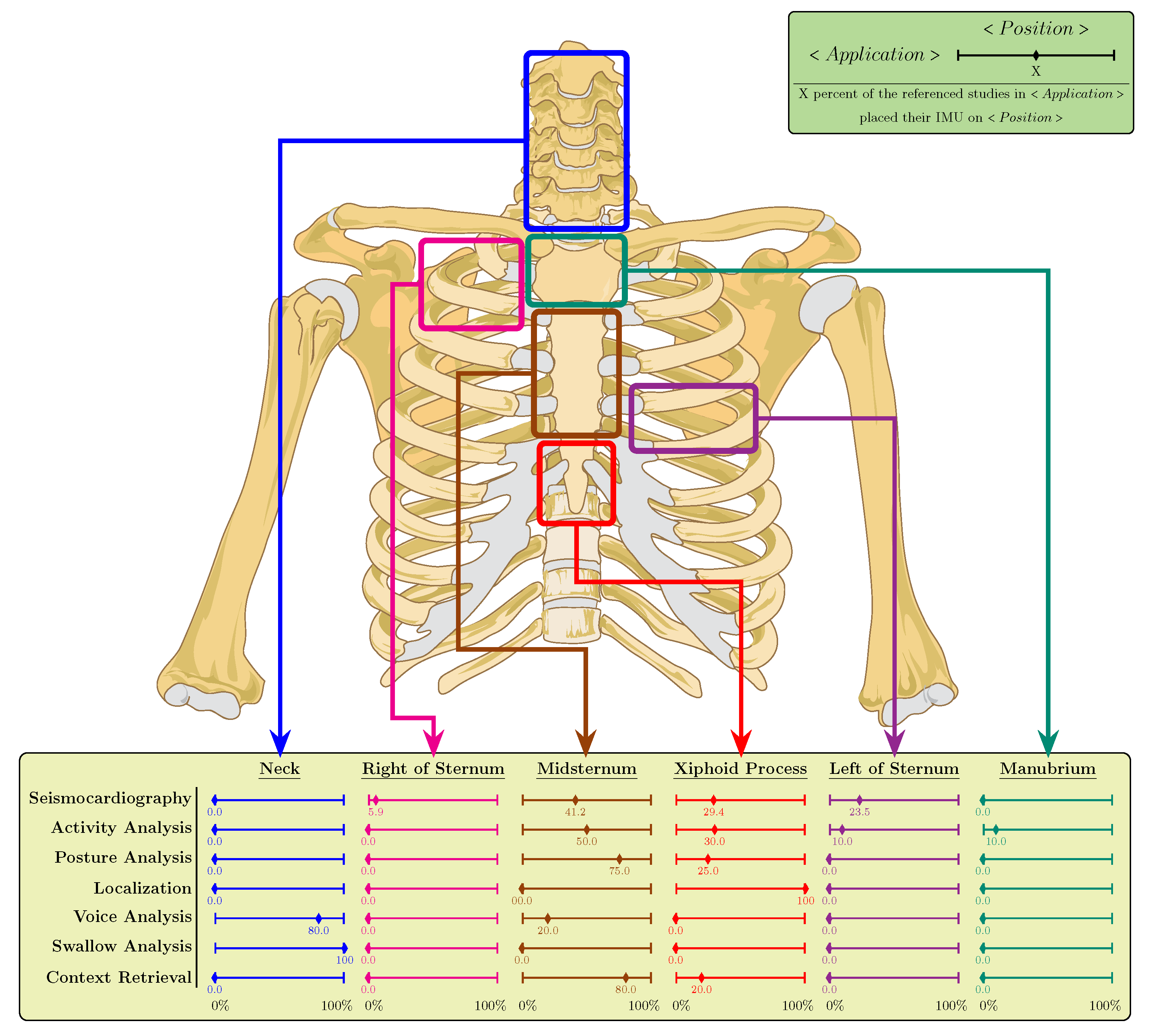
3.2. Sensor Placement
3.3. Validation Methods
4. Data Processing
4.1. Sensing-Processing Architecture
4.2. Machine Learning
4.3. Datasets
5. Research Challenges and Future Directions
5.1. Lack of Well-Acknowledged Benchmark Datasets
5.2. Robustness and Artifact Cancellation
5.3. Combined Applications
5.4. Sensor-Related Challenges
5.5. User Friendliness
6. Conclusions
Author Contributions
Funding
Institutional Review Board Statement
Informed Consent Statement
Conflicts of Interest
Abbreviations
| ACM | Accelerometer Contact Microphone |
| AdaBoost | Adaptive Boosting |
| ADC | Analog to Digital Converter |
| AI | Artificial Intelligence |
| ANN | Artificial Neural Network |
| AO | Aortic valve Opening |
| AR | Activity Recognition |
| ASR | Automatic Speech Recognition |
| BCG | Ballistocardiogram |
| BP | Blood Pressure |
| CAD | Coronary Artery Disease |
| CEBS | Combined measurement of ECG, Breathing and Seismocardiogram |
| CNN | Convolutional Neural Network |
| CV | Cross Validation |
| DaLiAc | Daily Life Activities |
| DAQ | Data Acquisition |
| DB | Database |
| DNN | Deep Neural Network |
| DoF | Degree of Freedom |
| ECG | Electrocardiography |
| EDA | Electro-dermal Activity |
| EE | Energy Expenditure |
| EMG | Elegtromyogram |
| ER | Emotion Recognition |
| GCG | Gyrocardiography |
| GMM | Gaussian Mixture Model |
| GNSS | Global Navigation Satellite System |
| gyr | Gyroscope |
| HF | Heart Failure |
| HR | Heart Rate |
| HRV | Heart Rate Variability |
| I2C | Inter-Integrated Circuit |
| IBI | Inter-beat Interval |
| ICG | Impedance Cardiogram |
| IMU | Inertial Measurement Unit |
| k-NN | k-Nearest Neighbor |
| LDA | Linear Discriminant Analysis |
| LVET | Left Ventricular Ejection Time |
| MCU | Microcontroller Unit |
| MEMS | Micro-electro-mechanical System |
| MET | Metabolic Equivalent of Task |
| mg | Magnetometer |
| ML | Machine Learning |
| MLP | Multilayer Perceptron |
| MS | Multiple Sclerosis |
| PCA | Principal Component Analysis |
| PCB | Printed Circuit Board |
| PCG | Phonocardiogram |
| PEP | Pre-Ejection Period |
| PDR | Pedestrian Dead Reckoning |
| PPG | Photoplethysmogram |
| RMSE | Root Mean Square Error |
| RSSI | Received Signal Strength Indicator |
| SCG | Seismocardiography |
| SPI | Serial Peripheral Interface |
| SoC | System on Chip |
| SVM | Support Vector Machine |
| TRL | Technology Readiness Level |
| VAD | Voice Activity Detection |
| VAE | Variational Autoencoder |
| XGBoost | Extreme Gradient Boosting |
| xl | Accelerometer |
Appendix A. Referenced Studies
| Reference | Sensor | Worn on | Fixation | Application |
|---|---|---|---|---|
| Seismocardiography | ||||
| Gupta et al. [36] | 3-ACM: Own fabrication | Midsternum | Elastic strap over skin | SCG for heart and respiration parameters and body motion |
| Yu and Liu [54] | 3-xl: ICM-20602 (TDK-InvenSense) | Left side of the sternum and right side of the back | Strap over skin | Motion artifact removal from SCG for heartbeat detection |
| Hersek et al. [59] | 3-xl: ADXL354 (Analog Devices) and a modified weighting scale for BCG measurement [91] | Midsternum | Kinesio tape | Mapping SCG to BCG |
| Sieciński et al. [16] | Used DB: Mechanocardiograms with ECG References [55,84] | HRV analysis | ||
| Mora et al. [21] | Used DB: CEBS [58,86] | SCG for heartbeat detection and IBI estimation | ||
| Choudhary et al. [22] | Used DB: CEBS [58,86] | SCG for detection of AO-peaks | ||
| Ahmaniemi et al. [24] | 3-xl: LSM6DS3 (STMicroelectronics) and PCG | Heart apex | Pocket of a belt | SCG for estimation of HR, PEP and LVET |
| Cocconcelli et al. [19] | 3-xl: ADXL355 (Analog Devices) | Midsternum | SCG for heartbeat detection | |
| Shandhi et al. [23] | 3-xl: ADXL354 (Analog Devices) and 3-gyr: QGYR330HA (Qualtre) | Midsternum | SCG for PEP estimation | |
| Dehkordi et al. [25] | 1-xl: ultra low-frequency piezoelectric crystal accelerometer (Seismed Instruments) | Xiphoid process | SCG to identify patients with CAD | |
| Hernandez and Cretu [20] | 1-gyr: MPU-9250 (TDK-InvenSense) | Xiphoid process | Elastic fabric belt | Estimation of HR during sleep |
| D’Mello et al. [30] | 3-xl: MPU-9250 (TDK-InvenSense) | Xiphoid process | Strap | Cardio-respiratory analysis |
| Kaisti et al. [55] | 3-xl: MMA8451Q (NXP Semiconductors); 3-gyr: MAX21000 (Maxim Integrated); | Midsternum | Double-sided tape | SCG for heartbeat detection |
| Sørensen et al. [60] | 1-xl × 2: 1521 (Silicon Designs) | Xiphoid process and fourth intercostal space | Double adhesive tape over skin | Relating SCG to ultrasound images |
| Inan et al. [26] | 3-xl: BMA280 (Bosch Sensortec) | Midsternum | Adhesive-backed gel electrodes | Identification of heart failure states |
| Selvaraj and Reddivari [56] | 3-xl and ECG and PPG | Left side of the chest | Adhered over skin | BP measurement |
| García-González et al. [58] | 3-xl: LIS344ALH (STMicroelectronics) | Chest | Heartbeat detection and RR time series analysis | |
| Skoric et al. [29] | 3-xl-gyr: MPU-9250 (TDK-InvenSense) | Xiphoid process | Double-sided tape | Respiration rate and volume |
| Cesareo et al. [28] | 9-IMU: LSM9DS0 (STMicroelectronics) [80] | Chest (right side), abdomen and coccyx | Respiration analysis | |
| Jafari Tadi et al. [57] | 3-xl: MMA8451Q (NXP Semiconductor) | MidSternum | Elastic strap | Gating nuclear imaging based on cardio-respiratory analysis |
| Activity Analaysis | ||||
| Barbareschi et al. [67] | 3-xl | Chest (manubrium) | Double-sided tape | Evaluating transfer skills of wheelchair users |
| Nazarahari and Rouhani [66] | 3-xl: Physilog system (GaitUp) | Chest (midsternum) | Medical tape | AR |
| Zhang et al. [34] | 3-xl: GT3X+ (Actigraph) | Chest (xiphoid process), wrist and waist | A soft nylon necklace underneath clothes | Physical activity measurement |
| Awais et al. [32] | Used DB: DaLiAc dataset [61] | AR with 13 classes | ||
| Altini et al. [11] | 3-xl: ADXL330 (Analog Devices) | Chest, Thigh, Ankle, Wrist and Waist | Elastic strap | EE estimation |
| Banos et al. [65] | 3-xl: Shimmer | Chest, Ankle and Wrist | Elastic strap | AR with 12 classes |
| Gao et al. [33] | 3-xl: Shimmer | Chest (midsternum), under-arm, waist and thigh | Fitted into a garment worn over other clothes | AR with 5 classes |
| Gjoreski et al. [35] | 3-xl: Shimmer | Chest (xiphoid process) and thigh | Elastic Velcro straps | AR with 6 classes and fall detection |
| Leutheuser et al. [61] | 3-xl-gyr: Shimmer | Chest (midsternum), wrist, hip and ankle | Embedded in special clothes | AR with 13 classes |
| Cleland et al. [62] | 3-xl: Shimmer | Chest (xiphoid process), wrist, lower back, hip, thigh and foot | Elastic strap and holster over clothes | AR with 7 classes |
| Godfrey et al. [63] | 3-xl: ADXL210 (Analog Devices); 3-gyr: ADXRS300 (Analog Devices) | Midsternum | Strap over clothes | AR with 8 classes |
| Atallah et al. [64] | 3-xl: ADXL330 (Analog Devices) | Chest (midsternum), ear, arm, wrist, waist, knee and ankle | Strap over clothes | AR with 5 classes |
| Posture Analysis | ||||
| Hsieh and Sosnoff [39] | 3-xl: Smartphone | Midsternum | Held along the sternum with hand | Postural control in MS patients |
| Reynard et al. [38] | 3-xl: Physilog system (GaitUp) | Midsternum | Belt over clothes | Medical approach (postural control) |
| Razjouyan et al. [37] | 3-xl: BioPatch ZephyrLife | Midsternum | Adhesive patch over skin | Posture detection for sleep analysis |
| Nam et al. [10] | 3-xl | Xiphoid process | Belt over clothes | Posture detection for sleep analysis |
| Localization | ||||
| Lu et al. [68] | 3-xl-gyr and barometer: NGIMU (x-io Technologies) | Xiphoid process | Stretching strap over clothes | Indoor positioning (PDR) |
| Tateno et al. [69] | 3-xl-gyr: MPU-9250 (TDK-InvenSense) and RSSI | Xiphoid process | Strap over clothes | Indoor positioning (PDR) |
| Hu et al. [70] | 3-xl: ADXL345 (Analog Devices); 3-gyr: ITG-3200 (TDK-InvenSense); 3-mg: HMC5883L(Honeywell) | Chest | Velcro belt over clothes | Indoor positioning (PDR) |
| Voice Analysis | ||||
| Dubey et al. [41] | 1-xl: BU-27135-000 (Knowles Electronics) | Neck | Double sided tape and Blenderm tape over skin | VAD (medical approach) |
| Mehta et al. [45] | 1-xl: BU-27135-000 (Knowles Electronics) | Neck | Hypoallergenic double-sided tape over skin | Measurement of vocal functions (medical approach) |
| Mehta et al. [46] | 1-xl: BU-27135-000 (Knowles Electronics) | Neck | Hypoallergenic double-sided tape over skin | Measurement of vocal functions (medical approach) |
| Vitikainen et al. [42] | 3-xl: ADXL330 (Analog Devices) | Neck | Adhesive tape over skin | Voice onset detection (medical approach) |
| Matic et al. [43] | 3-xl: Shimmer | Midsternum | Elastic strap over skin | VAD |
| Swallow Analysis | ||||
| Khalifa et al. [71] | 3-xl: ADXL327 (Analog Devices) contact microphone | Anterior neck overlying the cricoid cartilage | Swallow detection in patients | |
| Donohue et al. [72] | 3-xl: ADXL327 (Analog Devices) contact microphone | Anterior neck at the level of the cricoid cartilage | Adhesive tape | Swallow comparing between healthy people and Neurodegenerative patients |
| Donohue et al. [73] | 3-xl: ADXL327 (Analog Devices) | Anterior neck | Adhesive tape | Investigating swallowing vibrations |
| Steele et al. [74] | 2-xl | Anterior neck, below the palpable lower border of the thyroid cartilage | Single-use, disposable fixation unit | Swallow analysis for dysphagia detection |
| He et al. [75] | 3-xl: ADXL327 (Analog Devices) and contact microphone | Anterior neck over the palpable arch of the cricoid cartilage | Double-sided tape | Investigating swallowing vibrations |
| Li et al. [47] | 3-xl: MPU-6050 (TDK-InvenSense) [92] and PPG | Throat (cricoid cartilage) | Medical adhesive tape over skin | Swallow detection |
| Zahnd et al. [48] | 3-xl: ADXL327 (Analog Devices) | Throat (cricoid cartilage) | Adhesive tape over skin | Investigating swallowing vibrations |
| Context Retrieval | ||||
| Hashmi et al. [51] | 3-xl-gyr: Smartphone | Midsternum | Elastic strap over clothes | ER from gait analysis with 6 classes |
| Riaz et al. [53] | 3-xl-gyr: Smartphone and Opal (APDM) | Midsternum | Elastic strap over clothes | Age estimation from gait analysis |
| Uddin and Canavan [77] | Used DB: WESAD [85] | Stress and Meditation Detection | ||
| Riaz et al. [76] | 3-xl-gyr: Opal (APDM) | Chest (xiphoid process), wrist, ankle and lower back | Elastic strap over clothes | Estimation of age, gender and height from gait analysis |
| Matic et al. [44] | 3-xl: Shimmer | Midsternum | Elastic strap over skin | Correlation of VAD and mood changes |
| Vural et al. [78] | 3-xl | Midsternum | Strap over clothes | Biometric verification |
References
- Mück, J.E.; Ünal, B.; Butt, H.; Yetisen, A.K. Market and Patent Analyses of Wearables in Medicine. Trends Biotechnol. 2019, 38, 129–133. [Google Scholar] [CrossRef] [PubMed]
- Bonato, P. Advances in wearable technology and its medical applications. In Proceedings of the 2010 Annual International Conference of the IEEE Engineering in Medicine and Biology Society, Buenos Aires, Argentina, 31 August–4 September 2010; pp. 2021–2024. [Google Scholar] [CrossRef]
- Berglund, M.E.; Duvall, J.; Dunne, L.E. A survey of the historical scope and current trends of wearable technology applications. In Proceedings of the International Symposium on Wearable Computers, Heidelberg, Germany, 12–16 September 2016; IEEE Computer Society: New York, NY, USA, 2016; pp. 40–43. [Google Scholar] [CrossRef]
- Kröger, J.L.; Raschke, P.; Bhuiyan, T.R. Privacy implications of accelerometer data: A review of possible inferences. Acm Int. Conf. D Ser. 2019, 81–87. [Google Scholar] [CrossRef] [Green Version]
- You, Z. Space Microsystems and Micro/Nano Satellites; Elsevier Inc.: Amsterdam, The Netherlands, 2017; pp. 1–427. [Google Scholar] [CrossRef]
- Filippeschi, A.; Schmitz, N.; Miezal, M.; Bleser, G.; Ruffaldi, E.; Stricker, D. Survey of Motion Tracking Methods Based on Inertial Sensors: A Focus on Upper Limb Human Motion. Sensors 2017, 17, 1257. [Google Scholar] [CrossRef] [PubMed] [Green Version]
- Taebi, A.; Solar, B.; Bomar, A.; Sandler, R.; Mansy, H. Recent Advances in Seismocardiography. Vibration 2019, 2, 5. [Google Scholar] [CrossRef] [Green Version]
- Jones, S.E.; van Hees, V.T.; Mazzotti, D.R.; Marques-Vidal, P.; Sabia, S.; van der Spek, A.; Dashti, H.S.; Engmann, J.; Kocevska, D.; Tyrrell, J.; et al. Genetic studies of accelerometer-based sleep measures yield new insights into human sleep behaviour. Nat. Commun. 2019, 10, 1585. [Google Scholar] [CrossRef]
- Van Hees, V.T.; Sabia, S.; Jones, S.E.; Wood, A.R.; Anderson, K.N.; Kivimäki, M.; Frayling, T.M.; Pack, A.I.; Bucan, M.; Trenell, M.I.; et al. Estimating sleep parameters using an accelerometer without sleep diary. Sci. Rep. 2018, 8, 12975. [Google Scholar] [CrossRef]
- Nam, Y.; Kim, Y.; Lee, J. Sleep Monitoring Based on a Tri-Axial Accelerometer and a Pressure Sensor. Sensors 2016, 16, 750. [Google Scholar] [CrossRef] [Green Version]
- Altini, M.; Penders, J.; Vullers, R.; Amft, O. Estimating energy expenditure using body-worn accelerometers: A comparison of methods, sensors number and positioning. IEEE J. Biomed. Health Inform. 2015, 19, 219–226. [Google Scholar] [CrossRef]
- Elbasiony, R.; Gomaa, W. A Survey on Human Activity Recognition Based on Temporal Signals of Portable Inertial Sensors. In Advances in Intelligent Systems and Computing; Springer: Berlin/Heidelberg, Germany, 2020; Volume 921, pp. 734–745. [Google Scholar] [CrossRef]
- Inan, O.T.; Migeotte, P.F.; Park, K.S.; Etemadi, M.; Tavakolian, K.; Casanella, R.; Zanetti, J.; Tank, J.; Funtova, I.; Prisk, G.K.; et al. Ballistocardiography and Seismocardiography: A Review of Recent Advances. IEEE J. Biomed. Health Inform. 2015, 19, 1414–1427. [Google Scholar] [CrossRef] [Green Version]
- Zakeri, V.; Akhbardeh, A.; Alamdari, N.; Fazel-Rezai, R.; Paukkunen, M.; Tavakolian, K. Analyzing seismocardiogram cycles to identify the respiratory phases. IEEE Trans. Biomed. Eng. 2017, 64, 1786–1792. [Google Scholar] [CrossRef]
- Yang, C.; Tavassolian, N. Combined Seismo-and Gyro-Cardiography: A More Comprehensive Evaluation of Heart-Induced Chest Vibrations. IEEE J. Biomed. Health Inform. 2018, 22, 1466–1475. [Google Scholar] [CrossRef]
- Sieciński, S.; Kostka, P.S.; Tkacz, E.J. Heart Rate Variability Analysis on Electrocardiograms, Seismocardiograms and Gyrocardiograms on Healthy Volunteers. Sensors 2020, 20, 4522. [Google Scholar] [CrossRef] [PubMed]
- Lahdenoja, O.; Humanen, T.; Tadi, M.J.; Pänkäälä, M.; Koivisto, T. Heart rate variability estimation with joint accelerometer and gyroscope sensing. In Proceedings of the 2016 Computing in Cardiology Conference (CinC), Vancouver, BC, Canada, 11–14 September 2016; pp. 717–720. [Google Scholar]
- Cosoli, G.; Spinsante, S.; Scalise, L. Wrist-worn and chest-strap wearable devices: Systematic review on accuracy and metrological characteristics. Meas. J. Int. Meas. Confed. 2020, 159, 107789. [Google Scholar] [CrossRef]
- Cocconcelli, F.; Mora, N.; Matrella, G.; Ciampolini, P. Seismocardiography-based detection of heartbeats for continuous monitoring of vital signs. In Proceedings of the 2019 11th Computer Science and Electronic Engineering Conference, Nanjing, China, 8–10 November 2019; pp. 53–58. [Google Scholar] [CrossRef]
- Hernandez, J.E.; Cretu, E. Simple Heart Rate Monitoring System with a MEMS Gyroscope for Sleep Studies. In Proceedings of the 2018 IEEE 9th Annual Information Technology, Electronics and Mobile Communication Conference, Vancouver, BC, Canada, 1–3 November 2018; pp. 61–67. [Google Scholar] [CrossRef]
- Mora, N.; Cocconcelli, F.; Matrella, G.; Ciampolini, P. Detection and Analysis of Heartbeats in Seismocardiogram Signals. Sensors 2020, 20, 1670. [Google Scholar] [CrossRef] [PubMed] [Green Version]
- Choudhary, T.; Sharma, L.N.; Bhuyan, M.K. Automatic detection of aortic valve opening using seismocardiography in healthy individuals. IEEE J. Biomed. Health Inform. 2019, 23, 1032–1040. [Google Scholar] [CrossRef] [PubMed]
- Shandhi, M.M.H.; Semiz, B.; Hersek, S.; Goller, N.; Ayazi, F.; Inan, O.T. Performance Analysis of Gyroscope and Accelerometer Sensors for Seismocardiography-Based Wearable Pre-Ejection Period Estimation. IEEE J. Biomed. Health Inform. 2019, 23, 2365–2374. [Google Scholar] [CrossRef]
- Ahmaniemi, T.; Rajala, S.; Lindholm, H. Estimation of Beat-to-Beat Interval and Systolic Time Intervals Using Phono-and Seismocardiograms. In Proceedings of the 2019 41st Annual International Conference of the IEEE Engineering in Medicine and Biology Society (EMBC), Berlin, Germany, 23–27 July 2019; pp. 5650–5656. [Google Scholar] [CrossRef]
- Dehkordi, P.; Bauer, E.P.; Tavakolian, K.; Zakeri, V.; Blaber, A.P.; Khosrow-Khavar, F. Identifying patients with coronary artery disease using rest and exercise seismocardiography. Front. Physiol. 2019, 10, 1–10. [Google Scholar] [CrossRef]
- Inan, O.T.; Baran Pouyan, M.; Javaid, A.Q.; Dowling, S.; Etemadi, M.; Dorier, A.; Heller, J.A.; Bicen, A.O.; Roy, S.; De Marco, T.; et al. Novel Wearable Seismocardiography and Machine Learning Algorithms Can Assess Clinical Status of Heart Failure Patients. Circulation. Heart Fail. 2018, 11, e004313. [Google Scholar] [CrossRef]
- Telfer, B.A.; Williamson, J.R.; Weed, L.; Bursey, M.; Frazee, R.; Galer, M.; Moore, C.; Buller, M.; Friedl, K.E. Estimating Sedentary Breathing Rate from Chest-Worn Accelerometry from Free-Living Data. In Proceedings of the 2020 42nd Annual International Conference of the IEEE Engineering in Medicine & Biology Society (EMBC), Montreal, QC, Canada, 20–24 July 2020; pp. 4636–4639. [Google Scholar] [CrossRef]
- Cesareo, A.; Previtali, Y.; Biffi, E.; Aliverti, A. Assessment of Breathing Parameters Using an Inertial Measurement Unit (IMU)-Based System. Sensors 2018, 19, 88. [Google Scholar] [CrossRef] [Green Version]
- Skoric, J.; D’Mello, Y.; Aboulezz, E.; Hakim, S.; Clairmonte, N.; Lortie, M.; Plant, D.V. Relationship of the Respiration Waveform to a Chest Worn Inertial Sensor. In Proceedings of the 2020 42nd Annual International Conference of the IEEE Engineering in Medicine & Biology Society (EMBC), Montreal, QC, Canada, 20–24 July 2020; pp. 2732–2735. [Google Scholar] [CrossRef]
- D’Mello, Y.; Skoric, J.; Xu, S.; Akhras, M.; Roche, P.J.; Lortie, M.A.; Gagnon, S.; Plant, D.V. Autocorrelated Differential Algorithm for Real-Time Seismocardiography Analysis. IEEE Sens. J. 2019, 19, 5127–5140. [Google Scholar] [CrossRef]
- Pandia, K.; Inan, O.T.; Kovacs, G.T.; Giovangrandi, L. Extracting respiratory information from seismocardiogram signals acquired on the chest using a miniature accelerometer. Physiol. Meas. 2012, 33, 1643–1660. [Google Scholar] [CrossRef]
- Awais, M.; Palmerini, L.; Chiari, L. Physical activity classification using body-worn inertial sensors in a multi-sensor setup. In Proceedings of the 2016 IEEE 2nd International Forum on Research and Technologies for Society and Industry Leveraging a Better Tomorrow, Bologna, Italy, 7–9 September 2016. [Google Scholar] [CrossRef]
- Gao, L.; Bourke, A.K.; Nelson, J. Evaluation of accelerometer based multi-sensor versus single-sensor activity recognition systems. Med. Eng. Phys. 2014, 36, 779–785. [Google Scholar] [CrossRef] [PubMed]
- Zhang, J.H.; Macfarlane, D.J.; Sobko, T. Feasibility of a Chest-worn accelerometer for physical activity measurement. J. Sci. Med. Sport 2016, 19, 1015–1019. [Google Scholar] [CrossRef] [PubMed] [Green Version]
- Gjoreski, H.; Rashkovska, A.; Kozina, S.; Lustrek, M.; Gams, M. Telehealth using ECG sensor and accelerometer. In Proceedings of the 2014 37th International Convention on Information and Communication Technology, Electronics and Microelectronics (MIPRO), Opatija, Croatia, 26–30 May 2014; pp. 270–274. [Google Scholar] [CrossRef] [Green Version]
- Gupta, P.; Moghimi, M.J.; Jeong, Y.; Gupta, D.; Inan, O.T.; Ayazi, F. Precision wearable accelerometer contact microphones for longitudinal monitoring of mechano-acoustic cardiopulmonary signals. Npj Digit. Med. 2020, 3. [Google Scholar] [CrossRef] [Green Version]
- Razjouyan, J.; Lee, H.; Parthasarathy, S.; Mohler, J.; Sharafkhaneh, A.; Najafi, B. Information from postural/sleep position changes and body acceleration: A comparison of chest-worn sensors, wrist actigraphy, and polysomnography. J. Clin. Sleep Med. 2017, 13, 1301–1310. [Google Scholar] [CrossRef] [PubMed] [Green Version]
- Reynard, F.; Christe, D.; Terrier, P. Postural control in healthy adults: Determinants of trunk sway assessed with a chest-worn accelerometer in 12 quiet standing tasks. PLoS ONE 2019, 14, e0211051. [Google Scholar] [CrossRef] [PubMed] [Green Version]
- Hsieh, K.L.; Sosnoff, J.J. Smartphone accelerometry to assess postural control in individuals with multiple sclerosis. Gait Posture 2021, 84, 114–119. [Google Scholar] [CrossRef]
- Janssen, T.; Berkvens, R.; Weyn, M. RSS-Based Localization and Mobility Evaluation Using a Single NB-IoT Cell. Sensors 2020, 20, 6172. [Google Scholar] [CrossRef]
- Dubey, S.; Mahnan, A.; Konczak, J. Real-time voice activity detection using neck-mounted accelerometers for controlling a wearable vibration device to treat speech impairment. In Proceedings of the 2020 Design of Medical Devices Conference, Minneapolis, MS, USA, 6–9 April 2020. [Google Scholar] [CrossRef]
- Vitikainen, A.M.; Mäkelä, E.; Lioumis, P.; Jousmäki, V.; Mäkelä, J.P. Accelerometer-based automatic voice onset detection in speech mapping with navigated repetitive transcranial magnetic stimulation. J. Neurosci. Methods 2015, 253, 70–77. [Google Scholar] [CrossRef]
- Matic, A.; Osmani, V.; Mayora, O. Speech activity detection using accelerometer. In Proceedings of the 2012 Annual International Conference of the IEEE Engineering in Medicine and Biology Society, San Diego, CA, USA, 28 August–1 September 2012; pp. 2112–2115. [Google Scholar] [CrossRef]
- Matic, A.; Osmani, V.; Mayora, O. Automatic sensing of speech activity and correlation with mood changes. Smart Sens. Meas. Instrum. 2013, 2, 195–205. [Google Scholar] [CrossRef]
- Mehta, D.D.; Espinoza, V.M.; Van Stan, J.H.; Zañartu, M.; Hillman, R.E. The difference between first and second harmonic amplitudes correlates between glottal airflow and neck-surface accelerometer signals during phonation. J. Acoust. Soc. Am. 2019, 145, EL386–EL392. [Google Scholar] [CrossRef]
- Mehta, D.D.; Van Stan, J.H.; Hillman, R.E. Relationships between vocal function measures derived from an acoustic microphone and a subglottal neck-surface accelerometer. IEEE/ACM Trans. Audio Speech Lang. Process. 2016, 24, 659–668. [Google Scholar] [CrossRef]
- Li, W.; Pan, Y.; Zhong, Y.; Huan, R. A Novel Approach for Swallow Detection by Fusing Throat Acceleration and PPG Signal. In Proceedings of the 2018 40th Annual International Conference of the IEEE Engineering in Medicine and Biology Society (EMBC), Honolulu, HI, USA, 18–21 July 2018; pp. 4293–4296. [Google Scholar] [CrossRef]
- Zahnd, E.; Movahedi, F.; Coyle, J.L.; Sejdić, E.; Menon, P.G. Correlating tri-Accelerometer swallowing vibrations and hyoid bone movement in patients with dysphagia. In Proceedings of the ASME International Mechanical Engineering Congress and Exposition, Proceedings (IMECE), Phoenix, AZ, USA, 11–17 November 2016; Volume 3. [Google Scholar] [CrossRef]
- Zhang, Z.; Song, Y.; Cui, L.; Liu, X.; Zhu, T. Emotion recognition based on customized smart bracelet with built-in accelerometer. PeerJ 2016, 4, e2258. [Google Scholar] [CrossRef] [Green Version]
- Quiroz, J.C.; Yong, M.H.; Geangu, E. Emotion-recognition using smart watch accelerometer data: Preliminary findings. In Proceedings of the 2017 ACM International Joint Conference on Pervasive and Ubiquitous Computing and Proceedings of the 2017 ACM International Symposium on Wearable Computers, Maui, HI, USA, 11–15 September 2017; pp. 805–812. [Google Scholar]
- Hashmi, M.A.; Riaz, Q.; Zeeshan, M.; Shahzad, M.; Fraz, M.M. Motion Reveal Emotions: Identifying Emotions from Human Walk Using Chest Mounted Smartphone. IEEE Sens. J. 2020, 20, 13511–13522. [Google Scholar] [CrossRef]
- Ekman, P.; Oster, H. Facial expressions of emotion. Annu. Rev. Psychol. 1979, 30, 527–554. [Google Scholar] [CrossRef]
- Riaz, Q.; Hashmi, M.Z.U.H.; Hashmi, M.A.; Shahzad, M.; Errami, H.; Weber, A. Move your body: Age estimation based on chest movement during normal walk. IEEE Access 2019, 7, 28510–28524. [Google Scholar] [CrossRef]
- Yu, S.; Liu, S. A novel adaptive recursive least squares filter to remove the motion artifact in seismocardiography. Sensors 2020, 20, 1596. [Google Scholar] [CrossRef] [PubMed] [Green Version]
- Kaisti, M.; Tadi, M.J.; Lahdenoja, O.; Hurnanen, T.; Saraste, A.; Pänkäälä, M.; Koivisto, T. Stand-alone heartbeat detection in multidimensional mechanocardiograms. IEEE Sens. J. 2018, 19, 234–242. [Google Scholar] [CrossRef]
- Selvaraj, N.; Reddivari, H. Feasibility of Noninvasive Blood Pressure Measurement using a Chest-worn Patch Sensor. In Proceedings of the 2018 40th Annual International Conference of the IEEE Engineering in Medicine and Biology Society (EMBC), Honolulu, HI, USA, 18–21 July 2018; pp. 1–4. [Google Scholar] [CrossRef]
- Jafari Tadi, M.; Koivisto, T.; Pänkäälä, M.; Paasio, A. Accelerometer-based method for extracting respiratory and cardiac gating information for dual gating during nuclear medicine imaging. Int. J. Biomed. Imaging 2014, 2014. [Google Scholar] [CrossRef]
- García-González, M.A.; Argelagós-Palau, A.; Fernández-Chimeno, M.; Ramos-Castro, J. A comparison of heartbeat detectors for the seismocardiogram. Comput. Cardiol. 2013, 2013, 461–464. [Google Scholar]
- Hersek, S.; Semiz, B.; Shandhi, M.M.H.; Orlandic, L.; Inan, O.T. A Globalized Model for Mapping Wearable Seismocardiogram Signals to Whole-Body Ballistocardiogram Signals Based on Deep Learning. IEEE J. Biomed. Health Inform. 2020, 24, 1296–1309. [Google Scholar] [CrossRef]
- Sørensen, K.; Schmidt, S.E.; Jensen, A.S.; Søgaard, P.; Struijk, J.J. Definition of Fiducial Points in the Normal Seismocardiogram. Sci. Rep. 2018, 8, 1–11. [Google Scholar] [CrossRef] [Green Version]
- Leutheuser, H.; Schuldhaus, D.; Eskofier, B.M. Hierarchical, multi-sensor based classification of daily life activities: Comparison with state-of-the-art algorithms using a benchmark dataset. PLoS ONE 2013, 8, e75196. [Google Scholar]
- Cleland, I.; Kikhia, B.; Nugent, C.; Boytsov, A.; Hallberg, J.; Synnes, K.; McClean, S.; Finlay, D. Optimal placement of accelerometers for the detection of everyday activities. Sensors 2013, 13, 9183–9200. [Google Scholar] [CrossRef] [PubMed] [Green Version]
- Godfrey, A.; Bourke, A.K.; Ólaighin, G.M.; van de Ven, P.; Nelson, J. Activity classification using a single chest mounted tri-axial accelerometer. Med. Eng. Phys. 2011, 33, 1127–1135. [Google Scholar] [CrossRef] [PubMed]
- Atallah, L.; Lo, B.; King, R.; Yang, G.Z. Sensor positioning for activity recognition using wearable accelerometers. IEEE Trans. Biomed. Circuits Syst. 2011, 5, 320–329. [Google Scholar] [CrossRef] [PubMed]
- Banos, O.; Villalonga, C.; Garcia, R.; Saez, A.; Damas, M.; Holgado-Terriza, J.A.; Lee, S.; Pomares, H.; Rojas, I. Design, implementation and validation of a novel open framework for agile development of mobile health applications. BioMed. Eng. Online 2015, 14, S6. [Google Scholar] [CrossRef] [PubMed] [Green Version]
- Nazarahari, M.; Rouhani, H. Detection of daily postures and walking modalities using a single chest-mounted tri-axial accelerometer. Med. Eng. Phys. 2018, 57, 75–81. [Google Scholar] [CrossRef] [PubMed]
- Barbareschi, G.; Holloway, C.; Bianchi-Berthouze, N.; Sonenblum, S.; Sprigle, S. Use of a Low-Cost, Chest-Mounted Accelerometer to Evaluate Transfer Skills of Wheelchair Users During Everyday Activities: Observational Study. JMIR Rehabil. Assist. Technol. 2018, 5, e11748. [Google Scholar] [CrossRef] [Green Version]
- Lu, C.; Uchiyama, H.; Thomas, D.; Shimada, A.; Taniguchi, R.i. Indoor Positioning System Based on Chest-Mounted IMU. Sensors 2019, 19, 420. [Google Scholar] [CrossRef] [Green Version]
- Tateno, S.; Cho, Y.; Li, D.; Tian, H.; Hsiao, P. Improvement of pedestrian dead reckoning by heading correction based on optimal access points selection method. In Proceedings of the 2017 56th Annual Conference of the Society of Instrument and Control Engineers, Kanazawa, Japan, 19–22 September 2017; pp. 321–326. [Google Scholar] [CrossRef]
- Hu, W.Y.; Lu, J.L.; Jiang, S.; Shu, W.; Wu, M.Y. WiBEST: A hybrid personal indoor positioning system. In Proceedings of the 2013 IEEE Wireless Communications and Networking Conference (WCNC), Shanghai, China, 7–10 April 2013; pp. 2149–2154. [Google Scholar] [CrossRef] [Green Version]
- Khalifa, Y.; Coyle, J.L.; Sejdić, E. Non-invasive identification of swallows via deep learning in high resolution cervical auscultation recordings. Sci. Rep. 2020, 10, 8704. [Google Scholar] [CrossRef] [PubMed]
- Donohue, C.; Khalifa, Y.; Perera, S.; Sejdić, E.; Coyle, J.L. A Preliminary Investigation of Whether HRCA Signals Can Differentiate Between Swallows from Healthy People and Swallows from People with Neurodegenerative Diseases. Dysphagia 2020. [Google Scholar] [CrossRef]
- Donohue, C.; Mao, S.; Sejdić, E.; Coyle, J.L. Tracking hyoid bone displacement during swallowing without videofluoroscopy using machine learning of vibratory signals. Dysphagia 2020, 36, 1–11. [Google Scholar]
- Steele, C.M.; Mukherjee, R.; Kortelainen, J.M.; Pölönen, H.; Jedwab, M.; Brady, S.L.; Theimer, K.B.; Langmore, S.; Riquelme, L.F.; Swigert, N.B.; et al. Development of a Non-invasive Device for Swallow Screening in Patients at Risk of Oropharyngeal Dysphagia: Results from a Prospective Exploratory Study. Dysphagia 2019, 34, 698–707. [Google Scholar] [CrossRef] [Green Version]
- He, Q.; Perera, S.; Khalifa, Y.; Zhang, Z.; Mahoney, A.S.; Sabry, A.; Donohue, C.; Coyle, J.L.; Sejdić, E. The Association of High Resolution Cervical Auscultation Signal Features With Hyoid Bone Displacement During Swallowing. IEEE Trans. Neural Syst. Rehabil. Eng. 2019, 27, 1810–1816. [Google Scholar] [CrossRef] [PubMed]
- Riaz, Q.; Vögele, A.; Krüger, B.; Weber, A. One small step for a man: Estimation of gender, age and height from recordings of one step by a single inertial sensor. Sensors 2015, 15, 31999–32019. [Google Scholar] [CrossRef]
- Uddin, M.T.; Canavan, S. Synthesizing Physiological and Motion Data for Stress and Meditation Detection. In Proceedings of the 2019 8th International Conference on Affective Computing and Intelligent Interaction Workshops and Demos (ACIIW), Cambridge, UK, 3–6 September 2019; pp. 244–247. [Google Scholar] [CrossRef]
- Vural, E.; Simske, S.; Schuckers, S. Verification of individuals from accelerometer measures of cardiac chest movements. In Proceedings of the 2013 International Conference of the BIOSIG Special Interest Group (BIOSIG), Darmstadt, Germany, 5–6 September 2013; pp. 1–8. [Google Scholar]
- Burns, A.; Greene, B.R.; McGrath, M.J.; O’Shea, T.J.; Kuris, B.; Ayer, S.M.; Stroiescu, F.; Cionca, V. SHIMMER™—A Wireless Sensor Platform for Noninvasive Biomedical Research. IEEE Sens. J. 2010, 10, 1527–1534. [Google Scholar] [CrossRef]
- Cesareo, A.; Gandolfi, S.; Pini, I.; Biffi, E.; Reni, G.; Aliverti, A. A novel, low cost, wearable contact-based device for breathing frequency monitoring. In Proceedings of the 2017 39th Annual International Conference of the IEEE Engineering in Medicine and Biology Society (EMBC), Jeju, Korea, 11–15 July 2017; pp. 2402–2405. [Google Scholar] [CrossRef]
- Demrozi, F.; Pravadelli, G.; Bihorac, A.; Rashidi, P. Human Activity Recognition using Inertial, Physiological and Environmental Sensors: A Comprehensive Survey. IEEE Access 2020, 8, 210816–210836. [Google Scholar] [CrossRef]
- Hou, X.; Bergmann, J. Pedestrian Dead Reckoning with Wearable Sensors: A Systematic Review. IEEE Sens. J. 2020, 20, 6349. [Google Scholar] [CrossRef]
- Shweta; Khandnor, P.; Kumar, N. A survey of activity recognition process using inertial sensors and smartphone sensors. In Proceedings of the 2017 International Conference on Computing, Communication and Automation (ICCCA), Greater Noida, India, 5–6 May 2017; pp. 607–612. [Google Scholar] [CrossRef]
- Kaisti, M.; Tadi, M.J.; Lahdenoja, O.; Hurnanen, T.; Pänkäälä, M.; Koivisto, T. Mechanocardiograms with ECG reference. IEEE DataPort 2018. [Google Scholar] [CrossRef]
- Schmidt, P.; Reiss, A.; Duerichen, R.; Marberger, C.; Van Laerhoven, K. Introducing WESAD, a Multimodal Dataset for Wearable Stress and Affect Detection. In Proceedings of the 20th ACM International Conference on Multimodal Interaction, Boulder, CO, USA, 16–18 October 2018; ACM: New York, NY, USA, 2018. [Google Scholar]
- Goldberger, A.L.; Amaral, L.A.N.; Glass, L.; Hausdorff, J.M.; Ivanov, P.C.; Mark, R.G.; Mietus, J.E.; Moody, G.B.; Peng, C.K.; Stanley, H.E. PhysioBank, PhysioToolkit, and PhysioNet: Components of a new research resource for complex physiologic signals. Circulation 2000, 101, e215–e220. [Google Scholar] [CrossRef] [PubMed] [Green Version]
- Jafari Tadi, M.; Lehtonen, E.; Hurnanen, T.; Koskinen, J.; Eriksson, J.; Pänkälä, M.; Teräs, M.; Koivisto, T. A real-time approach for heart rate monitoring using a Hilbert transform in seismocardiograms. Physiol. Meas. 2016, 37, 1885–1909. [Google Scholar] [CrossRef]
- Di Rienzo, M.; Vaini, E.; Castiglioni, P.; Merati, G.; Meriggi, P.; Parati, G.; Faini, A.; Rizzo, F. Wearable seismocardiography: Towards a beat-by-beat assessment of cardiac mechanics in ambulant subjects. Auton. Neurosci. Basic Clin. 2013, 178, 50–59. [Google Scholar] [CrossRef] [PubMed]
- De Pinho André, R.; Diniz, P.H.F.S.; Fuks, H. Bottom-up investigation: Human activity recognition based on feet movement and posture information. In Proceedings of the 4th international Workshop on Sensor-based Activity Recognition and Interaction, Rostock, Germany, 21–22 September 2017; pp. 1–6. [Google Scholar]
- Ma, C.; Li, W.; Cao, J.; Du, J.; Li, Q.; Gravina, R. Adaptive sliding window based activity recognition for assisted livings. Inf. Fusion 2020, 53, 55–65. [Google Scholar] [CrossRef]
- Inan, O.T.; Etemadi, M.; Wiard, R.M.; Giovangrandi, L.; Kovacs, G.T.A. Robust ballistocardiogram acquisition for home monitoring. Physiol. Meas. 2009, 30, 169. [Google Scholar] [CrossRef]
- Zhong, Y.; Pan, Y.; Zhang, L.; Cheng, K. A wearable signal acquisition system for physiological signs including throat PPG. In Proceedings of the 2016 38th Annual International Conference of the IEEE Engineering in Medicine and Biology Society (EMBC), Orlando, FL, USA, 16–20 August 2016; pp. 603–606. [Google Scholar] [CrossRef]


| Reference | Year | Target Wearable | Scope |
|---|---|---|---|
| Cosoli et al. [18] | 2020 | Wrist- and chest-worn devices | Analysis of the accuracy and metrological characteristics of wearable devices for the purpose of activity monitoring. |
| Taebi et al. [7] | 2019 | Chest-worn SCG sensors | Advances in measurement and signal processing methods for the purpose of Seismocardiography. |
| Kröger et al. [4] | 2019 | Accelerometers carried out | Possible applications of the acceleration data and the privacy concerns associated with them. |
| Current survey | 2021 | Chest-worn inertial sensors | Existing applications of inertial sensors worn on the chest and their associated methods. |
| Sensor | Manufacturer | Type | Sensitivity | Use Case | |
|---|---|---|---|---|---|
| IMU | ICM-20602 | TDK-InvenSense | 6-MEMS-IMU | 131 LSB/(dps) 16,384 LSB/g | SCG [54] |
| MPU-6050 | TDK-InvenSense | 6-MEMS-IMU | 131 LSB/(dps) 16,384 LSB/g | Swallow detection [47] | |
| MPU-9250 | TDK-InvenSense | 9-MEMS-IMU | 131 LSB/(dps) 16,384 LSB/g 0.6 T/LSB | SCG [20,29,30] PDR [69] | |
| LSM9DS0 | STMicroelectronics | 9-MEMS-IMU | 8.75 mdps/LSB 0.061 mg/LSB 0.08 mgauss/LSB | SCG [80] | |
| LSM6DS3 | STMicroelectronics | 6-MEMS-IMU | 4.375 mdps/LSB 0.061 mg/LSB | SCG [24] | |
| Accelerometer | ADXL327 | Analog Devices | 3-MEMS-xl | 420 mV/g | Swallow detection [48,71,72,73,75] |
| ADXL345 | Analog Devices | 3-MEMS-xl | 256 LSB/g | PDR [70] | |
| ADXL354 | Analog Devices | 3-MEMS-xl | 400 mV/g | SCG [23,59] | |
| ADXL355 | Analog Devices | 3-MEMS-xl | 256,000 LSB/g | SCG [19] | |
| MMA8451Q | NXP Semiconductors | 3-MEMS-xl | 4096 counts/g | SCG [55,57] | |
| LIS344ALH | STMicroelectronics | 3-MEMS-xl | Vdd/5 V/g | SCG [58] | |
| 1521 | Silicon Designs | 1-MEMS-xl | 2000 mV/g | SCG [60] | |
| BMA280 | Bosch Sensortec | 3-MEMS-xl | 4096 LSB/g | SCG [26] | |
| BU-27135-000 | Knowles Electronics | 1-xl | −45.0 dB re 1V/g | Voice analysis [41,45,46] | |
| ADXL330 | Analog Devices | 3-MEMS-xl | 300 mV/g | Voice analysis [42] EE estimation [11] AR [64] | |
| ADXL210 | Analog Devices | 2-MEMS-xl | 100 mV/g | AR [63] | |
| Gyro. | ITG-3200 | TDK-InvenSense | 3-MEMS-gyr | 14.375 LSB/(dps) | PDR [70] |
| MAX21000 | Maxim Integrated | 3-MEMS-gyr | 960 digit/(dps) | SCG [55] | |
| ADXRS300 | Analog Devices | 1-MEMS-gyr | 1 (dps)/V | AR [63] | |
| Mg. | HMC5883L | Honeywell | 3-MEMS-mg | PDR [70] | |
| Use Case | |||||
| Platform | Smartphone | 9-IMU | Postural control [39] ER from gait analysis [51] Age estimation from gait analysis [53] | ||
| Opal | APDM | 9-IMU | Postural control in MS patients [39] Age [53], gender and height estimation [76] | ||
| BioPatch | ZephyrLife | 3-xl | Posture detection for sleep analysis [37] | ||
| Physilog system | GaitUp | 6-IMU | Postural control [38] AR [66] | ||
| GT3X+ | Actigraph | 3-xl | Physical activity measurement [34] | ||
| NGIMU | x-io Technologies | 9-IMU | PDR and indoor positioning [68] | ||
| Shimmer | Shimmer | 9-IMU | VAD [43,44] Detection of mood changes from VAD [44] AR [32,33,61,62,65] AR and fall detection [35] | ||
| Seismocardiography | Activity Analysis | Posture Analysis | Localization | Voice Analysis | Swallow Analysis | Context Retrieval | ||
|---|---|---|---|---|---|---|---|---|
| Total references screened | 20 | 13 | 4 | 3 | 5 | 7 | 6 | |
| Inertial Sensor | Accelerometer | |||||||
| uni-axial | 25% | 60% | ||||||
| bi-axial | 14.3% | |||||||
| tri-axial | 70% | 100% | 100% | 100% | 40% | 85.7% | 100% | |
| Gyroscope | ||||||||
| uni-axial | 10% | |||||||
| tri-axial | 20% | 23.1% | 100% | 50% | ||||
| Magnetometer | ||||||||
| tri-axial | 5% | 33.3% | ||||||
| Validation Method | Electrocardiography (ECG) | 80% | ||||||
| Impedance Cardiogram (ICG) | 5% | |||||||
| Sphygmomanometry | 5% | |||||||
| Spirometry | 5% | |||||||
| Blood Pressure Cuff | 5% | |||||||
| Optoelectronic Plethysmography | 5% | |||||||
| Respiration Belt | 5% | |||||||
| Electronic Stethoscope | 5% | |||||||
| Motion Capture System | 7.7% | |||||||
| Indirect Calorimetry | 7.7% | |||||||
| Multiple IMUs | 7.7% | 25% | ||||||
| Polysomnography | 50% | |||||||
| Microphone | 40% | |||||||
| Glottal airflow | 20% | |||||||
| Video Recordings | 20% | |||||||
| Videofluoroscopy | 85.7% | |||||||
| Emotion Elicitation | 33.3% | |||||||
| Self-reported questionnaires | 50% | |||||||
| Observer Assessment | 76.9% | 25% | 100% | 20% | 14.3% | 16.7% | ||
| Description | Sensing | Acquisition | Transmission | Storage | Preprocessing | Processing | Reference | |
|---|---|---|---|---|---|---|---|---|
| S.1 | Data are collected from the IMU on-body and transmitted to a middleware for preprocessing and storage. Data are then downloaded to a station for processing. | ✪ | ✪ | ✪ | ❈ | ❈ | ❢ | [28] |
| S.2 | Data are collected from the IMU on-body and transmitted to a middleware for storage. Raw data are then downloaded to a station for processing. | ✪ | ✪ | ✪ | ❈ | ❢ | ❢ | [38] |
| S.3 | A data acquisition middleware collects and stores the IMU data. Raw data are then downloaded to a station for processing. | ✪ | ❈ | ❈ | ❢ | ❢ | [45,46,63] | |
| S.4 | IMU data are collected by a data acquisition middleware and directly transmitted to a station for storage and any processing. | ✪ | ❈ | ❈ | ❢ | ❢ | ❢ | [23,36,48,58,59,60,71,72,73,75] |
| S.5 | IMU data are collected on-body and directly transmitted to a station for storage and any processing. | ✪ | ✪ | ✪ | ❢ | ❢ | ❢ | [10,19,29,30,33,35,47,54,62,65,67,68,74] |
| S.6 | IMU data are collected and stored on-body. Data are then downloaded to a station for any processing. | ✪ | ✪ | ✪ | ❢ | ❢ | [24,26,34,37,39,41,55,57,61,66] |
| Model | Manufacturer | Description | Use Case | Power Source |
|---|---|---|---|---|
| Off-the-shelf boards | ||||
| Uno R3 board | Arduino | Based on the ATmega328P (AVR RISC 8b, 32 KB ISP Flash, 1 KB EEPROM, 2 KB SRAM) | Data acquisition (1 kHz) and storage (memory card) [41] | |
| Leonardo board | Arduino | Based on the ATmega32u4 (AVR RISC 8b, 32 KB ISP Flash, 1 KB EEPROM, 2.5 KB SRAM) | Data acquisition (250 Hz) and transmission (serial) [30] | USB |
| Pro-Mini board | Arduino | Based on ATmega168 (Flash memory: 16 KB, SRAM: 1 KB, EEPROM: 512 bytes) | Data acquisition (I2C, 40 Hz) and transmission (BLE) [28] | Battery (Li-Po) |
| Mega board | Arduino | Based on ATmega2560 (Flash memory: 256 KB, SRAM: 8 KB, EEPROM: 4 KB) | Data acquisition and transmission (wireless) [70] | Battery |
| Raspberry Pi Zero W | Raspberry Pi | 1GHz, single-core CPU, 512 MB RAM, wireless LAN and Bluetooth connectivity | Data acquisition (550 Hz) and transmission (Wi-Fi) [29] | |
| FRDM-KL25Z | NXP Semiconductor | Based on MKL25Z128VLK4 (Arm Cortex-M0+, 48 MHz, 128 KB flash, 16 KB SRAM) | Data acquisition (800 Hz) and storage (memory card) [57] | |
| CC2650STK SimpleLink | Texas Instruments | Multi-sensor board with ARM Cortex-M3 processor | Data acquisition (250 Hz) and transmission [20] | Battery (CR2032) |
| Researcher-designed hardware | ||||
| STM32F411CEY6 | STMicroelectronics | Arm Cortex-M4 32b MCU+FPU, 125 DMIPS, 512 KB Flash, 128 KB RAM | Data acquisition (SPI, 800 Hz) and transmission (serial) [54] | USB |
| ATMEGA1284P | Microchip | AVR RISC 8b, 128 KB ISP Flash, 4 KB EEPROM, 16 KB SRAM | Data acquisition (500 Hz) and storage (memory card) [26] | Battery |
| MSP430 | Texas Instruments | 16-bit RISC CPU, up to 512 KB flash and 64 KB RAM | Data acquisition (60 Hz) and transmission (wireless) [10] | |
| Model | Manufacturer | Application |
|---|---|---|
| Data Acquisition System | ||
| MP150 | BIOPAC | Acquisition and transmission of acceleration, ECG and BCG [59]; |
| Acquisition and transmission of acceleration, gyration, ECG, BCG and ICG [23] | ||
| MP36 | BIOPAC | Acquisition and transmission of acceleration, respiration (thoracic piezoresistive band) and ECG [58] |
| IX-228/S | iWorx | Acquisition and transmission of acceleration and ECG [60] |
| 6210 DAQ | National Instruments | Acquisition and transmission of acceleration [48,71,72,73,75] and microphone [48,71,72,75] |
| Smartphone | ||
| Nexus S | Google/Samsung | Acquisition and storage of acceleration [45,46] |
| <not reported> | – | Gathering, storage and preprocessing signals from three IMUs [28] |
| ML Method | Seismocardiography | Activity Analysis | Posture Analysis | Localization | Voice Analysis | Swallow Analysis | Context Retrieval |
|---|---|---|---|---|---|---|---|
| AdaBoost | [61] | ||||||
| ANN, MLP | [33,62] | [53] | |||||
| CNN | [21] | [77] | |||||
| Decision Tree | [33,62] | [72] | |||||
| DNN | [71] | ||||||
| GMM | [78] | ||||||
| k-NN | [26] | [32,33,61,64] | [43] | [44] | |||
| LDA, PCA | [28] | [74] | |||||
| Naïve Bayes | [33,62,67] | [43] | [72] | [44] | |||
| Regression Models | [23,25,56] | [11,67] | [37,38] | [68,69] | [72] | ||
| Random Forest | [35,67] | [51,53,76,77] | |||||
| SVM | [33,61,62] | [43] | [47,72] | [44,51,53] | |||
| U-Net | [59] | ||||||
| VAE | [21] |
| Dataset | Sensor details Type: Part# (Manufacturer) | Participant Statistics Total (M:F) Item (Unit): Range (mean ± SD) | Description | Use Case |
|---|---|---|---|---|
| Mechanocardiograms with ECG References [55,84] | 3-xl: MMA8451Q (NXP) and 3-gyr: MAX21000 (Maxim) On sternum (upper chest); 2-lead ECG: ADS1293 (TI) | 29 (29 : 0) Age: 23-41 (29 ± 5) Height(cm): 170–190 (179 ± 5) Weight(kg): 60–98 (76 ± 11) BMI(kg/m2): 18–30 (24 ± 3.00) | Mechanocardiogram recordings (3-axis accelerometer and 3-axis gyroscope signals) with ECG reference were collected from healthy subjects lying either in the supine position or on their left or right side. Sensors attached to the subjects’ sternum using double-sided tape. | SCG [16] |
| WESAD [85] | 3-xl on lower chest and on wrist; ECG, EDA, EMG, respiration and temperature; | 15 (12 : 3) Age: (27.5 ± 2.4) | WESAD database is a collection of motion (acceleration) and physiological signals from both chest and wrist of the participants for stress and affect detection. The three affective states of neutral, stress and amusement were elicited in the participants, and the signals were recorded accordingly. | Context Retrieval [77] |
| MHEALTH [65] | 9-IMU: Shimmer (Shimmer) On chest left ankle, right wrist | 10 | Participants performed 12 daily living activities, including Walking, Sitting and relaxing, Standing still, Lying down, Climbing stairs, Running and Cycling. The dataset also includes 2-lead ECG recordings of the participants. | AR [65] |
| Combined measurement of ECG, Breathing and Seismocardiogram (CEBS) [58,86] | 3-xl: LIS344ALH (ST) On chest; Piezoresistor: SS5LB (BIOPAC) On Thorax; 2-lead ECG | 17 (11 : 6) Age: (24.7 ± 3.9) BMI(kg/m2): (24.7 ± 3.9) | ECG, respiration and acceleration of 17 subjects in supine position were collected. First the basal state of the subjects was recorded for 5 min. Then, the subjects listened to music for approximately 50 min. Finally, all 5 additional minutes of data were recorded from the subjects after the music ended. | SCG [21,22] |
| Daily Life Activities (DaLiAc) [61] | 6-IMU: Shimmer (Shimmer) On chest right hip, left ankle, right wrist | 23 (13 : 10) Age: (27 ± 7) BMI(kg/m2): (24.0 ± 3.5) | A total of 23 healthy subjects performed 13 daily life activities: Sitting, Lying, Standing, Washing dishes, Vacuuming, Sweeping, Walking outside, Ascending stairs, Descending stairs, Treadmill running (8.3 km/h), Bicycling (50 watt), Bicycling (100 watt) and Jumping rope chosen according to their MET values | AR [32] |
Publisher’s Note: MDPI stays neutral with regard to jurisdictional claims in published maps and institutional affiliations. |
© 2021 by the authors. Licensee MDPI, Basel, Switzerland. This article is an open access article distributed under the terms and conditions of the Creative Commons Attribution (CC BY) license (https://creativecommons.org/licenses/by/4.0/).
Share and Cite
Rahmani, M.H.; Berkvens, R.; Weyn, M. Chest-Worn Inertial Sensors: A Survey of Applications and Methods. Sensors 2021, 21, 2875. https://doi.org/10.3390/s21082875
Rahmani MH, Berkvens R, Weyn M. Chest-Worn Inertial Sensors: A Survey of Applications and Methods. Sensors. 2021; 21(8):2875. https://doi.org/10.3390/s21082875
Chicago/Turabian StyleRahmani, Mohammad Hasan, Rafael Berkvens, and Maarten Weyn. 2021. "Chest-Worn Inertial Sensors: A Survey of Applications and Methods" Sensors 21, no. 8: 2875. https://doi.org/10.3390/s21082875