Article

In Situ Oxidation of Cu2O Crystal for Electrochemical Detection of Glucose

1
State Key Lab of Chemical Engineering, Ministry of Science and Technology, Beijing 100084, China
2
Department of Chemical Engineering, Tsinghua University, Beijing 100084, China
3
College of Biological and Pharmaceutical Sciences, China Three Gorges University, Yichang 443002, China
*
Author to whom correspondence should be addressed.
Sensors 2019, 19(13), 2926; https://doi.org/10.3390/s19132926
Submission received: 12 May 2019 / Revised: 13 June 2019 / Accepted: 20 June 2019 / Published: 2 July 2019

Abstract

:
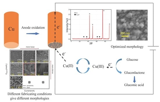
The development of a sensitive, quick-responding, and robust glucose sensor is consistently pursued for use in numerous applications. Here, we propose a new method for preparing a Cu2O electrode for the electrochemical detection of glucose concentration. The Cu2O glucose electrode was prepared by in situ electrical oxidation in an alkaline solution, in which Cu2O nanoparticles were deposited on the electrode surface to form a thin film, followed by the growth of Cu(OH)2 nanorods or nanotubes. The morphology and electrocatalytic activity of a Cu2O glucose electrode can be tuned by the current density, reaction time, and NaOH concentration. The results from XRD, SEM, and a Raman spectrum show that the electrode surface was coated with cubic Cu2O nanoparticles with diameters ranging from 50 to 150 nm. The electrode exhibited a detection limit of 0.0275 mM, a peak sensitivity of 2524.9 μA·cm−2·mM−1, and a linear response range from 0.1 to 1 mM. The presence of high concentrations of ascorbic acid, uric acid, dopamine and lactose appeared to have no effects on the detection of glucose, indicating a high specificity and robustness of this electrode.

Graphical Abstract

1. Introduction

Monitoring the concentration of glucose is essential for diabetes diagnosis [1], fermentation [2], chemical synthesis [3], and other industrial applications. In recent years, continuous effort has been made to develop electrochemical [4] and other methods including optical [5], acoustic [1], chemiluminescence [6], fluorescent nanogels [7], and near-infrared spectroscopy [8], for glucose detection. The electrochemical method is one with proven advantages in terms of accuracy, sensitivity, ease of operation, and portability. In 1962, Clark and Lyons proposed the concept of the glucose enzyme electrode and prepared the first glucose electrode [9]. In order to avoid the interference of background O2 in the sample, electron acceptors such as ferrocene derivatives are used to specify the electron transfer in GOx recycling [4,10]. More recently, a virgin electrode without mediators was used to regenerate GOx (flavin adenine dinucleotide) directly on the surface of the electrodes [11,12,13]. While enzymatic sensors are routinely used in blood glucose detection, their applications to on-site and in situ detection, as often requested by research and engineering practice, are hindered by the limited stability of GOx and unsatisfactory sensitivity due mainly to deficient electron transfer. To overcome these weaknesses, non-enzymatic glucose sensors [14] that directly oxidize glucose at the surface electrode are pursued, and numerous materials have been developed, such as palladium nanoparticles coated functional carbon nanotubes [15], Ti/TiO2 nanotube arrays/Ni composites [16], electrospun palladium (IV)-doped copper oxide composites in the form of nanofibers [17], nanoporous Au [18], and Cu nanoclusters/multi-walled carbon nanotubes (MWCNT) composites [19].
Compared with Pt and Au, Ni and Cu exhibit higher efficiency in the electrochemical detection of glucose because of the formation of Ni(II)/Ni(III)and Cu(II)/Cu(III) redox pairs [20,21]. CuO-based glucose sensors are also attractive due to their rich arability and low toxicity [22]. The oxidation of glucose on a CuO-based glucose electrode has not been fully elucidated [23,24]; however, it is suggested that Cu(II) forms Cu(III), which is capable of oxidizing glucose to gluconolactone and is reduced to Cu(II) after the glucose oxidation. The conventional method to fabricate a CuO glucose electrode consists of several steps, in which the first step is the hydrothermal synthesis of CuO nanoparticles, typically at approximately 100 °C [25,26]. The resulting CuO nanoparticles are then mixed with eutectic polymer binders, such as Nafion, Polyvinylidene fluoride (PVDF) and chitosan, forming an “ink”. Finally, the ink is coated onto the virgin noble electrode to form a thin layer [25,27,28]. The sensitivity of such an electrode is affected by the presence of binders with no oxidation capabilities.
Although nanomaterials such as Carbon nanotubes (CNT) and graphene are blended with CuO nanoparticles [28] giving an improved conductivity, a direct contact of CuO with glucose and an accelerated electron transfer remains desirable for improved sensitivity and responsive speed. In situ fabrication methods have been explored in previous studies by chemical or electrochemical oxidation under different conditions [29,30,31,32]. Here, we present a new method for an in situ fabrication of a Cu2O-based glucose electrode, in which a Cu2O electrode was prepared by electrooxidation of metallic copper as an anode in alkaline solution, forming a Cu2O layer which consists of particles with diameters of 30–150 nm on the surface of the Cu electrode. We expect the circumvention of the “ink” not only simplifies the preparation process but also, and more importantly, allows direct contact of glucose with the electrode and facilitates the electron transport, thereby promising an improved sensitivity and responsiveness. The sensitivity and specificity of the detection, as well as the stability of the electrode, were examined. The effects of the preparation conditions on the morphology and electrocatalytic activity of a Cu2O glucose electrode were investigated to understand the fabrication mechanism of high catalytic activity Cu2O nonenzymic glucose sensors. The results reveal that the morphology and crystal phase are determined by the ratio of current density and NaOH concentration (J/c). As J/c increases, the morphology changes from Cu2O nanoparticles to Cu(OH)2 nanotubes. Cu2O nanoparticles are deposited on the electrode surface, forming a thin film, followed by the growth of Cu(OH)2 nanorods or nanotubes; among them, Cu2O nanoparticles exhibit higher activity. These results provide a rapid and efficient method, using mild conditions, for the in situ synthesis of a Cu2O electrode with good stability and repeatability for electrochemical detection of glucose.

2. Materials and Methods

2.1. Materials and Reagents

A Cu electrode, Ag/AgCl electrode, and Pt electrode were purchased from Aidahengsheng, Tianjin, China. Ascorbic acid (99%, analytical pure) was purchased from Solarbio, Beijing, China. Lactose (99%, analytical pure) was purchased from Sinopharm Chemical Reagent, Beijing, China. Dopamine (99%, analytical pure) and uric acid (99%, analytical pure) were purchased from Aladdin, Shanghai, China. All agents were used without further purification.

2.2. Preparation of the Cu2O Electrode

The Cu2O electrode was synthesized via an in situ electrical oxidation method in alkaline solution. First, a disk Cu electrode with a diameter of 3 mm was polished using Al2O3 powders of 300 nm and 50 nm in diameter, respectively. After washing in ultrasonic baths of ethanol and distilled water consecutively and drying in a N2 atmosphere, the Cu electrode was implemented as the working electrode on CHI 852D (CH Instruments, Shanghai, China) with a 3-electrode system, in which the counter electrode was a Pt electrode and the reference electrode was Ag/AgCl, and oxidized for 300 s at a constant current density of 1.415 mA/cm2 (0.1 mA for a ϕ3 disk electrode) in 3 M NaOH solution. The obtained Cu2O electrode was washed with distilled water and dried in a N2 atmosphere.

2.3. Characterization Apparatus

A field-emission scanning electron microscope (FESEM) (Merlin, Carl Zeiss Jena, Germany) was used to observe the microstructures of Cu2O glucose electrode. The applied voltage was 15 kV. The crystalline phase of the structures was identified by X-ray diffraction (S2, Rigaku, Tokyo, Japan) with Cu Kα radiation. Raman spectroscopy was performed with a Raman microscopy system (LabRAM HR, HORIBA Jobin Yvon, Paris, France). A YAG laser served as an excitation source, and the applied wavelength was 532 nm.

2.4. Electrochemical Measurements

Electrochemical tests including cyclic voltammetry (CV) and Amperometric (it) measurements were conducted on CHI 852D (CH Instruments, Shanghai, China) with a 3-electrode system, in which the working electrode, the counter electrode, and the reference electrode were the Cu2O electrode synthesized as mentioned in Section 2.2, Pt electrode and Ag/AgCl (saturated) electrode, respectively.

3. Results and Discussion

3.1. Characterization of the Cu2O Glucose Electrode

The as-prepared Cu2O glucose electrode was characterized by XRD, SEM and a Raman spectrum, as shown in Figure 1.
From Figure 1a, it is interpreted that the major crystal phase of the Cu2O glucose electrode is the cubic phase of Cu2O (JCPDS card no.77-0199, Fm3m, a0 = b0 = c0 = 4.258 Å). The copper metal substrate (JCPDS card no.03-1005, Fm3, a0 = b0 = c0 = 3.6077 Å) is also present. Figure 1b shows the Raman spectrum of the Cu2O glucose electrode. The characteristic peaks at 108.8, 148.7, 218.3, 415, 520 and 630 cm−1 demonstrate the formation of Cu2O on the Cu electrode. It can be seen from Figure 1c that cubic nanoparticles with diameters of 30–150 nm are dispersed evenly on the electrode surface. As can be seen from Figure 1d, only Cu and O are displayed in the energy-dispersive X-ray spectroscopy (EDS) analysis; it is concluded that the cubic phase crystals on the Cu electrode are Cu2O, as described elsewhere [23,33,34].

3.2. Electrochemical Behavior of the Cu2O Electrode

Figure 2a shows the cyclic voltammetry of the Cu2O electrode prepared in a 50 mM NaOH solution at a scan rate of 50 mV/s, in the absence or presence of glucose. Here, five distinctive redox peaks can be observed on the Cu2O electrode. Peak 1 is assigned to the transition of Cu(0)/Cu(I), while peak 2 includes two transitions of Cu(0)/Cu(II) and Cu(I)/Cu(II). Peak 3 is related to the transition of Cu(II)/Cu(III) in alkaline solution [27]. Peak 4 and peak 5 are related to the transitions of Cu(III)/Cu(II) and Cu(II)/Cu(I), respectively [24]. In the presence of glucose, the CV profile also contains peak 1 and peak 2, while peak 3 disappears and is replaced by a shoulder peak around 0.5 V (vs. Ag/AgCl). The height of this peak is related to the glucose concentration.
Marioli and Kuwana [35] and Chen et al. [36] have attributed the difference in the cyclic voltammograms obtained in the absence and presence of glucose to the transition of Cu(II)/Cu(III). First, glucose turns to an enol form upon isomerization in an alkaline medium, then, the active intermediate is oxidized to gluconolactone by Cu(III) followed by the hydrolysis of gluconolactone and forms gluconic acid. The above-mentioned process is shown in Figure 2b.
Cyclic voltammetry curves were performed at different scan rates of 20–90 mV/s in the presence of 3 mM glucose and 50 mM NaOH solution, as shown in Figure 2c,d. It is shown that the intensity of the shoulder peak near 0.5 V (vs. Ag/AgCl) increases as the scan rate increases. Moreover, a linear relationship between current intensities of the peak at 0.5 V and the square root of scan rates is observed (Figure 2d). According to the Randles–Sevcik equation, good fit linearity reflects a sufficiently rapid electron transfer reaction rate, indicating the diffusion-controlled progression.

3.3. Amperometric Detection of Glucose

We then determined the current density at different glucose concentrations in 50 mM NaOH solution and obtained the results shown in Figure 3.
The amperometric J–t curve in Figure 3a shows that the current density responds to the change of glucose concentrations quickly. The current becomes stable within 3 s after each injection of fresh glucose solution. Figure 3b shows an almost ideal linear response of the current intensity within the glucose concentration range from 0.1 to 1.0 mM. The working voltage is 0.5 V vs. Ag/AgCl, which is higher than the standard potential of the oxygen evolution reaction (0.204 V vs. Ag/AgCl). Therefore, small oxygen bubbles accumulated on the surface of the electrode at a very slow rate. The unreleased small bubbles result in increased noise.
Figure 3b shows the linear dependency of the current response versus different glucose concentrations. The sensitivity of this Cu2O electrode is 2524.9 μA·cm−2·mM−1, when glucose concentrations are in the range of 0.1 to 1 mM. The limit of detection (LOD) of the Cu2O electrode is 2.57 µM, which is calculated by substituting the standard deviation of the background signal with the mean standard deviation of the fitting results (S/N = 3).

3.4. Stability and Selectivity

Figure 3c,d show that the current density decrease is only 6.09% after ten cycles. The recycle is critical for practical applications, and such a recycle time of Cu2O has not yet been reported before. The selectivity and specificity of the Cu2O glucose electrode were examined by adding other species in considerable concentrations. The current response was recorded in 50 mM NaOH solution with successive injection of 0.1 mM ascorbic acid (AA), 0.1 mM dopamine (DA), 0.1 mM uric acid (UA), 0.1 mM lactose (Lac) and 1 mM glucose (Glu), as shown in Figure 3e,f, from which it is concluded that these interferents have little effect on the detection of glucose since their physiological concentrations are AA (30 μM) [37], DA (0.14 nM) [38], UA (300 μM) [39] and Lac (1.5 μM) [40], respectively. The prepared Cu2O electrode can detect glucose in the presence of other traditional contaminants in their physiological concentrations.
Table 1 details the Cu2O electrodes and other metal-based electrodes utilized under alkaline conditions reported elsewhere, as well as their performance in terms of sensitivity, linear response range, detection limit, response time and cycles times. We profiled the data listed in Table 1, as shown in Figure S1. It can be concluded that the Cu2O electrode developed in the present study is superior in terms of both sensitivity and durability with regard to recycle times. The high sensitivity, being close to Cu2O/MoS2, we believe, is attributed to the circumvention of polymer binder, which leads to direct electron transport, as evidenced by the reduction of the overpotential [27]. The response time is a function of the thickness of the Cu2O layer and can be tuned by the current density and electro-oxidation time. This is advantageous for manipulating the structure of the electrode to meet the given requirement of sensitivity and response time. The electrode developed by the present study has the highest durability in terms of cycles times, which, we believe, benefits from the in situ generation of Cu2O nanostructures on the surface of the metallic copper electrode. Such high durability makes it promising for long-term detection.

3.5. Mechanism of In Situ Deposition of Cu2O

We also examined the effect of the fabrication conditions on the microstructure and electrochemical activity. The morphology and responsive behavior of the yielded Cu2O electrodes were determined, respectively. As shown in Figure 4a, their fabrication conditions were used to label the electrodes. For instance, 1M-0.1-100 indicates that the Cu2O electrode is fabricated in a condition of 1.0 M NaOH, 0.1 mA current and 100 s electrooxidation time. The SEM images of the 3M-0.1-300, 3M-0.1-1200, 5M-0.1-300 and 5M-0.1-1200 electrode (Figure S2) reveal that the electrooxidation time has little effect on the morphology of the electrodes. Therefore, we fixed the electro-oxidation time as 300 s for further discussion. To clarify, only the concentration of NaOH and the current density are labelled to indicate samples, as shown in Figure 4a.
Figure 4a shows the SEM images of the Cu2O electrodes fabricated under different conditions. The electrodes were characterized by XRD in Figure 4b and Raman in Figure S3. The main crystal phase of nanoparticles on 3M-0.1, 3M-0.3, 5M-0.1, 5M-0.3 and 5M-0.5 electrodes is face-centered cubic Cu2O (JCPDS card no.77-0199, Fm3m, a 0 = b 0 = c 0 = 4.258 Å), while the main crystal phase of the nanorod or nanotube on 3M-0.5 and 1M-0.1 electrodes is orthorhombic Cu(OH)2 (JCPDS card no.72-0140, Cmcm, Å, Å and Å).
The current density and the NaOH concentration may also significantly affect the morphology of the Cu2O electrode. It is observed that the current intensity and the NaOH concentration significantly affect the morphology of the Cu2O electrode. Here, we establish a model to elucidate this interesting phenomenon. The current density, which is denoted as J, is equivalent to the rate of Cu+/Cu2+ formation during the electro-oxidation reaction. The concentration of NaOH, which is denoted as c, is related to the rate of consuming Cu+ and Cu2+. In the case of a low value of J/c, the Cu2+ or Cu+, produced by electro-oxidation is immediately consumed to form nanoparticles due to excessive OH at the surface of the electrode. This prevents further nucleation and results in small polyhedral nanoparticles depositing on the surface of electrode, as shown in Figure 4a (e.g., 5M-0.1, 5M-0.3, 5M-0.5, and 3M-0.1). The polyhedral nanoparticles are face-centered cubic Cu2O. When the value of I/c increases, the rate of Cu+/Cu2+ formation gradually reaches that of Cu+/Cu2+ consumption, resulting in the formation of incomplete nanotubes (akin to a nanosheet), as shown in Figure 4a (e.g., 3M-0.3-300). If we further increase the value I/c by increasing the current intensity or reducing the concentration of NaOH, the intact nanotubes composed of Cu(OH)2 form on the surface of the Cu electrode (e.g., 3M-0.5-300), as shown in Figure 4a. According to the above discussion, the mechanism of forming different Cu2O nanostructures is illustrated in Figure 4d.
The oxidation time is vital for catalytic activities by determining the depth of the active material, which also relates to mass transfer resistance during the glucose oxidation reaction [61,62,63]. Figure 4c shows the relationship between sensitivity and charge consumption for each electrode. According to the similar catalytic activity at the optimum oxidation time and the fabricating mechanism shown in Figure 4d, the first step is the formation of the Cu2O film, followed by the reaction between Cu2O and OH to produce Cu(OH)2 nanorods or nanotubes. As a result, the electrodes prepared under different conditions contain a layer of Cu2O film. It could be inferred that cubic Cu2O exhibits a higher catalytic activity than orthorhombic Cu(OH)2 and the peak sensitivity is 2524.9 μA·cm−2·mM−1.

4. Conclusions

In this paper, Cu2O electrodes for glucose detection were prepared in situ by an electrochemical oxidation method. The electrode exhibits a high sensitivity of 2524.9 μA·cm−2·mM−1 for glucose detection, with a linear response range of 0.1–1 mM and a detection limit of 0.0275 mM. Moreover, the electrode showed good stability and repeatability. The electrodes fabricated in situ under different conditions were characterized by SEM, XRD, and a Raman spectrum. The mechanism relating preparation condition, morphology and catalytic activity was clarified. Here, we describe a promising method and underlying mechanism to fabricate cubic Cu2O electrodes with reduced resistance and overpotential but high sensitivity. Furthermore, the advantages of high repeatability, simple operation and low energy consumption make the described method possible for practical application.

Supplementary Materials

The following are available online at https://www.mdpi.com/1424-8220/19/13/2926/s1, Figure S1: Comparison of different glucose sensors’ performance, Figure S2: The SEM images of 5M-0.1-300, 5M-0.1-1200, 3M-0.1-300, 5M-0.1-1200, Figure S3: The Raman spectrum of the Cu2O electrodes fabricated under different conditions.

Author Contributions

Conceptualization, C.L.; methodology, Z.L. (Zhipeng Li), L.R. and N.S.; validation, Z.L. (Zheng Liu) and D.L.; formal analysis, C.L.; investigation, C.L.; resources, D.L and N.S.; data curation, C.L.; writing—original draft preparation, C.L.; writing—review and editing, D.L. and Z.L. (Zheng Liu); visualization, C.L.; supervision, D.L.; project administration, C.L.; funding acquisition, D.L.

Funding

This work was financially supported by the National Key Research and Development Program of China under grant No. 2016YFD0400203 and the Chinese National Natural Science Foundation under grant No. 21878175.

Conflicts of Interest

The authors declare no conflict of interest.

References

  1. Weiss, R.; Yegorchikov, Y.; Shusterman, A.; Raz, I. Noninvasive Continuous Glucose Monitoring Using Photoacoustic Technology—Results from the First 62 Subjects. Diabetes Technol. Ther. 2007, 9, 68–74. [Google Scholar] [CrossRef] [PubMed]
  2. Guerrasantos, L.; Käppeli, O.; Fiechter, A. Pseudomonas aeruginosa biosurfactant production in continuous culture with glucose as carbon source. Appl. Environ. Microbiol. 1984, 48, 301–305. [Google Scholar] [Green Version]
  3. Murphy, A.P.; Henthorne, L.R. One-Step Synthesis of Vitamin-C (l-Ascorbic Acid). U.S. Patent 5998634A, 15 March 1999. [Google Scholar]
  4. Wang, J. Electrochemical glucose biosensors. Chem. Rev. 2008, 108, 814–825. [Google Scholar] [CrossRef] [PubMed]
  5. Steiner, M.-S.; Duerkop, A.; Wolfbeis, O.S. Optical methods for sensing glucose. Chem. Soc. Rev. 2011, 40, 4805–4839. [Google Scholar] [CrossRef] [PubMed]
  6. Hao, M.; Liu, N.; Ma, Z. A new luminol chemiluminescence sensor for glucose based on pH-dependent graphene oxide. Analyst 2013, 138, 4393–4397. [Google Scholar] [CrossRef]
  7. Zhou, S.; Min, X.; Dou, H.; Sun, K.; Chen, C.-Y.; Chen, C.-T.; Zhang, Z.; Jin, Y.; Shen, Z. Facile fabrication of dextran-based fluorescent nanogels as potential glucose sensors. Chem. Commun. 2013, 49, 9473–9475. [Google Scholar] [CrossRef] [PubMed]
  8. Malin, S.F.; Ruchti, T.L.; Blank, T.B.; Thennadil, S.N.; Monfre, S.L. Noninvasive prediction of glucose by near-infrared diffuse reflectance spectroscopy. Clin. Chem. 1999, 45, 1651–1658. [Google Scholar]
  9. Clark, L.C.; Lyons, C. Electrode systems for continuous monitoring in cardiovascular surgery. Ann. N. Y. Acad. Sci. 2006, 102, 29–45. [Google Scholar] [CrossRef]
  10. Chen, C.; Xie, Q.; Yang, D.; Xiao, H.; Fu, Y.; Tan, Y.; Yao, S. Recent advances in electrochemical glucose biosensors. RSC Adv. 2013, 3, 4473–4491. [Google Scholar] [CrossRef]
  11. Si, P.; Ding, S.; Yuan, J.; Lou, X.W.; Kim, D.H. Hierarchically structured one-dimensional TiO2 for protein immobilization, direct electrochemistry, and mediator-free glucose sensing. ACS Nano 2011, 5, 7617–7626. [Google Scholar] [CrossRef]
  12. Alwarappan, S.; Singh, S.R.; Pillai, S.; Kumar, A.; Mohapatra, S. Direct electrochemistry of glucose oxidase at a gold electrode modified with graphene nanosheets. Anal. Lett. 2012, 45, 746–753. [Google Scholar] [CrossRef]
  13. Alwarappan, S.; Liu, C.; Kumar, A.; Li, C.-Z. Enzyme-doped graphene nanosheets for enhanced glucose biosensing. J. Phys. Chem. C 2010, 114, 12920–12924. [Google Scholar] [CrossRef]
  14. Tian, K.; Prestgard, M.; Tiwari, A. A review of recent advances in nonenzymatic glucose sensors. Mater. Sci. Eng. C 2014, 41, 100–118. [Google Scholar] [CrossRef] [PubMed]
  15. Chen, X.; Lin, Z.; Chen, D.-J.; Jia, T.; Cai, Z.; Wang, X.; Chen, X.; Chen, G.; Oyama, M. Nonenzymatic amperometric sensing of glucose by using palladium nanoparticles supported on functional carbon nanotubes. Biosens. Bioelectron. 2010, 25, 1803–1808. [Google Scholar] [CrossRef] [PubMed]
  16. Wang, C.; Yin, L.; Zhang, L.; Gao, R. Ti/TiO2 nanotube array/Ni composite electrodes for nonenzymatic amperometric glucose sensing. J. Phys. Chem. C 2010, 114, 4408–4413. [Google Scholar] [CrossRef]
  17. Wang, W.; Li, Z.; Zheng, W.; Yang, J.; Zhang, H.; Wang, C. Electrospun palladium (IV)-doped copper oxide composite nanofibers for non-enzymatic glucose sensors. Electrochem. Commun. 2009, 11, 1811–1814. [Google Scholar] [CrossRef]
  18. Yin, H.; Zhou, C.; Xu, C.; Liu, P.; Xu, X.; Ding, Y. Aerobic oxidation of D-glucose on support-free nanoporous gold. J. Phys. Chem. C 2008, 112, 9673–9678. [Google Scholar] [CrossRef]
  19. Kang, X.; Mai, Z.; Zou, X.; Cai, P.; Mo, J. A sensitive nonenzymatic glucose sensor in alkaline media with a copper nanocluster/multiwall carbon nanotube-modified glassy carbon electrode. Anal. Biochem. 2007, 363, 143–150. [Google Scholar] [CrossRef]
  20. Luo, P.F.; Kuwana, T. Nickel-titanium alloy electrode as a sensitive and stable LCEC detector for carbohydrates. Anal. Chem. 1994, 66, 2775–2782. [Google Scholar] [CrossRef]
  21. Zhao, J.; Wang, F.; Yu, J.; Hu, S. Electro-oxidation of glucose at self-assembled monolayers incorporated by copper particles. Talanta 2006, 70, 449–454. [Google Scholar] [CrossRef]
  22. Zaidi, S.A.; Shin, J.H. Recent developments in nanostructure based electrochemical glucose sensors. Talanta 2016, 149, 30–42. [Google Scholar] [CrossRef] [PubMed]
  23. Su, Y.; Guo, H.; Wang, Z.; Long, Y.; Li, W.; Tu, Y. Au@Cu2O core-shell structure for high sensitive non-enzymatic glucose sensor. Sens. Actuators B Chem. 2018, 255, 2510–2519. [Google Scholar] [CrossRef]
  24. Zhang, Y.; Su, L.; Manuzzi, D.; de los Monteros, H.V.E.; Jia, W.; Huo, D.; Hou, C.; Lei, Y. Ultrasensitive and selective non-enzymatic glucose detection using copper nanowires. Biosens. Bioelectron. 2012, 31, 426–432. [Google Scholar] [CrossRef] [PubMed]
  25. Reitz, E.; Jia, W.; Gentile, M.; Wang, Y.; Lei, Y. CuO nanospheres based nonenzymatic glucose sensor. Electroanalysis 2008, 20, 2482–2486. [Google Scholar] [CrossRef]
  26. Anu Prathap, M.U.; Kaur, B.; Srivastava, R. Hydrothermal synthesis of CuO micro-/nanostructures and their applications in the oxidative degradation of methylene blue and non-enzymatic sensing of glucose/H2O2. J. Colloid Interface Sci. 2012, 370, 144–154. [Google Scholar] [CrossRef] [PubMed]
  27. Zhao, Y.; Fan, L.; Zhang, Y.; Zhao, H.; Li, X.; Li, Y.; Wen, L.; Yan, Z.; Huo, Z. Hyper-Branched Cu@Cu2O Coaxial Nanowires Mesh Electrode for Ultra-Sensitive Glucose Detection. ACS Appl. Mater. Interfaces 2015, 7, 16802–16812. [Google Scholar] [CrossRef] [PubMed]
  28. Wang, X.; Hu, C.; Liu, H.; Du, G.; He, X.; Xi, Y. Synthesis of CuO nanostructures and their application for nonenzymatic glucose sensing. Sens. Actuators B Chem. 2010, 144, 220–225. [Google Scholar] [CrossRef]
  29. Babu, T.G.S.; Ramachandran, T. Development of highly sensitive non-enzymatic sensor for the selective determination of glucose and fabrication of a working model. Electrochim. Acta 2010, 55, 1612–1618. [Google Scholar] [CrossRef]
  30. Luo, Z.J.; Han, T.T.; Qu, L.L.; Wu, X.Y. A ultrasensitive nonenzymatic glucose sensor based on Cu2O polyhedrons modified Cu electrode. Chin. Chem. Lett. 2012, 23, 953–956. [Google Scholar] [CrossRef]
  31. Wang, L.; Fu, J.; Song, Y. A facile strategy to prepare Cu2O/Cu electrode as a sensitive enzyme-free glucose sensor. Int. J. Electrochem. Sci. 2012, 7, 12587–12600. [Google Scholar]
  32. Babu, T.G.S.; Ramachandran, T.; Nair, B. Single step modification of copper electrode for the highly sensitive and selective non-enzymatic determination of glucose. Microchim. Acta 2010, 169, 49–55. [Google Scholar] [CrossRef]
  33. Hovancová, J.; Šišoláková, I.; Oriňaková, R.; Oriňak, A. Nanomaterial-based electrochemical sensors for detection of glucose and insulin. J. Solid State Electrochem. 2017, 21, 2147–2166. [Google Scholar] [CrossRef]
  34. Gnana kumar, G.; Amala, G.; Gowtham, S.M. Recent advancements, key challenges and solutions in non-enzymatic electrochemical glucose sensors based on graphene platforms. RSC Adv. 2017, 7, 36949–36976. [Google Scholar] [CrossRef] [Green Version]
  35. Marioli, J.M.; Kuwana, T. Electrochemical characterization of carbohydrate oxidation at copper electrodes. Electrochim. Acta 1992, 37, 1187–1197. [Google Scholar] [CrossRef]
  36. Wei, H.; Sun, J.-J.; Guo, L.; Li, X.; Chen, G.-N. Highly enhanced electrocatalytic oxidation of glucose and shikimic acid at a disposable electrically heated oxide covered copper electrode. Chem. Commun. 2009, 20, 2842–2844. [Google Scholar] [CrossRef] [PubMed]
  37. Sargent, F. A study of the normal distribution of ascorbic acid between the red cells and plasma of human blood. J. Biol. Chem 1947, 171, 471–476. [Google Scholar]
  38. Ambade, V.; Arora, M.M.; Singh, P.; Somani, B.L.; Basannar, D. Adrenaline, noradrenaline and dopamine level estimation in depression: Does it help? Med. J. Armed Forces India 2009, 65, 216–220. [Google Scholar] [CrossRef]
  39. So, A.; Thorens, B. Science in medicine Uric acid transport and disease. J. Clin. Investig. 2010, 120, 1791–1799. [Google Scholar] [CrossRef]
  40. Arthur, P.G.; Kent, J.C.; Potter, J.M.; Hartmann, P.E. Lactose in blood in nonpregnant, pregnant, and lactating women. J. Pediatr. Gastroenterol. Nutr. 1991, 13, 254–259. [Google Scholar] [CrossRef]
  41. Tang, L.; Lv, J.; Kong, C.; Yang, Z.; Li, J. Facet-dependent nonenzymatic glucose sensing properties of Cu2O cubes and octahedra. New J. Chem. 2016, 40, 6573–6576. [Google Scholar] [CrossRef]
  42. Yazid, S.N.A.M.; Isa, I.M.; Hashim, N. Novel alkaline-reduced cuprous oxide/graphene nanocomposites for non-enzymatic amperometric glucose sensor application. Mater. Sci. Eng. C 2016, 68, 465–473. [Google Scholar] [CrossRef] [PubMed]
  43. Fang, L.; Wang, F.; Chen, Z.; Qiu, Y.; Zhai, T.; Hu, M.; Zhang, C.; Huang, K. Flower-like MoS2decorated with Cu2O nanoparticles for non-enzymatic amperometric sensing of glucose. Talanta 2017, 167, 593–599. [Google Scholar] [CrossRef] [PubMed]
  44. Li, Y.; Zhong, Y.; Zhang, Y.; Weng, W.; Li, S. Carbon quantum dots/octahedral Cu2O nanocomposites for non-enzymatic glucose and hydrogen peroxide amperometric sensor. Sens. Actuators B Chem. 2015, 206, 735–743. [Google Scholar] [CrossRef]
  45. Long, M.; Tan, L.; Liu, H.; He, Z.; Tang, A. Novel helical TiO2 nanotube arrays modified by Cu2O for enzyme-free glucose oxidation. Biosens. Bioelectron. 2014, 59, 243–250. [Google Scholar] [CrossRef] [PubMed]
  46. Liu, M.; Liu, R.; Chen, W. Graphene wrapped Cu2O nanocubes: Non-enzymatic electrochemical sensors for the detection of glucose and hydrogen peroxide with enhanced stability. Biosens. Bioelectron. 2013, 45, 206–212. [Google Scholar] [CrossRef] [PubMed]
  47. Cao, H.; Yang, A.; Li, H.; Wang, L.; Li, S.; Kong, J.; Bao, X.; Yang, R. A non-enzymatic glucose sensing based on hollow cuprous oxide nanospheres in a Nafion matrix. Sens. Actuators B Chem. 2015, 214, 169–173. [Google Scholar] [CrossRef]
  48. Yin, H.; Cui, Z.; Wang, L.; Nie, Q. In situ reduction of the Cu/Cu2O/carbon spheres composite for enzymaticless glucose sensors. Sens. Actuators B Chem. 2016, 222, 1018–1023. [Google Scholar] [CrossRef]
  49. Yang, Z.; Yan, X.; Li, Z.; Zheng, X.; Zheng, J. Synthesis of Cu2O on AlOOH/reduced graphene oxide for non-enzymatic amperometric glucose sensing. Anal. Methods 2016, 8, 1527–1531. [Google Scholar] [CrossRef]
  50. Zhou, D.L.; Feng, J.J.; Cai, L.Y.; Fang, Q.X.; Chen, J.R.; Wang, A.J. Facile synthesis of monodisperse porous Cu2O nanospheres on reduced graphene oxide for non-enzymatic amperometric glucose sensing. Electrochim. Acta 2014, 115, 103–108. [Google Scholar] [CrossRef]
  51. Neetzel, C.; Muench, F.; Matsutani, T.; Jaud, J.C.; Broetz, J.; Ohgai, T.; Ensinger, W. Facile wet-chemical synthesis of differently shaped cuprous oxide particles and a thin film: Effect of catalyst morphology on the glucose sensing performance. Sens. Actuators B Chem. 2015, 214, 189–196. [Google Scholar] [CrossRef] [Green Version]
  52. Khedekar, V.V.; Bhanage, B.M. Simple Electrochemical Synthesis of Cuprous Oxide Nanoparticles and Their Application as a Non-Enzymatic Glucose Sensor. J. Electrochem. Soc. 2016, 163, B248–B251. [Google Scholar] [CrossRef]
  53. He, J.; Jiang, Y.; Peng, J.; Li, C.; Yan, B.; Wang, X. Fast synthesis of hierarchical cuprous oxide for nonenzymatic glucose biosensors with enhanced sensitivity. J. Mater. Sci. 2016, 51, 9696–9704. [Google Scholar] [CrossRef]
  54. Wang, M.; Song, X.; Song, B.; Liu, J.; Hu, C.; Wei, D.; Wong, C.P. Precisely quantified catalyst based on in situ growth of Cu2O nanoparticles on a graphene 3D network for highly sensitive glucose sensor. Sens. Actuators B Chem. 2017, 250, 333–341. [Google Scholar] [CrossRef]
  55. Zhang, Y.; Xu, F.; Sun, Y.; Shi, Y.; Wen, Z.; Li, Z. Assembly of Ni(OH)2 nanoplates on reduced graphene oxide: A two dimensional nanocomposite for enzyme-free glucose sensing. J. Mater. Chem. 2011, 21, 16949–16954. [Google Scholar] [CrossRef]
  56. Zheng, Y.; Li, P.; Li, H.; Chen, S. Controllable growth of cobalt oxide nanoparticles on reduced graphene oxide and its application for highly sensitive glucose sensor. Int. J. Electrochem. Sci. 2014, 9, 7369–7381. [Google Scholar]
  57. Heidari, H.; Habibi, E. Amperometric enzyme-free glucose sensor based on the use of a reduced graphene oxide paste electrode modified with electrodeposited cobalt oxide nanoparticles. Microchim. Acta 2016, 183, 2259–2266. [Google Scholar] [CrossRef]
  58. Belkhalfa, H.; Teodorescu, F.; Quéniat, G.; Coffinier, Y.; Dokhan, N.; Sam, S.; Abderrahmani, A.; Boukherroub, R.; Szunerits, S. Insulin impregnated reduced graphene oxide/Ni(OH)2 thin films for electrochemical insulin release and glucose sensing. Sens. Actuators B Chem. 2016, 237, 693–701. [Google Scholar] [CrossRef]
  59. Zhang, H.; Liu, S. Nanoparticles-assembled NiO nanosheets templated by graphene oxide film for highly sensitive non-enzymatic glucose sensing. Sens. Actuators B Chem. 2017, 238, 788–794. [Google Scholar] [CrossRef]
  60. Lv, W.; Jin, F.M.; Guo, Q.; Yang, Q.H.; Kang, F. DNA-dispersed graphene/NiO hybrid materials for highly sensitive non-enzymatic glucose sensor. Electrochim. Acta 2012, 73, 129–135. [Google Scholar] [CrossRef]
  61. Guilbeau, E.J.; Towe, B.C.; Muehlbauer, M.J. Model for a Thermoelectric Enzyme Glucose Sensor. Anal. Chem. 1989, 61, 77–83. [Google Scholar]
  62. Schmidtke, D.W.; Freeland, A.C.; Heller, A.; Bonnecaze, R.T. Measurement and modeling of the transient difference between blood and subcutaneous glucose concentrations in the rat after injection of insulin. Proc. Natl. Acad. Sci. USA 1998, 95, 294–299. [Google Scholar] [CrossRef] [PubMed] [Green Version]
  63. Jablecki, M.; Gough, D.A. Simulations of the frequency response of implantable glucose sensors. Anal. Chem. 2000, 72, 1853–1859. [Google Scholar] [CrossRef] [PubMed]
Figure 1. (a) XRD pattern, (b) Raman spectrum, (c) SEM images of the newly in situ fabricated Cu2O electrode, and (d) energy-dispersive X-ray spectroscopy (EDS) analysis.
Sensors 19 02926 g001
Figure 2. (a) Cyclic voltammograms (cathodic sweep first) of a Cu2O electrode in 50 mM NaOH electrolyte. Scan rate: 50 mV/s. (b) Mechanism of glucose oxidation on the Cu2O electrode. (c) Cyclic voltammograms (CVs) of the Cu2O electrode at scan rates varying from 10–90 mV/s in 50 mM NaOH solution containing 3 mM glucose. (d) Linear regression of the peak current and square root of the scan rate.
Sensors 19 02926 g002
Figure 3. (a) Amperometric J–t test with successive addition of glucose at 0.5 V (vs. Ag/AgCl). (b) The calibration curve of (a). (c) Amperometric J–t tests repeated 10 times in 50 mM NaOH solution containing 1 mM glucose. (d) Current response percentage of each amperometric J–t test in (c). (e) Amperometric responses of the Cu2O electrode to the successive dropwise additions of interfering species (ascorbic acid (AA), dopamine (DA), uric acid (UA), lactose (Lac)) and glucose. (f) Current response percentage of (e).
Sensors 19 02926 g003
Figure 4. (a) SEM images. (b) XRD patterns of sensors prepared in different NaOH concentrations and current intensities. (c) The relationship between sensitivity and charge consumption during preparation for the Cu2O electrodes fabricated under different conditions. (d) Crystal growth mechanism of electro-oxidation of Cu as an anode under alkaline conditions.
Sensors 19 02926 g004
Table 1. Comparison of different glucose sensors performance.
ElectrodeSensitivity (μA·mM−1·cm−2)Linear Range (mM)Detection Limit (μM)Response Time (s)Cycles (times)Ref.
Octahedral Cu2O293.8930.1–55.1157[41]
Cu2O/graphene1330.050.01–3.00.3677[42]
Cu2O/MoS23108.870.01–4.01310[43]
CQDs/O-Cu2O2980.02–4.38.4103[44]
Cu2O/TiO2 nanotube14.563.0–9.06235[45]
rGOs wrapped Cu2O2850.3–3.33.391[46]
Hollow Cu2O2038.20.00125–0.03750.4135[47]
Cu/Cu2O/CS63.80.01–0.69551[48]
Cu2O/AlOOH/rGO155.10.005–14.772.653[49]
rGOs-porous Cu2O185.10.01–60.0531[50]
Au@Cu2O7150.05–2.018205[23]
polyhedral Cu2O300.961.2–2980.14441[51]
Cu2O/PC platinum5070.1–2.52653[52]
DH Cu2O/GCE1231.70.019–1.08918.532[53]
Nafion/Cu@Cu2O/GCE14200.0007–2.040 nm27[27]
Cu2O NPs/G3DN/CP23100.00048–1.8130.141.64[54]
Cu2O/Cu62.290.05–6.753752[31]
Cu2O/Cu728.670.01–7.5333.61[30]
CuO/Cu761.90.002–20217[32]
CuO/CuOx/Cu18900.002–150.0516[29]
Co3O4/GOH/GCE492.80.25–10-81[55]
Co3O4/rGO/GCE13660.0005–1.2770.1822[56]
Co3O4/rGOP1.210.04–41.4-1[57]
Ni(OH)2/insulin/rGO/Au18.90.005–10572[58]
NiO/rGO/GCE11380.001–0.40.18210[59]
NiO/DNA/graphene/GCE90.001–82.5810[60]
Cu2O@Cu2524.90.1–1.02.57310This work