Article

Two New Edible Lyophyllum Species from Tibetan Areas, China

1
Faculty of Environmental Sciences and Engineering, Kunming University of Science and Technology, Kunming 650500, China
2
Biotechnology and Germplasm Resources Institute, Yunnan Academy of Agricultural Sciences, Kunming 650223, China
*
Author to whom correspondence should be addressed.
Diversity 2023, 15(9), 1027; https://doi.org/10.3390/d15091027
Submission received: 18 August 2023 / Revised: 18 September 2023 / Accepted: 18 September 2023 / Published: 21 September 2023
(This article belongs to the Special Issue The Hidden Fungal Diversity in Asia 2.0)

Abstract

:
Two new species, Lyophyllum yiqunyang and L. heimogu, that belong to the section Difformia of the genus Lyophyllum, are described based on collections from Tibetan areas, China. The two species are delicious edible low-temperature mushrooms and are widely collected and eaten by local people. Lyophyllum yiqunyang sp. nov. is saprotrophic and has medium-sized basidiomata, olive-grey pileus, cheilocystidia, absent pleurocystidia, globose to subglobose basidiospores (6.12–6.31 × 6.02–6.23 μm) and clamp connections at the pileus context, hymenophoral trama and stipe. Lyophyllum heimogu sp. nov. is saprotrophic and has a dark grey to olive pileus, medium-sized basidiomata and globose to subglobose basidiospores (5.31–5.63 × 5.22–5.41 μm). In the phylogenetic analyses, our two new species formed distinct clades that are well supported by posterior probabilities and bootstrap proportions. Detailed descriptions, colour photos, illustrations and a phylogenetic tree to show the positions of the two new species are presented.

1. Introduction

The genus Lyophyllum P. Karst is classified under the family Lyophyllaceae, order Agaricales, http://www.indexfungorum.org/Names/Names.asp accessed on 10 June 2023, and is mainly characterised by basidiomata colour that is unchanging or changes to dark when injured; scattered, gregarious or solitary growth; a smooth pileus surface; a solid cylindrical or upwards-tapering stipe; basidiospores with variable shape (globose, ellipsoid or broadly fusiform); and saprophytic and symbiotic habits [1,2].
There is a complex without blackening when injured and with caespitose-growing basidiomata in the section Difformia [3]. Lyophyllum sect. Difformia includes 14 species worldwide, most of these species from Europe (L. brunneum Dähncke, Contu & Vizzini; L. calabrum Lavorato & Contu; L. cistophilum Vila & Llimona; L. decastes (Fr.) Singer; L. lanzonii Candusso; L. pergamenum [Sacc. & P. Syd.] Horniček; L. pseudoloricatum Dähncke, Contu & Vizzini; L. subglobisporum Consiglio & Contu; L. soniae Picillo & Contu) and a few from North/South America (L. multiforme [Peck] H.E. Bigelow and L. tucumanense Singer) and Asia (L. shimeji [Kawam.] Hongo and two novel species, L. yiqunyang and L. heimogu) [4,5,6,7,8,9,10,11]. Lyophyllum consists of 60 species worldwide [12].
Dai 1979 [13] identified the first Lyophyllum species in China, L. cinerascens (Bull.) Konrad & Maubl., which is now L. decastes. Since then, 20 additional Lyophyllum species have been recorded in China [14]. These species were identified based on phenotypic similarities to European Lyophyllum and lacked detailed descriptions and molecular data.
Many Lyophyllum species names have been combined and changed with the advancement of molecular biology methods. To date, 16 Lyophyllum species have been reported in China, viz. L. atrofuscum S.W. Wei, Q. Wang & Yu Li (Tibet); L. decastes (Yunnan, Sichuan, Qinghai, Liaoning and Fujian, etc.); L. deqinense Y.H. Ma, W.M. Chen & Y.C. Zhao (Yunnan); L. immundum (Berk.) Kühner (not clear); L. infumatum (Bres.) Kühner (not clear); L. loricatum (Fr.) Kühner (Tibet); L. macrosporum Singer (not clear); L. ochraceum (R. Haller Aar.) Schwöbel & Reutter (Sichuan); L. pallidofumosum (Yunnan); L. rhombisporum Shu H. Li & Y.C. Zhao (Yunnan); L. semitale (Fr.) Kühner (Yunnan, Tibet, Qinghai, Heilongjiang, Shanxi); L. subalpinarum S.W. Wei, Q. Wang & Yu Li (Tibet); L. subdecastes S.W. Wei, Q. Wang & Yu Li (Gansu); L. trigonosporum (Bres.) Kühner (Yunnan, Tibet); L. transforme (Lapl.) Singer (Yunnan, Tibet, Qinghai, Liaoning, Heilongjiang); and L. shimeji (Yunnan) [14,15,16,17,18].
Most species of Lyophyllum have been described as well-known edible mushrooms, such as L. shimeji, L. decastes and L. fumosum. Currently, the classification of this genus and the cultivation of L. shimeji have been studied extensively in Italy, Japan, Sweden and Switzerland [19].
During a survey of Lyophyllum in China, two saprotrophic new species, viz. L. yiqunyang and L. heimogu, belonging to sect. Difformia were collected and identified based on morphological features and molecular data.

2. Materials and Methods

2.1. Site Description

Jiuzhaigou and Bomi are important and well-preserved nature sites on the edge of the Tibetan Plateau. The Tibetan area is made up of high-altitude hydrological and tectonic activity. The rock strata are mostly carbonate rocks such as dolomite and tufa, as well as some sandstone and shales. Lyophyllum yiqunyang and L. heimogu were found growing on soil in mixed coniferous and broad-leaved forests dominated by Abies spp., Picea spp. and Salix cupularis, with an elevation of 2000–3000 m a.s.l., a temperate climate and annual rainfall of 550 mm.

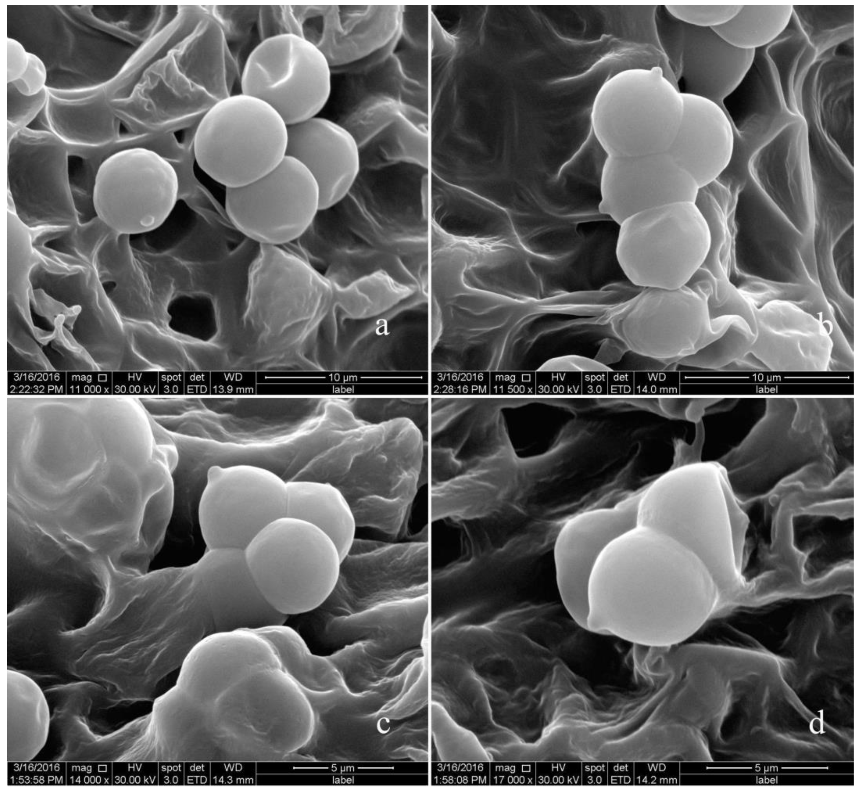
2.2. Morphological Studies

Five specimens were photographed in situ and collected from Tibetan areas in China. After collecting, samples were wrapped in aluminium foil and placed in a collection box until they were examined. Macro-morphological features were recorded from fresh collections, and colour was determined following Kornerup and Wanscher [20]. Specimens were dried at 50 °C in a food drier, stored in sealed plastic bags and deposited at the Biotechnology and Germplasm Resources Institute, Yunnan Academy of Agricultural Sciences, Kunming and Herbarium of Cryptogams at the Kunming Institute of Botany, Chinese Academy of Sciences (HKAS). Micromorphological features were observed under a Y-TV55 light microscope (Nikon, Tokyo, Japan) from the dried material that was studied. All tissues were revived in 5–10% KOH and mounted in Congo Red. Scanning electron microscopy (SEM) images were captured using Regulus 8100 (Hitachi, Tokyo, Japan). Twenty basidia and more than fifty basidiospores were measured and photographed using a Nikon Eclipse 80i microscope at magnifications up to ×1000. The notation [x/y/z] specified that measurements were made on ‘x’ basidiospores from ‘y’ basidiomata and ‘z’ collections. Basidiospore dimensions were given as ‘(a) b–n–c (d)’. Where ‘a’ and ‘d’ refer to the lower and upper extremes of all measurements, respectively, ‘b–c’ refers to the range of 95% of the measured basidiospores, ‘n’ refers to the average dimension, ‘Q’ is the length/width ratio of basidiospores and ‘Qm’ is the average Q of all basidiospores.

2.3. DNA Extraction, PCR Amplification and Sequencing

Molecular analyses were performed at the Yunnan Academy of Agricultural Sciences, China. Genomic DNA was extracted from dry specimens using the Ezup Column Fungi Genomic DNA Extraction Kit (Sangon Biotech, Shanghai, China) following the manufacturer’s protocol. Primer pairs (forward/reverse) used for PCR cycling of the ITS regions were ITS1/ITS4 [21]. PCR was carried out using a C1000 Thermal Cycler (Bio-Rad, Beijing, China) with a cycling program as follows. ITS: initial denaturation at 95 °C for 5 min, 35 cycles of denaturation at 95 °C for 30 s, annealing at 55 °C for 30 s, extension at 72 °C for 90 s and a final extension at 72 °C for 10 min [21].
PCR products were visualised via UV light after electrophoresis on 1% agarose gels stained with ethidium bromide. The PCR amplicons were sent to Sangon Biotech (Shanghai, China) for Sanger sequencing in both directions using the PCR primers.

2.4. Sequence Alignment and Phylogenetic Analyses

The sequences of new Lyophyllum identified in this study were submitted to the NCBI database. The Basic Local Alignment Search Tool for the GenBank database was used to check whether the newly generated sequences were amplified contaminant DNA and examine clusters with closely related sequences (see Table 1). DNA sequences were retrieved and assembled using SeqMan version 5.0. The sequence alignments were aligned using MAFFT version 7 (https://mafft.cbrc.jp/alignment/server/, accessed on 10 June 2023) [22], and each gene was analysed using BioEdit v. 7 [23]. Maximum likelihood (ML) analysis was performed using RAxML-HPC2 v. 8.2.12 [24] as implemented in the CIPRES (https://www.phylo.org/portal2/login!input.action, accessed on 10 June 2023) portal [25] using the GTR+G+I model and 1000 rapid bootstrap (BS) replicates for all genes. A reciprocal 70% bootstrap support approach was used to check for conflicts between the tree topologies from individual genes. Since there was no significant incongruence in topology between the ML trees, the ITS was partitioned for phylogenetic analyses. For Bayesian inference (BI), the best substitution model for each partition was determined by MrModeltest 2.2 [26]. The results suggested ITS1: JC+I, 5.8S: GTR+G+I, ITS2: K80+I+G. Bayesian analysis was performed using MrBayes ver. 3.2.7a [27] in the CIPRES portal. Four parallel runs, each consisting of one cold and three heated chains, were performed for 10 million generations with sampling every 100 generations for the single gene trees and 50 million generations with sampling every 1000 generations. Parameter convergence > 200 was verified in Tracer v. 1.7 [28]. Phylogenetic clades were strongly supported if bootstrap support value (BS) was ≥70% and/or posterior probability (PP) was ≥0.95.
Table 1. Names, specimen numbers, origin, references and corresponding GenBank accession numbers of Lyophyllum sequences used in the phylogenetic analyses. Newly generated sequences are in bold black, and a “*” before taxa indicates holotypes.
Taxon NameSpecimen NumberOriginITSReference
Calocybe gambosaHC78/64SwitzerlandAF357027[7]
C. carneaCBS552.50SwitzerlandAF357028[7]
C. persicolorHC80/99SwitzerlandAF357026[7]
Hypsizygus marmoreusV.1611GermanyAJ494834[11]
H. marmoreusV.3133ChinaFJ609271[11]
H. marmoreusHMW2MalaysiaHM561971[11]
H. marmoreusHZND-1ChinaJX046028[11]
H. marmoreus1-1KoreaKF192813[11]
H. tessulatusAFTOL-ID 1898USADQ917653[11]
H. tessulatusL2ChinaFJ467372[11]
H. ulmariusCBS 286.77KoreaAY265850[29]
H. ulmariusDUKE-JM/HWUSAEF421105[30]
L. ambustumCBS452.87SwitzerlandAF357057[7]
L. anthracophilumHC79/132SwitzerlandAF357055[7]
L. atratumCBS709.87SwitzerlandAF357053[7]
L. atrofuscumHMJAU63461ChinaOP605493[16]
L. atrofuscumHMJAU63456 *ChinaOP605494[16]
L. caerulescensHC80-140SwitzerlandAF357052[7]
L. caerulescensV.15759USAJF908339[11]
L. crassifoliumV.5077ItalyJF908331[11]
L. decastesdd08054ChinaFJ810160[11]
L. decastesLd418ChinaHM119485[11]
L. deqinenseYAASM6949 *ChinaOQ418117[18]
L. deqinenseYAASM6948ChinaOQ418116[18]
L. deliberatumV.15032SloveniaJF908338[11]
L. favreiBSI94cp2SwitzerlandAF357035[7]
L. favreiV.6334ItalyJF908333[11]
L. fumosumSJ02/006SwedenHM572539[11]
L. fumosumLAS00/144SwedenHM572541[11]
L. fumosumV.16077ItalyJF908340[11]
L. fumosumLfumNlf24JapanJN983977[11]
L. fumosumL2010512371ChinaJX966310[11]
L. fumosumYAASM6215ChinaON681708[17]
L. fumosumYAASM6340ChinaON681709[17]
L. gangraenosumV.12332ItalyJF908335[11]
L. heimoguL3026 *ChinaKY434100This study
L. heimoguL3033ChinaKY434101This study
L. heimoguL3035ChinaKY434102This study
L. infumatumV.10152ItalyJF908334[11]
L. leucophaeatumHae251.97SwitzerlandAF357032[7]
L. littoralisCA20091210ItalyJX280410[11]
L. loricatumV.13175USAJF908336[11]
L. loricatumCA20090202.03ItalyJX280406[11]
L. loricatum01.12.09ItalyJX280407[11]
L. moncalvoanumPDD 96328 *New ZealandNR_137615[2]
L. moncalvoanumPDD 102581New ZealandKJ461912[2]
L. ochraceumBSI94.cp1SwitzerlandAF357033[7]
L. ochraceumV.537ItalyJF908329[11]
L. rhombisporumL1762*ChinaJX966307[11]
L. rhombisporumL2082ChinaJX966308[11]
L. semitaleHC85/13SwitzerlandAF357049[7]
L. semitaleEL187-09SwedenHM572552[8]
L. shimejiOlsen821006SwedenHM572530[8]
L. shimejiNZ4Q88 New ZealandJN983985[11]
L. shimejiL2010512377ChinaJX966311[11]
L. sp.PBM 2688USADQ182502[11]
L. sp.SB102ChinaFJ687273[11]
L. sp.Aase811014SwedenHM572550[8]
L. sp.TO-2011ItalyJF908337[11]
L. sp. JN001ChinaFJ687270[11]
L. sp.O73586NetherlandsGU234137[11]
L. sp. Cultivar JpnSwedenHM572551[8]
L. sp. SL-2013ChinaJX966308[11]
L. sykosporumIFO30978 SwitzerlandAF357050[7]
L. subalpinarumHMJAU63447 *ChinaOP605490[16]
L. subalpinarumHMJAU63453ChinaOP605491[16]
L. subdecastesHMJAU63470ChinaOP605488[16]
L. subdecastesHMJAU63467 *ChinaOP605489[16]
L. turcicumKATO-2971 *TurkeyKJ158159[11]
L. yiqunyangL4206ChinaKY434104This study
L. yiqunyangL2989 *ChinaKY434103This study
Tephrocybe boudierBSI96/84USADQ825427[11]

3. Result

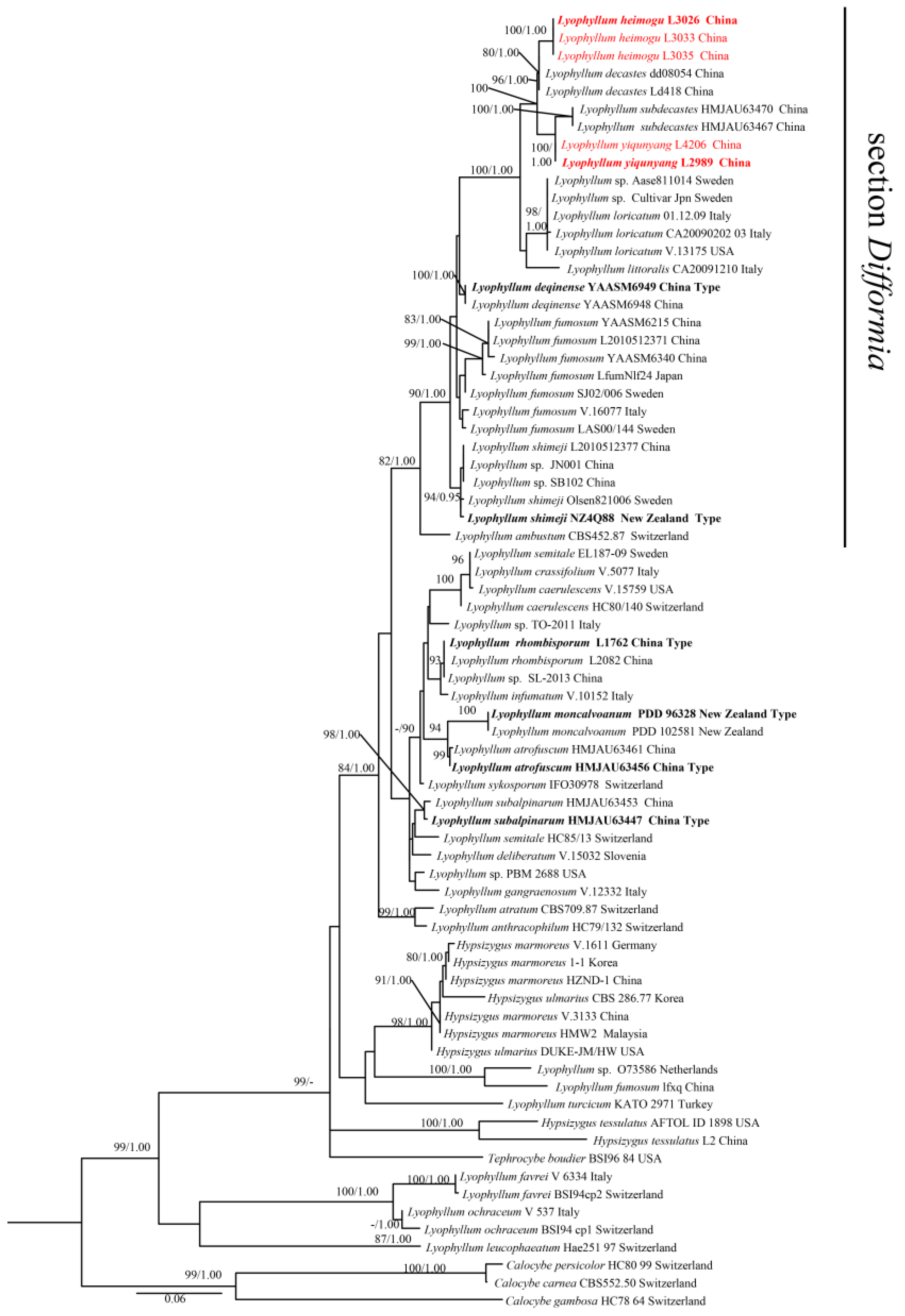
3.1. Phylogenetic Analysis

Five newly generated and 64 retrieved sequences from GenBank were used as the ingroup. Three sequences of Calocybe gambosa, C. carnea and C. persicolor retrieved from GenBank were used as the outgroup [30]. The ITS was 575 characters in length, of which 284 characters were constant, 291 were variable but parsimony-uninformative and 231 were parsimony-informative. Estimated base frequencies were as follows: A = 0.231384, C = 0.218313, G = 0.226647, T = 0.323655, substitution rates AC = 1.548998, AG = 4.813675, AT = 2.464671, CG = 0.797610, CT = 7.018015, GT = 1.000000; gamma distribution shape parameter α = 0.394616.
ML and BI analyses generated nearly identical tree topologies with minimal variation in statistical support values. Thus, only the ML tree is displayed (Figure 1). Phylogenetic data and thorough morphological analysis (see below) showed that the two newly described taxa in this study are significantly distinguished from other known Lyophyllum species.
In our phylogeny, our species L. yiqunyang (L4206 and L2989) and L. heimogu (L3026, L3033 and L3035) formed independent branches. Comparing the ITS sequences, there were 12/575 (2.09%), 18/575 (3.13%), 16/575 (2.78%) and 6/575 (1.04%) differences between L. yiqunyang (L2989, holotype) and L. heimogu (L3026, holotype), L. loricatum (V.13175), L. littoralis (CA20091210) and L. subdecastes (HMJAU 63467 holotype), respectively, as well as 15/575 (2.78%), 15/575 (2.78%), 13/575 (2.26%) and 9/575 (1.57%) ITS sequence differences between L. heimogu (L3026, holotype) and L. loricatum (V.13175), L. littoralis (CA20091210), L. subdecastes (HMJAU63467 holotype) and L. decastes (Ld418), respectively.

3.2. Taxonomy

Lyophyllum yiqunyang Shu H. Li sp. nov. Figure 2, Figure 3 and Figure 6.
MycoBank: 849794.
Holotype: China, Sichuan Province, Jiuzhaigou County, 103°54′37″ E, 33°10′34″ N, elev. 3251 m, in a forest dominated Abies spp., Picea spp. and Salix cupularis, Shu−Hong Li, 10 August 2014. (YAAS L2989, holotype!).
Etymology: “yiqunyang” refers to the common name used in mushroom markets in China.
Basidiomata mid-sized (Figure 2), Pileus 3.0–6.0 cm diameter, fleshy, fragile, variable in shape according to growth conditions, hemispherical when young, becoming convex with age; surface smooth, dry, olive-grey (2E-F), unchanging, without umbo, deflexed aspects of margin; pileus context thick, 0.1–0.3 cm wide, white (1A1), watery soaked in wet weather. Lamellae moderately close together, ventricose to broadly ventricose, adnate to narrowly adnate, broad, white (1A1) to pale grey (1B1), non-discolouring when bruised; edge even or entire. Stipe 3.0–7.0 × 0.5–1.5 cm, cylindrical to clavate, white or light grey, pruinose at the base, sometimes tapered towards at the base, often twisted, white (1A1) to pale grey (1B1), smooth, sometimes squamous on the surface, unchanging in colour when injured. Odour and taste not distinctive.
Basidiospores [120/2/2] 6.1–6.3 × 6.0–6.2 μm (Figure 3 and Figure 6), Q = 1.1–1.3, Qm = 1.18 ± 0.35, av. 6.21 ± 0.12 × 6.15 ± 0.24 μm, globose, subglobose to broadly ellipsoid, hyaline, smooth. Basidia 31.0–42.0 × 7.8–9.0 μm (N = 20), av. 38.1 ± 3.4 × 8.3 ± 1.28 μm, four–spored, sometime with basal clamp connections, clavate, siderophilous granulation. Subhymenium made up of moderately thin-walled hyphae, 5–15 μm across. Hymenophoral trama regular, consisting of thin and hyaline hyphae, some with clamp connection. Marginal cells absent. Pileipellis an interwoven trichodermium to a subcutis composed of almost hyaline interwoven filamentous hyphae, terminal cells 20–83 × 3–8 μm, almost cylindrical to subcylindrical, occasional hyphal tips flexuous and sometimes inflate, some with clamp connections. Stipitipellis a cutis of elongate hyphae. Clamp connection present at some septa. Thromboplerous hyphae present on the hymenophoral trama, 5–8 μm wide.
Ecology and distribution: saprotrophic (growing on fallen wood or soil); when growing on the soil, the soil’s upper layer has a relatively thick humus layer; gregarious in forests of Abies spp., Picea spp. or Salix cupularis. Summer to autumn (August to September). Known only from Sichuan Province, China.
Additional specimens examined: China, Sichuan province, Jiuzhaigou County, elev. 3200 m, latitude 103°54′37″ E and longitude 33°10′34″ N, in a forest dominated by Abies spp. and Salix cupularis, Shu-Hong Li, 8 August 2015, (L4206).
Notes: Lyophyllum yiqunyang is quite similar to L. deqinense in having an olive-greyish to greyish-orange pileus and subglobose to globose basidiospores, the L. deqinense basidia are shorter (24.1–33.8 × 7.3–9.9 μm). The pileipellis of L. deqinense is a cutis composed of parallel elements [18].
Lyophyllum yiqunyang is also similar to L. subalpinarum in having a greyish-yellow to olive-greyish pileus. Lyophyllum subalpinarum stipe is hollow, whitish-greyish to dark in the middle. The lamellae of L. subalpinarum change to black when touched or injured and present rounded-cylindrical basidiospores (6.9–8.7 × 4.3–5.1 μm) [16].
Lyophyllum yiqunyang is related to L. subdecastes; however, L. subdecastes has a yellowish-brown, brown, greyish-red pileus, relatively small basidiospores (3.9–5.0 × 3.7–5.0 μm) and larger basidia (36.7–50.6 × 8.4–10.9 μm) [16].
Lyophyllum heimogu S. H. Li sp. nov. Figure 4, Figure 5 and Figure 6.
MycoBank: 849795.
Holotype: China, Tibet Autonomous Region, Bomi County, elev. 2717 m, 95°43′06′′ E, 29°51′41′′ N, in a forest dominated by Abies spp. and Picea spp., Shu–Hong Li, 3 October 2013. (YAAS L3026, holotype!).
Etymology: “heimogu” refers to the common name used by local people.
Basidiomata mid-size (Figure 4), Pileus 3.0–6.5 cm wide, fleshy, fragile, hemispherical when young, becoming convex with age; surface smooth, dark grey (1F1) at the centre, becoming olive (1E-F3) towards the margin with an indistinctly striated margin, umbo papilla, dark (1F1), involute aspects of margin; pileus context thick, 0.1–0.2 cm wide, white (1A1). Lamellae moderately close together, subventricose to ventricose, free to adnexed, lamella edge even or entire, white when young, becoming yellowish-white with age. Context 0.3–0.8 cm thick, white to pale white, unchanging when injured. Stipe 2.5–7.0 × 0.5–2.0 cm, yellowish-brown (5E4-5), central, cylindrical to clavate, sometime bulbous at the base, solid, often twisted, smooth. Odour and taste not distinctive.
Basidiospores [120/2/2] 5.31–5.63 × 5.22–5.41 μm (Figure 5 and Figure 6), Q = 1.1–1.2, Qm = 1.12 ± 0.03, av. 5.5 ± 0.24 × 5.3 ± 0.36 μm, globose to subglobose, hyaline, smooth. Basidia 28.5–32.0 × 7.1–8.3 μm (N = 20), av. 30.2 ± 3.51 × 7.5 ± 0.82 μm, four–spored, sometime with basal clamp connections, clavate, siderophilous granulation. Subhymenium made up of moderately thin-walled hyphae, 4–10 μm across. Hymenophoral trama regular, consisting of thin and hyaline hyphae, some with clamp connection. Marginal cells absent. Pileipellis an interwoven trichodermium to a subcutis composed of almost hyaline interwoven filamentous hyphae, terminal cells 25–100 × 2–5 μm, almost cylindrical to subcylindrical, occasional hyphal tips flexuous and sometimes inflate, some with clamp connections. Stipitipellis a cutis of elongate hyphae. Clamp connection present at some septa. Thromboplerous hyphae present on the hymenophoral trama, 3–7 μm wide.
Ecology and distribution: saprotrophic (growing on fallen wood or soil); when growing on the soil, the soil has a relatively thick humus layer; clusters or gregarious in forests dominated by Abies or Picea. Summer to autumn (August to October). Known only from Tibet Autonomous Region, China.
Additional specimens examined: China, Tibet Autonomous Region, Bomi County, alt. 2700 m, in forest of Abies spp. and Picea spp., Shu–Hong Li, 95°43′15′′ E, 29°51′52′′ N, 18 September 2014 (YAAS L3035); ibid, Shu–Hong Li, 19 August 2014, YAAS L3533; ibid, Shu–Hong Li, 20 August 2014, YAAS L3033.
Notes: Lyophyllum heimogu is similar to L. subdecastes, having medium-sized basidiomata, brown to dark pileus and globose to subglobose basidiospores. However, L. subdecastes basidiospores are smaller (av. = 4.47 × 4.25 μm) and present fusoid-ventricose to broadly fusoid-ventricose pleurocystidia [16].
In our phylogenetic analyses, the phylogenetic positions of L. heimogu and L. decastes within Lyophyllum were well supported (80/1.00) as monophyletic clades. However, L. decastes was found in Poland and has a broad pileus, ellipsoid basidiospores (5.0–7.0 μm) [4]; ITS sequence differences between L. heimogu (L3026, holotype) and L. decastes (Bengtsson 19910929, HM572545 from Sweden) were 12/616 (1.95%).

4. Discussion

From a morphological perspective, L. yiqunyang and L. heimogu are very similar to L. decastes and L. shimeji but differ in their trophic modes. While L. yiqunyang and L. heimogu are saprotrophic, L. decastes and L. shimeji are symbiotic. Molecularly, L. yiqunyang and L. heimogu are closely related to L. decastes. However, the original description of L. decastes from Sweden in 1818 [4] was shorter. When comparing the ITS sequences of L. heimogu (L3026, holotype) and L. yiqunyang (L2989, holotype) with L. decastes (Bengtsson 19910929, HM572545 from Sweden), the differences are 12/616 (1.95%) and 13/616 (2.11%), respectively. Lyophyllum yiqunyang and L. heimogu are considered the most delicious mushrooms that can be stored at low temperatures, making them potential candidates for commercialisation. Detailed comparisons of the diagnostic characteristics of L. heimogu and L. yiqunyang with similar species are mentioned in Table 2.
Species of sect. Difformia are characterised by their caespitose growth [31], which is seen in L. decastes, L. fumosum and L. shimeji belonging to sect. Difformia. In our phylogenetic analyses (Figure 1), these three species formed a strongly supported group (90/100) in the sect. Difformia. Therefore, we believe that sect. Difformia is monophyletic with L. ambustum, L. decastes, L. fumosum, L. shimeji, L. loricatum, L. littoralis, L. subdecastes and the two new species studied here, L. yiqunyang and L. heimogu.
Most species of Lyophyllum are delicious edible mushrooms, such as L. decastes, L. shimeji and L. subdecastes. Cases of poisoning associated with Lyophyllum species have not been reported. Wu et al. [32] have indicated the diversity of Chinese macrofungi, including seven edible Lyophyllum species that are distributed in China. Furthermore, some species of Lyophyllum have been used in traditional medicine, for example, L. decastes, which has been used to treat hypoglycaemia [33].
Lyophyllum species are mainly distributed in temperate to subtropical regions [11,31,34]. Most species are saprotrophic, with few being symbiotic [4,11,16,31]. An important characteristic for identifying Lyophyllum species is the shape of their basidiospores. In most species of Lyophyllum, basidiospores are globose to subglobose (e.g., L. fumosum, L. heimogu, L. subdecastes, L. shimeiji, L. loricatum and L. yiqunyang, among others) [3,11,16,31,33,35,36]. However, in rare cases, species may have irregular rhombus basidiospores (e.g., L. subalpinarum and L. atrofuscum among others) [16].
Table 2. Comparison of the diagnostic characteristics of Lyophyllum heimogu and L. yiqunyang with similar species.
TaxaPileusStipeSporesBasidiaReference
L. decastesGreyish-brown to yellowish-brown or brown, usually darker when young, 4–12 cmWhitish-greyish, 5–10 cmBroadly ellipsoid, 5–7 × 5–7 µm -[4,33,34]
L. fumosumDark to light grey, brown, 2–10 cmCream-coloured to brown, 2.5–10 cmGlobose to subglobose, 5.5–7 × 5–7 µm40–45 × 8–10 µm[11]
L. heimoguDark grey to olive, 3.0–6.5 cmYellowish-brown, 2.5–7.0 × 0.5–2.0 cmGlobose to subglobose, 5.30–5.60 × 5.20–5.40 μm28.5–32.0 × 7.1–8.3 μmPresent study
L. subdecastesYellowish-brown, brown to greyish-redOrange-white, reddish-grey to greyish-red 2.7–6.6 × 0.5–1.5 cmGlobose to subglobose, 3.9–5.0 × 3.7–5.0 μm36.7–50.6 × 8.4–10.9 μm[16]
L. shimeijiDark grey to grey, brown, 2–8 cmWhite, 3–8 cmGlobose to subglobose, 4.0–6.0 × 4.0–6.0 μm-[11]
L. loricatumReddish-brown to chestnut brown 3–12 cmCream to pale brownish, grey-brown when old, 3.5–9 × 0.7–1.5 cmGlobose to subglobose, 5.0–6.0 × 4.5–5.3 μm28.0–32.0 × 7.0–8.0 µm[4]
L. littoralisGrey to brownish-grey, 5–15 cm Grey, 1.5–4 × 0.4–1.5 cmGlobose to subglobose, 4.5–5.5 × 4.5–5.5 μm-[36]
L. yiqunyangOlive-grey, 3.0–6.0 cmWhite or light grey, 3.0–7.0 × 0.5–1.5 cmGlobose to subglobose, 6.1–6.3 × 6.0–6.2 μm31.0–42.0 × 7.8–9.0 μmPresent study

Author Contributions

S.L. wrote the manuscript and phylogenetic analyses; S.T. and J.H. prepared the samples; D.Z. designed the article structure and correct manuscript. All authors have read and agreed to the published version of the manuscript.

Funding

This research was funded by the National Natural Science Foundation of China (Project ID: 32060006, 31560009, 31160010), China Agriculture Research System (Project ID: CARS-20), Central guidance for local scientific and technological development funds (202307AB110001).

Institutional Review Board Statement

Not applicable.

Informed Consent Statement

Not applicable.

Data Availability Statement

Not applicable.

Acknowledgments

We would like to thank the three anonymous reviewers for giving us comments that have improved our manuscript.

Conflicts of Interest

The authors declare no conflict of interest.

References

  1. Dähncke, R.M.; Contu, M.; Vizzini, A. Two new species of Lyophyllum s.l. (Basidiomycota, Agaricomycetes) from La Palma (Canary Islands, Spain). Mycotaxon 2011, 115, 65–71. [Google Scholar] [CrossRef]
  2. Cooper, J.A. New species and combinations of some New Zealand agarics belonging to Clitopilus, Lyophyllum, Gerhardtia, Clitocybe, Hydnangium, Mycena, Rhodocollybia and Gerronema. Mycosphere 2014, 5, 263–288. [Google Scholar] [CrossRef]
  3. Bon, M. Flore Mycologique d’Europe. Les collybiomarasmïoides et Ressemblants; Doc Mycol Mémoire Hors-Série. n. 5; Association d’Ecologie et de Mycologie: Amiens, France, 1999. [Google Scholar]
  4. Breitenbach, J. Fungi of Switzerland. Volume 3, Boletes and Agarics 1; Verlag Mycologia: Luzern, Switzerland, 1991. [Google Scholar]
  5. Clémençon, H.; Smith, A.H. New species of Lyophyllum (Agaricales) from North America and a key to the known staining species. Mycotaxon 1983, 18, 379–437. [Google Scholar]
  6. Clémençon, H. Schwarzende Lyophyllum-Arten Europas. Z. Mykol. 1986, 52, 61–84. [Google Scholar]
  7. Hofstetter, V.; Clémençon, H.; Vilgalys, R.; Moncalvo, J.M. Phylogenetic analyses of the Lyophylleae (Agaricales, Basidiomycota) based on nuclear and mitochondrial rDNA sequences. Mycol. Res. 2002, 106, 1043–1059. [Google Scholar] [CrossRef]
  8. Larsson, E.; Sundberg, H. Lyophyllum shimeji, a species associated with lichen pine forest in northern Fennoscandia. Mycoscience 2011, 52, 289–295. [Google Scholar] [CrossRef]
  9. Vizzini, A.; Contu, M. Lyophyllum rosae-mariae sp. nov. (Basidiomycota, Agaricomycetes) from La Palma (Canary Islands, Spain). Mycosphere 2010, 1, 83–86. [Google Scholar]
  10. Yamada, A.; Ogura, T.; Ohmasa, M. Cultivation of mushrooms of edible ectomycorrhizal fungi associated with Pinus densiflora by in vitro mycorrhizal synthesis: I. Primordium and basidiocarp formation in open-pot culture. Mycorrhiza 2001, 11, 59–66. [Google Scholar] [CrossRef]
  11. Sesli, E.; Vizzini, A.; Contu, M. Lyophyllum turcicum (Agaricomycetes: Lyophyllaceae), a new species from Turkey. Turk. J. Bot. 2015, 39, 512–519. [Google Scholar] [CrossRef]
  12. He, M.Q.; Zhao, R.L.; Kevin, D.H.; Dominik, B.; Martin, K.; Andrey, Y.; Eric, H.C.; Olivier, R.; Makoto, K.; Santiago, S.; et al. Notes, outline and divergence times of Basidiomycota. Fungal Divers. 2019, 99, 105–367. [Google Scholar] [CrossRef]
  13. Fanglan, D. A Summary of Chinese Fungi; Science Press: Beijing, China, 1979. [Google Scholar]
  14. Li, X.; Li, Y. Research progress on fungus of the genus Lyophyllum in China. J. Edible Fungi 2009, 16, 75–79. [Google Scholar]
  15. Wang, X.Q.; Zhou, D.Q.; Zhao, Y.C.; Zhang, X.L.; Li, L.; Li, S.H. Lyophyllum rhombisporum sp. nov. from China. Mycotaxon 2013, 123, 473–477. [Google Scholar] [CrossRef]
  16. Wei, S.W.; Lu, B.Y.; Wang, Y.; Dou, W.J.; Wang, Q.; Li, Y. Morphology and Phylogeny of Lyophylloid Mushrooms in China with Description of Four New Species. J. Fungi 2023, 9, 77. [Google Scholar] [CrossRef]
  17. Ma, Y.H.; Liu, P.; Zhao, Z.Y.; Chen, W.M.; Zhao, Y.C. Lyophyllum pallidofumosum sp. nov. (Lyophyllaceae, Agaricales), from southwestern China. Phytotaxa 2022, 576, 173–183. [Google Scholar] [CrossRef]
  18. Ma, Y.H.; Liu, P.; Zhao, Z.Y.; Chen, W.M.; Zhao, Y.C. Lyophyllum deqinense (Lyophyllaceae, Agaricales), a new species from southwestern China. Phytotaxa 2023, 598, 219–228. [Google Scholar] [CrossRef]
  19. Visnovsky, S.B.; Cummings, N.; Guerin-Laguette, A.; Wang, Y.; Yamada, A.; Kobayashi, H.; Kawai, M.; Pitman, A.R. Detection of the edible ectomycorrhizal fungus Lyophyllum shimeji colonising seedlings of cultivated conifer species in New Zealand. Mycorrhiza 2014, 24, 453–463. [Google Scholar] [CrossRef] [PubMed]
  20. Kornerup, A.; Wanscher, J.H. Methuen Handbook of Colour, 3rd ed.; Eyre Methuen: London, UK, 1978. [Google Scholar]
  21. White, T.J.; Bruns, T.D.; Lee, S.; Taylor, J.W. Amplification and direct sequencing of fungal ribosomal RNA genes for phylogenetics science direct. PCR Protoc. 1990, 18, 315–322. [Google Scholar]
  22. Katoh, K.; Standley, D.M. MAFFT multiple sequence alignment software version 7, improvements in performance and usability. Mol. Biol. Evol. 2013, 30, 772–780. [Google Scholar] [CrossRef]
  23. Hall, T. BioEdit v7. 2007. Available online: http://www.mbio.ncsu.edu/BioEdit/BioEdit.html (accessed on 10 June 2023).
  24. Stamatakis, A. RAxML version 8, a tool for phylogenetic analysis and post-analysis of large phylogenies. Bioinformatics 2014, 30, 1312–1313. [Google Scholar] [CrossRef]
  25. Miller, M.A.; Pfeiffer, W.; Schwartz, T. Creating the CIPRES Science Gateway for inference of large phylogenetic trees. In Proceedings of the Gateway Computing Environments Workshop (GCE), New Orleans, LA, USA, 14 November 2010. [Google Scholar]
  26. Nylander, J.A.A. MrModeltest, v. 2.2.; Program Distributed by the Author; Uppsala University, Department of Systematic Zoology: Uppsala, Sweden, 2004. [Google Scholar]
  27. Ronquist, F.; Huelsenbeck, J.; Teslenko, M. MrBayes Version 3.2 Manual: Tutorials and Model Summaries. 2011. Available online: https://brahms.biology.rochester.edu/software.html (accessed on 10 June 2023).
  28. Rambaut, A.; Drummond, A.J.; Xie, D.; Baele, G.; Suchard, M.A. Posterior summarization in Bayesian phylogenetics using Tracer 1.7. Syst. Biol. 2018, 67, 901–904. [Google Scholar] [CrossRef]
  29. Ravikumar, G.; Kalaiselvi, M.; Gomathi, D.; Vidhya, B.; Devaki, K.; Uma, C. Effect of laccase from Hypsizygus ulmarius in decolorization of different dyes. J. Appl. Pharm. Sci. 2013, 3, 150–152. [Google Scholar] [CrossRef]
  30. Hofstetter, V.; Redhead, S.A.; Kauff, F.; Moncalvo, J.M.; Matheny, P.B.; Vilgalys, R. Taxonomic revision and examination of ecological transitions of the Lyophyllaceae (Basidiomycota, Agaricales) based on a multigene phylogeny. Cryptogam. Mycol. 2014, 35, 399–425. [Google Scholar] [CrossRef]
  31. Singer, R. The Agaricales in Modern Taxonomy, 4th ed.; Koeltz Scientific Books: Koenigstein, Germany, 1986. [Google Scholar]
  32. Wu, F.; Zhou, L.W.; Yang, Z.L.; Bau, T.; Li, T.H.; Dai, Y.C. Resource diversity of Chinese macrofungi: Edible, medicinal and poisonous species. Fungal Divers. 2019, 98, 1–76. [Google Scholar] [CrossRef]
  33. Trudell, S.; Ammirati, J. Mushrooms of the Pacific Northwest. In Timber Press Field Guides; Timber Press: Portland, OR, USA, 2009; pp. 112–113. [Google Scholar]
  34. Davis, R.; Sommer, R.; John, A. Field Guide to Mushrooms of Western North America; University of California Press: Berkeley, CA, USA, 2012; p. 139. [Google Scholar]
  35. Miura, T.; Kubo, M.; Itoh, Y.; Iwamoto, N.; Kato, M.; Park, S.R.; Yuuichi, U.; Yukio, K.; Ikukatsu, S.; Suzuki, I. Antidiabetic activity of Lyophyllum decastes in genetically type 2 diabetic mice. Biol. Pharm. Bull. 2002, 2, 1234–1237. [Google Scholar] [CrossRef] [PubMed]
  36. Ballero, M.; Contu, M. A new species of Calocybe (Agaricales, Lyophylleae) from litoral pine woods of Sardinia (Italy). Mycotaxon 1990, 39, 473–476. [Google Scholar]
Figure 1. Phylogram generated from maximum likelihood (RAxML) analysis based on combined sequence data of ITS1−5.8S−ITS2 alignment of Lyophyllum. Calocybe persicolor, C. carnea and C. gambosa were used as the outgroup. ML bootstrap support values/Bayesian posterior probability greater than 70%/0.95 are indicated. Species names in red represent new species.
Diversity 15 01027 g001
Figure 2. Fresh basidiomata of Lyophyllum yiqunyang ((a) YAAS L2989; (b) YAAS L4206). Scale bars: (a,b) = 1 cm.
Diversity 15 01027 g002
Figure 3. Lyophyllum yiqunyang (YAAS L2989). (a) Pileipellis; (b) Thromboplerous hyphae; (c) Clamp connection; (d) Basidia; (e) Basidiospores. Scale bars: (a) = 20 μm, (be) = 5 μm.
Diversity 15 01027 g003
Figure 4. Fresh basidiomata of Lyophyllum heimogu ((a) YAAS L3026, holotype; (b) YAAS L3035; (c) YAAS L3533; (d) YAAS L3033). Scale bars: (ad) = 1 cm.
Diversity 15 01027 g004
Figure 5. Lyophyllum heimogu (YAAS L3026, holotype) (a) Pileipellis; (b) Thromboplerous hyphae (c) Clamp connection; (d) Basidia; (e) Basidiospores. Scale bars: (a) = 20 μm, (be) = 5 μm.
Diversity 15 01027 g005
Figure 6. SEM photo of Lyophyllum Basidiospores. (a,b) Lyophyllum heimogu (YAAS L3026), (c,d) Lyophyllum yiqunyang (YAAS L2989).
Diversity 15 01027 g006
Disclaimer/Publisher’s Note: The statements, opinions and data contained in all publications are solely those of the individual author(s) and contributor(s) and not of MDPI and/or the editor(s). MDPI and/or the editor(s) disclaim responsibility for any injury to people or property resulting from any ideas, methods, instructions or products referred to in the content.

