Article

Acanthocephalan Diversity and Host Associations Revealed from a Large-Scale Biodiversity Survey

Zoology Department, University of Otago, Dunedin 9016, New Zealand
*
Author to whom correspondence should be addressed.
Diversity 2023, 15(5), 665; https://doi.org/10.3390/d15050665
Submission received: 14 April 2023 / Revised: 11 May 2023 / Accepted: 12 May 2023 / Published: 14 May 2023
(This article belongs to the Special Issue Taxonomy, Biodiversity and Ecology of Parasites of Aquatic Organisms)

Abstract

:
Acanthocephalans constitute a relatively small phylum of dioecious helminths that infect invertebrate intermediate and vertebrate paratenic and definitive hosts. Like most parasites, acanthocephalans are usually overlooked in biodiversity studies, although they can have significant impacts on their host’s health and the structure of surrounding communities. In this study, we present morphological and molecular data from an extensive biodiversity survey of acanthocephalans infecting a range of marine animals in a coastal marine ecosystem in New Zealand. We recovered 13 acanthocephalan species infecting 32 of the 168 free-living animal species investigated, 1 of which is a new geographic record for New Zealand (Gorgorhynchoides queenslandensis), 9 of which constitute new host records, and at least 2 that are species new to science. The data presented here provide a baseline dataset to which future assessments of changes in diversity and distribution of acanthocephalans can be compared.

1. Introduction

Biodiversity loss is an urgent issue across the globe [1] and it is therefore increasingly important that patterns of biodiversity are documented and understood before species are lost. Without adequate records of species presence within ecosystems, it is not possible to assess which species will be most susceptible to loss, to align conservation efforts, or to predict the future of the world’s ecosystems [2]. Biodiversity surveys or studies that document species presence at one given point in time and space act as a starting point to address this problem. Unfortunately, biodiversity surveys tend to focus on certain conspicuous vertebrate taxa, consequently ignoring the vast majority of biodiversity within ecosystems [3]. Small, and often hidden to the naked eye, parasites are typically underrepresented in studies of biodiversity. It is, however, well documented that we are not able to appropriately understand the structure and functioning, or predict the future of natural systems without accounting for parasites [4,5,6,7,8,9].
Acanthocephalans are no exception to this and although they constitute a relatively small phylum of parasitic worms (approximately 1200–1500 species) [10] compared to other helminth taxa, they can exert large impacts on the structure of their surrounding communities. For example, Profilicollis antarcticus individuals influence the behavior of their crustacean intermediate hosts in a way that increases host susceptibility to predation by the parasite’s next host, the gull Larus dominicanus [11,12], thereby influencing energy flow within the food web. Acanthocephalans can also be significant components of natural systems due to their ability to cause, or be associated with, host mortality [13,14]. Recently, heavy burdens of acanthocephalans and associated peritonitis in New Zealand shags at least in part contributed to the death of some host individuals [15], highlighting the pathogenic potential of acanthocephalans.
The life cycle of acanthocephalans typically involves two or three hosts, including an arthropod intermediate host, a vertebrate definitive host, and sometimes a paratenic host such as a teleost fish. About 25% of all acanthocephalans infect marine animals, and all marine vertebrate taxa are known to host adult acanthocephalans. Proboscis hooks can be used for species delineation for most acanthocephalan species and are retained throughout each stage in their life cycles. Therefore, acanthocephalan life cycles and species resolution are relatively well elucidated, at least in comparison to other helminths [16]. New Zealand is currently home to a total of 26 known marine acanthocephalan parasites, of which 20 have been identified to species level [16]. These records result from a few small-scale surveys or reports on specific acanthocephalans or host species.
All-inclusive localized biodiversity studies are a next step in discovering and documenting parasite biodiversity, especially those incorporating multiple host and parasite taxa [17]. Not only do they reveal what species are present in a given ecosystem, biodiversity surveys act as a starting point to track changes in parasite diversity and distribution through time [17,18]. This may, in turn, aid in the conservation of endemic or threatened host species. In addition, large-scale biodiversity surveys can provide insights into previously unknown disease-causing or disease-associated agents [19]. Lastly, parasite surveys can advance our understanding of the processes behind species diversification, co-evolution, and the evolution of parasites [20].
The aim of this study was to characterize the biodiversity of parasitic Acanthocephala infecting marine animals from Otago’s coastal ecosystem in New Zealand. We present morphological and molecular data on acanthocephalan species parasitizing a range of marine animals from an extensive, collaborative, and opportunistic biodiversity survey carried out between 2019 and 2021. Thirteen acanthocephalan species were recovered, providing nine new host records, at least one new geographic record, Gorgorhynchoides queenslandensis, and at least two species new to science. Our findings serve as a baseline for future studies to assess changes in the diversity and distribution of parasitic acanthocephalans in New Zealand waters.

2. Materials and Methods

2.1. Host and Acanthocephalan Collection

Between June 2019 and August 2021, the authors dissected a total of 6826 individuals belonging to 168 animal species, which with the aim of characterizing helminth biodiversity in Otago’s coastal marine ecosystem, New Zealand. Data regarding other helminth groups are, or will be, published elsewhere (e.g., [17,21,22]). Host individuals were acquired deceased as by-catch or as a by-product of other research, except for a few inter- and sub-tidal fish species collected using hand nets and euthanized under a University of Otago Animal Use Protocol (permit AUP-19-190). All host animals were collected from within the Otago coastal marine ecosystem (OCME) as defined by the Otago regional Council [23]. The host taxa dissected included 81 species of vertebrates (31 seabird species, 40 teleost fish species, 9 elasmobranch species, and 1 marine mammal species, see Table 1 of Bennett et al. [17]) and 87 invertebrate species (see Bennett et al. [21] for a list of invertebrate species investigated).
Hosts were defrosted if frozen, or dissected fresh. For vertebrates, the gastrointestinal tracts and body cavities were investigated for adult and larval acanthocephalans. For invertebrates, internal cavities were examined for larval acanthocephalans.

2.2. Molecular Data

Representatives of each acanthocephalan species, when numbers and conditions allowed it, were DNA sequenced. Genomic DNA was extracted using the Dneasy Blood and Tissue kit (Qiagen, Hilden, Germany) according to the manufacturer’s protocol. Genes cox1, 28S, and 18S were targeted depending on the acanthocephalan taxa. Polymerase chain reaction (PCR) protocols for the 28S and cox1 gene follow those of Bennett et al. [17] using primer pairs T16 and T30 [24] and JB3 and JB4 [25], respectively. PCR conditions for the 18S gene consisted of 5 min at 94 °C, followed by 30 cycles of 94 °C for 1 min, 50 °C for 1 min, 72 °C for 1 min, and a final extension step at 72 °C for 7 min, using the primer pair 18SA and 18SB [26]. PCR products were cleaned using EXOSAP™ Express PCR Product Cleanup Reagent (USB Corporation, Cleveland, OH, USA) following the manufacturer’s instructions. Sanger sequencing by capillary electrophoresis was performed by the Genetic Analysis Service, Department of Anatomy, University of Otago (Dunedin, New Zealand), Macrogen Incorporated (Seoul, Republic of Korea) or by Massey Genome Services, School of Fundamental Science, Massey University (Palmerston North, New Zealand).
The sequences were imported into Geneious Prime(R) v1.2, trimmed using the trim function with default parameters, and manually edited for incorrect or ambiguous bases. An alignment was created for each of the main acanthocephalan groups recovered, together with sequences of close relatives downloaded from GenBank following BLASTn searches to confirm the lowest taxonomic identification possible. Three alignments were used for phylogenetic analysis, including a concatenated cox1, 28S, and 18S alignment of representatives from Family Polymorphidae, an 18S alignment for representatives of Leptorhynchidae, and a 28S alignment for representatives of Neoechinorhynchidae. In groups, outgroups, and their GenBank accession numbers used in phylogenies are presented in the respective figures. The program JModelTest v2.1.6 was used to estimate the model of evolution for two alignments (Leptorhynchidae and Neoechinorhynchidae), restricted to three substitution models compatible with MrBayes. The models selected were GTR + I + G for the 18S Leptorhynchidae alignment and GTR + I + G for the 28S Neoechinorhynchidae alignment. For the concatenated cox1, 18S, and 28S Polymorphidae alignment, a mixed nucleotide substitution rate was used to model average all possible models of evolution. Bayesian inference was conducted for each alignment using the online interface: Cyberinfrastructure for Phylogenetic Research Science Gateway (CIPRES) [27]. The analyses performed had random starting trees for two runs (each with one cold and three heated chains), employing a Markov Chain Monte Carlo approach for sampling the joint posterior probability distribution across 10,000,000 generations at heating temperatures of 0.01, 0.02, and 0.02 for the Polymorphidae, Leptorhynchidae, and Neoechinorhynchidae alignments, respectively. The first 25% of samples were discarded as burnin. After each analysis, mixing and convergence estimates were evaluated through CIPRES output files and Tracer v1.6.0 [28] to ensure the appropriateness of each estimated phylogeny. The resulting trees were summarized in a 50% majority-rule consensus tree with clade credibility support values (Bayesian posterior probability, BPP) and branch length information. The trees were visualized in FigTree v1.4.4 [29] and edited in Inkscape v1.1 (downloaded from https://inskcape.org). A BPP higher than 0.8 was considered moderately supported and greater than 0.95 was considered high nodal support. Uncorrected pairwise genetic distances were estimated in MEGA v11 [30].

2.3. Morphological Data

Morphological data were gathered from representative acanthocephalan specimens to allow identification to the lowest taxonomic level possible in combination with the genetic data obtained. For light microscopy, specimens were cleared in beechwood creosote as temporary mounts. Measurements were made using ImageJ software (Wayne Rasband, NIH, Maryland, United States of America) from photographs taken on an Olympus BX51 compound microscope mounted with a DP25 camera attachment (Tokyo, Japan). When available, specimens were chosen for scanning electron microscopy (SEM). These specimens were fixed in 2.5% glutaraldehyde in 0.1 M phosphate buffer, then post-fixed in 1% osmium tetroxide and dehydrated through a gradient series of ethanol, and then critical-point dried in a CPD030 BalTec critical-point dryer (BalTec AG, Balzers, Liechtenstein) using carbon dioxide, mounted on aluminium stubs, and sputter coated with gold/palladium (60:40) to a thickness of 10 nm in an Emitech K575X Peltier-cooled high-resolution sputter coater (EM Technologies, Ashford, Kent, UK). The specimens were viewed with a Zeiss Sigma VP variable-pressure scanning electron microscope (Carl Zeiss Inc., Oberkocken, Germany) at the Otago Centre for Electron Microscopy (University of Otago, New Zealand).

3. Results

In total, 35% of seabird species (11 of 31), 33% of fish species (13 of 40), 1 marine mammal (1 of 1), and 5% of invertebrate species (4 of 87) dissected from Otago’s coastal marine ecosystem hosted parasitic acanthocephalans. No acanthocephalans were recovered from elasmobranch hosts. We recovered 13 acanthocephalan species (Figure 1), all of which were recovered as adults within 18 definitive/accidental hosts (11 seabirds, 6 fish and leopard seal species), 3 were found as larvae within 4 invertebrate intermediate hosts (2 amphipod and 2 crab species), and 3 were found as larvae within 12 teleost paratenic hosts (Table 1). Overall, 44 unique acanthocephalan–host interactions are reported here, of which 9 are new host records (Table 1). Acanthocephalans recovered belonging to orders Neoechinorhynchida and Echinorhynchida were found infecting fish definitive hosts, while those belonging to Polymorphida infected seabirds and the one marine mammal, with one exception: the echinorhynchidan Rhadinorhynchus sp. (Family Rhadinorhynchidae Lühe, 1912) infected a flesh-footed shearwater, and was probably ingested with its fish host. We provide genetic data for 7 of the 13 acanthocephalan species from cox1, 18S, or 28S data (Table 2).
Below, we present remarks on each species recovered during this survey, including morphological descriptions where appropriate, information regarding the prevalence and intensity of infections, host specificity, and new genetic data and illustrative phylogenies.
Table 1. Acanthocephalan species recovered from a range of marine animals in Otago’s coastal ecosystem, including data on host species, life stages recovered (Ad = adult, Cy = cystacanth), host type (D = definitive, P = paratenic, A = accidental, and I = Intermediate), and if the host–acanthocephalan association is new or not.
Acanthocephalan SpeciesHostStageHost TypeNew RecordRef
Andracantha leucocarboiLittle shag, Microcarbo melanleucos brevirostrisAdDNo[15,31]
Otago shag Leucocarbo chalconotusAdDNo[15,31]
Spotted shag Phalacrocorax punctatusAdDNo[15,31]
Andracantha sigmaLittle blue penguin Eudyptula novaehollandiaeAdDNo[15,31,32]
Otago shag L. chalconotusAdDNo[15,31]
Spotted shag P. punctatusAdDNo[15,31]
Sprat Sprattus antipodumCyPNo[22]
Aspersentis sp.NZ sole Peltorhampus novaezeelandiaeAdDYes
Bolbosoma balaenaeLittle blue penguin E. novaehollandiaeAdANo[32]
Sprat S. antipodumCyPNo[22]
Amphipod Themisto sp.CyINo[21,22]
Corynosoma hannaeLeopard seal Hydrurga leptonyxAdDNo[22]
Otago shag L. chalconotusAdANo[22]
Spotted shag P. punctatusAdANo[22]
Yellow-eyed penguin Megadyptes antipodesAdANo[22]
Banded wrasse Pseudolabrus fucicolaCyPNo[22]
Blue cod Parapercis coliasCyPNo[22]
Brill Colistium guntheriCyPNo[22]
Crested bellowsfish Notopogon lillieiCyPNo[22]
Lemon sole Pelotretis flavilatusCyPNo[22]
NZ sole P. novaezeelandiaeCyPNo[22]
Pigfish Congiopodus leucopaecilusCyPNo[22]
Scaly gurnard Lepidotrigla brachyopteraCyPNo[22]
Scarlet wrasse Pseudolabrus milesCyPNo[22]
Sprat S. antipodumCyPNo[22]
Stargazer Genyagnus monopterygiusCyPNo[22]
Tarahiki Nemadactylus macropterusCyPNo[22]
Echinorhynchus sp.Pigfish C. leucopaecilusAdDYes
Gorgorhynchoides queenslandensisLemon sole P. flavilatusAdDYes
Neoechinorhynchus sp.Mullet Aldrichetta forsteriAdDYes
Sprat S. antipodumAdDYes
Plagiorhynchus allisonaePied stilt Himantopus himantopusAdDYes
South Island pied oystercatcher Haematopus finschiAdDNo[33]
Amphipod Transorchestia serrulataCyINo[21,22]
Profilicollis novaezealandensisBlack-backed gull Larus dominicanusAdDNo[22,34]
Little shag M. melanleucos brevirostrisAdDNo[15,22]
Royal spoonbill Platalea regiaAdDNo[22]
Spotted shag P. punctatusAdDNo[15,22]
Purple shore crab Hemigrapsus sexdentatusCyINo[21,22]
Stalk-eyed mud crab Hemiplax hirtipesCyINo[21,22]
Rhadinorhynchus sp.Flesh-footed shearwater Puffinus carneipesAdAYes
Tegorhynchus sp. Scarlet wrasse P. milesAdDYes
Tenuisoma tarapungiBlack-backed gull L. dominicanusAdDYes
Red-billed gull Chroicocephalus scopulinusAdDNo[35]
Table 2. Newly produced gene sequences of acanthocephalans infecting marine animals in Otago, New Zealand.
Acanthocephalan SpeciesHostIsolateGenbank Accession
28S18Scox1
Aspersentis sp.NZ soleFF1aca1_10OQ947383OQ942219OQ947286
Corynosoma hannaeLeopard sealLSL1aca1OQ947384
Otago shagOSH1aca1OQ947385
Spotted shagSSH1aca1OQ947386
Yellow-eyed penguinYEP1aca1OQ947387
Gorgorhynchoides queenslandensisLemon soleFF2aca2_17OQ947388OQ942220
Neoechinorhynchus sp.MulletMull1aca1OQ947389 OQ947287
SpratSPRAaca4OQ947390
Plagiorhynchus allisonaePied stiltPsti1aca1OQ947391
Profilicollis novaezealandensisRoyal spoonbillSPB1aca1OQ947392
Tegorhynchus sp.Scarlet wrasseFish3Baca1OQ947393OQ942221
Tenuisoma tarapungiBlack-backed gullBBG40aca1OQ947394
Figure 1. Plate of scanning electron or photo micrographs of acanthocephalan diversity in Otago’s coastal marine ecosystem. (a) Andracantha leucocarbo ex spotted shag [31]; (b) Andracantha sigma ex spotted shag [31]; (c) Bolbosoma balaenae ex little blue penguin [32]; (d) Corynosoma hannae ex Otago shag [36]; (e) Profilicollis novaezealandensis ex Royal spoonbill; (f) Tenuisoma tarapungi ex red-billed gull [35]; (g) Plagiorhynchus allisonae ex amphipod Transorchestia serrulata; (h) Aspersentis sp. ex NZ sole; (i) Gorgorhynchoides queenslandensis ex lemon sole; (j) Echinorhynchus sp. ex pigfish; (k) Tegorhynchus sp. ex scarlet wrasse; (l) Rhadinorhynchus sp. ex flesh-footed shearwater; (m) Neoechinorhynchus sp. ex yellow-eyed mullet. Scale bars (ag) = 1 mm, (hm) = 100 µm.
Diversity 15 00665 g001

3.1. Class Palaeacanthocephala: Order Polymorphida Petrochenko, 1956: Family Polymorphidae Meyer, 1931

Almost half of the acanthocephalan species recovered in this survey belonged to Family Polymorphidae (Figure 2). The phylogeny inferred from cox1, 18S, and 28S sequence data provides high nodal support for most species within Polymorphidae except for some representatives of Corynosoma and Bolbosoma (Figure 2). The relationships between taxa are comparable to those reported in other studies using the same molecular markers (e.g., Garcia-Varela et al. [37]; Presswell et al. [35]). All polymorphid acanthocephalans were identified to species level.

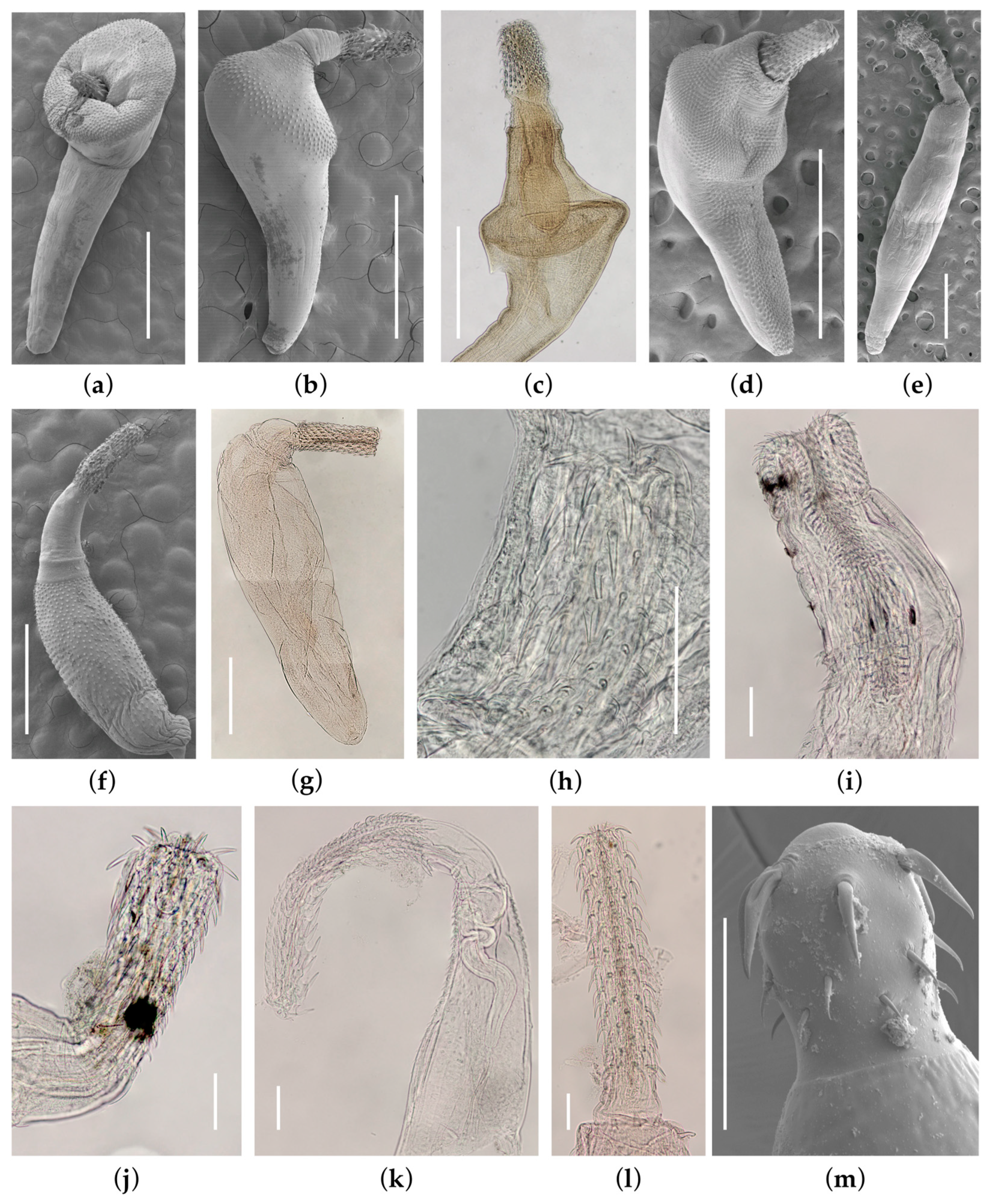
3.1.1. Andracantha leucocarboi Presswell, García-Varela & Smales, 2018 (Figure 1a)

In this survey, A. leucocarboi specimens were recovered from three definitive shag hosts (little pied Microcarbo melanoleucos (Gould), Otago Leucocarbo chalconotus (Gray), and spotted shags Phalacrocorax punctatus (Sparrman)). The prevalence of A. leucocarboi was highest in little pied shags (86%, n = 8), followed by Otago (50%, n = 16) and spotted (35%, n = 34) shags. Similarly, the intensity of A. leucocarboi was also highest in little pied shags with an average of 44 (1–100) individuals per infected shag, followed by spotted and Otago shags (mean intensity = 20 (1–90) and 5 (1–14), respectively). Andracantha leucocarboi was previously reported in the same hosts when originally described by Presswell et al. [31] and again when reported more recently by Presswell and Bennett [15].

3.1.2. Andracantha sigma Presswell, García-Varela & Smales, 2018 (Figure 1b)

This species was described from the spotted shag Phalacrocorax punctatus, Otago shag Leucocarbo chalconotus, and little blue penguin Eudyptula novaehollandiae (Stephens) (as E. minor Forster) [31], but no intermediate or paratenic hosts were known at the time. This study recovered cystacanths of A. sigma from the sprat Sprattus antipodum (Hector), as reported in Bennett et al. [22]. As of yet, no invertebrate first intermediate host is known for this species. Spotted shags hosted the highest prevalence and intensity of A. sigma (prevalence = 82%, n = 34, intensity = 40.5 individuals on average per infected host). On average, little blue penguins hosted 3.3 A. sigma individuals per infected host and Otago shags hosted 3.5 individuals with a prevalence of 47% and 38%, respectively, which was markedly lower than that of spotted shags.

3.1.3. Bolbosoma balaenae (Gmelin, 1790) Porta, 1908 (Figure 1c)

In this study, we recovered one specimen of an adult Bolbosoma balaenae infecting a little blue penguin, Eudyptula novaehollandiae, as reported and briefly described in Bennett et al. [32]. Bolbosoma balaenae usually occurs in baleen whales (Mysticeti) and is the largest of all the Bolbosoma species, reaching a length of over 200 mm in its natural hosts [38]. The single specimen from the little blue penguin was smaller and not mature, but it was found fully everted (a sign that it was alive when it passed into the intestine of the bird) and in good condition, so the penguin was undoubtedly an accidental host [32]. Bolbosoma balaenae was also found in one fish paratenic host (sprat Sprattus antipodum) and its first intermediate host, an amphipod Themisto sp., as first reported in Bennett et al. [22], which represents the complete life cycle elucidated for this species. A previous study found cystacanths in euphausiid shrimp Nyctiphanes couchii (Bell) from Spain [39], which suggests a relatively broad host choice for this species in its first intermediate host.

3.1.4. Corynosoma hannae Zdzitowiecki, 1984 (Figure 1d)

First described from a leopard seal Hydrurga leptonyx (Blainville) in Australia [40], the adult stage of C. hannae has since been reported in New Zealand from two pinnipeds: NZ fur sea lion Phocarctos hookeri (Gray) and NZ fur seal Arctophoca forstseri (Lesson) [41]. Adult C. hannae were recovered from an individual leopard seal and immature adults have also been recovered from the Otago shag (prevalence = 62.5%, n = 16 and intensity = 19.1) and spotted shag species (prevalence =14.7%, n = 34 and intensity = 1.4) and yellow-eyed penguins (prevalence = 50%, n = 6 and intensity = 78) [15,22,36]. Corynosoma hannae cystacanths have been previously reported from two flatfish species, brill Colistium guntheri (Hutton), and New Zealand sole Peltorhamphus novaehollandiae Günther [36,42] and, in this study, we reported an additional 10 paratenic fish hosts (see Table 1 and Bennett et al. [22]). Larval C. hannae exhibited the lowest host specificity of all acanthocephalans present in Otago that were recovered from 30% of all teleost fish species investigated in this study.

3.1.5. Profilicollis novaezelandensis Brockerhoff & Smales, 2002 (Figure 1e)

Profilicollis novaezelandensis was described from a South Island pied oystercatcher Haematopus finschi Martens and bar-tailed godwit Limosa lapponica (Linnaeus) [43]. The species has also been reported from two gull and two shag species [15,34,44]. Adult Profilicollis novaezelandensis were recovered in this survey from Royal spoonbill Platalea regia Gould (prevalence = 66.6%, n = 3 and mean intensity = 17.5), as reported in Presswell and Bennett [15], as well as all of the above definitive hosts. Cystacanths have been found in five different intertidal crab species [21]. Surprisingly, no new specimens were found of P. antarcticus Zdzitowiecki, 1985, which was previously described as common in oystercatchers Haematopus finschi Martens and H. unicolor Forster, and bar-tailed godwit Limosa lapponica L. [43]. We have dissected a number of oystercatchers without finding this species. Neither have we uncovered any specimens from the three crab species that are said to harbor this acanthocephalan [43].

3.1.6. Tenuisoma tarapungi Presswell, Bennett & Smales, 2020 (Figure 1f)

Tenuisoma tarapungi was originally described from the red-billed gull by Presswell et al. [35]. The present survey also recovered this acanthocephalan from a second definitive host species, the black-backed gull. Sequences of 28S rDNA from the latter were identical to those provided by Presswell et al. [35]. The prevalence was higher in red-billed gulls (16.6%) than in black-backed gulls (6.2%), although the intensity was relatively low for both species (mean intensity 2.7 and 1.6, respectively). No intermediate hosts have yet been identified for this species.

3.2. Class Palaeacanthocephala: Order Polymorphida Petrochenko, 1956: Family Plagiorhynchidae Golvan, 1960

Plagiorhynchus allisonae Smales 2002 (Figure 1g)

Plagiorhynchus allisonae Smales 2002, described from South Island pied oystercatcher Haematopus finschi, and variable oystercatcher H. unicolor [33], was also recovered in this survey from a pied stilt Himantopus himantopus (Linnaeus) which constitutes a new host record. Based on the data from this study, the mean intensity of P. allisonae was much higher in variable oystercatchers (439.5 based on 2/6 infected hosts) than South Island pied oystercatchers (60 based on 1/6 infected hosts) and pied stilts (5 based on 2/2 infected hosts). The intermediate host is known to be the amphipod Transorchestia serrulata (Dana) (formerly T. chilensis). [45]. Newly generated 28S sequences from a pied stilt matched, with 100% identity, those supplied in Lagrue et al. [45].

3.3. Class Palaeacanthocephala: Order Echinorhynchida Southwell & Macfie, 1925: Family Echinorhynchidae Cobbold, 1876

Echinorhynchus sp. (Figure 1j)

A single adult male specimen of an unknown Echinorhynchus species was recovered from a pigfish Congiopodus leucopaecilus (Richardson), constituting the first record of an acanthocephalan infection for this host. The specimen was in fair condition, but with the proboscis only partly inverted. The features were as follows: body length 5.49 mm, maximum width 720 µm; proboscis length 662, proboscis width 213; proboscis receptacle 959 long, 283 wide; hook rows 14, hooks per row 9–11; largest hooks 69 µm (dorsal) and 89 µm (ventral), smallest hooks 25 µm (dorsal) and 32 µm (ventral) long; testes in third quarter of body length, tandem; anterior testis 462 × 237, posterior testis 385 × 206. Comparing the hook rows and number per row, the size of hooks, size and shape of proboscis, position of testes, habitat, host, and locality with all other marine species currently accepted in the genus [46,47] leads to the conclusion that this specimen does not belong to any known named species. Further specimens are required to name and describe this species. Unfortunately, no DNA sequence could be obtained in this study.

3.4. Class Palaeacanthocephala: Order Echinorhynchida Southwell & Macfie, 1925: Family Heteracanthocephalidae Petrochenko, 1956

Aspersentis sp. (Figure 1h)

A single gravid female specimen belonging to genus Aspersentis was recovered from a NZ sole Peltorhamphus novaezeelandiae Günther in our survey. This species is morphologically closest to Aspersentis peltorhamphi (Baylis, 1944) (syns. Rhadinorhynchus peltorhamphi Baylis, 1944; Heteracanthocephalus peltorhamphi (Baylis, 1944) Petrochenko, 1956) also found and described from the NZ sole in 1944 [48], and more recently in Anglade and Randhawa in 2018 [42]. However, this species differs with considerably smaller proboscis hooks compared to those reported for A. peltoramphi and all other Aspersentis species [48,49,50,51,52,53]. Its features are as follows: body length 6 mm, maximum width 1.2 mm; proboscis 340 µm long; proboscis receptacle 818 µm long; hook rows 12–14, hooks in row 9 approx. 8; ventral hooks 35–62 µm, dorsal hooks 18–28 long; eggs 66 × 15 µm. These specimens, recovered from one of three NZ sole individuals, therefore appear to represent an undescribed species, although the material was not sufficient to allow a formal description at this time.

3.5. Class Palaeacanthocephala: Order Echinorhynchida Southwell & Macfie, 1925: Family Isthmosacanthidae Smales, 2012

Gorgorhynchoides queenslandensis Smales, 2014 (Figure 1i)

A specimen of Gorgorhynchoides queenslandensis was recovered from one lemon sole Pleotretis flavilatus Waite, out of six individuals investigated. The 18S and 28S sequences obtained here were identical to those of the same species from a yellowtail amberjack Seriola lalandi Valenciennes in Australia [54] (Genbank accessions MN705838 [18S] and MN705858 [28S]). This constitutes a new host record for lemon sole and a new geographic record for G. queenslandensis in New Zealand, extending its currently known range, and it represents the first record of any species within family Isthmosacanthidae in New Zealand.

3.6. Class Palaeacanthocephala: Order Echinorhynchida Southwell & Macfie, 1925: Family Leptorhynchoididae Witenberg, 1923

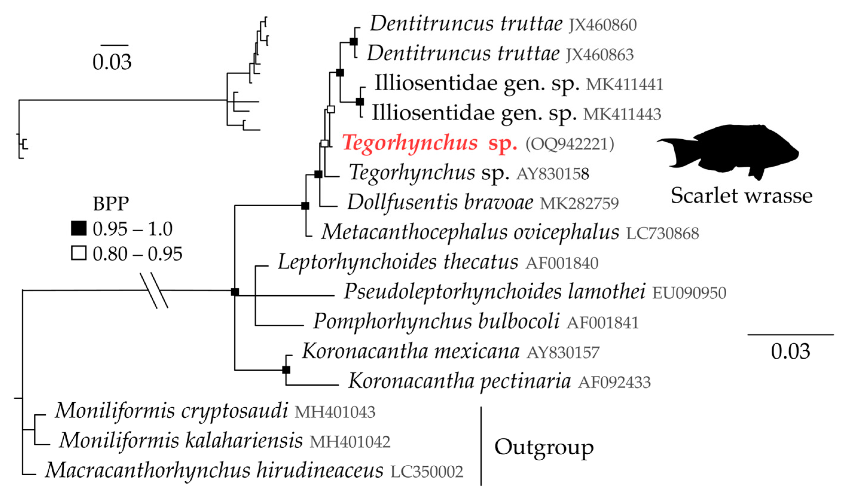
Tegorhynchus sp. (Figure 1k and Figure 3)

A single-part specimen belonging to family Leptorhynchoididae (syn. Illiosentidae) was recovered from a scarlet wrasse Pseudolabrus miles (Schneider & Forster). Morphologically, this specimen belongs to the genus Tegorhynchus (syn. Illiosentis see Kita et al. [55]), but the proboscis hook formation does not conform to any other species description and it is therefore assumed that this is an undescribed species. Formal description awaits the discovery of further specimens. Its total body length is not known. Its trunk spines extend from the neck to 590 µm (dorsal) and 450 µm (ventral) lengths of the body. Its other features are as follows: proboscis length 862 µm; proboscis receptacle length 724 µm; neck length 226 µm; proboscis hooks in 16 rows; 12+ hooks per row (ventral), 7 posterior spines and 20+ rooted hooks (dorsal); dorsal hooks 20–30 µm, ventral hooks 45–77 µm. Our 18S sequence of this species places Tegorhynchus sp. within Family Leptorhynchidae with high nodal support (Figure 3). The amplified 28S sequences place this species within Leptorhynchoididae with 0.54% genetic divergence to the closest sequence, an unnamed species of Tegorhynchus from an unknown host, provided by García-Varela and Nadler [56]. This is the first record of a species within family Leptorhynchoididae for New Zealand.
Figure 3. Bayesian phylogenetic tree of acanthocephalans of Family Leptorhynchoididae illustrating the position of Tegorhynchus sp. ex scarlet wrasse recovered in this study (in red), inferred from 18S gene data. Scale represents substitution per base. BPP denoted by black and black-outlined white squares.
Diversity 15 00665 g003

3.7. Class Palaeacanthocephala: Order Echinorhynchida Southwell & Macfie, 1925: Family Rhadinorhynchidae Lühe, 1911

Rhadinorhynchus sp. (Figure 1l)

A single male specimen of an unknown species of Rhadinorhynchus was found in a flesh-footed shearwater Ardenna carneipes (Gould). The species of this family are parasites of fish [57], so its discovery in a seabird was unexpected. It is assumed that this specimen was ingested with its fish host. The only fish reported as a host of Rhadinorhynchus in New Zealand is albacore tuna Thunnus alalunga, which was infected by Rhadinorhynchus sp. [58,59]. It is unlikely that this species from the shearwater is the same rhadinorhynchid as in the tuna species, as they are too large a prey item for shearwater to predate; thus, the identity of the fish host of Rhadinorhynchus sp. remains a mystery. Its features are as follows: total body length > 9 mm; trunk spines in two fields, with anterior field consisting of two circles of bluntly tipped spines, 47–58 µm long, and the second field being unclear due to damage; proboscis cylindrical, length 1737 µm; proboscis receptacle length 1792 µm; proboscis hooks in 14 rows of 20+ per row (estimated from partially inverted proboscis); dorsal hook 59–75 µm, ventral hooks 57–67 µm long; testes oval and tandem; anterior testis 880 µm long, 520 µm wide; posterior testis 754 µm long, 506 µm wide. Unfortunately, the specimen was too damaged to attempt identification to species level and the DNA amplification of remaining tissue was unsuccessful. We did not find any specimens of rhadinorhynchid in any of the pelagic fish sampled; therefore, this species remains intriguing because of its apparent rarity.

3.8. Class Eoacanthocephala: Order Neoechinorhynchida Southwell & Macfie, 1925: Family Neoechinorhynchidae (Ward, 1917)

Neoechinorhynchus sp. (Figure 1m and Figure 4)

Specimens of genus Neoechinorhynchus were recovered from 10 out of 11 yellow-eyed mullet Aldrichetta forsteri (Valenciennes) and 1 of 10 sprat Sprattus antipodum (Hector) in this survey, often in large numbers, with the largest infection comprising over 350 individuals in one mullet intestine. The specimens recovered from mullet and sprat were genetically identical at the level of the 28S gene. These specimens were closest morphologically to N. aldrichettae Edmonds 1971, described from the same host (A. forsteri) in Australia and reported also from New Zealand [60,61]. Recently, specimens collected in Tasmania and identified as N. aldrichettae from A. forsteri were redescribed along with DNA sequences presented by Huston et al. [62]. However, a partial 28S sequence for Huston et al.’s specimens (OM103598) exhibited 12.4% uncorrected pairwise genetic divergence from our newly produced sequences of 28S (Figure 4). Within our presented phylogeny, relationships were well supported within the family, with high support for topologies for most species presented, although Neoechinorhynchus appears paraphyletic (Figure 4). Neoechinorhynchus sp. recovered from sprat and mullet were also highly supported as sister taxa to N. aldrichettae from mullet in Australia (Figure 4). Based on our phylogenetic tree, Neoechinorhynchus species exhibit between 12.4 and 46.6% intraspecific genetic variation in 28S (uncorrected pairwise p distance). Reports from other studies illustrate interspecific divergences ranging from 1.65 to 32.9% [63], and so, based on this, the specimens recovered here may or may not represent a sister species to N. aldrichettae. For the purpose of this study we will consider them as Neoechinorhynchus sp. indet. In the overall size of the trunk, proboscis, and proboscis hooks, the range of measurements is within that given by Edmonds [60] and Huston et al. [62]. However, there are characters, such as the length of the lemnisci and proboscis receptacle, that appear to differ from the descriptions of N. aldrichettae.
Figure 4. Bayesian phylogenetic tree of acanthocephalans from Family Neoechinorhynchidae illustrating the position of Neoechinorhynchus sp. ex mullet and sprat recovered in this study (in red), inferred from 28S gene data. Scale represents substitution per base. BPP denoted by black and black-outlined white squares.
Diversity 15 00665 g004

4. Discussion

We recovered a phylogenetically diverse range of acanthocephalans infecting marine animals in Otago’s coastal marine ecosystem. In total, 13 acanthocephalan species were identified from 18 definitive or accidental host species and 16 intermediate host species, including 9 new host records, at least 1 new geographic record for New Zealand, at least 2 species new to science, and 17 newly generated DNA sequences. The data presented here provide insights into patterns of acanthocephalan biodiversity and host use across species in a coastal ecosystem. We hope these baseline data will provide a starting point for future comparisons pertaining to how acanthocephalan diversity and distributions may change in future.
The acanthocephalans recovered may have a range of impacts on their hosts’ biology and ecology. These impacts are well documented for larval acanthocephalans within invertebrate intermediate and fish paratenic hosts, including species recovered here such as Profilicollis. [11,12]. Such influences are likely exerted in all acanthocephalan host-associations identified here of intermediate/paratenic hosts and larval acanthocephalans. Lesser known are the impacts of adult acanthocephalans on their definitive hosts, especially in seabirds. Various polymorphids, including species recovered here, have previously been associated with host mortality, especially in high infection intensities where acanthocephalans penetrate the intestinal wall causing peritonitis [15,64]. Sub-lethal impacts of acanthocephalans in definitive hosts are much less documented, perhaps because they are difficult to quantify, although consideration should undoubtedly be given to such subtle impacts [65].
A large proportion of species recovered belonged to Family Polymorphidae (6 of 13 species), of which all were identified to a species level. The seven remaining acanthocephalans include five that remain taxonomically unresolved (i.e., only identified to genus level). This taxonomic imbalance may be in part due to the authors’ previous experience in avian polymorphid systematics (e.g., [31,35]). A lack of taxonomic resolution is also a result of a paucity of specimens; some acanthocephalans have low prevalence and intensity in the studied definitive hosts compared to the polymorphids, which were particularly prevalent with sometimes high intensities. In fact, four of the five unresolved species consisted of only an individual specimen within one definitive host individual. Therefore, although a large effort was expended here to identify acanthocephalans from a range of marine animals within one whole coastal ecosystem, much work is still required to provide a resolution of the species recovered here, in particular those acanthocephalans that infect fish.
Taxonomic resolution is not the only limiting factor in our characterization of acanthocephalan biodiversity within Otago, or even New Zealand’s marine environment. Based on the data obtained here, is it likely that all acanthocephalans occurring in Otago’s coastal marine ecosystem, or New Zealand, have been discovered? For acanthocephalans that use fish as definitive hosts, by dissecting 40 fish species in this survey, we recovered at least 2 new species. Considering that there are over 1100 species of marine fish in New Zealand, most of which are yet to be investigated for their parasitic fauna [16], a simple extrapolation means that there could be as many as 57 acanthocephalan species infecting just marine fish in New Zealand yet to be discovered, which would almost double the currently known total species in New Zealand [16]. This only takes into account acanthocephalans maturing in fish definitive hosts, not those species infecting marine mammals and seabirds. Only further large-scale biodiversity surveys can allow an estimate of the actual number of acanthocephalans present in New Zealand marine animals.
Investigations into a wide range of definitive hosts have provided insights into patterns of acanthocephalan host specificity. With the exception of B. balaenae, all polymorphids infected between two and four definitive hosts, and these definitive hosts were exclusively coastal seabirds, except for C. hannae, which was also identified from a leopard seal. Conversely, acanthocephalans maturing in fish appeared to exhibit a higher host specificity with most species infecting only one fish host, e.g., Echinorhynchus sp., Gorgorhynchoides queenslandensis, and Tegorhynchus sp. Not only do these data provide insights into which species are acanthocephalan hosts, but they also indicate which host species are not yet considered hosts of acanthocephalans. Of course, low sample sizes limit our ability to conclude which free-living taxa do not host acanthocephalans; however, these findings provide a starting point to answer this question.
Once it is established which free-living species are infected with acanthocephalan species, it is possible to gain insight into which host species may be more important than others, or which are likely “natural” hosts as opposed to accidental ones. To determine this, we must look towards the prevalence and intensity data, which in this study vary a lot between and within acanthocephalan taxa. For instance, as mentioned earlier, fish acanthocephalans tended to have high host specificity and so only infected one or two definitive hosts. Acanthocephalans infecting seabirds, however, vary in their prevalence and intensity between different host species. For instance, spotted shags, Otago shags, and little blue penguins are currently the only known definitive hosts of A. sigma. However, the prevalence in spotted shags is much higher (82%) than in Otago shags and little blue penguins (38 and 47% prevalence, respectively). The intensity shows a similar pattern with an average of 40 individuals found per infected spotted shag compared to less than 4 individuals per infected Otago shag and little blue penguins. This suggests that spotted shags are likely the preferred host of A. sigma. Alternatively, this pattern could indicate that spotted shags simply consume more of the paratenic or intermediate hosts in their diet, or that spotted shag immune systems are not able to prevent acanthocephalan establishment compared to other potential hosts [66]. Regardless of the processes determining which host species are most appropriate for acanthocephalan infection, our study highlights that infection parameters can provide insights into the factors that determine which host species are more likely to be infected or not.
Although acanthocephalans constitute a relatively small phylum of parasitic helminths, they deserve effort to document their occurrence for their contribution to natural biodiversity and their impacts on host populations. Carlson et al. [67] estimated that by 2070, up to 5% of all currently known acanthocephalan species will be committed to extinction. Without baseline inventories such as this, our ability to understand how patterns of acanthocephalan diversity and distribution will change in response to natural and anthropogenic pressures may be greatly limited.

Author Contributions

J.B., B.P. and R.P. conceived and designed the study. J.B. and B.P. conducted data gathering. J.B. and B.P. wrote the article with input from R.P. All authors have read and agreed to the published version of the manuscript.

Funding

This research was funded in part by a Ministry of Business, Innovation and Employment Endeavour Fund, Emerging Aquatic Diseases: A novel diagnostic pipeline and management framework, Award number CAWX2207, Recipient: Cawthron Institute, in part by an Otago University PhD scholarship to J Bennett, and in part by Zoology PBRF funds to B Presswell and R Poulin.

Institutional Review Board Statement

This work complies with the University of Otago animal ethics standards under Animal Use Protocol AUP-19-190 and a Department of Conservation permit 65658-DOA.

Data Availability Statement

All DNA sequences generated in this study are available for download on GenBank. Figure 1a–e scanning electron microscopy and photo micrograph images have been previously published in publications authors JB and/or BP are authors of, including Presswell et al. [31], Bennett et al. [32], Hernández-Orts et al. [36], and Presswell et al. [35].

Acknowledgments

We thank Philippe Vieira Alves and José Luis Luque for inviting us to contribute to the Diversity Special Issue on ‘Taxonomy, Biodiversity and Ecology of Parasites of Aquatic Organisms’. We thank all the persons who provided specimens for this project including Pablo Escobar-Flores, Darren Stevens, and other scientists and crew aboard Kaharoa East Coast and Tangaroa Chatham Rise trawl surveys from the National Institute of Water and Atmospheric Research; Lisa Argilla, Angelina Martelli, and Emily Brewer from the Dunedin Wildlife Hospital; Biz Bell and the team from Wildlife Management International; Clément Lagrue, Karen Middlemiss, and others from the Department of Conservation; and Rob Lewis, Adelle Heineman, Sally Carson, Bill Dickson, and others from the Marine Studies Centre, Department of Marine Science, University of Otago. We also thank Jan Littleton, Shelley Cameron, and Tania King for their assistance in the lab, and Fátima Jorge for her assistance in developing ideas for this project. We also thank Kate Hutson, Ian Davidson, and Patrick Cahill for their support in the production of this manuscript through a collaboration between Otago University and the Cawthron Institute.

Conflicts of Interest

The authors declare no conflict of interest.

References

  1. Butchart, S.H.M.; Walpole, M.; Collen, B.; van Strien, A.; Scharlemann, J.P.W.; Almond, R.E.A.; Baillie, J.E.M.; Bomhard, B.; Brown, C.; Bruno, J.; et al. Global Biodiversity: Indicators of Recent Declines. Science 2010, 328, 1164–1168. [Google Scholar] [CrossRef] [PubMed]
  2. Cowart, D.A.; Pinheiro, M.; Mouchel, O.; Maguer, M.; Grall, J.; Miné, J.; Arnaud-Haond, S. Metabarcoding Is Powerful yet Still Blind: A Comparative Analysis of Morphological and Molecular Surveys of Seagrass Communities. PLoS ONE 2015, 10, e0117562. [Google Scholar] [CrossRef] [PubMed]
  3. Titley, M.A.; Snaddon, J.L.; Turner, E.C. Scientific Research on Animal Biodiversity Is Systematically Biased towards Vertebrates and Temperate Regions. PLoS ONE 2017, 12, e0189577. [Google Scholar] [CrossRef] [PubMed]
  4. Lafferty, K.D.; Dobson, A.P.; Kuris, A.M. Parasites Dominate Food Web Links. Proc. Natl. Acad. Sci. USA 2006, 103, 11211–11216. [Google Scholar] [CrossRef]
  5. Wood, C.L.; Byers, J.E.; Cottingham, K.L.; Altman, I.; Donahue, M.J.; Blakeslee, A.M.H. Parasites Alter Community Structure. Proc. Natl. Acad. Sci. USA 2007, 104, 9335–9339. [Google Scholar] [CrossRef]
  6. Kuris, A.M.; Hechinger, R.F.; Shaw, J.C.; Whitney, K.L.; Aguirre-Macedo, L.; Boch, C.A.; Dobson, A.P.; Dunham, E.J.; Fredensborg, B.L.; Huspeni, T.C.; et al. Ecosystem Energetic Implications of Parasite and Free-Living Biomass in Three Estuaries. Nature 2008, 454, 515–518. [Google Scholar] [CrossRef]
  7. Thompson, R.M.; Mouritsen, K.N.; Poulin, R. Importance of Parasites and Their Life Cycle Characteristics in Determining the Structure of a Large Marine Food Web. J. Anim. Ecol. 2005, 74, 77–85. [Google Scholar] [CrossRef]
  8. Dunne, J.A.; Lafferty, K.D.; Dobson, A.P.; Hechinger, R.F.; Kuris, A.M.; Martinez, N.D.; McLaughlin, J.P.; Mouritsen, K.N.; Poulin, R.; Reise, K.; et al. Parasites Affect Food Web Structure Primarily through Increased Diversity and Complexity. PLoS Biol. 2013, 11, e1001579. [Google Scholar] [CrossRef]
  9. Sato, T.; Egusa, T.; Fukushima, K.; Oda, T.; Ohte, N.; Tokuchi, N.; Watanabe, K.; Kanaiwa, M.; Murakami, I.; Lafferty, K.D. Nematomorph Parasites Indirectly Alter the Food Web and Ecosystem Function of Streams through Behavioural Manipulation of Their Cricket Hosts. Ecol. Lett. 2012, 15, 786–793. [Google Scholar] [CrossRef]
  10. Kennedy, C.R. Ecology of the Acanthocephala; Cambridge University Press: New York, NY, USA, 2006. [Google Scholar]
  11. Haye, P.A.; Ojeda, F.P. Metabolic and Behavioral Alterations in the Crab Hemigrapsus crenulatus (Milne-Edwards 1837) Induced by Its Acanthocephalan Parasite Profilicollis antarcticus (Zdzitowiecki 1985). J. Exp. Mar. Biol. Ecol. 1998, 228, 73–82. [Google Scholar] [CrossRef]
  12. Latham, A.; Poulin, R. Effect of Acanthocephalan Parasites on the Behaviour and Coloration of the Mud Crab Macrophthalmus hirtipes (Brachyura: Ocypodidae). Mar. Biol. 2001, 139, 1147–1154. [Google Scholar] [CrossRef]
  13. Richardson, D.J.; Nickol, B.B. Acanthocephala. In Parasitic Diseases of Wild Birds; Atkinson, C.T., Thomas, N.J., Hunter, D.B., Eds.; Wiley-Blackwell: Oxford, UK, 2009; pp. 277–288. ISBN 978-0-8138-0462-0. [Google Scholar]
  14. Shanebeck, K.M.; Thacker, C.; Lagrue, C. Corynosoma strumosum (Acanthocephala) Infection in Marine Foraging Mink (Neogale vison) and River Otter (Lontra canadensis) and Associated Peritonitis in a Juvenile Mink. Parasitol. Int. 2022, 89, 102579. [Google Scholar] [CrossRef]
  15. Presswell, B.; Bennett, J. Helminth Parasites of Shags (Phalacrocoracidae) from the Otago Region of Southern New Zealand. J. Helminthol. 2021, 95, e9. [Google Scholar] [CrossRef]
  16. Bennett, J.; Presswell, B.; Poulin, R. Biodiversity of Marine Helminth Parasites in New Zealand: What Don’t We Know? N. Z. J. Mar. Freshw. Res. 2022, 56, 175–190. [Google Scholar] [CrossRef]
  17. Bennett, J.; Poulin, R.; Presswell, B. Large-Scale Genetic Investigation of Nematode Diversity and Their Phylogenetic Patterns in New Zealand’s Marine Animals. Parasitology 2022, 149, 1794–1809. [Google Scholar] [CrossRef]
  18. Faltýnková, A.; Georgieva, S.; Kostadinova, A.; Bray, R.A. Biodiversity and Evolution of Digeneans of Fishes in the Southern Ocean. In Biodiversity and Evolution of Parasitic Life in the Southern Ocean; Klimpel, S., Kuhn, T., Mehlhorn, H., Eds.; Parasitology Research Monographs; Springer International Publishing: Cham, Switzerland, 2017; Volume 9, pp. 49–75. ISBN 978-3-319-46342-1. [Google Scholar]
  19. Mattiucci, S.; Cipriani, P.; Paoletti, M.; Levsen, A.; Nascetti, G. Reviewing Biodiversity and Epidemiological Aspects of Anisakid Nematodes from the North-East Atlantic Ocean. J. Helminthol. 2017, 91, 422–439. [Google Scholar] [CrossRef]
  20. Santacruz, A.; Barluenga, M.; Pérez-Ponce de León, G. The Macroparasite Fauna of Cichlid Fish from Nicaraguan Lakes, a Model System for Understanding Host–Parasite Diversification and Speciation. Sci. Rep. 2022, 12, 3944. [Google Scholar] [CrossRef]
  21. Bennett, J.; Poulin, R.; Presswell, B. Annotated Checklist and Genetic Data for Parasitic Helminths Infecting New Zealand Marine Invertebrates. Invertebr. Biol. 2022, 141, e12380. [Google Scholar] [CrossRef]
  22. Bennett, J.; Presswell, B.; Poulin, R. Tracking Life Cycles of Parasites across a Broad Taxonomic Scale in a Marine Ecosystem. Int. J. Parasitol. 2023; in press. [Google Scholar] [CrossRef]
  23. Otago Regional Council. Regional Plan: Coast of Otago; Otago Regional Council: Dunedin, New Zealand, 2012; p. 196. [Google Scholar]
  24. Harper, J.T.; Saunders, G.W. Molecular Systematics of the Florideophyceae (Rhodophyta) Using Nuclear Large and Small Subunit RDNA Sequence Data. J. Phycol. 2001, 37, 1073–1082. [Google Scholar] [CrossRef]
  25. Bowles, J.; Blair, D.; Mcmanus, D. Genetic Variants within the Genus Echinococcus Identified by Mitochondrial DNA Sequencing. Mol. Biochem. Parasitol. 1992, 54, 165–173. [Google Scholar] [CrossRef]
  26. Littlewood, D.T.J.; Olson, P.D. Small Subunit RDNA and the Platyhelminthes: Signal, Noise, Conflict and Compromise. In Interrelationships of the Platyhelminthes; CRC Press: London, UK, 2001; p. 376. [Google Scholar]
  27. Miller, M.A.; Pfeiffer, W.; Schwartz, T. Creating the CIPRES Science Gateway for Inference of Large Phylogenetic Trees. In Proceedings of the 2010 Gateway Computing Environments Workshop (GCE), New Orleans, LA, USA, 14 November 2010; pp. 1–8. [Google Scholar]
  28. Rambaut, A.; Drummond, A.J.; Xie, D.; Baele, G.; Suchard, M.A. Posterior Summarization in Bayesian Phylogenetics Using Tracer 1.7. Syst. Biol. 2018, 67, 901–904. [Google Scholar] [CrossRef] [PubMed]
  29. Rambaut, A. FigTree; Institute of Evolutionary Biology, University of Edinburgh: Edinburgh, UK, 2010. [Google Scholar]
  30. Tamura, K.; Stecher, G.; Kumar, S. MEGA11: Molecular Evolutionary Genetics Analysis Version 11. Mol. Biol. Evol. 2021, 38, 3022–3027. [Google Scholar] [CrossRef] [PubMed]
  31. Presswell, B.; García-Varela, M.; Smales, L.R. Morphological and Molecular Characterization of Two New Species of Andracantha (Acanthocephala: Polymorphidae) from New Zealand Shags (Phalacrocoracidae) and Penguins (Spheniscidae) with a Key to the Species. J. Helminthol. 2018, 92, 740–751. [Google Scholar] [CrossRef] [PubMed]
  32. Bennett, J.; McPherson, O.; Presswell, B. Gastrointestinal Helminths of Little Blue Penguins, Eudyptula novaehollandiae (Stephens), from Otago, New Zealand. Parasitol. Int. 2021, 80, 102185. [Google Scholar] [CrossRef] [PubMed]
  33. Smales, L.R. Plagiorhynchidae Meyer, 1931 (Acanthocephala) from Australasian Birds and Mammals, with Descriptions of Plagiorhynchus (Plagiorhynchus) menurae (Johnston, 1912) and P. (P.) allisonae n. Sp. Syst. Parasitol. 2002, 51, 207–216. [Google Scholar] [CrossRef]
  34. Latham, A.D.M.; Poulin, R. New Records of Gastrointestinal Helminths from the Southern Black-backed Gull (Larus dominicanus) in New Zealand. N. Z. J. Zool. 2002, 29, 253–257. [Google Scholar] [CrossRef]
  35. Presswell, B.; Bennett, J.D.L.; Smales, L.R. Morphological and Molecular Characterisation of a New Genus and Species of Acanthocephalan, Tenuisoma tarapungi n. g., n. Sp. (Acanthocephala: Polymorphidae) Infecting Red-Billed Gulls in New Zealand, with a Key to the Genera of the Polymorphidae Meyer, 1931. Syst. Parasitol. 2020, 97, 25–39. [Google Scholar] [CrossRef]
  36. Hernández-Orts, J.S.; Smales, L.R.; Pinacho-Pinacho, C.D.; García-Varela, M.; Presswell, B. Novel Morphological and Molecular Data for Corynosoma hannae Zdzitowiecki, 1984 (Acanthocephala: Polymorphidae) from Teleosts, Fish-Eating Birds and Pinnipeds from New Zealand. Parasitol. Int. 2017, 66, 905–916. [Google Scholar] [CrossRef]
  37. García-Varela, M.; Pérez-Ponce de León, G.; Aznar, F.J.; Nadler, S.A. Phylogenetic Relationship among Genera of Polymorphidae (Acanthocephala), Inferred from Nuclear and Mitochondrial Gene Sequences. Mol. Phylogenet. Evol. 2013, 68, 176–184. [Google Scholar] [CrossRef]
  38. Santoro, M.; Palomba, M.; Gili, C.; Marcer, F.; Marchiori, E.; Mattiucci, S. Molecular and Morphological Characterization of Bolbosoma balaenae (Acanthocephala: Polymorphidae), a Neglected Intestinal Parasite of the Fin Whale Balaenoptera physalus. Parasitology 2021, 148, 1293–1302. [Google Scholar] [CrossRef]
  39. Gregori, M.; Aznar, F.; Abollo, E.; Roura, Á.; González, Á.; Pascual, S. Nyctiphanes couchii as Intermediate Host for the Acanthocephalan Bolbosoma balaenae in Temperate Waters of the NE Atlantic. Dis. Aquat. Organ. 2012, 99, 37–47. [Google Scholar] [CrossRef]
  40. Zdzitowiecki, K. Some Antarctic Acanthocephalans of the Genus Corynosoma Parasitizing Pinnipedia, with Descriptions of Three New Species. Acta Parasitol. Pol. 1984, 29, 359–377. [Google Scholar]
  41. Aznar, F.J.; Hernández-Orts, J.; Suárez, A.A.; García-Varela, M.; Raga, J.A.; Cappozzo, H.L. Assessing Host–Parasite Specificity through Coprological Analysis: A Case Study with Species of Corynosoma (Acanthocephala: Polymorphidae) from Marine Mammals. J. Helminthol. 2012, 86, 156–164. [Google Scholar] [CrossRef]
  42. Anglade, T.; Randhawa, H. Gaining Insights into the Ecological Role of the New Zealand Sole (Peltorhamphus novaezeelandiae) through Parasites. J. Helminthol. 2018, 92, 187–196. [Google Scholar] [CrossRef]
  43. Brockerhoff, A.M.; Smales, L.R. Profilicollis novaezelandensis n. Sp. (Polymorphidae) and Two Other Acanthocephalan Parasites from Shore Birds (Haematopodidae and Scolopacidae) in New Zealand, with Records of Two Species in Intertidal Crabs (Decapoda: Grapsidae and Ocypodidae). Syst. Parasitol. 2002, 52, 55–65. [Google Scholar] [CrossRef]
  44. Fredensborg, B.L.; Latham, A.D.; Poulin, R. New Records of Gastrointestinal Helminths from the Red-billed Gull (Larus novaehollandiae scopulinus). N. Z. J. Zool. 2004, 31, 75–80. [Google Scholar] [CrossRef]
  45. Lagrue, C.; Heaphy, K.; Presswell, B.; Poulin, R. Strong Association between Parasitism and Phenotypic Variation in a Supralittoral Amphipod. Mar. Ecol. Prog. Ser. 2016, 553, 111–123. [Google Scholar] [CrossRef]
  46. WoRMS Editorial Board World Register of Marine Species. Available online: https://www.marinespecies.org (accessed on 20 January 2022).
  47. Smales, L.R. Echinorhynchus lesteri N. SP. (Acanthocephala: Echinorhynchidae) Parasitic in the Intestine of the Orange Roughy Hoplostethus atlanticus. Trans. R. Soc. S. Aust. 2012, 136, 50–55. [Google Scholar] [CrossRef]
  48. Baylis, H.A. XLIII.—Three New Acanthocephala from Marine Fishes of Australasia. Ann. Mag. Nat. Hist. 1944, 11, 462–472. [Google Scholar] [CrossRef]
  49. Van Cleave, H.J. A New Genus and New Species of Acanthocephala from the Antarctic. Ann. Mag. Nat. Hist. 1929, 4, 229–331. [Google Scholar] [CrossRef]
  50. Pichelin, S.; Smales, L.; Bray, R.A. A Discussion on the Heteracanthocephalidae Petrochenko, 1956 (Acanthocephala: Palaeacanthocephala). Syst. Parasitol. 2002, 52, 145–152. [Google Scholar] [CrossRef] [PubMed]
  51. Edmonds, S.J.; Smales, L.R. A New Species of Acanthocephala from the Greenback Flounder, Rhombosolea tapirina Günther, 1862. Trans. R. Soc. S. Aust. 1992, 116, 35–38. [Google Scholar]
  52. Baylis, H.A. Parasitic Nematoda and Acanthocephala Collected in 1925–1927. Discov. Rep. 1929, 1, 541–560. [Google Scholar]
  53. Smales, L.R. A Redescription of Aspersentis zanclorhynchi (Johnston and Best, 1937) Comb. Nov. (Heteracanthocephalidae: Acanthocephala). Trans. R. Soc. S. Aust. 1996, 120, 167–171. [Google Scholar]
  54. Smales, L.R. New Species of Gorgorhynchoides (Acanthocephala: Isthmosacanthidae) from Australian Fish (Carangidae) with a Key to the Species of the Genus. Comp. Parasitol. 2014, 81, 240–247. [Google Scholar] [CrossRef]
  55. Kita, Y.; Hiruta, S.F.; Sasaki, M.; Kajihara, H. Systematic Position of the Genus Metacanthocephalus Yamaguti, 1959 (Palaeacanthocephala: Echinorhynchida) Inferred from Molecular Evidence, with a Redescription of Metacanthocephalus ovicephalus (Zhukov, 1960). Parasitol. Int. 2023, 94, 102731. [Google Scholar] [CrossRef]
  56. García-Varela, M.; Nadler, S.A. Phylogenetic Relationships of Palaeacanthocephala (Acanthocephala) Inferred from SSU and LSU RDNA Gene Sequences. J. Parasitol. 2005, 91, 1401–1409. [Google Scholar] [CrossRef]
  57. Smales, L. The Genus Rhadinorhynchus (Acanthocephala: Rhadinorhynchidae) from Marine Fish in Australia with the Description of Four New Species. Acta Parasitol. 2014, 59, 721–736. [Google Scholar] [CrossRef]
  58. Jones, J.B. Movements of Albacore Tuna (Thunnus alalunga) in the South Pacific: Evidence from Parasites. Mar. Biol. 1991, 111, 1–9. [Google Scholar] [CrossRef]
  59. Gibson, D.; Jones, J.B. Fed up with Parasites? A Method for Estimating Asymptotic Growth in Fish Populations. Mar. Biol. 1993, 117, 495–500. [Google Scholar] [CrossRef]
  60. Edmonds, S.J. Australian Acanthocephala No. 13: Three New Species. Trans. R. Soc. S. Aust. 1971, 95, 55–60. [Google Scholar]
  61. Boustead, N.C. Fish Diseases Recorded in New Zealand, with a Discussion on Potential Sources and Gertification Procedures; Fisheries Research Division Occasional Publication; New Zealand Ministry of Agriculture and Fisheries: Wellington, New Zealand, 1982. [Google Scholar]
  62. Huston, D.C.; Cutmore, S.C.; Smales, L.R. Molecular Characterisation and Updated Description of Neoechinorhynchus aldrichettae Edmonds, 1971 (Acanthocephala: Neoechinorhynchidae), Based on Material from Aldrichetta Forsteri (Valenciennes) Collected in Tasmania, Australia. Syst. Parasitol. 2022, 99, 241–251. [Google Scholar] [CrossRef]
  63. Pinacho-Pinacho, C.D.; Sereno-Uribe, A.L.; García-Varela, M. Morphological and Molecular Data Reveal a New Species of Neoechinorhynchus (Acanthocephala: Neoechinorhynchidae) from Dormitator maculatus in the Gulf of Mexico. Parasitol. Int. 2014, 63, 763–771. [Google Scholar] [CrossRef]
  64. Shanebeck, K.; Lakemeyer, J.; Siebert, U.; Lehnert, K. Novel Infections of Corynosoma enhydri and Profilicollis sp. (Acanthocephala: Polymorphidae) Identified in Sea Otters Enhydra lutris. Dis. Aquat. Organ. 2020, 137, 239–246. [Google Scholar] [CrossRef]
  65. Shanebeck, K.M.; Besson, A.A.; Lagrue, C.; Green, S.J. The Energetic Costs of Sub-lethal Helminth Parasites in Mammals: A Meta-analysis. Biol. Rev. 2022, 97, 1886–1907. [Google Scholar] [CrossRef]
  66. Thompson, R.M.; Poulin, R.; Mouritsen, K.N.; Thieltges, D.W. Resource Tracking in Marine Parasites: Going with the Flow? Oikos 2013, 122, 1187–1194. [Google Scholar] [CrossRef]
  67. Carlson, C.J.; Burgio, K.R.; Dougherty, E.R.; Phillips, A.J.; Bueno, V.M.; Clements, C.F.; Castaldo, G.; Dallas, T.A.; Cizauskas, C.A.; Cumming, G.S.; et al. Parasite Biodiversity Faces Extinction and Redistribution in a Changing Climate. Sci. Adv. 2017, 3, e1602422. [Google Scholar] [CrossRef]
Figure 2. Bayesian phylogenetic tree of acanthocephalans of Family Polymorphidae infecting marine animals in Otago’s coastal marine ecosystem, inferred from concatenated cox1, 18S, and 28S gene data. Each colored species was found in Otago and each silhouette represents a host (black silhouettes denote definitive hosts and grey silhouettes denote intermediate or paratenic hosts). Scale represents substitution per base. BPP denoted by black and black-outlined white squares. Genbank accession numbers follow each taxon name as cox1/18S/28S.
Diversity 15 00665 g002
Disclaimer/Publisher’s Note: The statements, opinions and data contained in all publications are solely those of the individual author(s) and contributor(s) and not of MDPI and/or the editor(s). MDPI and/or the editor(s) disclaim responsibility for any injury to people or property resulting from any ideas, methods, instructions or products referred to in the content.

