Article

Analysis of the Anomalies in a Middle Eocene Shell of Neochelys (Pleurodira, Podocnemididae) from the Duero Basin (Zamora, Spain)

by
Andrea Guerrero
1,2,*,
Francisco Ortega
1,
Santiago Martín de Jesús
3 and
Adán Pérez-García
1,*
1
Grupo de Biología Evolutiva, Facultad de Ciencias, Universidad Nacional de Educación a Distancia, Avda. de Esparta s/n, Las Rozas, 28232 Madrid, Spain
2
Escuela Internacional de Doctorado, Universidad Nacional de Educación a Distancia, Calle Bravo Murillo, 38, 28015 Madrid, Spain
3
Colección de Vertebrados Fósiles de la Cuenca del Duero-Sala de las Tortugas de la Universidad de Salamanca, Departamento de Geología, Facultad de Ciencias, Universidad de Salamanca, 37008 Salamanca, Spain
*
Authors to whom correspondence should be addressed.
Diversity 2023, 15(3), 314; https://doi.org/10.3390/d15030314
Submission received: 31 December 2022 / Revised: 3 February 2023 / Accepted: 13 February 2023 / Published: 21 February 2023
(This article belongs to the Special Issue Fossil Reptiles and Associated Faunal Record)

Abstract

:
Turtle shells frequently exhibit anomalous osseous modifications on their surface which can sometimes compromise the survival of the organism. Nowadays, despite the large number of anomalies identified in both extant and extinct turtle shells, the etiology, as well as the pathogenesis, of the various osseous modifications remains unknown in most documented extinct representatives. In fact, the interpretation of these anomalies in most fossil turtles is often speculative, the great majority of anomalous osseous modifications being attributed to vertebrate feeding traces, without considering other potential causative agents. In this context, we herein re-analyzed the shell anomalies recognized in an individual determined as Neochelys sp. (Pleurodira, Podocnemididae) from the middle Eocene (Lutetian) of the El Tejar fossil site (Corrales del Vino, Zamora, Spain), previously proposed as traumatic injuries resulting from a crocodile attack. The re-evaluation of these osseous alterations through detailed physical examination, comparison through direct observation and from the literature on extant turtles, and the use of the files obtained from a computerized axial tomography scan, allows the proposal of diverse causal agents, none of them supporting the previous interpretation. In addition, information regarding the pathogenesis and stages of the healing of the shell anomalies studied herein is provided.

1. Introduction

Turtle shells are frequently characterized by exhibiting anomalous osseous modifications on their surface (e.g., pitting, [1]; grooves, [2]; scratches, [3]; punctures, [4]; and lacerations, [5]), which can be variable both in depth (i.e., severity) and extent (i.e., surface area). Such modifications have been recorded in a wide variety of both extant and extinct turtle taxa, with different ecological adaptations (i.e., terrestrial, [6,7]; freshwater, [8,9]; and fully aquatic, [10,11]). However, despite their high occurrence in the different turtle lineages, the etiology, as well as the pathogenesis, of most osseous modifications remain unknown for most extinct representatives in which they have been evidenced. In this sense, studies of anomalous shell modifications performed in extant turtles are generally characterized as taxon-specific reports (e.g., [12]), without detailed morphological descriptions of the lesions (e.g., [13]), illustrations of the osseous modifications (e.g., [14]), or discussions concerning pathogenesis (e.g., [15]). The absence of such data in the literature on extant turtles makes it difficult to carry out comparative studies with shell anomalies identified in extinct specimens. As a consequence, nowadays, it is relatively complex to achieve reliable interpretations and diagnoses of anomalous shell modifications in the turtle fossil record. The lack of understanding of the significance of these shell anomalies, together with difficulty in their justification and interpretation of the fossil record, makes diagnoses in extinct specimens speculative. Thus, the etiology for most of the shell anomalies is generally attributed to vertebrate feeding traces, without considering other potential factors. However, this supposed unequivocal attribution can be wrong in many cases.
In this context, here, we performed a detailed study of the anomalous conditions (i.e., different typologies of shell marks and the anomalous lack of the posterior region of the carapace) of an individual (Figure 1A and Figure 2A) determined as Neochelys sp. (Pleurodira, Podocnemididae), from the middle Eocene (Lutetian) of the El Tejar fossil site, at Corrales del Vino (Zamora, Spain) ([16], Figure 1, Figure 2 and Figure 3). The anomalies of this specimen were previously analyzed and diagnosed as traumatic injuries resulting from a crocodile attack, being interpreted as non-fatal for the turtle ([17]; for more details see the Discussion section). However, the re-study of these bone alterations through a detailed physical examination, comparison through direct observation and with the literature on extant turtles, and the use of a computerized axial tomography scan allows us to refute the previous hypothesis, proposing the combination of several causal agents. Thus, this study focused on improving our understanding of the significance (i.e., etiology and pathogenesis) of anomalous turtle shell modifications in the fossil record.

2. Materials and Methods

Institutional Abbreviations
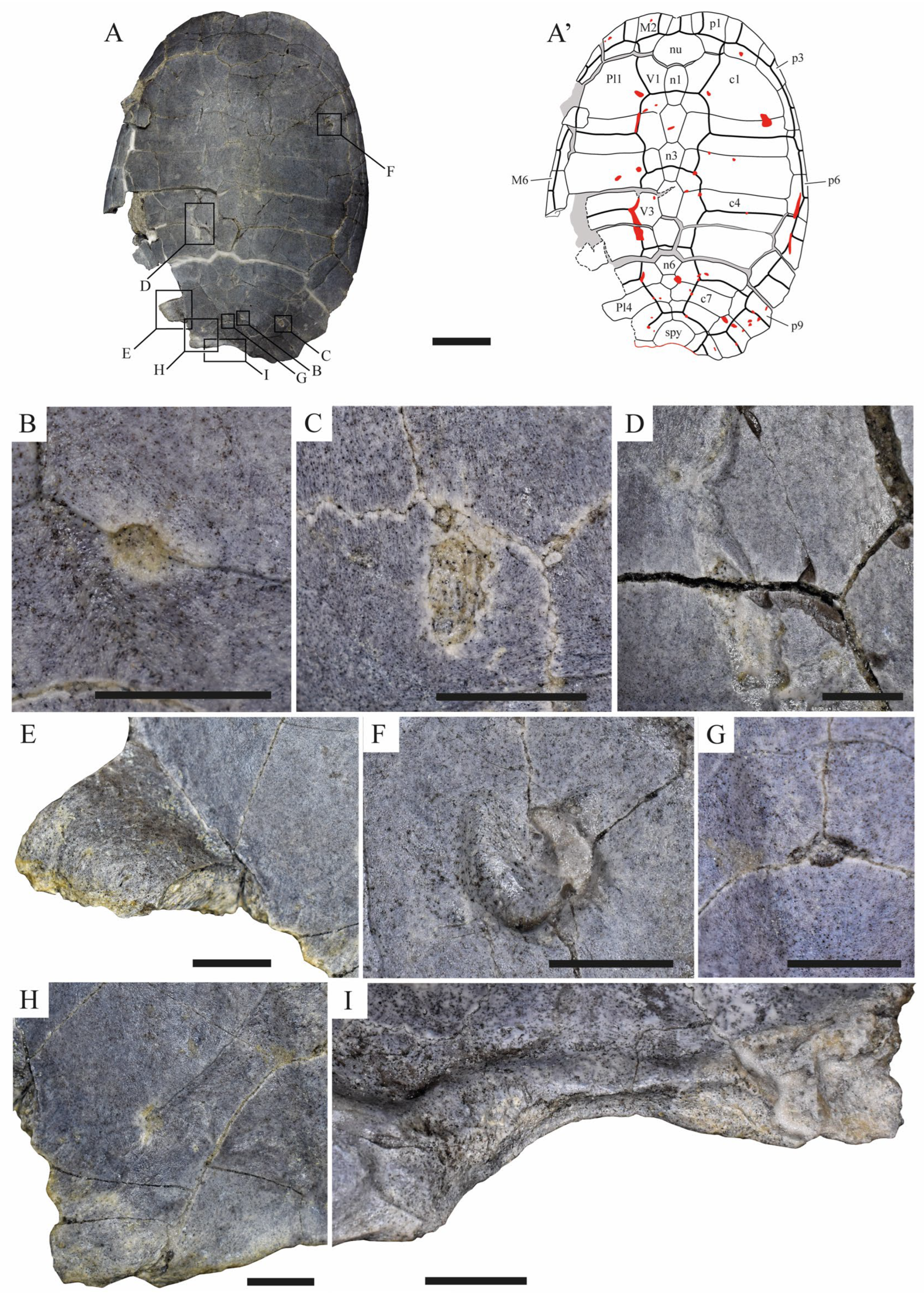
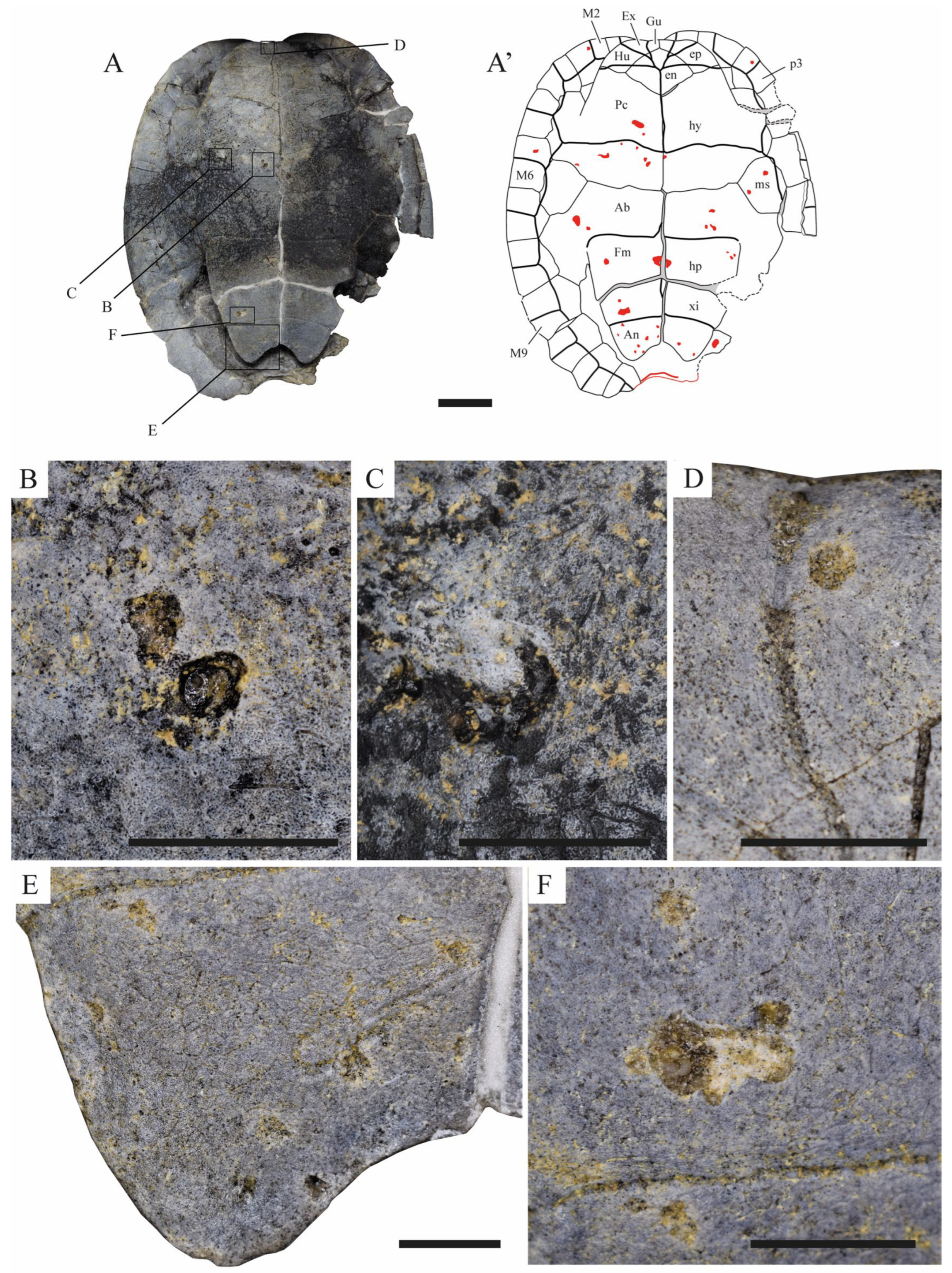
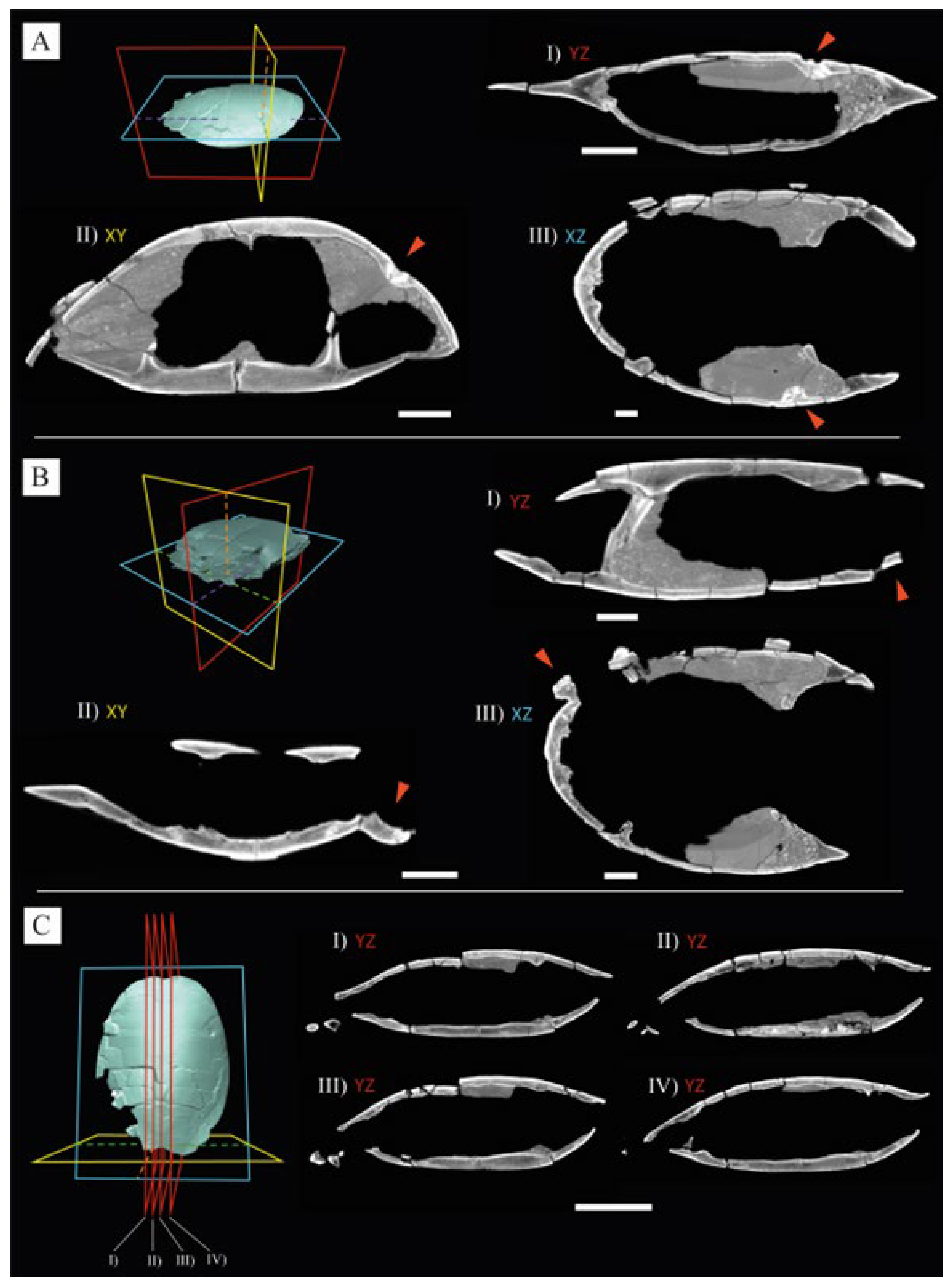
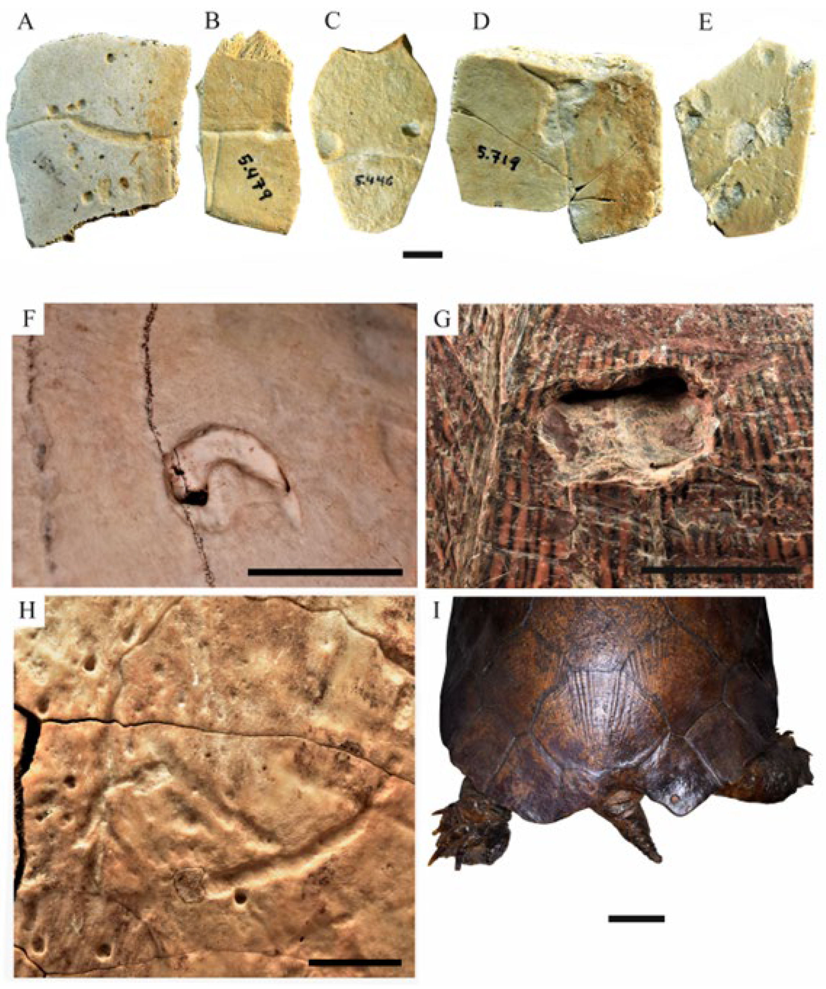
MNHN.RA, Collection of Reptiles and Amphibians, National Museum of Natural History, Paris, France; MNHN.ZA.AC, Collection of Reptiles, Comparative Anatomy, National Museum of Natural History, Paris, France; STUS, Sala de las Tortugas de la Universidad de Salamanca, Salamanca, Spain.
The specimen analyzed here, STUS 14024, was recently determined as Neochelys sp. (Pleurodira, Podocnemididae) (see discussion in [16]), a systematic attribution that is here supported, and was found on the Lutetian (middle Eocene) El Tejar fossil site, at Corrales del Vino (Zamora, Spain). STUS 14024 is deposited in the Sala de las Tortugas de la Universidad de Salamanca (Salamanca University, Salamanca, Spain), and corresponds to a well-preserved and almost complete shell (Figure 1A and Figure 2A). The pathological conditions documented in the specimen were previously analyzed by [17], attributing the shell injuries to trauma resulting from the attack of a predator, specifically a crocodile of the genus Asiatosuchus (see [17] for more details).
The specimen was analyzed through physical examination, a detailed description of the pathological conditions being presented. Photographs (taken with a monochrome background with a Nikon D3500 18–55 mm f0.82 digital camera) and schematic drawings (performed with Adobe Illustrator CS6) of the shell are included in this study. Computerized axial tomography (Figure 3) was used to confirm and further characterize the origin of the anomalous conditions. This also allowed us to provide additional information on the potential causes of the alterations, as well as on the nature of the healing processes. Thus, externally visible pathological osseous modifications are commonly associated with changes in skeletal microstructure (see [18] and references therein). The specimen was scanned with Optima CT600 scanner at the “Clínica Quirón Juan Bravo” (Madrid, Spain), at a voltage of 135 kV and a current of 75 mA, obtaining 950 images in Dicom format. The data were imported to the Avizo 7.1 software (VSG, Germany) for visualization and analysis. Images were subjected to a differential diagnosis procedure, which constitutes one of the methodological tools of the paleopathology [19]. The discussion was based on the comparison of both medical and veterinary bone pathology literature as well as by the direct observation of extant turtle individuals, especially those corresponding to the same lineage as STUS 14024 (i.e., Podocnemididae), deposited in the Comparative Anatomy and Zoology of Reptiles and Amphibians collections of the Muséum national d’Histoire Naturelle (Paris, France).
The description of the shell anomalies of STUS 14024 was performed following the terminology described by [20]. Thus, the term “hole” is used in this study to describe a perforation that penetrates from the external to the internal surfaces of the shell bone, whereas “pit” is used to identify the erosions that do not fully penetrate the bone (for more details see [20] and references therein). Likewise, the term “groove” is used for linear to sub-linear furrows or indentations in the bone surface [20].

3. Results

Several alterations are recognized in the specimen studied here (Figure 1 and Figure 2) which imply, in all cases, bone loss. They can be grouped into two typologies of anomalies: shell marks (i.e., shell pitting and grooves, Figure 1B–D,F–H and Figure 2B–F) and the complete loss of the posterior region of the carapace (Figure 1E,H,I).
The shell marks are identified, in this Neochelys specimen, as disseminated anomalies throughout the carapace (Figure 1A) and plastron (Figure 2A), without specific patterns on the plates outer surface. Most of the marks on STUS 14024 correspond to shell pitting, which varies in morphology (i.e., amorphous, Figure 2B; sub-circular, Figure 1H; and circular, Figure 2D). Almost all the pittings on the shell are characterized as shallow (i.e., the erosions do not fully penetrate the external cortical bone layer), with sub-straight lateral margins, defining hemispherical-to-flat bottoms of 1 to 5 mm in diameter. The only exception is the hole located between the first and second costal plates (Figure 1F). This mark fully penetrates the internal cortex layer of the bone (Figure 3A) and is characterized as presenting sub-straight lateral margins, with a hemispherical bottom, and of 12 mm in diameter. Radiological imaging revealed, as a pathological response associated with the hole, new bone growth developed on the ventral surface of the carapace, internal to the penetration of the plate. Such osseous growth presents a different density both in relation to the rest of the shell plates as well as to other regions of the same plate. Apart from this bone growth, no other macroscopic signs of new bone formation were identified in response to the anomalous shell marks. Most of the shell pittings do not coincide with the sulci between scutes or the plate sutures (e.g., Figure 2E), although, in some cases, they were also identified in such regions (Figure 1H,G, respectively). Grooves were also identified among the shell marks observed in the specimen studied here (Figure 1D and Figure 2C). These range from straight to curved linear excavations and are located in several areas of the shell (i.e., between the plate sutures, the sulci of the scutes, or in any of these mentioned regions, Figure 1A and Figure 2A). The grooves in the shell of STUS 14024 are from 20 mm to 45 mm in length and from 2 mm to almost 10 mm in width. CT-scan images show that grooves correspond to shallow erosions. Therefore, they do not reach the cancellous layer of the plates.
The other typology of an erosive alteration in STUS 14024 corresponds to the abnormal bone destruction of the posterior region of the carapace (Figure 1E,H,I). This anomaly implies the complete loss of the pygal plate, the partial destruction of the suprapygal and the most posterior peripherals (Figure 1A), as well as the anomalous osseous modification of the posterior area of the last two costal plates (Figure 1E,H). Likewise, it should be noted that the postero-lateral region of the sulci between the last vertebral and last left pleural scutes is missing (Figure 1H). The anomalous region is characterized by displaying a smooth and irregular contour. Radiological imaging evidences a homogeneous density of the anomalous region to the rest of the shell (Figure 3B,C). Throughout this erosive alteration, slight bone remodeling was observed on the ventral surface of the anomalous region (Figure 1I), in response to the bone destruction.

4. Discussions

The abnormal absence of the posterior region of the carapace (Figure 1E,H,I) and the main hole (Figure 1F) identified on the shell of the middle Eocene turtle analyzed here, STUS 14024, have been interpreted as unequivocally caused by pre-mortem damage. Both anomalies present evidence of bone remodeling (Figure 1F,I), which demonstrate a response by the bone tissue of the specimen to the action of the injurious agent and, therefore, indicate that the organism was alive during that process. The remaining abnormalities observed (i.e., shallow pits and grooves in the shell, Figure 1B–D,G,H and Figure 2B–F), although similar to some observed in the post-mortem stages of other organisms (e.g., [21,22,23]), also coincide with the pre-mortem marks commonly described in the shell of extant turtles. Thus, although it is not possible to confirm with certainty whether the specimen was alive when these shallow marks were originated on its shell, it is possible that they were originated as pre-mortem marks, like the other typologies of the anomalies observed on the shell of the Neochelys specimen.

4.1. Etiology of the Anomalous Shell Marks of STUS 14024

The first typology of the anomalies observed in STUS 14024, the shell marks (Figure 1B–D,F–H and Figure 2B–F), has been associated, in the literature of both extant and extinct turtles, with a wide variety of causative agents: vertebrate feeding marks [24], epibiont organisms (i.e., bryozoa, [25]; barnacles, [26]), ecto- and endoparasites (i.e., leeches, [27]; ticks, [28]; spirorchid liver flukes, [29]; cestodes, [30]), fungi [31], bacteria [32], and algae [33]). Considering the interpreted ecology for Neochelys (i.e., freshwater turtle), as well as the nature of the shell damage (i.e., the agent acted from outside of the host), three of these etiologies can be a-priori considered compatible with the anomalies of the specimen studied here: predator attack; fungi, bacteria, and algae; and ectoparasites.
Vertebrate feeding marks are, by far, the most common putative causative agents proposed for the extinct turtle specimens with this anomalous condition (e.g., [34,35,36,37,38,39,40,41,42,43,44,45]). In this sense, the lack of detailed studies on the etiology of the shell modifications (e.g., pits, grooves, and lacerations) of extant turtles corresponding to vertebrate feeding traces motivated many studies on extinct specimens to unquestioningly attribute shell marks to putative feeding traces (e.g., [36,46,47,48,49]), without taking into account other possible causal agents (e.g., parasitism, [27]; or microbial activity, [31,32,33]), which are very common in extant turtles. This is the case with the specimen studied herein, STUS 14024, whose shell anomalies were recognized as a clear result of the attack of the eusuchian crocodyliform Asiatosuchus [17]. Specifically, the inference concerning the nature of these anomalies was based on the supposed pattern interpreted for them and its comparison with the dental morphology of this crocodyliform species, also identified in the fossil site of El Tejar, at Corrales del Vino, where it was found (for more details see [17]). That hypothesis is refuted here. In this sense, the strongly distinctive marks and patterns commonly reported as a result of extant crocodile feeding behavior (e.g., [50,51,52,53,54,55]) are not observed in STUS 14024. Such diagnostic features described in the bones as crocodile feeding marks are usually a combination of bisected pits (for more details, see [52]), hook marks (i.e., J- or L-shaped marks), drag snags, and striation pivots [52,54,55]. However, in the Neochelys specimen, none of the more common marks resulting from the crocodile feeding behavior are identified, and, although the morphology of the pits and holes of STUS 14024 can potentially fit with some crocodile-induced osseous modifications (i.e., rounded pits; Figure 1B and Figure 2D), its presence can be justified by other causative agents (see below). Likewise, the marks present in STUS 14024 are disseminated over the shell (Figure 1 and Figure 2), without a specific pattern and, therefore, without evidencing any arrangement of the teeth rows that would be expected of a potential predator. The comparison of several shells of extant podocnemidids with the specimen studied here evidenced that similar marks to those of STUS 14024 (i.e., pits and grooves) are very common among extant freshwater turtles (Figure 4F–H), and do not correspond to vertebrate feeding traces. Moreover, marks similar to those of STUS 14024 have been observed in many other Neochelys individuals from various Eocene sites of the Duero Basin, also deposited in STUS (Figure 4A–E). These other specimens also do not present other evidence pointing to tooth marks as a potential agent for their anomalies (Figure 4A–E), and, hence, these were probably produced by other causal agents. Therefore, based on this interpretation, it is improbable that the shell marks of STUS 14024 were the result of a predator attack.
Fungi, bacteria, and algae are other putative agents for the anomalous marks on the shell of the Neochelys specimen. They cause erosive lesions similar to some observed on the fossil (i.e., the shallow amorphous pitting and grooves, Figure 1C,D and Figure 2B,C,F) in the shell of extant turtles (e.g., [29,56]). The action of these agents is common in aquatic and semiaquatic turtles, as is the case of Neochelys. As a result, fungi, bacteria and algae can invade an organism in damp habitats and cause shell injuries (e.g., [57,58]). The anomalous conditions of bacterial, algal, and fungal origins are commonly known as septicaemic cutaneous ulcerative disease (SCUD), ulcerative shell disease (USD), carapacial shell disease (CSD), or necrotizing shell disease (NSD) [20,57,59,60,61,62]. The clinical signs of these affections include microscopic to large amorphous pits, commonly with the presence of irregular and amorphous regions of bone necrosis (e.g., [57,63,64]). In this sense, some of the anomalies observed in the carapace and plastron of STUS 14024 (i.e., the amorphous pits and the irregular grooves with poorly defined margins of the plastron, Figure 1C,D and Figure 2B,C,F) fit with those observed in these affections. Specifically, these coincide both in morphology (i.e., amorphous), depth (i.e., restricted to the external cortical layer), and size with those described and illustrated as a result of fungal, bacterial, and algal agents (e.g., [20,28,65]). The absence of large areas of amorphous erosive bone in STUS 14024 indicates that the shell of the individual could be in the first stages of the condition since in cases where the affection is severe, the necrotic areas are usually laterally extensive [32,56,66]). The shell pitting (both pits and holes) of a sub-circular and circular morphology with well-defined margins (e.g., Figure 1B,F–H and Figure 2D) are excluded from this etiology since, despite fitting the size and depth of some of those originated by fungal, bacterial, and algal agents, the morphology is significantly different.
Attachment scars of ectoparasites are also one of the most common diagnostics for the shell marks of extant freshwater turtles, mostly for shell pitting (e.g., [20,67,68,69,70]). Among the potential parasitic organisms, Hirudinidae (leeches) are the most compatible agents with the circular and sub-circular shell marks observed in STUS 14024. Such organisms cause shell-bone decalcification through their salivary secretions, generating relatively penetrative holes [71,72], such as the main hole observed in the Neochelys specimen (Figure 1F). Specifically, the lesions produced by leeches are recognized as focal holes, often reaching the internal cortex of the bone, approximately perpendicular to the shell surface, with straight or convex lateral margins (e.g., [68,73]), generating a circular to sub-circular morphology of a few millimeters in diameter (for more details, see [4,73,74]). This is compatible with the pits and main hole of STUS 14024 (e.g., Figure 1B,F–H and Figure 2D). Likewise, solitary circular and sub-circular pits such as the main hole of STUS 14024 (Figure 1F), have been described in the literature as a shell region where a single leech fed for a significant period of time [20]. By contrast, areas with several pits, a-priori similar to the outer surface of the posterior region of the carapace studied here (Figure 1A,B,G,H), have been interpreted as areas where leeches shifted their position over a protracted interval or, alternatively, as generated by a bacterium or other microbial agents that were carried by leeches, causing the degradation of the bone proximal to the parasite attachment site [20]. The morphology, size, and depth fit with the features of the shell marks produced by these organisms. Therefore, the ectoparasites, specifically leeches, are considered here as the more probable etiological agent of these anomalies.

4.2. Etiology of the Anomaly of the Posterior Region of the Carapace STUS 14024

The second typology of anomalies observed in STUS 14024 corresponds to the abnormal bone destruction of the posterior region of the carapace, which shows a slight bone remodeling on the ventral surface (Figure 1E,H,I). The lack of relatively large shell areas, as in the case of the Neochelys specimen, is mostly attributed in extant turtles to trauma (i.e., physical injury caused by a violent force applied to the body; [75]), as well as to diseases generated by fungi, bacteria or algae (e.g., [28,29]).
As commented above, shell diseases associated with a fungal, bacterial, or algal infection are very common among freshwater turtle species and clinical signs include from small pitting lesions to the necrosis of extensive shell areas [28,32]. The use of CT scans in the study of extant turtles has revealed that some specimens with no clinical or gross signs of shell disease can have internal manifestations of this condition, which produces a change in the bone structure (e.g., see [18] and references therein), the affected area being more prone to fractures and, therefore, to the loss of a shell region. As discussed, some of the shell marks of this Neochelys specimen are consistent with those that can originate and develop from this disease.
Turtle traumatisms are common both in extant (e.g., [29,76,77]) and extinct representatives (e.g., [45,78]), and are usually the result of predation marks [51,75], intraspecific aggressions (e.g., [79]), or other traumatism typologies (e.g., falling, [76]). All these factors mentioned are, a-priori, compatible with the loss of a considerable region of the shell, such as that of STUS 14024, which can be gradual (i.e., begins as a fracture where, due to the habitat conditions, the cracked area ends up rotting and, therefore, the specimen loses the affected part, [76]) or abrupt (i.e., in the case of an attack, [51]). Except in very clear cases, it is very complex to establish the causative agent of a trauma in extant turtles (e.g., [80]) and, above all, in extinct ones. In this context, traumatism cannot be confirmed with certainty as a causative agent in the abnormal absence of the posterior region of the carapace of STUS 14024. Therefore, we consider improbable the previous interpretation suggested by [17] (i.e., result of a predator attack, see below) which is, therefore, also incompatible with the interpretations previously performed by other authors from the analysis of similar pathologies in the fossil record of turtles (e.g., [45,78]). Specifically, very similar shell anomalies (i.e., the anomalous absence of a significant portion of the shell together with new bone formation) to that observed in the specimen STUS 14024 were reported in two extinct individuals, which were also attributed as a crocodyliform predation attempt: in an Upper Cretaceous panchelid turtle (i.e., Rionegrochelys caldieroi), from Patagonia [78]; and a podocnemidid (i.e., Podocnemis) from the late Miocene of the Peruvian Amazonia [45]. As in the case of STUS 14024, we consider it unlikely that the absence of part of their shell was due to an attack by a predator. Specifically, R. caldieroi, like in extant turtles, presents, on the dorsal surface of its anterior plastral lobe, a sulcus which marks the limit of the scute and coincides with the beginning of the zone of insertion of soft tissues. Therefore, a significant region of the anterior plastral lobe is missing, beyond said sulcus in R. caldieroi; if it was an attack, the predator would have damaged a considerable portion of the soft tissue, involving sensitive areas of the individual ([78]: Figure 3), which most likely would have caused the death of the turtle without the possibility of regeneration. The same can be applied for the posterior region of the carapace both for the specimen studied here as well as for the late Miocene podocnemidid discussed by [45] (Figure 2), in which a potential bite of the region would impact the soft tissues of the turtle (i.e., since the limit of the marginal scutes coincides with the beginning of the area occupied by soft tissues and, in both individuals, a considerable part of the carapace is missing, beyond this limit), which would have probably been lethal to an individual.
In addition, neither of the three cases present diagnostic tooth marks (i.e., in the case of R. caldieroi and Podocnemis, no other marks are present over the shell), which are very characteristic and a clear indication of crocodile attack marks in extant representatives, nor secondary-impact damage (e.g., depressed fractures or crushing of some regions of the carapace or plastron, [54]). Likewise, the abnormal absence of the posterior region of the carapace has been also identified in extant podocnemidids (e.g., Figure 4I), evidencing the incompatibility with the predator attack hypothesis due to the better preservation of their shells. Therefore, for all these reasons, and contrary to the interpretation suggested by [17], the predator attack as a potential etiology has been considered unlikely for the specimen studied here. As a result, the abnormal absence of the posterior region of the carapace of STUS 14024 could be both a caused by a fungal, bacterial, or algal disease, as well as a consequence of some type of traumatism, with the exception of a predator attack. However, it cannot be determined which of the two conditions is the cause, since both etiologies can produce similar pathological changes in a turtle shell.
Figure 4. (AE), Anomalous shell plates of the Spanish podocnemidid turtle genera Neochelys. (A) STUS 1502, ventral view of an hypoplastron of Neochelys sp. From La Laguna—Casaseca de Campeán (Zamora); (B) STUS 5479, dorsal view of a peripheral plate of Neochelys sp. From La Laguna—Casaseca de Campeán (Zamora); (C) STUS 5446, dorsal view of a neural of Neochelys sp. From La Laguna—Casaseca de Campeán (Zamora); (D) STUS 5719, dorsal view of a peripheral plate of Neochelys sp. From La Laguna—Casaseca de Campeán (Zamora); (E) STUS 8255, ventral view of a xiphiplastron of Neochelys salmanticensis from Cabrerizos (Salamanca). (FI) Erymnochelys madagascariensis specimens (Pleurodira, Podocnemididae), of the Muséum national d’Histoire naturelle (París, France), with similar anomalies to those of STUS 14024. F, MNHN.ZA.AC 1897-80; (G) MNHN.ZA.AC 1946-71; H, MNHN.ZA.AC 1923-430; I, MNHN.RA To.0609-2. Scale bars equal 1 cm in (AE), and 2 cm in (FI).
Diversity 15 00314 g004

5. Pathogenesis

Both the main hole and the anomalous absence of the posterior region of the carapace of the turtle analyzed herein, STUS 14024, attributed to the activity of an ectoparasite and to a traumatism, respectively, fully penetrated the shell bone, from the external to the internal cortex (Figure 1A and Figure 3). Although the two damages occurred whilst the studied Neochelys specimen was alive, it cannot be confirmed if they were synchronic to each other. Likewise, none of these anomalies involved vital areas of STUS 14024 since the specimen had enough time to produce a physiological response to the injuries. Specifically, differences observed in the density of some areas of the bone growth of the main hole (Figure 3A) evidence an incomplete healing process since, in general, over time, the new tissue tends to restore its physical and mechanical properties [81]. This indicates that the organism died before the healing process of the pathology was completed. In contrast, the radiological images of the posterior region do not show significant density variations relative to the remaining areas of the carapace (Figure 3B,C). There is no evidence of an incomplete healing process. Likewise, the loss of the sulci between the last pleural and vertebral scutes (Figure 1H) indicates that such an area experienced bone remodeling, thus losing part of the suture. In no case is it possible to establish whether the death of the organism was related to any of these injuries.

6. Conclusions

The anomalies present on a shell of a podocnemidid turtle identified as Neochelys sp. (Pleurodira, Podocnemididae), from the middle Eocene (Lutetian) of the El Tejar fossil site, at Corrales del Vino (Zamora, Spain), have been analyzed here. The anomalies involve bone loss (i.e., shell marks and the anomalous absence of the posterior region of the carapace), and new bone formation (i.e., bone healing traces). These abnormal bone conditions of the turtle specimen were previously analyzed and diagnosed as traumatic injuries resulting from a crocodile attack, being interpreted as non-fatal for the turtle. Thus, the re-study of the different bone alterations through a detailed physical examination, the comparison through direct observation and from the literature on extant turtles, and the use of a computerized axial tomography scan proposed alternative causal agents (i.e., other typologies of traumatisms; mixed bacterial, fungal, and algal agents; and parasites).
As a result, two potential etiologies were suggested for the shell marks of the Neochelys specimen. The shallow amorphous pits and irregular erosive areas located on the carapace and plastron make fungal, bacterial, and algal diseases the most parsimonious hypothesis for these marks. In the case of the circular to sub-circular pits and the focal hole of the shell, an ectoparasite origin is considered the more compatible etiology. Specifically, leeches are the most likely ectoparasite since the feeding marks commonly produced by these organisms coincide in morphology, size, and depth with the circular to sub-circular shell damage of the specimen herein examined. With regards to the abnormal bone destruction of the posterior region of the carapace, this was considered to be due to some type of trauma or as a cause of a bacterial, fungal or algal disease. Although it was not possible to specify the origin of the anomaly, the hypothesis of a predator attack, as proposed in the previous study of the Neochelys specimen, STUS 14024, was ruled out.
The detailed macroscopic examination of the shell anomalies of the Neochelys specimen, as well as the use of computerized axial tomography, allowed us to obtain information regarding its pathogenesis. The presence of bone growth in two of the anomalies (i.e., the hole and the posterior margin of the carapace) demonstrates a response by the osteological system of the specimen to the action of the external agent and, therefore, indicates that the organism was alive at the time the shell injuries were inflicted. Likewise, the changes in density observed by radiological imaging of the hole reflect an incomplete healing process. In this case, as evidenced by the lack of homogeneous bone growth density, this individual had no time to complete the healing process. However, by contrast, the radiological images of the posterior region do not show significant density variations with the rest of the carapace; therefore, there is no evidence of an incomplete healing process. Thus, it cannot be confirmed whether the death of the specimen was due to any of the anomalies of its shell either.

Author Contributions

Writing—original draft preparation, A.G.; supervision, F.O., S.M.d.J. and A.P.-G.; funding acquisition, A.G. and A.P.-G. All authors have read and agreed to the published version of the manuscript.

Funding

This research was funded by an FPI UNED fellowship and by the Ministerio de Ciencia e Innovacion (PID2019-111488RB-I00).

Institutional Review Board Statement

Not applicable.

Data Availability Statement

No new data were created or analyzed in this study. Data sharing is not applicable to this article.

Acknowledgments

We acknowledge Salvador Bailón (MNHN.ZA.AC), Mathilde Aladini (MNHN.RA), and Stéphane Grosjean (MNHN.RA) for access to material of extant pleurodiran taxa. This research was funded by an FPI UNED fellowship and by the Ministerio de Ciencia e Innovacion (PID2019-111488RB-I00). The authors thank the Clínica Quiron Juan Bravo (Madrid, Spain) for CT scanning the specimen studied here. The authors also thank the editor T. Jumpamoon as well as À. Hernández Luján (Institut Català de Paleontologia Miquel Crusafont) and two anonymous reviewers for their useful comments and constructive reviews.

Conflicts of Interest

The authors declare no conflict of interest.

References

  1. Collareta, A.; Casati, S.; Zuffi, M.A.; Di Cencio, A. First authentic record of the freshwater turtle Mauremys from the upper Pliocene of Italy, with a new occurrence of the rarely reported ichnotaxon Thatchtelithichnus holmani. Carnets Geol. 2020, 20, 301–313. [Google Scholar] [CrossRef]
  2. Reolid, M.; Santos, A.; Mayoral, E. Grazing activity as taphonomic record of necrobiotic interaction: A case study of a sea turtle carapace from the Upper Jurassic of the Prebetic (south Spain). Rev. Mex. de Cienc. Geol. 2015, 32, 21–28. [Google Scholar]
  3. Jagt, J.W.; Deckers, M.J.; De Leebeeck, M.; Donovan, S.K.; Nieuwenhuis, E. Episkeletozoans and bioerosional ichnotaxa on isolated bones of Late Cretaceous mosasaurs and cheloniid turtles from the Maastricht area, the Netherlands. Geologos 2020, 26, 39–49. [Google Scholar] [CrossRef]
  4. Zonneveld, J.P.; AbdelGawad, M.K.; Miller, E.R. Ectoparasite borings, mesoparasite borings, and scavenging traces in early Miocene turtle and tortoise shell: Moghra Formation, Wadi Moghra, Egypt. J. Paleontol. 2022, 96, 304–322. [Google Scholar] [CrossRef]
  5. Hutchison, J.H.; Frye, F. Evidence of pathology in early Cenozoic turtles. PaleoBios 2001, 21, 12–19. [Google Scholar]
  6. Homer, B.L.; Berry, K.H.; Brown, M.B.; Ellis, G.; Jacobson, E.R. Pathology of diseases in wild desert tortoises from California. J. Wildl. Dis. 1998, 34, 508–523. [Google Scholar] [CrossRef]
  7. Scheyer, T.M.; Delfino, M.; Klein, N.; Bunbury, N.; Fleischer-Dogley, F.; Hansen, D.M. Trophic interactions between larger crocodylians and giant tortoises on Aldabra Atoll, Western Indian Ocean, during the Late Pleistocene. R. Soc. Open Sci. 2018, 5, 171800. [Google Scholar] [CrossRef] [Green Version]
  8. Garner, M.M.; Herrington, R.; Howerth, E.W.; Homer, B.L.; Nettles, V.F.; Isaza, R.; Shotts, E.B.; Jacobson, E.R. Shell disease in river cooters (Pseudemys concinna) and yellow–bellied turtles (Trachemys scripta) in a Georgia (USA) lake. J. Wildl. Dis. 1997, 33, 78–86. [Google Scholar] [CrossRef] [Green Version]
  9. Pereda-Suberbiola, X.; Astibia, H.; Murelaga, X.; Elorza, J.J.; Gomez-Alday, J.J. Taphonomy of the Late Cretaceous dinosaur-bearing beds of the Laño Quarry (Iberian Peninsula). Palaeogeogr. Palaeoclimatol. Palaeoecol. 2000, 157, 247–275. [Google Scholar] [CrossRef]
  10. Hayashi, R.; Chan, B.K.K.; Simon-Blecher, N.; Watanabe, H.; Guy-Haim, T.; Yonezawa, T.; Levy, Y.; Shuto, T.; Achituv, Y. Phylogenetic position and evolutionary history of the turtle and whale barnacles (Cirripedia: Balanomorpha: Coronuloidea). Mol. Phylogenet. Evol. 2013, 67, 9–14. [Google Scholar] [CrossRef]
  11. Agha, M.; Riley, M.K.; Sanford, E.; Carlton, J.T.; Newman, W.A.; Todd, B.D. A review of epizoic barnacles reported from freshwater turtles with a new record from California. Herpetol. Rev. 2018, 49, 25–28. [Google Scholar]
  12. Lafortune, M.; Wellehan, J.F.; Terrell, S.P.; Jacobson, E.R.; Heard, D.; Kimbrough, J.W. Shell and systemic hyalohyphomycosis in Fly River turtles, Carettochelys insculpta, caused by Paecilomyces lilacinus. J. Herpetol. Med. Surg 2005, 15, 15–19. [Google Scholar] [CrossRef]
  13. Pfaller, J.B.; Frick, M.G.; Reich, K.J.; Williams, K.L.; Bjorndal, K.A. Carapace epibionts of loggerhead turtles (Caretta caretta) nesting at Canaveral National Seashore, Florida. J. Nat. Hist. 2008, 42, 1095–1102. [Google Scholar] [CrossRef]
  14. McCoy, J.C.; Failey, E.L.; Price, S.J.; Dorcas, M.E. An assessment of leech parasitism on semi-aquatic turtles in the western Piedmont of North Carolina. Southeast. Nat. 2007, 6, 191–202. [Google Scholar] [CrossRef]
  15. Ryan, T.J.; Lambert, A. Prevalence and colonization of Placobdella on two species of freshwater turtles (Graptemys geographica and Sternotherus odoratus). J. Herpetol. 2005, 39, 284–287. [Google Scholar] [CrossRef]
  16. Ortega, F.; Armenteros, I.; de Celis, A.; Escaso, F.; Huerta, P.; Martín de Jesús, S.; Narváez, I.; Pérez-García, A.; Sanz, J.L. Crocodyliformes and Testudines from the Eocene of the Duero Basin (northwestern Spain): An update of their diversity and stratigraphic context. Hist Biol. 2022, 34, 1560–1581. [Google Scholar] [CrossRef]
  17. Jiménez-Fuentes, E. Predación crocodiliana a quelonios. Un Neochelys (Pelomedusidae), del Eoceno de Zamora, lisiado por un Asiatosuchus. Stud. Geol. Salmant. 2003, 39, 11–23. [Google Scholar]
  18. Hallock, L.A.; McMillan, A.; Wiles, G.J. Draft Periodic Status Review for the Western Pond Turtle in Washington; Washington Department of Fish and Wildlife: Olympia, WA, USA, 2016; p. 18. [Google Scholar]
  19. Rothschild, B.M.; Martin, L.D. Skeletal impact of disease. Bull. N. M. Mus. Nat. Hist. Sci. 2006, 33, 1–226. [Google Scholar]
  20. Zonneveld, J.P.; Bartels, W.S. The occurrence of bone modification features in the carapace and plastron of the extant red-eared Slider Trachemys scripta elegans (Wied-Neuwied, 1839): Implications for paleoecological analyses of fossil turtle assemblages. Palaios 2022, 37, 499–519. [Google Scholar] [CrossRef]
  21. Bader, K.S.; Hasiotis, S.T.; Martin, L.D. Application of forensic science techniques to trace fossils on dinosaur bones from a quarry in the Upper Jurassic Morrison Formation, northeastern Wyoming. Palaios 2009, 24, 140–158. [Google Scholar] [CrossRef]
  22. Fernandez-Jalvo, Y.; Andrews, P. Atlas of Taphonomic Identifications: 1001+ Images of Fossil and Recent Mammal Bone Modification; Springer: Dordrecht, The Netherlands, 2016; p. 359. [Google Scholar] [CrossRef]
  23. Höpner, S.; Bertling, M. Holes in bones: Ichnotaxonomy of bone borings. Ichnos 2017, 24, 259–282. [Google Scholar] [CrossRef]
  24. Emmons, L.H. Jaguar predation on chelonians. J. Herpetol. 1989, 23, 311–314. [Google Scholar] [CrossRef]
  25. Frazier, J.G.; Winston, J.E.; Ruckdeschel, C.A. Epizoan communities on marine turtles. III. Bryozoa. Bull. Mar. Sci. 1992, 51, 1–8. [Google Scholar]
  26. Jackson, C.G.; Ross, A. The occurrence of barnacles on the alligator snapping turtle, Macroclemys temminckii (Troost). J. Herpetol. 1971, 5, 188–189. [Google Scholar] [CrossRef]
  27. Bielecki, A.; Cichocka, J.; Jabłonski, A.; Jelén, I.; Ropelewska, E.; Biedunkiewicz, A.; Terlecki, J.; Nowakowski, J.J.; Pakulnicka, J.; Szlachciak, J. Coexistence of Placobdella costata (Fr. Müller, 1846) (Hirudinida: Glossiphoniidae) and mud turtle Emys orbicularis. Biologia 2012, 67, 731–738. [Google Scholar] [CrossRef]
  28. Jacobson, E.R. Infectious Diseases and Pathology of Reptiles: Color Atlas and Text; CRC Press: Boca Raton, FL, USA, 2007; p. 736. [Google Scholar] [CrossRef]
  29. Mader, D.R. Reptile Medicine and Surgery; Saunders Elsevier: St. Louis, MI, USA, 2006; p. 1242. [Google Scholar]
  30. Johnson, C.A.; Griffith, J.W.; Tenorio, P.; Hytrek, S.; Lang, C.M. Fatal trematodiasis in research turtles. Comp. Med. 1998, 48, 340–343. [Google Scholar]
  31. Schumacher, J. Fungal diseases of reptiles. Vet. Clin. N. Am.-Exot. Anim. Pract. 2003, 6, 327–335. [Google Scholar] [CrossRef]
  32. Granados, J.L.; Moreno, O.G.; Brieva, C.I. Lesiones ulcerativas cutaneas en tortugas dulceaquícolas. Rev. Med. Vet. Zoot. 2013, 60, 61–70. [Google Scholar]
  33. Fayolle, S.; Moriconi, C.; Oursel, B.; Koenig, C.; Suet, M.; Ficheux, S.; Logez, M.; Olivier, A. Epizoic algae distribution on the carapace and plastron of the European pond turtle (Emys orbicularis, Linnaeus, 1758): A study from the Camargue, France. Cryptogam. Algol. 2016, 37, 221–232. [Google Scholar] [CrossRef]
  34. Jiménez-Fuentes, E. Los reptiles fósiles del Valle del Duero: Podocnemis carbajosai nov. sp. del Eoceno de Salamanca. Estud. Geol. 1971, 27, 85–93. [Google Scholar]
  35. Erickson, B.R. Chelonivorous habits of the Paleocene crocodile Leidyosuchus formidabilis; Science Museum of Minnesota: Saint Paul, MN, USA, 1984; Volume 5, pp. 3–9. [Google Scholar]
  36. Antunes, M.T.; De Broin, F. Le Crétacé terminal de Beira Litoral, Portugal: Remarques stratigraphiques et écologiques, étude complémentaire de Rosasia soutoi (Chelonii, Bothremydidae). Earth Sci. 1998, 9, 153–200. [Google Scholar]
  37. Jiménez-Fuentes, E.; Martín de Jesús, S.; Mulas Alonso, E. Malformaciones y deformaciones patológicas en tortugas fósiles. Notas Informativas Sala de las Tortugas 1987, 3, 1–4. [Google Scholar]
  38. Mead, J.I.; Cubero, R.; Zamora, A.L.V.; Swift, S.L.; Laurito, C.; Gomez, L.D. Plio-Pleistocene Crocodylus (Crocodylia) from southwestern Costa Rica. Stud. Neotrop. Fauna Environ. 2006, 41, 1–7. [Google Scholar] [CrossRef]
  39. Steadman, D.W.; Franz, R.; Morgan, G.S.; Albury, N.A.; Kakuk, B.; Broad, K.; Franz, S.E.; Tinker, K.; Pateman, M.P.; Lott, T.A.; et al. Exceptionally well preserved late Quaternary plant and vertebrate fossils from a blue hole on Abaco, The Bahamas. Proc. Natl. Acad. Sci. USA 2007, 104, 19897–19902. [Google Scholar] [CrossRef] [Green Version]
  40. Lehman, T.M.; Wick, S.L. Chupacabrachelys complexus, n. gen. n. sp. (Testudines: Bothremydidae), from the Aguja Formation (Campanian) of West Texas. J. Vertebr. Paleontol. 2010, 30, 1709–1725. [Google Scholar] [CrossRef]
  41. Schwimmer, D.R. Bite marks of the giant crocodylian Deinosuchus on Late Cretaceous (Campanian) bones. N. M. Mus. Nat. Hist. Sci. Bull. 2010, 51, 183–190. [Google Scholar]
  42. MCCoy, M.R.; Karl, H.V.; Tichy, G.; Steinbacher, J.; Aigner, G.; Cemper-Kisslich, J. Radiological evaluation of a fossil turtle trauma from the Upper Jurassic of Eichstätt (Testudines: Cryptodira). Stud. Geol. Salmant. 2012, 48, 37–44. [Google Scholar]
  43. Noto, C.R.; Main, D.J.; Drumheller, S.K. Feeding traces and paleobiology of a Cretaceous (Cenomanian) crocodyliform: Example from the Woodbine Formation of Texas. PALAIOS 2012, 27, 105–115. [Google Scholar] [CrossRef]
  44. Smith, H.F.; Hutchison, J.H.; Townsend, K.E.B.; Adrian, B.; Jager, D. Morphological variation, phylogenetic relationships, and geographic distribution of the Baenidae (Testudines), based on new specimens from the Uinta Formation (Uinta Basin), Utah (USA). PLoS ONE 2017, 12, e0180574. [Google Scholar] [CrossRef] [Green Version]
  45. Pujos, F.; Salas-Gismondi, R. Predation of the giant Miocene caiman Purussaurus on a mylodontid ground sloth in the wetlands of proto-Amazonia. Biol. Lett. 2020, 16, 20200239. [Google Scholar] [CrossRef]
  46. Joyce, W.G. The first complete skeleton of Solnhofia parsonsi (Cryptodira, Eurysternidae) from the Upper Jurassic of Germany and its taxonomic implications. J. Paleontol. 2000, 74, 684–700. [Google Scholar] [CrossRef]
  47. Alonso-Santiago, L.; Alonso-Andres, L.; Jiménez-Fuentes, E. Análisis de varios casos de zoopaleopatología del Eoceno medio de Zamora (Espana). Stud. Geol. Salmant. 2006, 42, 97–112. [Google Scholar]
  48. Karl, H.-V.; Tichy, G. The structure of fossil teeth of chelonophagous crocodiles (Diapsida: Crocodylia). Stud. Geol. Salmant. 2004, 40, 115–124. [Google Scholar]
  49. Karl, H.-V. Bite traces in a turtle shell fragment from the Kimmeridgian (Upper Jurassic) of northern Germany. Stud. Geol. Salmant. 2012, 9, 25–30. [Google Scholar]
  50. Ortiz, R.M.; Plotkin, P.T.; Owens, D.W. Predation upon olive ridley sea turtles (Lepidochelys olivacea) by the American crocodile (Crocodylus acutus) at Playa Nancite, Costa Rica. Chelonian Conserv. Biol. 1997, 2, 585–586. [Google Scholar]
  51. Milàn, J.; Kofoed, J.; Bromley, R.G. Crocodylian-chelonian carnivory: Bite traces of Dwarf Caiman, Paleosuchus palpebrosus in red-eared slider, Trachemys scripta carapaces. M. Mus Nat. Hist. Sci. 2010, 51, 195–200. [Google Scholar]
  52. Drumheller-Horton, S.K. An Actualistic and Phylogenetic Approach to Identifying and Interpreting Crocodylian Bite Marks. Ph.D. Thesis, University of Iowa, Iowa City, IA, USA, 2012; p. 170. [Google Scholar] [CrossRef]
  53. Njau, J.K.; Blumenschine, R.J. A diagnosis of crocodile feeding traces on larger mammal bone, with fossil examples from the Plio-Pleistocene Olduvai Basin, Tanzania. J. Hum. Evol. 2006, 50, 142–162. [Google Scholar] [CrossRef]
  54. Drumheller, S.K.; Brochu, C.A. A Diagnosis of Alligator mississippiensis bite marks with comparisons to existing crocodylian datasets. Ichnos 2014, 21, 131–146. [Google Scholar] [CrossRef]
  55. Njau, J.; Gilbert, H. Standardizing terms for crocodile-induced bite marks on bone surfaces in light of the frequent bone modification equifinality found to result from crocodile feeding behavior, stone tool modification, and trampling. FOROST Occas. Publ. 2016, 3, 1–13. [Google Scholar]
  56. Lescano, J.; Quevedo, M.; Fernández, V. Enfermedad ulcerativa cutánea septicémica en una colección multi-especie de tortugas semiacuáticas. Rev. Investig. Vet. Perú 2013, 24, 561–564. [Google Scholar] [CrossRef] [Green Version]
  57. Johnson, J.H. Husbandry and medicine of aquatic reptiles. Semin. Avian Exot. Pet Med. 2004, 13, 223–228. [Google Scholar] [CrossRef]
  58. Aleksić-Kovačević, S.; Oezvegy, J.; Krstić, N.; Rusvai, M.; Jakab, C.; Stanimirović, Z.; Becskei, Z. Skin and skeletal system lesions of european pond turtles (Emys orbicularis) from natural habitats. Acta Vet. Hung. 2014, 62, 180–193. [Google Scholar] [CrossRef] [Green Version]
  59. Assan Kasim, M.; Senthil Kumar, K.; Palanivelrajan, M. Shell rot infection in red eared turtle. Int. J. Adv. Biol. Res. 2017, 7, 634–635. [Google Scholar]
  60. Barnett, S. Shell infections: When there are chinks in the armor. In Terrapin Tales (USA); MAATS: Highland, MD, USA, 2003; pp. 18–42. [Google Scholar]
  61. Wallach, J.D. The pathogenesis and etiology of Ulcerative Shell Disease in turtles. J. Zoo Anim. Med. 1975, 6, 11–13. [Google Scholar] [CrossRef]
  62. Woodburn, D.B.; Kinsel, M.J.; Poll, C.P.; Langan, J.N.; Haman, K.; Gamble, K.C.; Madox, C.; Jeon, A.B.; Wellehan, J.F.X.; Ossiboff, R.J.; et al. Shell lesions associated with Emydomyces testavorans infection in freshwater aquatic turtles. Vet. Pathol. 2021, 58, 578–586. [Google Scholar] [CrossRef] [PubMed]
  63. Lovich, J.E.; Gotte, S.W.; Ernst, C.H.; Harshbarger, J.C.; Laemmerzahl, A.F.; Gibbons, J.W. Prevalence and histopathology of shell disease in turtles from Lake Blackshear, Georgia. J. Wildl. Dis. 1996, 32, 259–265. [Google Scholar] [CrossRef] [PubMed]
  64. Lambert, M.R.; Hernández-Gómez, O.; Krohn, A.R.; Mutlow, A.; Patterson, L.; Rosenblum, E.B.; Timmer, M.; Willis, J.; Bushell, J. Turtle Shell Disease Fungus (Emydomyces testavorans): First documented occurrence in California and prevalence in free-living turtles. Ichthyol. Herpetol. 2021, 109, 958–962. [Google Scholar] [CrossRef]
  65. Frye, F.L.; Williams, D.L. Self-Assessment Colour Review of Reptiles and Amphibians; Manson Publishing: Stanford-le-Hope, UK, 1995; p. 233. [Google Scholar]
  66. Rothschild, B.M.; Schultze, H.-P.; Pellegrini, R. Osseous and other hard tissue pathologies in turtles and abnormalities of mineral deposition. In Morphology and Evolution of Turtles, Vertebrate Paleobiology and Paleoanthropology; Brinkman, D.B., Ed.; Springer Science+Business Media: Heidelberg, Germany, 2013; pp. 501–534. [Google Scholar]
  67. Fediras, S.; Roang, R.; Ziane, N.; Olivier, A.; Bechet, A.; Benyacoub, S. Prevalence of Placobdella costata (Fr. Mûller, 1846) (Hirudinida: Glossiphoniidae) on the European pond turtle (Emys orbicularis) in northeast Algeria. Herpetol. Notes 2017, 10, 3–8. [Google Scholar]
  68. Trivalairat, P.; Chiangkul, K.; Purivirojkul, W. Parasitism of Placobdelloides siamensis (Oka, 1917) (Glossiphoniidae: Hirudinea) in snail-eating turtles, Malayemys spp., and the effects of host and aquatic environmental factors. Biodivers. Data J. 2020, 8, e57237. [Google Scholar] [CrossRef]
  69. Chiangkul, K.; Trivalairat, P.; Kunya, K.; Purivirojkul, W. Placobdelloides tridens sp. n., a new species of glossiphoniid leech (Hirudinea: Rhynchobdellida) found feeding on captive Orlitia borneensis in Thailand, and an update to the host distribution of P. siamensis. Syst. Parasitol. 2021, 98, 141–154. [Google Scholar] [CrossRef]
  70. Guerrero, A.; Ortega, F.; Pérez-García, A. Mark of ectoparasitic activity on a bothremydid (Pleurodira) turtle shell from the Lo Hueco site (Upper Cretaceous, Central Spain). Cretac. Res. 2022, 138, 105297. [Google Scholar] [CrossRef]
  71. Ernst, C.H.; Ernst, E.M. Ectoparasites associated with neotropical turtles of the genus Callopsis (Testudines, Emydidae, Batagurinae). Biotropica 1977, 9, 139–142. [Google Scholar] [CrossRef]
  72. Siddall, M.E.; Gaffney, E.S. Observations on the leech Placobdella ornata feeding from bony tissues of turtles. J. Parasitol. 2004, 90, 1186–1188. [Google Scholar] [CrossRef]
  73. Zonneveld, J.-P.; Bartels, W.S.; Gunnell, G.F.; McHugh, L.P. Borings in early Eocene turtle shell from the Wasatch Formation, South Pass, Wyoming. J. Paleontol. 2015, 89, 802–820. [Google Scholar] [CrossRef]
  74. Chiangkul, K.; Trivalairat, P.; Purivirojkul, W. Redescription of the Siamese shield leech Placobdelloides siamensis with new host species and geographic range. Parasite 2018, 25, 56. [Google Scholar] [CrossRef] [PubMed] [Green Version]
  75. Kirchgessner, M.; Mitchell, M.A. Chelonians. In Manual of Exotic Pet Practice; Mitchell, M., Tully, T.N., Eds.; Elsevier Health Sciences: Amsterdam, The Netherlands, 2009; p. 560. [Google Scholar]
  76. Girling, S.J.; Raiti, P. BSAVA Manual of Reptiles; No. Ed. 3; British Small Animal Veterinary Association: Quedgeley, UK, 2019; p. 512. [Google Scholar]
  77. Vella, D. Management of aquatic turtle shell fractures. Lab Anim. 2009, 38, 52–53. [Google Scholar] [CrossRef]
  78. de Valais, S.; Díaz-Martínez, I.; Citton, P.; Maniel, I.; de la Fuente, M. A predation attempt in a Late Cretaceous pleurodire turtle from Patagonia. Cretac. Res. 2020, 107, 104290. [Google Scholar] [CrossRef]
  79. Garner, M.M.; Jacobson, E.R. Noninfectious Diseases and Pathology of Reptiles: Color Atlas and Text, Diseases and Pathology of Reptiles; CRC Press: Boca Raton, FL, USA, 2020; Volume 2, p. 512. [Google Scholar]
  80. Dodd Jr, C.K.; Franz, R.; Johnson, S.A. Shell injuries and anomalies in an insular population of Florida box turtles (Terrapene carolina bauri). Herpetol. Nat. Hist. 1997, 51, 66–72. [Google Scholar]
  81. Ghiasi, M.S.; Chen, J.; Vaziri, A.; Rodriguez, E.K.; Nazarian, A. Bone fracture healing in mechanobiological modeling: A review of principles and methods. Bone Rep. 2017, 6, 87–100. [Google Scholar] [CrossRef]
Figure 1. STUS 14024, anomalous carapace of a Neochelys sp. (Pleurodira, Podocnemididae) from the middle Eocene (Lutetian) of El Tejar fossil site, at Corrales del Vino (Zamora, Spain). (A,A’) dorsal view. (BI) detail of some of the anomalies identified on the dorsal surface of the carapace: pits (B,G,H), grooves (C,D), a hole (F), and the abnormal absence of the posterior region of the carapace (E,H,I). Scale bars equal 5 cm in (A,A’), and 1 cm in (BI). Abbreviations for the plates (in lowercase and normal type): c, costal; n, neural; nu, nuchal; p, peripheral; spy, suprapygal; Abbreviations for the scutes (in uppercase and in bold type): M, marginal; Pl, pleural; V, vertebral.
Diversity 15 00314 g001
Figure 2. STUS 14024, anomalous plastron of a Neochelys sp. (Pleurodira, Podocnemididae) from the middle Eocene (Lutetian) of El Tejar fossil site, at Corrales del Vino (Zamora, Spain). (A,A’) dorsal view. (BF) detail of some of the anomalies identified on the ventral surface of the plastron: pits (B,D,E) and grooves (C,F). Scale bars equal 5 cm in (A,A’), and 1 cm in (BF). Abbreviations for the plates (in lowercase and normal type): ent, entoplastron; ep, epiplastron; hp, hypoplastron; hy, hyoplastron; ms, mesoplastron; p, peripheral; xi, xiphiplastron. Abbreviations for the scutes (in uppercase and in bold type): Ab, abdominal; An, anal; Ex, extragular; Fe, femoral; Gu, gular; Hu, humeral; M, marginal; Pc, pectoral.
Diversity 15 00314 g002
Figure 3. Cross-section slices of the shell of anomalous carapace of a Neochelys sp. (Pleurodira, Podocnemididae), STUS 14024, from the middle Eocene (Lutetian) El Tejar fossil site, at Corrales del Vino (Zamora, Spain). (A) cross-section of the hole: (I) and (II), perpendicular view of the shell anomaly; (III), parallel view of the shell anomaly. (B) cross-section of the seventh costal plate, which also lacks its posterior region. (C) cross-section of the abnormal condition of the posterior region of the carapace: (I–IV) perpendicular view of the carapace. Scale bars equal to 2.5 cm in (A,B), and 10 cm in (C).
Diversity 15 00314 g003
Disclaimer/Publisher’s Note: The statements, opinions and data contained in all publications are solely those of the individual author(s) and contributor(s) and not of MDPI and/or the editor(s). MDPI and/or the editor(s) disclaim responsibility for any injury to people or property resulting from any ideas, methods, instructions or products referred to in the content.

