Article

Characterization of Tomato Brown Rugose Fruit Virus (ToBRFV) Detected in Czech Republic

1
Mendeleum—Institute of Genetics, Mendel University in Brno, Valticka 334, 691 44 Lednice, Czech Republic
2
Division of Plant Pests Diagnostics, National Reference Laboratory, Central Institute for Supervising and Testing in Agriculture, Šlechtitelů 773/23, 779 00 Olomouc, Czech Republic
3
Department of Chemistry and Toxicology, Veterinary Research Institute, Hudcova 296/70, 621 00 Brno, Czech Republic
*
Author to whom correspondence should be addressed.
Diversity 2023, 15(2), 301; https://doi.org/10.3390/d15020301
Submission received: 29 November 2022 / Revised: 23 January 2023 / Accepted: 16 February 2023 / Published: 18 February 2023
(This article belongs to the Special Issue Recent Advances in Plant-Pathogen Interactions)

Abstract

:
Tomato is the most consumed vegetable in the world. The tomato brown rugose fruit virus (ToBRFV) is an important destructive virus that damages tomatoes and peppers with significant economic impact. The detection and characterization of this important viral pathogen were evaluated at the molecular and morphological level. The viral isolate was purified and inoculated on tomato and pepper plants. Small RNAs were sequenced in both plants and the profiles were compared. The complete genome of the isolate was obtained, and microRNA (miRNA) profiles were unveiled by small RNA sequencing. Symptoms caused by the isolate were also described and the morphology of the isolate was observed by transmission electron microscopy. Our results contribute to further understanding of the role of miRNAs in ToBRFV pathogenesis, which may be crucial for understanding disease symptom development in tomatoes and peppers.

1. Introduction

Tobamoviruses are mechanically transmitted plant viruses that cause severe economic damage to vegetables worldwide. Except for tomato mosaic virus (ToMV) and tobacco mosaic virus (TMV) that are the most commonly spread, there is a new threat, namely the tomato brown rugose fruit virus (ToBRFV) (Martellivirales; Virgaviridae; Tobamovirus), a recently described tobamovirus first detected in the Middle East [1,2]. Pepper can be also infected by ToBRFV [3,4] and eggplant is considered an unconfirmed host [2]. ToBRFV was initially detected in Jordan and Israel in 2014 and 2015, respectively, and recently it has been detected in tomato-production areas worldwide. The occurrence of ToBRFV in tomato-production areas was confirmed in the USA (California and Florida), China, Iran, Israel, Jordan, Saudi Arabia, Syria, Turkey, the United Kingdom, and in many EU countries, such as Italy, Germany, Greece, the Netherlands, Belgium, Poland, and Austria [5,6,7,8,9,10,11]. The spread of the virus has accelerated, representing a major economic concern, which has consequently led to phytosanitary regulation in the EU [12]. ToBRFV was detected in the Czech Republic for the first time in 2020 (https://gd.eppo.int/reporting/article-6901—accessed 29 November 2022) [3]. There is no known resistance to ToBRFV, even in cultivars previously resistant to other tobamoviruses.
Thus far, because of extensive crop handling and manipulation, ToBRFV has primarily been a threat to tomato production in protected cultures (greenhouses, screenhouses, and high tunnels), although outbreaks in open fields have been reported.
The genome of ToBRFV is ~6.4 nt long and has four open reading frames, 183 kDa and 126 kDa replication proteins, a movement protein (MP), a coat protein (CP), and 5′ and 3′ untranslated regions [2]. Small noncoding RNAs called microRNAs (miRNAs) play an important role in posttranscriptional gene regulation related to diverse biological processes, including development, immune system responses, and cell death [13]. Viral replication and proliferation included in host antiviral responses and the pathogenesis of the virus may be influenced by miRNAs. Perfect binding in the seed region has a major impact on the regulatory functions of a miRNA. The seed sequence or seed region in miRNA is represented by a conserved heptametrical sequence. Perfect binding in the seed region has a major impact on the regulatory function of a miRNA. Even if the base pairing between the miRNA and its target messenger RNA (mRNA) does not match perfectly, the seed sequence must always be perfectly complementary [14]. miRNAs can hold a negative or positive role in virus-related processes in three manners as follows: direct binding to the viral genome; binding to viral transcripts; or binding to host transcripts [14].
For plant virologists, high-throughput sequencing is a powerful type of technology that provides rapid and comprehensive information on the infectious agents (viruses and viroids) present in explored tissues [14,15]. Therefore, this technology is being increasingly used for the quick identification of viruses replicating in plant tissues, starting either from the analysis of small interfering RNA (sRNA) populations [16] or from sequenced libraries of fragmented double-stranded RNAs (dsRNAs) of viral origins [17,18] extracted from infected tissues.
Computational analyses of high-throughput sequencing data, followed by experimental validation, have been used to identify highly conserved miRNAs [19,20,21,22].
The aim was to describe the Czech ToBRFV isolate at the molecular and morphological level to determine its pathogenicity and to characterize the miRNA profiles in infected tomato and pepper plants.

2. Materials and Methods

2.1. Plant Material and Real-Time RT-PCR Detection

2.1.1. Origin of ToBRFV Isolate

The isolate of ToBRFV was obtained from a tomato plant (Solanum lycopersicum L., an unspecified variety with small yellow fruits), sampled during a phytosanitary check [3] carried out in a greenhouse located in the northeastern part of Moravia, Czech Republic. The tomato plant had fully developed asymptomatic fruits and showed no specific symptoms of a virus infection except necrosis of older leaves corresponding to the late sampling date (September).

2.1.2. Indicator Plants and Symptoms on Inoculated Plants

A bioassay was carried out on the following indicator plants: tobacco (Nicotiana benthamiana Domin, which can be seen in Supplementary Figure S1, Nicotiana clevelandii Gray × glutinosa L.); tomato (Solanum lycopersicum cv. Vilma, isolate TT2); and pepper plants (Capsicum annuum L. cv. Oraneta, isolate PP1). The tobacco plants were propagated from the laboratory’s seed source and the tomato and pepper plants were grown from seeds that tested negative for ToBRFV. The inoculation of indicator plants was performed in a phytotron under following environmental conditions: 16 h/24 °C during the day; 8 h/22 °C at night.
For the inoculation, a total of 0.5 g of tomato leaf tissue was homogenized with 10 mL of the inoculation buffer (4.48 g NA2HPO4. 12 H2O was filled up to 25 mL with demineralized water and 0.78 g NAH2PO4. 2 H2O was filled up to 10 mL with demineralized water, then both solutions were mixed; before the inoculation, 10 mL was filled up to 250 mL with demineralized water and 0.5 g of polyvinyl pyrrolidone was added) in an extraction bag (BIOREBA). The homogenate was transferred to a Petri dish with an abrasive (Celit) and mixed. All leaves of each indicator plant were gently rubbed by gloved fingers dipped in the inoculum. After the inoculation, plants were rinsed with tap water to remove the abrasive. Non-inoculated indicator plants of each species and variety, which served as the negative controls, were grown in the same conditions.

2.1.3. Real-Time RT-PCR Detection

For ToBRFV detection and confirmation of the infection, two specific real-time RT-PCR tests [23,24] were performed according to the Commission Implementing Regulation (EU) 2020/1191 (which applies until 31 May 2023), as amended by Commission Implementing Regulation (EU) 2021/74 and Commission Implementing Regulation (EU) 2021/1809).
RNA extraction was performed using a RNeasy Plant Mini Kit (Qiagen) as follows: A total of 0.5 g of tomato leaves was homogenized with 5 mL of the extraction buffer in an extraction bag (BIOREBA) using semi-automatic homogenizer Homex 6 (BIOREBA). A total of 50 µL of the homogenate were transferred into a 1.5 mL tube and mixed with 450 µL of the RLT buffer (included in the kit) with β-mercaptoethanol. According to the manufacturer’s instructions, the extraction process was followed by the final RNA elution using 2 × 30 µL of RNase-free water.
A one-step real-time RT-PCR using CaTa28 primers and probes developed by ISHI-Veg [23] was performed to detect the virus. The total volume of 10 µL of the reaction mix contained 1× Luna Universal Probe One-Step Reaction Mix (New England Biolabs), 0.3 µM of each primer (CaTa28 F: GGTGGTGTCAGTGTCTGTTT, CaTa28 R: GCGTCCTTGGTAGTGATGTT), 0.2 µM of the probe (CaTa28 P: FAM—AGAGAATGGAGAGAGCGGACGAGG—BHQ1) [25], and 2 µL of the undiluted RNA extract. The reaction was performed in a StepOnePlus Real-Time PCR System (Applied Biosystems) under the following reaction conditions: reverse transcription for 10 min at 55 °C; initial denaturation for 1 min at 94 °C and 45 cycles of 10 s at 94 °C denaturation; and 1 min of 60 °C extension. During the test validation in the laboratory, the experimental Ct cut-off value was set to 34. Simultaneously, the internal positive control RT-qPCR test was performed in a separate reaction using the primers and probe to detect the plant cytochrome oxidase gene (COX—F: CGTCGCATTCCAGATTATCCA, COX—R: CAACTACGGATATATAAGAGCCAAAACTG, COX-P: HEX—TGCTTACGCTGGATGGAATGCCCT—BHQ1) [26] under the same reaction conditions to check the quality and quantity of the extracted RNA.

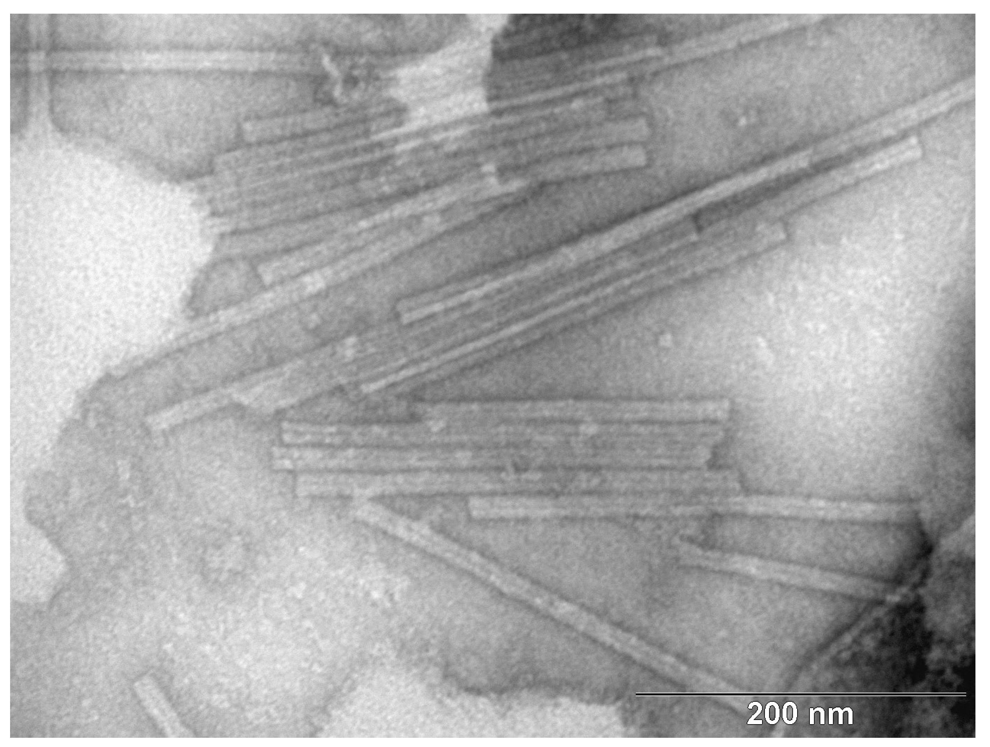
2.1.4. Transmission Electron Microscopy

For TEM, a total of 0.5 g of the tomato leaf tissue was homogenized with 5 mL of demineralized water in an extraction bag (BIOREBA). The homogenate was centrifuged on an Airfuge Air-Driven ultracentrifuge at 90,000 rpm for 120 min (Beckman Coulter, Brea, CA, USA). The resulting suspension was covered with an electron microscopic grid (300 Old Mesh, Agar Scientific), coated with a formvar film (Sigma-Aldrich) and carbon. The grid was removed from the suspension after 10–15 s, and the residual water was dried with a strip of filtration paper. For negative staining, a drop of NH4MoO4 (Serva, Germany) was placed onto the grid for a few seconds, then the excess stain was dried with filtration paper. The sections prepared in this way were observed under a Philips 208 S Morgagni transmission electron microscope (FEI, Brno, Czech Republic) at 18,000× magnification and with an accelerating voltage of 80 kV.

2.2. Small RNA Sequencing

The same RNA as for real-time RT-PCR detection was used for small RNA sequencing. The amount and quality of RNA were determined using an Agilent Small RNA kit (Agilent, Santa Clara, USA), and the precise concentration was measured using a Modulus™ Single Tube Multimode Reader (Turner Biosystems, Sunnyvale, CA, USA). The small RNA library was constructed using a NEBNext® Small RNA Library Prep Set (NEB, Ipswich, UK) and purification was conducted with a TailorCut Gel Extraction Tool Set (SeqMatic, Fremont, CA, USA). The quality and quantity of the library were determined using an Agilent High Sensitivity DNA Kit (Agilent, Santa Clara, CA, USA). All the kits were used according to the manufacturers’ instructions. For the sequencing run, the final pooled library of small RNAs consisted of 2 samples. Sample PP1 was labelled with index 12 (CTTGTA) and sample TT2 was labelled with index 6 (GCCAAT). The libraries were pooled at a concentration of 2 nM according to fluorimetry measurements, assuming that the final cloned small RNA products were ~150 bp. The libraries were sequenced with a MiniSeq (Illumina, San Diego, CA, USA), using a MiniSeq High Output Reagent Kit, 75 cycles (Illumina, San Diego, CA, USA) providing 36 nt long reads.

2.3. Bioinformatics and Data Evaluation

The quality of sequences was controlled by using a FastQC-0.10.1 [20]. A FASTX-Toolkit (http://hannonlab.cshl.edu/fastx_toolkit/ accessed on 10 September 2022), specifying the Q33 parameter, was used to obtain fasta format from fastq and to remove the adaptors (TGGAATTC). Sequences shorter than 15 nucleotides were discarded. The clipped reads of both isolates (PP1, TT2) were mapped onto reference sequence Acc. No. NC_028478 using CLC Genomics Workbench 22.0.2 (CLC Bio, Aarhus, Denmark) with the following parameters: mismatch cost = 2 (the cost of a mismatch between the read and the reference sequence); insertion cost = 3 (the cost of an insertion in the read, causing a gap in the reference sequence); and deletion cost = 3 (the cost of having a gap in the read). Furthermore, the reads were mapped randomly.
The phylogenetic analysis was applied using following parameters: Three method; Fast Minimum Evolution; and Max Seq Difference, 0.75 (ncbi.nlm.nih.gov/blast/treeview accessed on 12 August 2022). The 50 most similar ToBRFV genomic sequences were included.
The total number of known miRNAs was counted and annotated using miRbase Release 22.1 (Solanum lycopersicum) in CLC Genomics Workbench 6.5.1 (CLC Bio, Aarhus, Denmark). The statistical method to quantify differential expression in CLC Genomics Workbench 22.0.2 was used as follows: transcriptomic analyses; small RNA analyses; and extract and count, and annotate and merge counts. The numbers of miRNA sequences were normalized to one million reads (RPM) in order to enable comparative analyses.

3. Results

3.1. Real-Time RT-PCR Detection and Correlation with the Symptoms

Ten days after inoculation, the tomato and pepper plants did not show any symptoms of a viral infection compared to the negative controls. Only scratchings were observed on the inoculated leaves. These scratchings probably corresponded to the inoculation wounds. Necrotizing local chlorotic spots were observed on the leaves of the Nicotiana clevelandii × glutinosa plants. The Nicotiana benthamiana plants stopped growing and became chlorotic.
Twenty days after inoculation, some tomato leaves were strongly deformed. Chlorotic spots and slight deformations were observed on the leaves of the pepper plants, accompanied with the oldest leaves dying. The Nicotiana clevelandii × glutinosa plants stopped growing and necrosis and stunting developed on young leaves. The plants of Nicotiana benthamiana stopped growing, became strongly chlorotic, and the oldest leaves died.
Thirty days after inoculation, mild blistering was observed on the leaves of the tomato plant. Dark green mottling appeared on the pepper plant. Both inoculated and non-inoculated tomato and pepper plants seemed to be stunted and bushy. All inoculated tobacco plants completely died within 30 days of inoculation.
One hundred days after inoculation, the tomato plant was slightly stunting, and its leaves were deformed and blistered. The plant bloomed and produced fruits. Viral symptoms on the fruits were not observed. The symptoms in the pepper plant included stunting and leaf mottling. The pepper plant bloomed and produced fruits. Viral symptoms on the fruits were not observed.
The young leaves of the inoculated tomato and pepper plants were tested 30 days after inoculation and showed very low Ct values (˂5 for tomato/TT2 and ˂8 for pepper/PP1), indicating a high concentration of the virus in the inoculated plants. One hundred days after inoculation, the bioassay was completed, and different parts of the tomato and pepper plants were tested. In pepper/PP1 (Figure 1), roots, young leaves, fruits, and flowers were tested. The pepper seeds could not be tested because the harvested fruits were not yet ripe. The Ct values of all tested parts were low (˂9), indicating a high concentration of viral titer. In tomato/TT2 (Figure 2), a high concentration of the virus was detected (Ct ˂ 6) in all tested parts (roots, young leaves, fruits, and seeds obtained from harvested fruits). Despite that, the fruits did not show any viral symptoms.

3.2. Description of the Molecular Level of the Czech ToBRFV Isolate

The small RNA sequencing run on a MiniSeq (Illumina) provided a total of 55,100,547 single-end reads and 35,747,500 reads passed filter (PF), namely the chastity filter.

3.2.1. ToBRFV Isolate PP1 and TT2

The small RNA sequencing of the PP1 isolate extracted from the inoculated pepper provided 26,110,206 PF reads. After a basic analysis including the Q33 parameter and clipping, the final number of the reads was 23,306,421. These reads were used for mapping. In total, 2,421,807 reads were mapped on the reference ToBRFV sequence Acc. No. NC_028478. The complete viral genomic sequence was obtained, and the sequence is available under Acc. No. OP413740. The size of the genomic sequence was 6370 nts. The small RNA sequencing of the TT2 isolate extracted from the inoculated tomato provided 8,359,292 PF reads. After a basic analysis including Q33 and clipping, the final number of reads was 7,426,077. These were used for mapping. In total, 1,154,559 reads were mapped on reference sequence Acc. No. NC_028478. The size of the genomic sequence was 6368 nts. The phylogenetic analyses showed a similarity within the cluster contained in isolates 2020015323_A (Acc. No. OM515231) and 2020015323_B (Acc. No. OM515232) from the UK, obtained from tomato, and isolate Tom-BA21 (OK624678) from Italy, also obtained from tomato. The phylogenetic tree of the 50 most similar genomic sequences is shown in Figure 3.

3.2.2. Comparison of Isolates PP1 and TT2

Based on the results (Section 3.1), the newly described Czech ToBRFV isolate inoculated twice, once on pepper (PP1) and once on tomato (TT2), was evaluated as identical at the level of genomic sequence. This was also confirmed by the phylogenetic analyses. This newly described ToBRFV isolate is genetically stable. The obtained sequences of isolates PP1 and TT2 were analyzed using online tools available through NCBI (blastN). Supplementary Figure S2 shows a dot plot graph. The two isolates are clearly identical. Within the whole genome sequence, four open reading frames (ORFs) were identified, found at positive strands (nucleotide positions: 2–3427; 3428–4924; 5675–6193; and 4911–5711). As the sequences of the two isolates were identical, only one genomic sequence of the newly described ToBRFV isolate was submitted to GenBank, Acc. No. OP413740.
  • coverage = (read count * read length)/total genome size
  • coverage for PP1 = (2,421,807 * 36)/6368 = 13,691
  • coverage for TT2 = (1,154,559 * 36)/6370 = 6524

3.2.3. Conserved miRNAs

The total number of known miRNAs was counted and annotated using miRBase Release 22.1 (Solanum lycopersicum) in CLC Genomics Workbench 6.5.1 (CLC Bio, Aarhus, Denmark). The statistical method used to quantify differential expression in CLC Genomics Workbench 6.5.1 was used as follows: transcriptomic analyses; small RNA analyses; and extract and count, and annotate and merge counts. The number of miRNA sequences was normalized to one million reads (RPM) in order to enable comparative analyses. Particular miRNAs were identified using CLC Genomics Workbench 22.0.2 (CLC Bio, Aarhus, Denmark) according to miRBase 22.1 miRNAs of the PP1 and TT2 isolates (Table 1 and Table 2). The RNA of the PP1 isolate contained 27 miRNAs, and the RNA of the TT2 isolate contained 42 miRNAs, including their precursor variants, precursors, mature 3’ supers, and mature 5’ subs.

3.2.4. Transmission Electron Microscopy

Transmission electron microscopy is the only imaging technique that yields the direct visualization of viruses, due to its nanometer-scale resolution. ToBRFV particles have a rod-like shape and are ~274.8 nm in length and 13.9 nm in width, measured across 36 particles. The TEM observation confirmed the typical appearance of ToBRFV particles (Figure 4).

4. Discussion

According to the phylogenetic analyses based on the whole genome of the virus reisolated from the tomato and pepper indicator plants, the Czech ToBRFV isolate belongs to the cluster with isolates 2020015323_A (Acc. No. OM515231) and 2020015323_B (Acc. No. OM515232) from the UK, obtained from tomato, and isolate Tom-BA21 (OK624678) from Italy, also obtained from tomato. This cluster is a standalone cluster and is different from the majority of the other European ToBRFV isolates according to the Nextstrain build, a new tool that is available at https://nextstrain.nrcnvwa.nl/ToBRFV/20220412, accessed on 10 August 2022 [27].
This attempt to use the small RNA high-throughput sequencing technique to identify conserved miRNAs differentially expressed in pepper and tomato plants, combined with the whole viral ToBRFV genome description and electron microscopy, was carried out for the first time. The experimental strategy of this study was designed to investigate the profile of pepper and tomato miRNAs.
In the case of pepper, 27 miRNAs were detected and in the case of tomato, 42 miRNAs were detected, including their precursor variants, precursors, mature 3’ supers, and mature 5’ subs. The only known publication about ToBRFV miRNAs was published by Gaafar and H. Ziebell (2020) [28], but they only use in silico predictions of miRNAs targeting different loci in the genome of ToBRFV.
The most abundant MIR396b, including precursor variants 17 nts and 21 nts, was detected in the pepper plant. This sequence belongs to the MIR396 family of miRNAs, which are predicted to target mRNAs coding for growth-regulating factors (GRFs), transcription factors, rhodanese-like proteins, and a kinesin-like protein [29]. Regarding the tomato plant, the second most detected miRNA was MIR396a, including precursor 15 nts and precursor variant 25 nts. MIR369a-5p induces tomato‘s susceptibility to Phytophthora infestans and Botrytis cinerea infections and enhances the tendency to produce reactive oxygen species (ROS) under pathogen-related biotic stress by suppressing target genes and upregulating salicylic acid [30]. It was found that after water stress, MIR396a-5p was downregulated in the IL9-1 drought-tolerant tomato, while it was upregulated in the M82 sensitive genotype as determined by high-throughput sequencing [31]. In general, expression of MIR396s is probably more linked with water stress than with the presence of ToBRFV [32].
The tomato plant showed the highest abundancy of MIR6023 (71.235 RPM) and the pepper plant showed a very low frequency (0.0429 RPM) in both hosts as precursor/precursor variants. The tomato Hcr9 (Homologs of Cladosporium fulvum resistance 9) gene family is targeted by MIR6023 [14]. An example of sequence diversity generated from a single MIR locus is MIR6023, encoding canonical MIR6023, a well-characterized miRNA regulating R genes in tomato [14]. Prigigallo et al. [33] demonstrated that MIR6023 is specifically associated with the PVY (Potato virus Y) infection of tomato and indicates that the wide diversification of this miRNA family is a direct consequence of the viral infection. The analyses of the high-throughput sequencing data obtained from a PSTVd (Potato spindle tuber viroid)-variant-infected tomato plant’s leaves and stems revealed an alteration in the miRNAs involved in diverse functions, such as disease resistance [34]. This information proves that the accumulation of MIR6023 could be associated with the regulation of R genes if the tomato is infected by ToBRFV. However, this phenomenon is not proved in case of pepper. Seo et al. [35] implied that MIR6023 in pepper might have evolved independently, and their findings indicate that miRNA genes have undergone a dynamic evolution in pepper.
The numbers of detected miRNAs were not dependent on the total number of the reads per sample and were not dependent on the Ct values reached by real-time RT-PCR.

5. Conclusions

The Czech ToBRFV isolate shows the typical morphology of a ToBRFV virion. The size of the genomic sequence revealed by small RNA sequencing was 6368 nts. The phylogenetic analyses showed a similarity within the cluster containing two isolates from the UK and one isolate from Italy, all obtained from tomatoes. We detected 27 miRNA forms (PP1) and 42 miRNA forms (TT2), including their precursor variants, precursors, mature 3’ supers, and mature 5’ subs. The most accumulated miRNA that is probably associated with ToBRFV presence was MIR6023 in the tomato plant but not in the pepper plant. MiRNAs from the MIR396 family were expressed in both plants significantly, but it is not clear if their expression is linked with ToBRFV expression.

Supplementary Materials

The following supporting information can be downloaded at: https://www.mdpi.com/article/10.3390/d15020301/s1.

Author Contributions

Conceptualization, A.E.; methodology, E.H., J.C., M.H., H.O. and P.K.; software, A.E.; validation, A.E., J.C. and E.H.; formal analysis, A.E.; investigation, E.H.; resources, S.L.; data curation, A.E.; writing—original draft preparation, A.E., L.F., M.H., H.O., V.C. and P.K.; writing—review and editing, K.T. and S.L.; visualization, A.E. and P.K.; supervision, A.E.; project administration, J.C.; funding acquisition, J.C. All authors have read and agreed to the published version of the manuscript.

Funding

This research was supported by the project of MENDELU No. IGA-ZF/2021-ST2002: Tomato brown rugose fruit virus—actual threat of tomatoes and peppers production in European Union and project of MZe No. QK22010031.

Institutional Review Board Statement

The experiment described in this study does not include human participants and/or animals.

Data Availability Statement

Not applicable.

Acknowledgments

We thank Nora Hodeckova for professional editing of English.

Conflicts of Interest

The authors declare no conflict of interest.

References

  1. Salem, N.; Mansour, A.; Ciuffo, M.; Falk, B.W.; Turina, M. A new tobamovirus infecting tomato crops in Jordan. Arch. Virol. 2016, 161, 503–506. [Google Scholar] [CrossRef]
  2. Luria, N.; Smith, E.; Reingold, V.; Bekelman, I.; Lapidot, M.; Levin, I.; Elad, N.; Tam, Y.; Sela, N.; Abu-Ras, A.; et al. A New Israeli Tobamovirus Isolate Infects Tomato Plants Harboring Tm-22 Resistance Genes. PLoS ONE 2017, 12, e0170429. [Google Scholar] [CrossRef] [Green Version]
  3. Tomato Brown Rugose Fruit Virus (TOBRFV)[Overview]|EPPO Global Database. Available online: https://gd.eppo.int/taxon/tobrfv (accessed on 15 November 2022).
  4. Salem, N.M.; Cao, M.; Odeh, S.; Turina, M.; Tahzima, R. First Report of Tobacco Mild Green Mosaic Virus and Tomato Brown Rugose Fruit Virus Infecting Capsicum annuum in Jordan. Plant Dis. 2020, 104, 601. [Google Scholar] [CrossRef]
  5. Cambrón-Crisantos, J.M.; Rodríguez-Mendoza, J.; Valencia-Luna, J.B.; Alcasio-Rangel, S.; García-Ávila, C.D.J.; López-Buenfil, J.A.; Ochoa-Martínez, D.L. Primer reporte de Tomato brown rugose fruit virus (ToBRFV) en Michoacán, México. Mex. J. Phytopathol. 2019, 37, 185–192. [Google Scholar] [CrossRef] [Green Version]
  6. Fidan, H.; Sarikaya, P.; Calis, O. First report of Tomato brown rugose fruit virus on tomato in Turkey. New Dis. Rep. 2019, 39, 18. [Google Scholar] [CrossRef] [Green Version]
  7. Panno, S.; Caruso, A.G.; Davino, S. First Report of Tomato Brown Rugose Fruit Virus on Tomato Crops in Italy. Plant Dis. 2019, 103, 1443. [Google Scholar] [CrossRef]
  8. Ling, K.-S.; Tian, T.; Gurung, S.; Salati, R.; Gilliard, A. First Report of Tomato Brown Rugose Fruit Virus Infecting Greenhouse Tomato in the United States. Plant Dis. 2019, 103, 1439. [Google Scholar] [CrossRef]
  9. Menzel, W.; Knierim, D.; Winter, S.; Hamacher, J.; Heupel, M. First report of Tomato brown rugose fruit virus infecting tomato in Germany. New Dis. Rep. 2019, 39, 1. [Google Scholar] [CrossRef] [Green Version]
  10. Yan, Z.-Y.; Ma, H.-Y.; Han, S.-L.; Geng, C.; Tian, Y.-P.; Li, X.-D. First Report of Tomato brown rugose fruit virus Infecting Tomato in China. Plant Dis. 2019, 103, 2973. [Google Scholar] [CrossRef]
  11. Beris, D.; Malandraki, I.; Kektsidou, O.; Theologidis, I.; Vassilakos, N.; Varveri, C. First Report of Tomato Brown Rugose Fruit Virus Infecting Tomato in Greece. Plant Dis. 2020, 104, 2035. [Google Scholar] [CrossRef] [Green Version]
  12. EUR-Lex—32020R1191—EN—EUR-Lex. Available online: https://eur-lex.europa.eu/eli/reg_impl/2020/1191/oj (accessed on 28 November 2022).
  13. O’Brien, J.; Hayder, H.; Zayed, Y.; Peng, C. Overview of MicroRNA Biogenesis, Mechanisms of Actions, and Circulation. Front. Endocrinol. 2018, 9, 402. [Google Scholar] [CrossRef] [PubMed] [Green Version]
  14. Li, F.; Pignatta, D.; Bendix, C.; Bruncard, J.O.; Cohn, M.M.; Baker, B. MicroRNA regulation of plant innate immune receptors. Proc. Natl. Acad. Sci. USA 2012, 109, 1790–1795. [Google Scholar] [CrossRef] [Green Version]
  15. Wu, Q.; Luo, Y.; Ru, R.; Lau, N.; Lai, E.C.; Li, W.-X.; Ding, S.-W. Virus discovery by deep sequencing and assembly of virus-derived small silencing RNAs. Proc. Natl. Acad. Sci. USA 2010, 107, 1606–1611. [Google Scholar] [CrossRef] [PubMed] [Green Version]
  16. Kreuze, J.F.; Perez, A.; Untiveros, M.; Quispe, D.; Fuentes, S.; Barker, I.; Simon, R. Complete viral genome sequence and discovery of novel viruses by deep sequencing of small RNAs: A generic method for diagnosis, discovery and sequencing of viruses. Virology 2009, 388, 1–7. [Google Scholar] [CrossRef] [Green Version]
  17. Al Rwahnih, M.; Daubert, S.; Golino, D.; Rowhani, A. Deep sequencing analysis of RNAs from a grapevine showing Syrah decline symptoms reveals a multiple virus infection that includes a novel virus. Virology 2009, 387, 395–401. [Google Scholar] [CrossRef] [PubMed] [Green Version]
  18. Coetzee, B.; Freeborough, M.-J.; Maree, H.J.; Celton, J.-M.; Rees, D.J.G.; Burger, J.T. Deep sequencing analysis of viruses infecting grapevines: Virome of a vineyard. Virology 2010, 400, 157–163. [Google Scholar] [CrossRef] [PubMed] [Green Version]
  19. Abreu, P.M.V.; Gaspar, C.G.; Buss, D.S.; Ventura, J.A.; Ferreira, P.C.G.; Fernandes, P.M.B. Carica papaya MicroRNAs Are Responsive to Papaya meleira virus Infection. PLoS ONE 2014, 9, e103401. [Google Scholar] [CrossRef]
  20. Eichmeier, A.; Kiss, T.; Kocanova, M.; Hakalova, E.; Spetik, M.; Cechova, J.; Tichy, B. Conserved MicroRNAs in Human Nasopharynx Tissue Samples from Swabs Are Differentially Expressed in Response to SARS-CoV-2. Genes 2022, 13, 348. [Google Scholar] [CrossRef]
  21. Eichmeier, A.; Kiss, T.; Penazova, E.; Pecenka, J.; Berraf-Tebbal, A.; Baranek, M.; Pokluda, R.; Cechova, J.; Gramaje, D.; Grzebelus, D. MicroRNAs in Vitis vinifera cv. Chardonnay Are Differentially Expressed in Response to Diaporthe Species. Genes 2019, 10, 905. [Google Scholar] [CrossRef] [Green Version]
  22. Yang, Y.; Huang, J.; Sun, Q.; Wang, J.; Huang, L.; Fu, S.; Qin, S.; Xie, X.; Ge, S.; Li, X.; et al. microRNAs: Key Players in Plant Response to Metal Toxicity. Int. J. Mol. Sci. 2022, 23, 8642. [Google Scholar] [CrossRef]
  23. International Seed Federation. Available online: https://worldseed.org/ (accessed on 28 November 2022).
  24. Menzel, W.; Jelkmann, W.; Maiss, E. Detection of four apple viruses by multiplex RT-PCR assays with coamplification of plant mRNA as internal control. J. Virol. Methods 2002, 99, 81–92. [Google Scholar] [CrossRef] [PubMed]
  25. Leichtfried, T.; Reisenzein, H.; Steinkellner, S.; Gottsberger, R.A. Improvement in the sensitivity of viroid detection by adapting the reverse transcription step in one-step RT-qPCR assays. J. Virol. Methods 2021, 292, 114123. [Google Scholar] [CrossRef] [PubMed]
  26. Cullen, D.W.; Toth, I.K.; Pitkin, Y.; Boonham, N.; Walsh, K.; Barker, I.; Lees, A.K.; Van der Heyden, H.; Bilodeau, G.J.; Carisse, O.; et al. Use of Quantitative Molecular Diagnostic Assays to Investigate Fusarium Dry Rot in Potato Stocks and Soil. Phytopathology 2005, 95, 1462–1471. [Google Scholar] [CrossRef] [Green Version]
  27. Botermans, M.; de Koning, P.P.; Oplaat, C.; Fowkes, A.; McGreig, S.; Skelton, A.; Adams, I.; Fox, A.; De Jonghe, K.; Demers, J.; et al. Tomato brown rugose fruit virus Nextstrain build version 3: Rise of a novel clade. PhytoFrontiers 2022. ISSN 2690-5442. [Google Scholar] [CrossRef]
  28. Gaafar, Y.Z.A.; Ziebell, H. Novel targets for engineering Physostegia chlorotic mottle and tomato brown rugose fruit virus-resistant tomatoes: In silico prediction of tomato microRNA targets. Peerj 2020, 8, e10096. [Google Scholar] [CrossRef]
  29. Jones-Rhoades, M.W.; Bartel, D.P. Computational Identification of Plant MicroRNAs and Their Targets, Including a Stress-Induced miRNA. Mol. Cell 2004, 14, 787–799. [Google Scholar] [CrossRef]
  30. Chen, L.; Meng, J.; Zhai, J.; Xu, P.; Luan, Y. MicroRNA396a-5p and -3p induce tomato disease susceptibility by suppressing target genes and upregulating salicylic acid. Plant Sci. 2017, 265, 177–187. [Google Scholar] [CrossRef]
  31. Liu, M.; Yu, H.; Zhao, G.; Huang, Q.; Lu, Y.; Ouyang, B. Profiling of drought-responsive microRNA and mRNA in tomato using high-throughput sequencing. BMC Genom. 2017, 18, 481. [Google Scholar] [CrossRef] [Green Version]
  32. Pantaleo, V.; Vitali, M.; Boccacci, P.; Miozzi, L.; Cuozzo, D.; Chitarra, W.; Mannini, F.; Lovisolo, C.; Gambino, G. Novel functional microRNAs from virus-free and infected Vitis vinifera plants under water stress. Sci. Rep. 2016, 6, 20167. [Google Scholar] [CrossRef]
  33. Prigigallo, M.I.; Križnik, M.; De Paola, D.; Catalano, D.; Gruden, K.; Finetti-Sialer, M.M.; Cillo, F. Potato Virus Y Infection Alters Small RNA Metabolism and Immune Response in Tomato. Viruses 2019, 11, 1100. [Google Scholar] [CrossRef] [Green Version]
  34. Adkar-Purushothama, C.R.; Perreault, J. Current overview on viroid–host interactions. Wiley Interdiscip. Rev. RNA 2020, 11, e1570. [Google Scholar] [CrossRef] [PubMed]
  35. Seo, E.; Kim, T.; Park, J.H.; Yeom, S.I.; Kim, S.; Seo, M.-K.; Shin, C.; Choi, D. Genome-wide comparative analysis in Solanaceous species reveals evolution of microRNAs targeting defense genes in Capsicum spp. DNA Res. 2018, 25. [Google Scholar] [CrossRef] [PubMed] [Green Version]
Figure 1. Symptomatic Capsicum annuum cv. Oraneta with PP1 isolate.
Diversity 15 00301 g001
Figure 2. Symptomatic Solanum lycopersicum cv. Vilma with TT2 isolate.
Diversity 15 00301 g002
Figure 3. Phylogenetic analyses of ToBRFV genomic sequences and the sequence of PP1/TT2 isolates. The scale bar represents a genetic distance of 0.0007. The phylogenetic analyses were applied using the following parameters: Three method; Fast minimum Evolution; Max Seq Difference, 0.75, 50 most similar ToBRFV genomic sequences were included.
Diversity 15 00301 g003
Figure 4. TEM figure of ToBRFV particles: flexuous, tobamovirus-like, and rod-shaped particles observed in the leaf extract. The scale bar represents a distance of 200 nm.
Diversity 15 00301 g004
Table 1. Particular miRNAs of PP1 isolate, normalized per one million reads (RPM).
miRNARPMMatch TypeLength
MIR166b0.042906631Precursor variant18
MIR167b0.042906631Precursor variant17
MIR171c0.042906631Precursor variant17
MIR172b0.171626523Precursor19
MIR396a0.471972938Precursor15
MIR396a0.085813262Precursor variant25
MIR396b0.514879569Precursor variant17
MIR396b0.042906631Precursor variant21
MIR482d0.128719892Precursor variant21
MIR482e0.429066308Precursor20
MIR482e0.128719892Precursor23
MIR482e0.085813262Precursor variant20
MIR482e0.042906631Precursor variant20
MIR482e0.042906631Precursor17
MIR482e0.042906631Precursor variant20
MIR53000.042906631Precursor variant20
MIR53030.042906631Precursor variant20
MIR53030.042906631Precursor variant23
MIR60230.386159677Precursor variant20
MIR60230.171626523Precursor variant18
MIR60230.042906631Precursor variant17
MIR60230.042906631Precursor variant19
MIR7981d0.042906631Precursor variant23
MIR94690.042906631Precursor variant18
Table 2. Particular miRNAs of TT2 isolate, normalized per one million reads (RPM).
miRNARPMMatch TypeLength
MIR10532//MIR7981f//MIR7981d0.134660602Precursor variant20
MIR105400.269321204Precursor22
MIR156a//MIR156b//MIR156c0.269321204Precursor variant25
MIR1590.134660602Precursor15
MIR166c0.538642408Precursor variant25
MIR168a0.269321204Precursor15
MIR168b0.269321204Precursor16
MIR396a5.386424084Precursor15
MIR396a0.942624215Precursor variant25
MIR396a0.134660602Precursor variant25
MIR396a0.134660602Mature 3’ super23
MIR396b0.134660602Precursor15
MIR396b0.134660602Precursor variant25
MIR602371.23545851Precursor16
MIR60230.538642408Precursor15
MIR60230.269321204Precursor20
MIR60230.134660602Precursor21
MIR60230.134660602Precursor25
MIR60230.134660602Precursor15
MIR60270.134660602Mature 5’ sub19
MIR60270.134660602Mature 5’ sub20
MIR7981c0.134660602Precursor variant21
MIR7981d0.538642408Precursor17
MIR7981d0.269321204Precursor16
MIR7981e//MIR105321.211945419Precursor18
MIR7981e//MIR105320.67330301Precursor variant23
MIR7981e//MIR105320.134660602Precursor variant20
MIR7981e//MIR105320.134660602Precursor16
MIR7981e//MIR105320.134660602Precursor variant18
MIR7981e//MIR10532//MIR7981c//MIR7981d0.134660602Precursor variant18
MIR7981e//MIR10532//MIR7981d0.134660602Precursor variant18
MIR7981e//MIR10532//MIR7981f2.423890838Precursor18
MIR7981e//MIR10532//MIR7981f1.346606021Precursor15
MIR7981e//MIR10532//MIR7981f0.942624215Precursor16
MIR7981e//MIR10532//MIR7981f0.807963613Precursor17
MIR7981e//MIR10532//MIR7981f0.538642408Precursor variant17
MIR7981e//MIR10532//MIR7981f0.134660602Precursor variant18
MIR7981f0.134660602Precursor16
MIR7981f0.134660602Precursor15
MIR7981f0.134660602Precursor variant22
MIR7981f//MIR7981b0.134660602Precursor variant22
MIR9471a0.134660602Precursor variant25
Disclaimer/Publisher’s Note: The statements, opinions and data contained in all publications are solely those of the individual author(s) and contributor(s) and not of MDPI and/or the editor(s). MDPI and/or the editor(s) disclaim responsibility for any injury to people or property resulting from any ideas, methods, instructions or products referred to in the content.

